Introduction
Acute appendicitis (AA) is the most commonly occurring abdominal acute condition in emer-gency departments1,2.
It can present at all ages, although it occurs more frequently during the 2ndand 3rd decades of life. Although it is relatively rare among extreme age-groups, it is more complicated when it does occur, due to difficulty in pinpointing exact pain location and in giving an accurate description of symptoms.
The overall lifetime risk of deveopling ap-pendicitis is estimated to be of 7%. Around 1% of outpatients presenting with abdominal pain have acute appendicitis (2.3% in the case of children)3,4. The mortality rate in non-complited cases is of 0.3%, increasing to 1-3% in
ca-ses of perforation and to 5-15% among the el-derly.
It is traditionally believed that acute appendici-tis is caused by an infection due to an obstructive problem. The main cause for obstruction in 60% of cases is hyperplasia of submucosal lymphoid fo-llicles. In 30-40% of cases, it is due to a faecalith or appendicolith (rarely visible via x-ray), with the remaining 4% being attributed to foreign bodies. In exceptional cases (1%), it is the form of pre-sentation of appendicular tumours.
Typical symptoms include pain initially centred in the epigastric region and subsequently moving to the right iliac fossa (RIF), presenting along with fever, nausea and vomiting, although this only oc-curs in 70% of cases5.
Until recently, surgical treatment was recom-mended for any case of RIF pain with a
reasona-acute appendicitis at the emergency department
REBECAPINTADO GARRIDO1, MARTAMOYA DE LA CALLE2, SUSANASÁNCHEZ RAMÓN2, MIGUELÁNGELCASTRO VILLAMOR2, SARAPLAZA LOMA1, MARCELINOMENDO GONZÁLEZ1
1Radiodiagnosis Department. 2Emergency Department. Río Hortera University Hospital. Valladolid, Spain.
Background: Appendicitis is the most common cause of acute abdominal pain and
subsequent surgery. For that reason the diagnosis of this condition is a cause of big concern in emergency departments.
Objective: The aim of the present study was to assess the usefulness of
ultrasono-graphy in the diagnosis of acute appendicitis.
Methods:Retrospective study which included patients presented in the emergency
de-partment with abdominal pain of suspected acute abdominal disorder origin and re-mitted to undergone ultrasonography to rule out appendicitis from January to July 2004.
Results:Among 2015 ultrasonography scans 296 were performed to exclude a
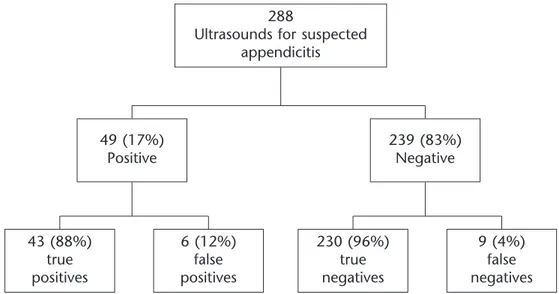
diagno-sis of acute appendicitis. 288 could be interpreted and the diagnodiagno-sis of acute appendi-citis was established in 52. In 15 cases the ultrasonography and the definite diagnosis differed. Ultrasonography and surgical diagnosis were different in 6 patients. In 9 pa-tients the ultrasonography was not diagnostic. Ultrasound sensitivity, specificity, positi-ve predictipositi-ve value, and negatipositi-ve predictipositi-ve value were 83.7%, 97.4%, 87.7% and 96.2%, respectively.
Conclusions:The global cost-effectiveness of ultrasonography to diagnose appendicitis
is good. Due to its availability and its low cost, ultrasonography is an accurate test for the diagnosis of acute appendicitis in emergency departments, specially in uncertain cases. [Emergencias 2008;20:81-86]
Key words:Acute appendicitis. Acute abdominal disorder. Ultrasonography.
CORRESPONDENCE: Susana Sánchez Ramón C/ Ciudad de la Habana 19, 2ºA 47016 Valladolid
E-mail: [email protected]
RECEIVED: 26-2-2007
ACCEPTED: 25-9-2007
ble suspicion of AA, which led to a very high rate of unnecessary appendectomies (10-30%)6.
The main clinical problems currently posed by appendicitis are its significant rate of post-surgical morbidity (18%)7 and that diagnostic and thera-peutic delay can lead to the onset of a histologi-cally more severe appendicitis as a result of an in-creased risk of perforation6,8,9.
The highly accurate diagnostic imaging me-thods currently available help to improve the ma-nagement of patients with suspected AA10. Both ultrasound and CT scans have proven to be highly reliable methods for diagnosing appendicitis11, but the indications and circumstances dictating which method should be used remain subject to discus-sion3. The initial diagnostic imaging test in most centres is ultrasonography6,12. This study assesses the need for ultrasonography in the diagnosis of AA in an emergency department.
Method
This is a descriptive, observational and retros-pective study, involving the review of clinical his-tories of patients attended in the emergency de-partment of our hospital from January to June 2004. A total of 2015 emergency abdominal ul-trasounds were performed during this period, 296 being requested for suspected AA.
Initial clinical assessments and ultrasound re-quests were made in all cases by an emergency department physician.
The following data were collected: epidemiolo-gical (sex and age), clinical (pain in lower right hemi-abdomen, fever, physical exploration and analyses), ultrasonographic and histopathological.
The diagnosis of clinically suspected appendici-tis was made on the basis of pain in the lower right hemi-abdomen and the presence or absence of one or more of the following criteria: fever (de-emed to be a body temperature at physical explo-ration above 37.5ºC), leukocytosis (>10.000 U/ml) and Blumberg’s sign (pain following abdominal decompression).
The final diagnosis was made comparing the ultrasonographic findings with the histopathologi-cal study results, except for cases of appendiceal adhesion masses for which post-surgical findings were taken into consideration. A clinical radiologi-cal follow-up was performed in non-surgiradiologi-cal pa-tients.
The ultrasonographic exploration of the right iliac fossa is performed by gradual compression: continuous, uniform pressure is applied to the
ex-ploration area to displace the air from the intesti-nal ansae and to minimise pain caused to the pa-tient, avoiding rebound pain due to successive compression and decompression. High resolution transducers, and occasionally Doppler-colour, are used.
The ultrasonographic criteria used by the Ra-diology Department to diagnose acute appendici-tis were: identification of a tubular intestinal struc-ture located in the lower right hemi-abdomen, closed at one end, with a transverse diameter ex-ceeding 6 mm, not compressible and aperistaltic, the appearance of appendicolith and/or the pre-sence of extra-appendicular alterations such as in-flamed peri-enteric fat, phlegmon or peri-appen-dicular abscess.
Variables were compiled in Excel tables and analysed and processed via the Windows SPSS programme version 11.0. Discrete variables were described using absolute frequencies (percenta-ges) and continuous variables as means and stan-dard deviations. The χ2test (or Fisher exact test in calculated values under 5) for discrete variables and the Student t test for continuous variables were applied when required. Statistical significan-ce was set at p < 0.05. Diagnostic performansignifican-ce markers were sensitivity, specificity, positive pre-dictive value and diagnostic efficacy.
Results
Pain in RIF with clinical suspicion of acute ap-pendicitis accounted for 15% of the emergency ultrasound explorations.
Of the 296 emergency ultrasounds requested due to suspected appendicitis, this diagnosis could not be evaluated in 8 cases: 7 due to the presence of abundant abdominal gas preventing adequate observation of intestinal ansae and in one case due to obesity. As for the 288 remaining cases, the age ranged between 2 to 92 years (me-an 31.4 years) (me-and 162 were women (56%) (me-and 126 men (44%).
Table 1 shows the various end diagnoses arri-ved at for the patients following clinical assess-ment and ultrasound performance. The diagnosis of non-specific abdominal pain was the most fre-quent of the pathologies found, accounting for 67% thereof.
pre-dictive value, 96.2% negative prepre-dictive value and 95% diagnostic efficacy (Figure 2).
The ultrasonographic findings in patients un-dergoing diagnostic ultrasonography were: identi-fication of a tubular structure exceeding 6 mm in diameter (97.7%), abdominal free fluid (42.8%), inflamed echogenic surrounding mesoappendix (39.5%), presence of appendicolith (25.6%) and appendiceal adhesions or abscess (6.9%).
In all cases in which the ultrasound was perfor-med for suspicion of acute appendicitis which was subsequently not confirmed, the observations in-cluded a tubular structure over 6 mm, inflamed surrounding mesoappendix in 66.7%, free fluid in 33.3% and appendicolith in 16.7% of cases. The final diagnosis in these cases was: non-specific ab-dominal pain in 3 cases, pelvic inflammatory dise-ase in 1 cdise-ase, acute gastroenteritis in 1 cdise-ase and cecal diverticulitis in the last case.
Of the 9 false negative cases, 7 showed a to-tally normal ultrasonographic exploration, free fluid was observed in the pelvis in one case and in the last case the ultrasound was performed for suspected terminal ileitis. Of the true negatives 83% were finally diagnosed as non-specific abdo-minal pain.
Discussion
Despite being one of the most frequent diag-noses among surgical emergencies, AA continues to pose significant diagnostic problems.
The diagnosis of AA in most cases is based on clinical history and physical exploration13.
At our hospital emergency department, in ca-ses with a medical history and physical explora-tion suggestive of possible appendicitis, chest and abdominal radiography and blood analyses are routinely performed. Of all signs and symptoms, only the presence of leukocytosis and a positive Blumberg’s sign show a statistically significant as-sociation with the diagnosis of AA.
Thirty percent of clinical cases are atypical and confusing5, leading to diagnostic errors and an in-crease in the number of unnecessary laparotomies. This situation may prove particularly problematic for women who may present with acute gynaeco-logical symptoms which could, to a large extent, simulate those of acute appendicitis. It is in these atypical cases where many studies6,14,15 show that ultrasonography is useful in arriving at a diagnosis of appendicitis, whilst proving less useful in pa-tients with a high clinical probability of appendi-citis who require immediate surgical assessment and for whom any delay in treatment should be avoided to reduce potential complications.
Image testing on suspicion of AA should be used as a diagnostic complement in selected cases and not as a routine tool in the initial clinical ex-ploration.
Table 1. Final clinical diagnoses following ultrasound
performance in the 288 patients included in the study
Diagnosis Number of patients Percentage
Non-specific abdominal pain 194 67.4
Appendicitis 52 18
Gastrointestinal disorders 24 8.4
Gynaecological disorders 9 3.1
Other 9 3.1
Figure 1. Results of ultrasounds performed.
288
Ultrasounds for suspected appendicitis
49 (17%) Positive
43 (88%) true positives
6 (12%) false positives
230 (96%) true negatives
9 (4%) false negatives 239 (83%)
The only specific sign of AA that can be obser-ved via a simple abdominal radiography is the presence of appendicolith (provided there are other compatible symptoms). Other signs yielded by abdominal radiography, such as the presence of a dilated ansa, hydroaerial levels, antalgic sco-liosis and erased psoas, are less specific9.
Another complementary method is abdominal ultrasonography, which in our case is usually the final diagnostic test. Among our patients we have confirmed the usefulness of ultrasonography as a diagnostic confirmation test (high positive predic-tive value, negapredic-tive predicpredic-tive value and specifi-city). Observation of an enlarged and non-com-pressible appendix is a sign of high positive predictive value. Nevertheless, the main difficulty posed by appendicitis ultrasound lies in elimina-ting the presence thereof. The usual negative diagnostic criterion (lack of visual appreciation of an inflamed appendix) may be due to the non-existence of appendicitis or to the impossibility of confirmation thereof which has often led to nega-tive predicnega-tive ultrasound values to be lower that the positive values7. The literature reports a rate of perforated acute appendicitis of 21%16, but the existence of false negatives rises to 44% due to the reduction in sensitivity caused by the perfora-tion, that is, the identification of a non-compressi-ble tubular structure of a +6 mm. diameter – the ultrasonographic sign most indicative of appendi-citis2 – is no longer visible as it becomes concea-led within the adhesion mass that is formed by the perforation. Another cause of ultrasound false negatives is the precocity in many cases when performing the test17. It is therefore very impor-tant to interpret the ultrasonography in each indi-vidual clinical context, particularly in cases in the early stages.
A great number of studies6,14,17,18 recommend that if clinical findings still suggest appendicitis despite negative ultrasound results, the patient should remain under hospital observation and subject to clinical exploration and, in some cases, successive ultrasonographies and even surgery.
It must not be forgotten that the ultrasonogra-phic results of the physician performing the test since the results may vary between those of an in-experienced physician and those of one familiar with the test, often explaining the false negatives and positives reported, and the lower number of false results obtained by more experienced ultra-sound physicians12,17,19.
The diagnostic values obtained in this study are similar to those published to date1,11.
Another complementary imaging test that can be performed for an emergency diagnosis of AA is Computed Tomography (CT). The higher sensiti-vity provided by CT makes it more useful accor-ding to various studies9,13,14 when diagnosing AA. Moreover, CT is less painful and offers a better performance in the diagnosis of other abdominal diseases, thus leading to many more alternative diagnoses9,13. Its main disadvantages are a higher radiation level, higher cost and the need, accor-ding to some authors, to use contrast material9.
We believe that the use of CT is advisable in cases of inconclusive results from analyses, routine radiographies and ultrasonographies performed, as well as in the case of an evolutive or complex disease, as it enables a more reliable detection of the presence of appendicular adhesions or abs-cess, and particularly in the elderly, for whom the risk of radiation exposure is minimal. In most of the cases in our series, the ultrasound proved suf-ficient to achieve a diagnosis as has been shown in the results, moreover offering the possibility of making alternative diagnoses20.
We believe that the false positives obtained are related to the inexperience of the ultrasonogra-pher performing the test plus the fact that many appendicitis cases do not undergo surgical inter-vention but rather are treated with antibiotics. We consider that the false negatives obtained are related to appendicular perforation, as in such ca-ses the sensitivity is reduced due to the enlarged tubular structure being less visible. This hypothe-sis, however, has not been tested in our study.
We thus conclude that abdominal ultrasono-graphy is the most useful complementary explora-tion in emergency departments for diagnosing acute appendicitis (95% accuracy) especially in cases of doubtful diagnosis. The accessibility and low cost of this approach make it ideal for
emer-Figure 2. Calculation of sensitivity, specificity, positive and
gency diagnosis, offering high levels of sensitivity and specificity. Its contribution often renders it es-sential in the study of acute appendicitis, as it helps achieve a large number of alternative diag-noses thereby assisting physicians in decision ma-king in doubtful cases and reducing the rate of unnecessary laparotomies.
References
1 Birnbaum BA, Wilson SR. Appendicitis at the milennium. Radiology 2000;215:337-48.
2 Kessler N, Cyteval C Gallix B, Lesnik A, Blayac PM, Pujol J, et al. Appendicitis: Evaluation of Sensitivity, Specificity and Predictive values of US, doppler US and laboratory finfing. Radiology 2004;230:472-9.
3 Del Cura Jl, Oleaga L, Grand D. Indicaciones de las técnicas de diagnóstico por la imagen en la sospecha de apendicitis aguda: propuesta de protocolo diagnóstico. Radiología 2001;43:478-89.
4 Rioux M. Sonographic detection of the normal and abnor-mal appendix. Am J Roentgenorl 1992;158:773-8. 5 Rosengren D, Brown AFT, Chu K. Radiological imaging to
improve the emergency department diagnosis of acute ap-pendicitis. Emergency Medicine Australasia 2004;16:410-6. 6 Bianchi A, Heredia A, Hidalgo LA, García F, Armella C, Su-ñol X. ¿Es suficiente la observación clínica en los casos du-dosos de apendcitis? Emergencias 2005;17:176-9.
7 Del Cura Jl, Oleaga L. La radiología en urgencias. Temas de actualidad. Monografía Seram 2006;79-86.
8 Gutierrez CJ, Mariano MC, Faddis DM, Sullivan RR, Wong RS, Lourie DJ, et al. Doppler ultrasound accurately screens patients with appendicitis. Am J Surg 1999;65:1015-7. 9 Marincek B. Nontraumatic abdominal emergencies: acute
abdominal pain: diagnostic strategies. Eur Radiol 2002;12:2136-50.
10 Paulson E, Kalady M, Pappas T. Suspected appendicitis. N J Med 2003;3:236-43.
11 Del Cura Jl, Oleaga L, Grande D, Fariña MA, Isusi M. Com-paración de la ecografía y la tomografía computarizada en el diagnóstico de la apendicitis aguda. Radiología 2001;43:175-86.
12 Chen SC, Wang HP, Hsu HY, Huang PM, Lin FY. Accuracy of ED Sonography in the diagnosis of acute appendicitis. Am J Emerg Med 2000;18:449-52.
13 Vázquez MA, Monteruel E, García E, Mintegui S, Canapé S, Benito J. Rendimiento de la ecografía abdominal en el diagnóstico de apendicitis aguda. Anales de Pediatría 2003;58:556-61.
14 Steven L, Ho S. Ultrasonography and computed tomo-graphy in suspected acute appendicitis. Semin Ultrasound CT MR 2003;24:69-73.
15 Teruhiko MD, Craig C. Sistematic review: Computed tomo-graphy and Ultrasonotomo-graphy to detect acute appendicitis in adults and adolescents. Ann Int Med 2004;141:537-46. 16 Bendeck SE N-MMBGJRJ. Imaging for suspected
appendici-tis: negative appendectomy and perforation rates. Radio-logy 2002;225:131-6.
17 Gallinas F, Garce C, Pérez A. La ecografía en la selección del dolor abdominal quirúrgico urgente. Estudio prospecti-vo. Cir Pediatr 2004;17:141-4.
18 Garcia FJ, Gil P. Sonography in acute appendicitis: diagno-sis utility and influence upon management and outcome. Eur Radiol 2000;10:1886-93.
19 Skaane P, Schistad O, Amland PF, Solheim K. Routine Ultra-sonography in the diagnosis of the acute appendicitis: A valuable tool in the daily practice? Am Surg 1997;63:937-42.
20 Alonso JM, Sandoval E. Valor de la ecografía en el diagnós-ticos diferencial de la apendicitis. Radiología 2001;40:307-13.
Indicación y utilidad de la ecografía urgente en la sospecha de apendicitis aguda
Pintado Garrido R, Moya de la Calle M, Sánchez Ramón S, Castro Villamor MA, Plaza Loma S, Mendo González M
Objetivo:La apendicitis aguda es la patología quirúrgica aguda abdominal más frecuente. Su diagnóstico constituye
uno de los problemas más habituales en los servicios de urgencias. El objetivo del presente estudio es evaluar la utili-dad de la ecografía abdominal en el diagnóstico de esta entiutili-dad.
Método:Estudio retrospectivo realizado entre enero y junio del 2004 de todas las consultas realizadas en el servicio de
urgencias por dolor abdominal indicativo de probable abdomen agudo, en los que se realizó una ecografía abdominal para descartar apendicitis aguda.
Resultados:Se realizaron 2.015 ecografías abdominales urgentes de las cuales 296 fueron solicitadas para descartar el
diagnóstico de apendicitis aguda, de éstas 288 fueron valorables. En 52 pacientes la ecografía fue indicativa de apendi-citis aguda. En 15 casos el diagnóstico ecográfico fue discordante con el diagnóstico final. En 6 pacientes el informe ecográfico de apendicitis no se confirmó a posteriori. En 9 casos la ecografía fue no diagnóstica pese al diagnóstico quirúrgico de apendicitis aguda. Con estos datos, el rendimiento global de la ecografía para el diagnóstico de apendi-citis aguda, se tradujo en una sensibilidad del 83,7%, especificidad del 97,4%, valor predictivo positivo del 87,7%, va-lor predictivo negativo del 96,2%.
Conclusiones:El rendimiento global de la ecografía abdominal en el diagnóstico de apendicitis aguda en nuestro
me-dio es aceptable. Debido a su accesibilidad y bajo coste es la prueba idónea para el diagnóstico en urgencias, sobre to-do en casos duto-dosos. [Emergencias 2008;20:81-86]