Effect of plasma rich in growth factors on the early phase of healing of surgically severed Achilles tendon in sheep : histological study / Daniel Aguilar García et al
9
0
0
Texto completo
(2) JOURNAL OF APPLIED ANIMAL RESEARCH, 2018 VOL. 46, NO. 1, 471–478 https://doi.org/10.1080/09712119.2017.1337017. Effect of plasma rich in growth factors on the early phase of healing of surgically severed Achilles tendon in sheep: histological study Daniel Aguilar-Garcíaa, José Andrés Fernández-Sarmientoa, María del Mar Granadosa, Juan Morgaza, Rocío Navarretea, José M. Carrillob, José M. Vilarc, Ramón Cugatd and Juan Manuel Domíngueza a. Department of Animal Medicine and Surgery, University of Córdoba, Córdoba, Spain; bDepartment of Animal Medicine and Surgery, CEU Cardenal Herrera University, Valencia, Spain; cDepartment of Animal Pathology, University Institute of Health and Biomedical Research, University of Las Palmas de Gran Canaria, Arucas, Spain; dArtroscopia GC, Hospital Quirón, Barcelona, Spain ABSTRACT. ARTICLE HISTORY. Tendon injuries are the most frequent musculoskeletal problems, constituting 30–50% of all sport-related lesions. Efficient handling of early stage tendon injury and healing can accelerate tendon repair as well as improve the quality of newly formed tendons. Plasma rich in growth factors (PRGF) is an autologous biological therapy that has been proposed to treat tendon injuries. To elucidate the effect of this treatment on the early stage of tendon healing, 14 sheep were used to perform the present study. The right Achilles tendon was surgically severed and repaired with sutures. Seven animals were treated with PRGF and the other seven with saline solution after surgical tendon reconstruction. Tendons were ultrasound-guide infiltrated 1 week after the induced injury. Two weeks later, the sheep were euthanized and histochemical and immunohistochemical analyses were performed. The tendons with PRGF showed reduced infiltration of inflammatory cells compared to those treated with saline solution. Analysis of the blood vessels, morphometric data of fibroblast nuclei, and collagen fibres in the extracellular matrix did not show statistical differences between groups. These findings suggest the important role that PRGF therapy plays in the modulation of inflammatory response in Achilles tendon injuries, leading to acceleration of the tendon healing process, shortening the convalescence period.. Received 13 March 2017 Accepted 26 May 2017. 1. Introduction Musculoskeletal injuries present a significant social issue because of its substantial impact on quality of life, and subsequent economic consequences (Anitua, Sanchez, Nurden, Nurden, et al. 2006). Tendon injuries are the primary musculoskeletal issues, constituting 30–50% of all sport-related lesions (Jarvinen et al. 2005), and present a considerable challenge due to slow tendon healing capacity and high re-injury rate (Sharma & Maffulli 2006; Hope & Saxby 2007; James et al. 2008). Plasma rich in growth factors (PRGF) is a type of platelet-rich plasma (PRP), characterized by a moderate platelet concentration (two- to threefold above peripheral blood), absence of leukocytes and erythrocytes (Anitua et al. 2012), and apparently innocuous nature (Vilar et al. 2017). PRGF is considered a useful autologous therapy proposed to improve tendon repair (Sanchez et al. 2007; Fernandez-Sarmiento et al. 2013; Seijas et al. 2015; Lopez-Najera et al. 2016). During the inflammatory phase of tendon healing, different subtypes of inflammatory cells are sequentially attracted to the damaged area (Marsolais et al. 2001). In addition, an angiogenic stimulus is produced, and the synthesis of new capillaries allows for the transport of oxygen and nutrients to the developing tissue (Butler et al. 2004; Hope & Saxby 2007). Previous studies have shown the importance of inflammation and angiogenesis modulation in tendon injuries (Dakin et al. 2014;. KEYWORDS. PRGF; Achilles tendon; inflammation; regeneration. Manning et al. 2014). The growth factors and other active proteins secreted by platelets play an important role in the modulation of inflammatory response and the induction of a rapid increase in specific cell populations, including endothelial cells, migrating fibroblasts, and resident cells of the tendon (Andia et al. 2010; Zhang et al. 2013). Some in vitro and in vivo PRP studies reported anti-inflammatory effects mainly explained by the activity of Hepatocyte growth factor (HGF) and transforming growth factor beta 1 (TGF-β1) (Bendinelli et al. 2010; Fernandez-Sarmiento et al. 2013; Mazzocca et al. 2013; Zhang et al. 2013). In tendon rupture, the extracellular matrix (ECM) is synthesized in order to fill the gap in between the tendinous stumps. At the molecular level, collagen type III synthesized as the primary ECM protein is replaced by collagen type I, which gives rise to natural tendon mechanical strength (Sharma & Maffulli 2006; Wang 2006; Hope & Saxby 2007). Morphometric parameters of fibroblast nuclei have been evaluated as indicators of maturation of tendon tissue during the healing process (Fernandez-Sarmiento et al. 2013). Additionally, the packing and orientation of collagen fibres have been used as a parameter to assess tendon ECM, as well as the proportion of collagen types I and III in the newly formed tendon (Kaux et al. 2012; Fernandez-Sarmiento et al. 2013; Lane et al. 2013). Furthermore, several animal models have been designed with. CONTACT José M. Vilar [email protected] Department of Animal Pathology, University Institute of Health and Biomedical Research, University of Las Palmas de Gran Canaria, 35416 Trasmontaña S/N, Arucas, Spain © 2017 The Author(s). Published by Informa UK Limited, trading as Taylor & Francis Group This is an Open Access article distributed under the terms of the Creative Commons Attribution License (http://creativecommons.org/licenses/by/4.0/), which permits unrestricted use, distribution, and reproduction in any medium, provided the original work is properly cited..
(3) 472. D. AGUILAR-GARCÍA ET AL.. the aim of studying the influence of PRP on several tendo-ligamentous structures (Kaux et al. 2012; Lane et al. 2013; Yoshioka et al. 2013). Histological results have varied depending on the study. These differences may be attributed to measurements at different time points of study and/or different methodologies used to obtain PRP. Long-term effects of PRGF application on Achilles tendon injury in human medicine have been studied in the clinical setting (Sanchez et al. 2007). To our knowledge, the effect of PRGF at the early stage of Achilles tendon reparation has not been studied in acute tendon injury. Previously, our group studied the effects of PRGF at 4 and 8 weeks after a surgically induced Achilles tendon injury in a sheep model. The aim of this study was to evaluate the histological effect of PRGF on tendon healing at 2 weeks. Our hypothesis was that PRGF leads to tendons with less inflammatory cell infiltration, therefore accelerating the beginning of proliferative and remodelling phases of tendon repair, thereby improving overall tendon healing.. 2. Methods 2.1. Animals Fourteen mature ewes weighing 50–55 kg and aged 24–30 months were included in the study. The animals belonged to the Experimental Farm of the University of Córdoba. All sheep used in this study were deemed healthy beforehand and did not suffer from any orthopaedic or systemic conditions. The animals were randomly placed into one of two groups: PRGF group (n = 7) or Saline group (n = 7), depending on receiving a subsequent treatment with PRGF or saline solution, respectively. The study was supervised and accepted by the Bioethics Committee in animal research at our institution.. 2.2. Experimental model The surgical procedure was performed following an animal model previously reported (Fernandez-Sarmiento et al. 2013). The animals were sedated intramuscularly with xylazine 0.1 mg/kg and morphine 0.3 mg/kg. Subsequently, anaesthesia was induced using propofol, intubated via endotracheal tube, and maintained under general anesthesia using isoflurane. The right hindlimb was clipped and aseptically prepared. A lateral incision across the Achilles tendon was performed in order to completely severe it. The tendon and peritenon were sutured as previously described elsewhere (FernandezSarmiento et al. 2013). In order to protect the repaired tendon, the tarsal joint was immobilized using a medial type I transarticular external fixator. Benzylpenicillin plus dihydrostreptomycin, at doses of 15,000 IU/kg and 15 mg/kg, respectively (Shotapen LA™; Virbac Animal Health, Barcelona, Spain), was administered intramuscularly at the time of surgery and at 72 h post-surgery to prevent infections. Analgesic buprenorphine 0.02 mg/kg/8h (Buprex™ 0.3 mg; Schering-Plough, Madrid, Spain) was administered intramuscularly for 5 days after surgery. No anti-inflammatory drug was used. The sheep were kept under surveillance and post-operative care during hospitalization.. 2.3. PRGF preparation and injection on repaired area Twenty millilitres of blood was collected from the jugular vein of each animal in four 5 mL sterile tubes containing 3.8% trisodium citrate as anticoagulant. The blood was centrifuged at 630 g for 8 min according to the method reported to obtain PRGF in ovines (Anitua, Sanchez, Nurden, Zalduendo, et al. 2006). The fraction considered PRGF was selected with the use of a pipette, obtaining a total volume of 2 mL per animal. Surgically severed Achilles tendons in the PRGF group were injected with activated PRGF before closing the peritendon. Activation of PRGF was performed using 0.1 mL of 10% calcium chloride. Afterwards 1 mL of PRGF was inoculated intratendinously into each stump using a 23-gauge needle. In the saline group, 1 mL of saline solution plus 0.05 mL of 10% calcium chloride was injected into each side of the surgically severed Achilles tendon. PRGF or saline solution infiltration was repeated 1 week later by ultrasonographic guidance with the animals under deep sedation. At both infiltrations, during the surgery and 1 week later, the syringe was covered with tape in order to double-blind the experiment. All the animals were humanely euthanized at 2 weeks, 1 week after the second infiltration, by using a standard protocol with T61™ (Intervet Schering-Plough Animal Health; Salamanca, Spain).. 2.4. Histological study After the animals were euthanized, both Achilles tendons were fixed in 10% formalin. The intact tendon was used as a positive control. Two paraffin blocks, proximal and distal stumps, were obtained from each surgically treated tendon and one block from the positive control. From each block, eight 4-µm-thick non-consecutive paraffin sections were cut. The sections were mounted on slides, and two were stained with haematoxylin and eosin (HE), two with Masson trichrome (MT), two were used for immunohistochemical (IHC) analysis of collagen type I, and two for IHC analysis of collagen type III. Histological images were obtained via a photomicroscope (Axiophot; Carl-Zeiss, Oberkochen, Germany) with an attached digital camera controller (DS-L1 camera control unit, Nikon). Slide sampling was performed in a systematic randomized manner by superimposing dotted transparent acetate gel onto each slide. From MT-stained sections and IHC-stained sections for collagen types I and III, 10 histological images per slide were obtained at 200× magnification. From HE-stained sections, 10 histological images per slide were obtained at 100× magnification, 10 histological images per slide at 200× magnification, and 10 histological images per slide at 630× magnification. These histological images were analysed in a computer equipped with image-analysis software (Image Pro Plus, version 6.0; Media Cybernetics, Rockville, Maryland). In order to not influence the histological evaluation, a number was randomly assigned to each slide and assessed by two independent pathologists. After the evaluation, the author determined which group each slide belonged to. The morphometric parameters of fibroblast nuclei were evaluated by using the images captured at 630× magnification from HE-stained sections. The following quantitative parameters were calculated for each fibroblast nucleus: area.
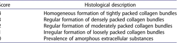
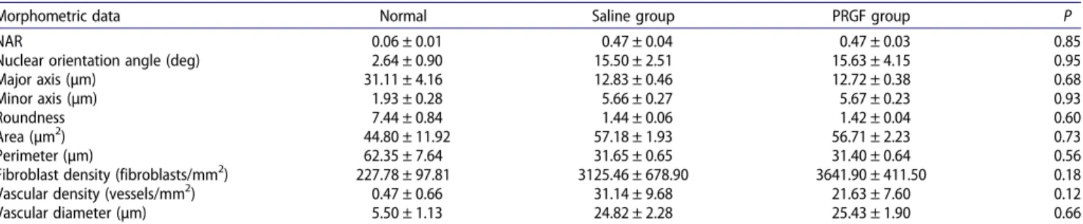
(4) JOURNAL OF APPLIED ANIMAL RESEARCH. (µm2), perimeter (µm), major axis (µm), minor axis (µm), and roundness [(perimeter)2/4 × Pi × area]. Nuclear aspect ratio (NAR) was also calculated as the fraction between the major and minor axes, being used as an indicator of nuclear shape, from spindle to circular shape, the latter corresponding to a value of 1.00. Nuclear orientation angle (NOA) was defined as the angle between the major axis of the fibroblast nuclei and the longitudinal tendon axis. Fibroblast density was calculated by the use of histological images from HE-stained sections captured at 630× magnification. Furthermore, vascular density and vascular diameter were quantified with the use of histological images captured at 100× magnification from HE-stained sections. The grade of inflammation was evaluated by using histological images from HE-stained sections captured at 200× magnification. A semi-quantitative grading scale of 5 points was designed considering the distribution and intensity of inflammatory cell infiltration, where 0 corresponded to the absence of inflammation and 4 to severe inflammation (Table 1). The scale was a modification of a previously reported model (Manning et al. 2011). Histochemical study of collagen fibres was performed on images captured at 200× magnification from MT-stained slides. A 5-point semi-quantitative grading scale reported in previous studies (Fernandez-Sarmiento et al. 2013) was used to evaluate both the packing and orientation of collagen fibres along the longitudinal tendon axis (Tables 2 and 3). IHC analysis was performed for collagen type I (Rabbit antibovine collagen type I; Millipore; California, USA) and type III (Rabbit anti-rat collagen type III; AbD Serotec; Kidlington, UK). Positive reaction was detected by using chromogen 3,3-diaminobenzidine tetrahydrochloride (Sigma) before counterstaining with Harris’s haematoxylin. A 5-point semi-quantitative grading scale, previously reported (Crovace et al. 2008), was used to evaluate both the extension and intensity of the positive reaction against collagen types I and III (Table 4).. 2.5. Statistical analysis The data were statistically evaluated using SPSS 17.0 software (SPSS Inc., Chicago, USA). All variables were analysed for normality of distribution by the Kolmogorov–Smirnov test. The morphometric data of fibroblast nuclei, fibroblast density, vascular density, and vascular diameter are expressed as mean ± standard deviation. The quantitative parameters were analysed Table 1. Semi-quantitative 5-point grading scale to evaluate the grade of inflammation from inflammatory cell infiltration. Score (0–4) 0 1 2 3. 4. Histological description Absence of inflammatory cells Slight: less than 10 isolated inflammatory cells per field, without the formation of aggregation Moderate: less than 10 isolated inflammatory cells per field, with 1–3 minor inflammatory cell aggregations of perivascular distribution Strong: less than 10 isolated inflammatory cells per field, with 1–2 major inflammatory cell aggregations of perivascular distribution, and with or without 1–3 minor inflammatory cell aggregations of perivascular distribution Severe: inflammatory cell infiltration homogeneously spread in the entire field, with minor and major inflammatory cell aggregations. 473. Table 2. Semi-quantitative 5-point grading scale to evaluate the packing of collagen fibres. Score 4 3 2 1 0. Histological description Homogeneous formation of tightly packed collagen bundles Regular formation of densely packed collagen bundles Regular formation of moderately packed collagen bundles Irregular formation of loosely packed collagen bundles Prevalence of amorphous extracellular substances. with a non-paired t-test. Non-parametric variables (grade of inflammation, packing and orientation of collagen fibres, and IHC study of collagen types I and III) were analysed with the Mann–Whitney test. Values are expressed as median ± range. The significance level was standardized at P < 0.05.. 3. Results There were no preoperative or post-operative complications and all animals remained healthy during the period of study. The data on morphometric changes in fibroblast nuclei for the PRGF and saline groups at 2 weeks are presented in Table 5. Unoperated positive control tendons showed spindleshaped tenocyte nuclei. The NAR value increased significantly in both PRGF- and saline-treated tendons, where fibroblast nuclei exhibited an oval shape characterized by an NAR value higher than in normal tendons. The tenocyte nuclei in normal tendons were highly aligned with respect to the longitudinal tendon axis. The NOA showed a significant increase for PRGF and saline groups in comparison with normal control tendons, which indicated that the fibroblast nuclei were not aligned with respect to the longitudinal tendon axis. None of the remaining morphometric variables of fibroblast nuclei evaluated (major axis, minor axis, roundness, area, or perimeter) presented statistical differences between PRGF- and salinetreated tendons. Normal Achilles tendon is characterized by low cellular density. After surgically severing the Achilles tendon, a significant increase in fibroblast density was detected in all the tendons injected with either PRGF or saline solution. Statistically significant differences were not registered between PRGF and saline groups (Table 5). Blood supply was evaluated by quantifying the vascular density and diameter of the capillaries. Normal tendons exhibited a low vascular density with small diameter capillaries. Two weeks after surgery, the vascular density and diameter in PRGF and saline groups showed a significant increase compared with normal tendons. No statistically significant differences were observed between PRGF and saline groups (Table 5). Inflammatory status 2 weeks after surgically severing Achilles tendons was evaluated by using a semi-quantitative grading scale (Table 6). Normal tendons exhibited the absence of Table 3. Semi-quantitative 5-point grading scale to evaluate the orientation of collagen fibres. Score 4 3 2 1 0. Histological description Collagen fibres parallel to the longitudinal tendon axis Collagen fibres slightly angled to the longitudinal tendon axis Collagen fibres with a moderate angle to the tendon axis Collagen fibres markedly angled to the longitudinal tendon axis Collagen fibres without any specific orientation.
(5) 474. D. AGUILAR-GARCÍA ET AL.. Table 4. Semi-quantitative 5-point grading scale to evaluate both the extension and intensity of the immunohistochemically positive reaction against collagen types I and III.. Table 6. Inflammatory cell infiltration in normal, saline, and PRGF groups.. Score. Severe (4 points) – 16.0% – Strong (3 points) – 28.0% – Moderate (2 points) – 56% 4.0% Slight (1 point) – – 56.0% Absent (0 point) 100% – 40.0% Median score (points) 0±0 2±1 1±0 *Significant difference between PRGF and saline groups (P < 0.05).. 4 3 2 1 0. Histological description Intense: more than 80% with high intensity staining Moderate: more than 80% with moderate intensity staining Slight: 50–80% with mild intensity staining Very slight: less than 50% with very mild intensity staining Absent: without positive reaction. inflammatory cell infiltration. Both PRGF-treated tendons and those injected with saline solution showed a significant increase in the amount of inflammatory cells infiltrated along the tendinous tissue in comparison with healthy tendons. Lymphocytes were the main inflammatory cells observed within the inflammatory infiltrate. PRGF-treated tendons exhibited significantly lower inflammatory cells when compared to the saline group. Histologically, tendons treated with PRGF did not exhibit strong or severe inflammation (Figures 1(a,b) and 2). Collagen fibre analysis was carried out using histological images stained with MT (Table 7). The packing and orientation of collagen fibres in the intact tendon presented as homogeneous collagen fibre bundles, tightly packed and parallel to the longitudinal tendon axis. At 2 weeks post-injury, a disorganization of collagen fibres was observed in both groups of study, showing a decrease in both packing and orientation variables. The statistical comparison between the PRGF and saline groups did not show a detectable difference (Figure 1(c,d)). The IHC assessment revealed an increase in collagen type III in both PRGF and saline groups in comparison to normal tendons. However, no statistically significant differences were observed between PRGF and saline groups. With regard to collagen type I, a positive reaction was observed for both PRGF and saline groups, although there was no statistical difference in the extension of the positively stained area (Figure 3).. 4. Discussion Tendon healing with no appearance of recurrences is the objective of an effective therapeutic strategy. PRGF is an autologous treatment that accelerates tissue regeneration and the recovery period, improving the quality of the tissue repaired to decrease relapse incidence (Anitua 2001; Sanchez et al. 2007; Fernandez-Sarmiento et al. 2013; Anitua et al. 2015; Seijas et al. 2015). Few studies have been performed to evaluate the effect of PRP on early stages of tendon regeneration (Lyras, Kazakos, Verettas, Botaitis, et al. 2009; Lyras, Kazakos, Verettas, Polychronidis, et al. 2009; Spang et al. 2011;. Percentage of specimens receiving each score. Normal. Saline group. PRGF group. P. 0.004*. Kaux et al. 2012; Lane et al. 2013). To our knowledge, PRGF has not been employed to evaluate its histological effect on the early stage of Achilles tendon healing at 2 weeks after surgery. In our study, a significant increase in fibroblast density was observed in the PRGF and saline groups compared to the control group. Fernandez-Sarmiento et al. (2013) observed a lower fibroblast density in tendons injected with PRGF than in those treated with saline solution at both 4 and 8 weeks. This difference can be explained by the more advanced healing stage in PRGF-treated tendons. Therefore, PRGF induces noticeable histological changes at the level of cellular components more than 2 weeks after injection in surgically severed Achilles tendons. The morphometric parameters of fibroblast nuclei were also evaluated in the current study. The NAR value increased in surgically severed tendons in both treatment groups, indicating that injured tendons had ovoid-shaped cells, in contrast to the spindle-shaped morphology seen in intact tendons. This increase in the nuclear aspect is associated with higher cellular activity, in order to produce the ECM, mainly collagen, to fill and repair the discontinuity created by surgical injury (Platt 2005; Sharma & Maffulli 2006; Wang 2006; Hope & Saxby 2007). The NOA parameter of tenocytes in intact tendons was aligned along the axis of tendon tension. At 2 weeks after tendon division, disorganization in fibroblast nuclei was observed in all the injured tendons. An increase in NOA value was observed in both PRGF and saline groups. In concordance with this finding, Spang et al. (2011) reported no histological differences in surgically damaged patellar tendons in rats treated with PRP compared with saline at 2 weeks. Fernandez-Sarmiento et al. (2013) did not detect any statistical differences between PRGF- and saline-treated tendons in NAR or NOA variables at 4 weeks after surgery. In contrast, significant changes were observed in fibroblast nuclei of the PRGFtreated group at 8 weeks, which had a more elongated shape and were highly aligned with respect to the longitudinal tendon axis than in tendons injected with saline solution.. Table 5. Morphometric data from fibroblast nuclei, fibroblast density, vascular density, and vascular diameter in normal, saline, and PRGF groups. Morphometric data NAR Nuclear orientation angle (deg) Major axis (µm) Minor axis (µm) Roundness Area (µm2) Perimeter (µm) Fibroblast density (fibroblasts/mm2) Vascular density (vessels/mm2) Vascular diameter (µm). Normal. Saline group. PRGF group. P. 0.06 ± 0.01 2.64 ± 0.90 31.11 ± 4.16 1.93 ± 0.28 7.44 ± 0.84 44.80 ± 11.92 62.35 ± 7.64 227.78 ± 97.81 0.47 ± 0.66 5.50 ± 1.13. 0.47 ± 0.04 15.50 ± 2.51 12.83 ± 0.46 5.66 ± 0.27 1.44 ± 0.06 57.18 ± 1.93 31.65 ± 0.65 3125.46 ± 678.90 31.14 ± 9.68 24.82 ± 2.28. 0.47 ± 0.03 15.63 ± 4.15 12.72 ± 0.38 5.67 ± 0.23 1.42 ± 0.04 56.71 ± 2.23 31.40 ± 0.64 3641.90 ± 411.50 21.63 ± 7.60 25.43 ± 1.90. 0.85 0.95 0.68 0.93 0.60 0.73 0.56 0.18 0.12 0.66.
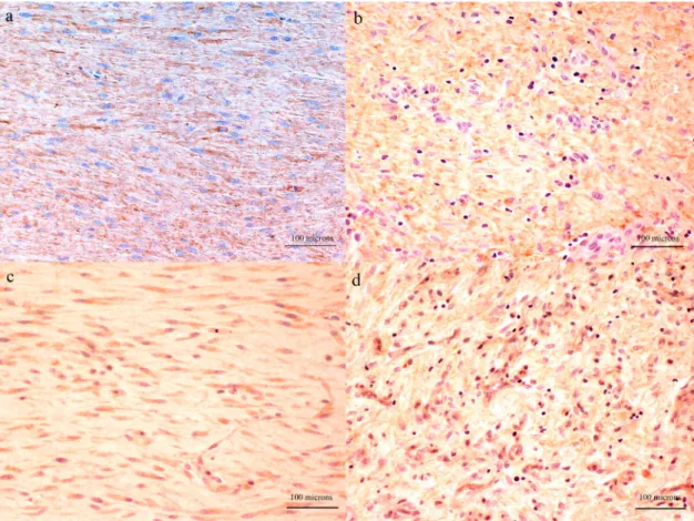
(6) JOURNAL OF APPLIED ANIMAL RESEARCH. 475. Figure 1. Representative histological sections stained with HE (a and b) and MT (c and d), from surgically severed Achilles tendons in sheep, treated with PRGF (a and c) or saline solution (b and d). Inflammatory cells infiltration was clearly higher in the saline group (arrow) than in the PRGF group.. Thus, PRGF therapy induced a favourable effect at the level of maturation in healing tissues at later stages (Fernandez-Sarmiento et al. 2013). After Achilles tendon damage, an initial inflammatory phase occurs and vasoactive agents and a wide variety of cytokines. are released (Sharma & Maffulli 2006; Hope & Saxby 2007). In this study, capillaries in the histological analysis of normal intact Achilles tendons were nearly inexistent. However, a significant increase in capillary density and diameter was observed in both PRGF and saline groups. According to this finding, Fernandez-Sarmiento et al. (2013) reported no differences in these parameters at 4 weeks of injury. In contrast, at 8 weeks PRGFtreated tendons showed faster vascular regression than saline solution-treated tendons, with a significant decline in vascularization in the PRGF group. This may be indicative of a more. Table 7. Packing and orientation of collagen fibres in normal, saline, and PRGF groups.. Figure 2. Distribution of frequencies of grade of inflammation shown in histological images captured from normal, saline-, and PRGF-treated tendons. The tendons treated with PRGF did not exhibit strong or severe inflammation.. Packing of collagen fibres Percentage of specimens receiving each score Grade 4 Grade 3 Grade 2 Grade 1 Grade 0 Median grade. Normal 100% – – – – 4±0. Saline group – 10.5% 80% 9.5% – 2±2. PRGF group – 2% 83.5% 14.5% – 2±2. 1.00. Orientation of collagen fibres Percentage of specimens receiving each score Grade 4 Grade 3 Grade 2 Grade 1 Grade 0 Median grade. 100% – – – – 4±0. – 9.5% 63.5% 24.5% 2.5% 2±3. – 11.5% 68.5% 19.0% 1.0% 2±3. 1.00. P.
(7) 476. D. AGUILAR-GARCÍA ET AL.. Figure 3. Representative histological sections immunostained for collagen types I (a and b) and III (c and d), from surgically severed Achilles tendons in sheep treated with PRGF (a and c) or saline solution (b and d).. advanced tendon healing status in comparison with the saline group (Fernandez-Sarmiento et al. 2013). Inflammatory response is necessary in tendon repair, although high levels of pro-inflammatory cytokines may result in collateral tissue damage and impair tendon healing (Manning et al. 2014). Thus, modulation of the inflammatory phase is beneficial in the repair of a tendon injury. In our study, normal Achilles tendons had no inflammatory cell infiltration at the histological level. Two weeks after surgically induced injury, Achilles tendons treated with PRGF showed significantly reduced inflammatory cell infiltration than those injected with only saline solution. These data may suggest an anti-inflammatory effect of PRGF on the early stage of Achilles tendon healing. In addition, Fernandez-Sarmiento et al. (2013) described the same tendency at the later stages of healing (4 and 8 weeks after the surgery), with lower inflammatory cell infiltration in Achilles tendons treated with PRGF. It may suggest a role of PRGF on the modulation of the inflammatory phenomenon during the early phase of tendon repair. HGF and TGF-β, growth factors contained in PRGF, have been associated with a significant role in the anti-inflammatory effect in human cartilage and tendon injuries in in vitro experiments (Bendinelli et al. 2010; Zhang et al. 2013). The balance between the positive and negative effects which diminish or inhibit inflammation in tissue repair indicates that fine modulation of the early inflammatory phase of healing may be beneficial to tendon healing (Dakin et al. 2014; Manning et al. 2014). Consequently, the anti-inflammatory effect of PRGF on early stage Achilles tendon healing at 2 weeks may promote. the repair process of the tendon reported at 4 and 8 weeks (Fernandez-Sarmiento et al. 2013). In this way, a moderate supraphysiological concentration of growth factors might accelerate the early healing process, as well as initiate a more advanced stage of tissue repair maturation in surgically severed Achilles tendons. PRP, and particularly PRGF, has been studied in clinical settings to stimulate patellar tendon healing on the donor site in bone–tendon–bone anterior cruciate ligament reconstruction (De Almeida et al. 2012; Seijas et al. 2015). De Almeida et al. (2012) noted that there were less anterior knee pain and shorter recovery times in patients treated with PRP with respect to the control group. It is well known that tissue inflammation is frequently associated with pain, which is of primary concern in clinical practice (Ferreira et al. 1978). The anti-inflammatory effect induced by PRGF may promote local analgesia. Tendons are rich in collagens, the most abundant being type I, constituting 95% of the total collagen (Wang 2006). Intact tendons in our study were formed by the homogeneous formation of tightly packed collagen bundles, and collagen fibres were oriented parallel to the longitudinal tendon axis. Analysis of collagen fibres showed a loss in both packing and orientation after 2 weeks in both the PRGF- and saline-treated animals. In concordance with the present study, Spang et al. (2011) showed no differences in collagen fibres of patellar tendons treated with PRP or saline after 2 weeks of an acute injury in rats. However, PRGF-treated tendons were reported to have significantly more well-organized packing and orientation of collagen fibres than in the saline group at both 4.
(8) JOURNAL OF APPLIED ANIMAL RESEARCH. and 8 weeks (Fernandez-Sarmiento et al. 2013). In our study, tendon healing evaluated at 2 weeks was too immature to perceive remarkable changes in ECM collagen fibre organization between the PRGF and saline groups. An IHC analysis was performed to estimate the amount of collagen types I III present in the healing tendon. Our results showed no differences between the study groups for either collagen type III or I estimation. These results are supported by another study previously performed (Kaux et al. 2012). No differences between PRP and saline groups were detected in the expression level of collagen types I and III measured by RTPCR at 2 weeks after the complete separation of Achilles tendon in rats (Kaux et al. 2012). This result supports that in a precocious stage of tendon healing, PRGF does not trigger a detectable difference in collagen type I or III production. Two limitations may be considered in the present study. First, the study involves just one time-point analysis of the tendon healing process, at 2 weeks after the surgically induced injury. Our group performed this study as a continuation of a previous similar experiment. The goal of the current study was to clarify the role that PRGF plays in the early tendon healing stage. Second, biomechanical testing was not performed to check the functional effect of PRGF on injured Achilles tendons. The fact that we observed no differences in fibroblasts and collagen fibre evaluation between the PRGF and placebo groups suggests that no differences should be detected at the biomechanical level.. 5. Conclusion In conclusion, PRGF did not lead to remarkable differences regarding the morphometric data of fibroblasts, fibroblast density, vascular density and vascular diameter, as well as ECM evaluation. However, the evaluation of the inflammatory status showed reduced inflammatory cell infiltration in severed Achilles tendons treated with PRGF, compared to the tendons treated with saline solution. This may suggest the active role of PRGF in the modulation of inflammatory response in early stage tendon healing. Thus, the anti-inflammatory effect of PRGF on early phase tendon regeneration may help accelerate the early healing process and play a critical role in more advanced stages of tissue repair.. Disclosure statement No potential conflict of interest was reported by the authors.. Funding D. A.-G. was the recipient of a grant from the Ministry of Education, Culture and Sport of Spanish government [FPU Program AP2012-1787].. References Andia I, Sanchez M, Maffulli N. 2010. Tendon healing and platelet-rich plasma therapies. Expert Opin Biol Ther. 10:1415–1426. Anitua E. 2001. The use of plasma-rich growth factors (PRGF) in oral surgery. Pract Proced Aesthet Dent. 13:487–493. Anitua E, Murias-Freijo A, Alkhraisat MH, Orive G. 2015. Clinical, radiographical, and histological outcomes of plasma rich in growth factors in. 477. extraction socket: a randomized controlled clinical trial. Clin Oral Investig. 19:589–600. Anitua E, Prado R, Sánchez M, Orive G. 2012. Platelet-rich plasma: preparation and formulation. Oper Tech Orthop. 22:25–32. Anitua E, Sanchez M, Nurden AT, Nurden P, Orive G, Andia I. 2006. New insights into and novel applications for platelet-rich fibrin therapies. Trends Biotechnol. 24:227–234. Anitua E, Sanchez M, Nurden AT, Zalduendo M, de la Fuente M, Orive G, Azofra J, Andia I. 2006. Autologous fibrin matrices: a potential source of biological mediators that modulate tendon cell activities. J Biomed Mater Res A. 77:285–293. Bendinelli P, Matteucci E, Dogliotti G, Corsi MM, Banfi G, Maroni P, Desiderio MA. 2010. Molecular basis of anti-inflammatory action of platelet-rich plasma on human chondrocytes: mechanisms of NF-kappaB inhibition via HGF. J Cell Physiol. 225:757–766. Butler DL, Juncosa N, Dressler MR. 2004. Functional efficacy of tendon repair processes. Annu Rev Biomed Eng. 6:303–329. Crovace A, Lacitignola L, Francioso E, Rossi G. 2008. Histology and immunohistochemistry study of ovine tendon grafted with cBMSCs and BMMNCs after collagenase-induced tendinitis. Vet Comp Orthop Traumatol. 21:329–336. Dakin SG, Dudhia J, Smith RK. 2014. Resolving an inflammatory concept: the importance of inflammation and resolution in tendinopathy. Vet Immunol Immunopathol. 158:121–127. De Almeida AM, Demange MK, Sobrado MF, Rodrigues MB, Pedrinelli A, Hernandez AJ. 2012. Patellar tendon healing with platelet-rich plasma: a prospective randomized controlled trial. Am J Sports Med. 40:1282– 1288. Fernandez-Sarmiento JA, Dominguez JM, Granados MM, Morgaz J, Navarrete R, Carrillo JM, Gomez-Villamandos RJ, Munoz-Rascon P, Martin de Las Mulas J, Millan Y, et al. 2013. Histological study of the influence of plasma rich in growth factors (PRGF) on the healing of divided Achilles tendons in sheep. J Bone Joint Surg Am. 95:246–255. Ferreira SH, Nakamura M, de Abreu Castro MS. 1978. The hyperalgesic effects of prostacyclin and prostaglandin E2. Prostaglandins. 16:31–37. Hope M, Saxby TS. 2007. Tendon healing. Foot Ankle Clin. 12:553–567. James R, Kesturu G, Balian G, Chhabra AB. 2008. Tendon: biology, biomechanics, repair, growth factors, and evolving treatment options. J Hand Surg Am. 33:102–112. Jarvinen TA, Kannus P, Maffulli N, Khan KM. 2005. Achilles tendon disorders: etiology and epidemiology. Foot Ankle Clin. 10:255–266. Kaux JF, Drion PV, Colige A, Pascon F, Libertiaux V, Hoffmann A, Janssen L, Heyers A, Nusgens BV, Le Goff C, et al. 2012. Effects of platelet-rich plasma (PRP) on the healing of Achilles tendons of rats. Wound Repair Regen. 20:748–756. Lane JG, Healey RM, Chase DC, Amiel D. 2013. Use of platelet-rich plasma to enhance tendon function and cellularity. Am J Orthop (Belle Mead NJ). 42:209–214. Lopez-Najera D, Rubio-Zaragoza M, Sopena-Juncosa JJ, Alentorn-Geli E, Cugat-Bertomeu R, Fernandez-Sarmiento JA, Dominguez-Perez JM, Garcia-Balletbo M, Primo-Capella VJ, Carrillo-Poveda JM. 2016. Effects of plasma rich in growth factors (PRGF) on biomechanical properties of Achilles tendon repair. Knee Surg Sports Traumatol Arthrosc. 24:3997– 4004. Lyras DN, Kazakos K, Verettas D, Botaitis S, Agrogiannis G, Kokka A, Pitiakoudis M, Kotzakaris A. 2009. The effect of platelet-rich plasma gel in the early phase of patellar tendon healing. Arch Orthop Trauma Surg. 129:1577–1582. Lyras DN, Kazakos K, Verettas D, Polychronidis A, Tryfonidis M, Botaitis S, Agrogiannis G, Simopoulos C, Kokka A, Patsouris E. 2009. The influence of platelet-rich plasma on angiogenesis during the early phase of tendon healing. Foot Ankle Int. 30:1101–1106. Manning CN, Havlioglu N, Knutsen E, Sakiyama-Elbert SE, Silva MJ, Thomopoulos S, Gelberman RH. 2014. The early inflammatory response after flexor tendon healing: a gene expression and histological analysis. J Orthop Res. 32:645–652. Manning CN, Kim HM, Sakiyama-Elbert S, Galatz LM, Havlioglu N, Thomopoulos S. 2011. Sustained delivery of transforming growth factor beta three enhances tendon-to-bone healing in a rat model. J Orthop Res. 29:1099–1105..
(9) 478. D. AGUILAR-GARCÍA ET AL.. Marsolais D, Cote CH, Frenette J. 2001. Neutrophils and macrophages accumulate sequentially following Achilles tendon injury. J Orthop Res. 19:1203–1209. Mazzocca AD, McCarthy MB, Intravia J, Beitzel K, Apostolakos J, Cote MP, Bradley J, Arciero RA. 2013. An in vitro evaluation of the anti-inflammatory effects of platelet-rich plasma, ketorolac, and methylprednisolone. Arthroscopy. 29:675–683. Platt MA. 2005. Tendon repair and healing. Clin Podiatr Med Surg. 22:553– 560. Sanchez M, Anitua E, Azofra J, Andia I, Padilla S, Mujika I. 2007. Comparison of surgically repaired Achilles tendon tears using platelet-rich fibrin matrices. Am J Sports Med. 35:245–251. Seijas R, Rius M, Ares O, Garcia-Balletbo M, Serra I, Cugat R. 2015. Healing of donor site in bone-tendon-bone ACL reconstruction accelerated with plasma rich in growth factors: a randomized clinical trial. Knee Surg Sports Traumatol Arthrosc. 23:991–997.. Sharma P, Maffulli N. 2006. Biology of tendon injury: healing, modeling and remodeling. J Musculoskelet Neuronal Interact. 6:181–190. Spang JT, Tischer T, Salzmann GM, Winkler T, Burgkart R, Wexel G, Imhoff AB. 2011. Platelet concentrate vs. saline in a rat patellar tendon healing model. Knee Surg Sports Traumatol Arthrosc. 19:495–502. Vilar JM, Damiá E, Rubio M, Santana A, Sopena J, Ceron J, Tvarijonaviciute A, Cugat R, Carrillo JM. 2017. Therapeutic doses of plasma rich in growth factors cannot provoke cancer by means of the IGF-1 pathway or inflammation in dogs. J Appl An Res. 45:490–493. Wang JH. 2006. Mechanobiology of tendon. J Biomech. 39:1563–1582. Yoshioka T, Kanamori A, Washio T, Aoto K, Uemura K, Sakane M, Ochiai N. 2013. The effects of plasma rich in growth factors (PRGF-Endoret) on healing of medial collateral ligament of the knee. Knee Surg Sports Traumatol Arthrosc. 21:1763–1769. Zhang J, Middleton KK, Fu FH, Im HJ, Wang JH. 2013. HGF mediates the antiinflammatory effects of PRP on injured tendons. PLoS One. 8:e67303..
(10)
Figure

Documento similar
Plotinus draws on Plato’s Symposium (206c4–5) – “procreate in what is beautiful” – 3 in order to affirm that mixed love (which is also a love of beauty) is fecund, although
Carmignani(2003) found that the effect of fragmentation on growth is negative. In this study, the relation between political instability and economic growth in countries will
In the “big picture” perspective of the recent years that we have described in Brazil, Spain, Portugal and Puerto Rico there are some similarities and important differences,
Female genital mutilation, daughter- and wife-beating, child and arranged marriage, polygamy, purdah, easy divorce for men, female sexual and domestic slavery, veiling, routine rape
In the preparation of this report, the Venice Commission has relied on the comments of its rapporteurs; its recently adopted Report on Respect for Democracy, Human Rights and the Rule
The draft amendments do not operate any more a distinction between different states of emergency; they repeal articles 120, 121and 122 and make it possible for the President to
Abstract: Transepidermal water-loss (TEWL), stratum-corneum hydration (SCH), erythema, elas- ticity, pH and melanin, are parameters of the epidermal barrier function and
Inclusion criteria were: 2) acute rupture of the Achilles tendon, 2) closed and complete rupture of the Achilles tendon, 3) rupture between the Achilles tendon distal 2
