Identification and evaluation of genes involved in seed development and seedlessness phenotype in Vitis viniera L
163
0
0
Texto completo
(2) IDENTIFICACIÓN Y EVALUACIÓN DE GENES INVOLUCRADOS EN EL DESARROLLO DE LA SEMILLA Y EL FENOTIPO DE APIRENIA EN Vitis vinifera L.. Tesis entregada a la Pontificia Universidad Católica de Chile en cumplimiento parcial de los requisitos para optar al grado de Doctor en Ciencias Biológicas con mención en Genética Molecular y Microbiología.. Por EVELYN ANDREA POBLETE REY. Director de tesis: Dr. Patricio Arce-Johnson Co- director de tesis: Dra. Carmen Espinoza Cancino Julio, 2019.
(3) To my parents and brothers, for their unconditional love and support. To my nieces for always cheering me up and gave me the best moments. To my partner for accompanying me at every moment of this adventure..
(4) 1. ACKOWLEDGMENTS. Thanks to the thesis advisor, Dr. Patricio Arce- Johnson for receiving me at his laboratory and for their supervision of this thesis during all these years. To Dr. Carmen Espinoza, co- tutor of this investigation, for their advice and critical correction of my thesis. I am also grateful to Dr. Beth Krizek, for the constant concern during my internship at the University of South Carolina. I would like to express my gratitude to all the members of the “Laboratorio de Biología Molecular y Biotecnología Vegetal”, especially to my friends Daniela Muñoz, Claudia Santibañez, Litsy Martínez, Valeria Borjas, Francisca Parada and Brenda Riquelme, for their advice and help, for the company during this period and for encouraging me in the most challenging moments. Thanks to SINLÍMITES family, for supporting me in the final stage of this thesis, for the joy and optimism. I especially want to thank my friend Pía Kochifas, for helping me during the collection of vegetal samples at Curacaví. This was an essential part of my thesis. Infinite thanks to Enyel, for accompanying and encourage me, even in the most bitter moments. Also, for the beautiful moments that we will continue building together. And most importantly, to my parents, for always supporting my decisions and believe in me..
(5) 2. This work was supported by Scholarship of Comisión Nacional de Investigación Científica y Tecnológica (CONICYT) No 21120262, Operational Expenses of CONICYT year 2014 and 2015, No 21120262, Fondecyt 1150220 and Genetic Improvement Program: PMG Vides CORFO 13 CTI 18862.
(6) 3. TABLE OF CONTENTS ABBREVIATIONS ................................................................................................................... 9 RESUMEN .............................................................................................................................. 11 ABSTRACT............................................................................................................................. 14 CHAPTER 1 ............................................................................................................................ 17 GENERAL INTRODUCTION .............................................................................................. 17 1. 2.. Seed development in angiosperms ................................................................................. 18 Main characteristics of the ovule and its development .................................................. 21 2.1. Ovule characteristics and developmental stages during ovule formation ............. 21 2.2. Genes involved in ovule development ..................................................................... 24 3. Vitis vinifera as a study model ....................................................................................... 27 3.1. Morphological characteristics of grapevine reproductive organs ......................... 28 3.2. Seed and berry development ................................................................................... 30 3.3 Seedlessness phenotype .......................................................................................... 33 4. Expression profile to identify candidate genes involved in the formation of the seed and seedlessness in Vitis vinifera ................................................................................................. 37 4.1. VvAGL6 .................................................................................................................. 39 4.2. VvAIL5 ................................................................................................................... 40 4.3. VvLUG .................................................................................................................... 43 4.4. VvINO ..................................................................................................................... 44 CHAPTER 2 ............................................................................................................................ 48 PROBLEM STATEMENT: HYPOTHESIS AND OBJECTIVES .................................... 48 1. 2.. Hypothesis ..................................................................................................................... 49 Objectives ...................................................................................................................... 49. CHAPTER 3 ............................................................................................................................ 50 MATERIALS .......................................................................................................................... 50 1.. Biological Material ........................................................................................................ 51 1.1. Plant material ......................................................................................................... 51 1.2. Bacterial strains ..................................................................................................... 52 1.3. Plasmids ................................................................................................................. 52 2. Culture media ................................................................................................................. 54.
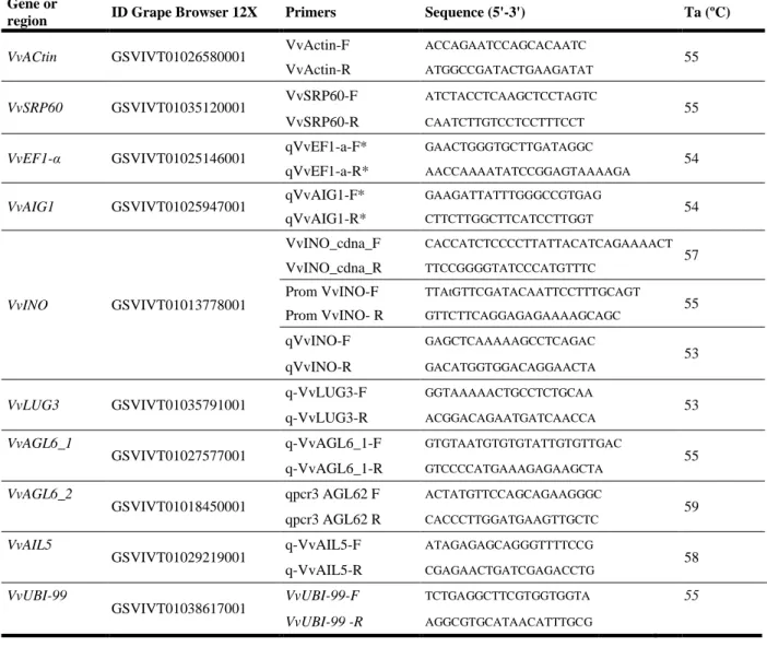
(7) 4. 2.1. Culture media for plants ......................................................................................... 54 2.2. Culture media for bacteria ..................................................................................... 54 3. General reagents ............................................................................................................ 55 3.1. Enzymes .................................................................................................................. 55 3.2. Commercial Kits ..................................................................................................... 55 3.3. General Reagents ................................................................................................... 55 3.4. Oligonucleotides ..................................................................................................... 57 4. Equipment ...................................................................................................................... 58 5. Web sites, databases and softwares ............................................................................... 59 CHAPTER 4 ............................................................................................................................ 60 METHODS .............................................................................................................................. 60 1.. Collection of Plant Material ........................................................................................... 61 1.1. Sampling of Vitis vinifera ....................................................................................... 61 1.2. Sampling of Arabidopsis thaliana .......................................................................... 63 2. Nucleic acid extraction, cDNA synthesis and gene expression analysis ....................... 63 2.1. DNA extraction ....................................................................................................... 63 2.2. RNA isolation, cDNA synthesis and q RT- PCR ..................................................... 64 3. DNA recombinant techniques ........................................................................................ 67 3.1. DNA fragments amplification by Polymerase chain reaction (PCR) ..................... 67 3.2. DNA electrophoresis in agarose and acrylamide geles ......................................... 68 3.3. Extraction and sequencing of plasmidial DNA ...................................................... 69 3.4. Binary vector constructions .................................................................................... 69 3.5. Transformation of competent cells of Rhizobium radiobacter ............................... 70 4. Transformation of Arabidopsis Plants ........................................................................... 70 4.1. Transformation of Arabidopsis using Floral Dip ................................................... 70 4.2. Selection of transgenic plants ................................................................................. 71 5. Genotyping of ino– 1 mutants using ‘Derived Cleaved Amplified Polymorphic Sequences’............................................................................................................................. 72 6. Phenotypic analysis of Arabidopsis plants .................................................................... 73 7. Statistical analysis .......................................................................................................... 73 8. In silico analysis............................................................................................................. 75 CHAPTER 5 ............................................................................................................................ 76 RESULTS ................................................................................................................................ 76 1. Correlate the seeded and seedless phenotype and the expression of the selected genes in the early stages of the development of the grape berries ...................................................... 77 1.1. Determine the expression profile of the candidate genes during the early berry development, in different varieties of table grapes ........................................................... 80.
(8) 5. 1.2. Compare the accumulation of transcripts in grapevine plants with seedless and seeded phenotype, during early developmental stages...................................................... 91 2. To functionally evaluate the participation of one selected gene in the formation of the seeds in the model plant Arabidopsis thaliana. .................................................................. 103 2.1. Isolation and in silico characterization of VvINO ............................................... 103 2.2. Functional evaluation of VvINO through ectopic expression in Arabidopsis thaliana ............................................................................................................................ 108 CHAPTER 6 .......................................................................................................................... 121 DISCUSSION ........................................................................................................................ 122 CHAPTER 7 .......................................................................................................................... 136 CONCLUSIONS ................................................................................................................... 137 ANNEXES ............................................................................................................................. 140 BIBLIOGRAPHY ................................................................................................................. 148.
(9) 6. LIST OF FIGURES. FIGURE 1. Arabidopsis female reproductive organs. .............................................................. 19 FIGURE 2. Schematic representation of female gametophyte development in Arabidopsis. .. 23 FIGURE 3. Principal stages of carpel and ovule development and genes identified. .............. 26 FIGURE 4. Flower, ovule and seed of grapevine. .................................................................... 29 FIGURE 5. Flower, berry and seed development in Vitis vinifera. .......................................... 31 FIGURE 6. Profile expression of candidate genes in different tissues. .................................... 42 FIGURE 7. Phenological stages of Vitis vinifera used for this study ....................................... 62 FIGURE 8. Phenotypic Evaluation of Arabidopsis ovules and seeds. ..................................... 74 FIGURE 9. Characteristics of table grape varieties using in this study. .................................. 81 FIGURE 10. Relative quantification of VvAGL6_1 and VvAGL6_2 transcripts during early stages of flower and berry development. ........................................................................... 84 FIGURE 11. Relative quantification of VvAIL5 transcripts during early stages of flower and berry development. ............................................................................................................ 86 FIGURE 12. Relative quantification of VvLUG transcripts during early stages of flower and berry development. ............................................................................................................ 88 FIGURE 13.Relative quantification of VvINO transcripts during early stages of flower and berry development. ..................................................................................................................... 90 FIGURE 14. Phenotype of segregating lines from Moscatel rosada x Flame seedless. ........... 92 FIGURE 15. Comparison of the VvAGL6_1 transcripts accumulation in grapevine plants with seeded and seedless phenotype. ......................................................................................... 94 FIGURE 16. Comparison of the VvAGL6_2 transcripts accumulation in grapevine plants with seeded and seedless phenotype. ......................................................................................... 95 FIGURE 17. Comparison of the VvAIL5 transcripts accumulation in grapevine plants with seeded and seedless phenotype .......................................................................................... 97.
(10) 7. FIGURE 18. Comparison of the VvLUG transcripts accumulation in grapevine plants with seeded and seedless phenotype. ......................................................................................... 99 FIGURE 19. Comparison of the VvINO transcripts accumulation in grapevine plants with seeded and seedless phenotype.................................................................................................... 101 FIGURE 20. VvINO amplification and final P35S::VvINO construction. ............................. 105 FIGURE 21. Diagram of plant cis-acting regulatory DNA elements in VvINO Promoter. .... 107 FIGURE 22. Genotypification of ino-1 mutants. ................................................................... 109 FIGURE 23. Vegetative growth of ino-1 -/+ and wild type plants. ....................................... 110 FIGURE 24. Silique size differences in ino-1+/- mutants and wilt type plants. .................... 112 FIGURE 25. Phenotypic evaluation of ino-1 mutants. ........................................................... 114 FIGURE 26. Selection and expression analysis of P35S::VvINO Arabidopsis lines. ............ 116 FIGURE 27. Silique size in P35S::VvINO and wild type Arabidopsis plants. ...................... 118 FIGURE 28. Phenotypic evaluation of P35S::VvINO Arabidopsis lines ............................... 120 FIGURE 29. Representation of VvINO and VvAIL5 expression profiles. .............................. 128 FIGURE 30. PK7FWG2 vector used for ectopic expression in Arabidopsis plants. ............. 145 FIGURE 31. Aligning between the isolated VvAGL6_1 and the predicted sequence. ........... 146 FIGURE 32. Aligning between the isolated VvINO and the predicted sequence. .................. 147.
(11) 8. LIST OF TABLES. TABLE 1. Vectors used in this work. ................................................................................... 53 TABLE 2. Oligonucleotides used for PCR and RT-qPCR. .................................................. 65 TABLE 3. Characteristics of candidate genes. ..................................................................... 78 TABLE 4. Predicted functions of candidate genes. .............................................................. 79 TABLE 5. Plant cis-acting regulatory DNA elements in VvINO Promoter ....................... 141.
(12) 9. ABBREVIATIONS. A. :. Auxin. AG. :. AGAMOUS. AGL13. :. AGAMOUS LIKE 13. AGL6. :. AGAMOUS LIKE 6. AIL5. :. AINTEGUMENTA LIKE 5. ANT. :. AINTEGUMENTA. ATS. :. ABERRANT TESTA SHAPE. Bp. :. Base pairs. C. :. Cytokinins. CTAB. :. Cetyltrimethylammonium bromide. cDNA. :. Complementary Deoxyribonucleic acid.. CDS. :. Coding sequence. Col-0. :. Columbia ecotype.. CRC. :. CRABS CLAW. CUC. :. CUP- SHAPED COTYLEDON 1. Cv. :. cultivar. dCAPS. :. Derived cleaved amplified polymorphic sequences. DNA. :. Deoxyribonucleic acid. FS. :. Flame seedless. i.i.. :. inner integument. INO. :. INNER NO OUTER. LB. :. Luria-Bertani. Ler-0. :. Landsberg erecta ecotype. LG. :. Linkage group.
(13) 10. LUG. :. LEUNIG. MMC. :. Megaspore mother cell. mRNA. :. messenger Ribonucleic acid. MS. :. Murashige and Skoog media.. o.i.. :. outer integument.. PCR. :. Polymerase chain reaction. qRT-PCR :. quantitative Real time PCR. QTL. :. Quantitative trait locus. RG. :. Red globe. RNA. :. Ribonucleic acid. SHP1. :. SHATTERPROOF1. SHP2. :. SHATTERPROOF2. SIN. :. SHORT INTEGUMENTS. SPT. :. SPATULA. STK. :. SEED-STICK. SUP. :. INNER NO OUTER. TS. :. Thompson seedless. Ts. :. Thai seedless.
(14) 11. RESUMEN Las semillas, desarrolladas a partir de los óvulos florales, son estructuras con una importante función biológica, ya que permiten que las plantas puedan generar descendencia. La formación de los óvulos y las semillas, son por lo tanto, procesos complejos en el que participan múltiples genes. La apirenia es un fenotipo caracterizado por la presencia de frutas sin semillas, con muy pocas semillas o con trazas seminales. Esta característica es altamente deseada por los consumidores de fruta. Vitis vinifera L. es uno de los cultivos más importantes económicamente a nivel mundial. Naturalmente, las bayas de vid apirénicas se generan por dos mecanismos, Partenocarpia y Estenoespermocarpia, en los cuales se producen uvas sin semillas o con rudimentos seminales, respectivamente. Ambos mecanismos son el resultado de algún defecto ocurrido en etapas tempranas del desarrollo de la flor o la baya de vid. El desarrollo del óvulo/semilla y el fenotipo de apirenia en V. vinifera, es un proceso que ha sido escasamente estudiado. Se cree que al igual que en otras especies como Arabidopsis thaliana, este es un proceso complejo en el cual participarían múltiples genes. A la fecha sólo han sido sugerido algunos genes candidatos , de los cuales sólo 3 han sido caracterizados..
(15) 12. Previamente, se realizó un análisis transcripcional masivo en nuestro laboratorio, utilizando tejido de flores y bayas jóvenes de vid, el cual permitió identificar genes candidatos que presentaron perfiles de expresión diferencial entre plantas con bayas semilladas y apirénicas. En este contexto, la investigación realizada en esta tesis, consistió en caracterizar cuantitativamente el perfil de expresión de los genes candidatos que codifican para factores de transcripción putativos como VvAGL6_1, VvAGL6_2, VvAIL5, VvINO, o el correpresor transcripcional VvLUG, en el desarrollo temprano de la baya de vid y posteriormente, en determinar si alguno de estos genes afecta la formación de la semilla en la planta modelo Arabidopsis thaliana. El estudio realizado permitió la identificación de dos genes, VvAIL5 y VvINO, los cuales presentan una acumulación diferencial de transcritos entre individuos con fenotipo semillado/apirénico. En flores pre-polinizadas ambos genes exhibieron una disminución en la cantidad de transcritos en vides apirénicas, con respecto a vides semilladas. Por otra parte, en el estado de frutos en cuaja, sólo el gen VvINO exhibió disminución de los niveles de transcrito en bayas apirénicas en comparación a bayas semilladas. Estos resultados sugieren que existe una correlación entre una menor expresión de VvAIL5 y VvINO y el desarrollo de bayas apirénicas, y a su vez, que ambos genes podrían contribuir a la formación de la semilla, participando en diferentes etapas del desarrollo de la flor y la baya. Posteriormente, se seleccionó y evaluó la funcionalidad del gen VvINO, mediante expresión ectópica en la planta modelo Arabidopsis thaliana. A través del análisis in silico de la secuencia genómica de VvINO, se identificó múltiples elementos regulatorios en la región promotora que sugieren una interacción de este gen con otros factores de transcripción, así como regulaciones.
(16) 13. a nivel hormonal. Finalmente, se obtuvieron plantas de Arabidopsis sobreexpresoras de VvINO, las cuales presentaron silicuas más grandes y con un mayor número de semillas en comparación a las wild type. Estos resultados, en conjunto con trabajos previamente publicados, permiten sugerir que VvINO afecta la formación del primordio del óvulo y por lo tanto, influye en el número total de las semillas en frutos maduros de Arabidopsis thaliana. Los resultados generados en el marco de esta investigación constituyen una primera aproximación al estudio de genes de Vitis vinifera, candidatos a participar en el desarrollo reproductivo de la semilla, tema que hasta el momento ha sido escasamente estudiado. Así, este trabajo deja abiertas nuevas interrogantes para profundizar en el estudio de estos genes a futuro, lo que sin duda contribuirá a lograr una comprensión más acabada sobre los mecanismos moleculares y genéticos involucrados en la formación de la semilla en vid..
(17) 14. ABSTRACT The seeds, developed from floral ovules, are structures with an essential biological role since they allow plants to generate offspring. The formation of ovules and seeds is, therefore, a complex process in which multiple genes participate. Seedlessness is a phenotype characterized by the presence of fruits without seeds, with very few seeds or with seminal traces. This feature is highly desired by fruit consumers. Vitis vinifera L. is one of the most economically important crops worldwide. In nature, seedless grapevine berries can be obtained by two mechanisms, Parthenocarpy and Stenospermocarpy, which generate berries completely seedless or with seminal rudiments, respectively. Both mechanisms are the consequence of a defect occurred in early reproductive stages across flower or berry development. The ovule/seed development and seedlessness phenotype in grapevine is a process that has been poorly studied. It is believed that in other species such as Arabidopsis thaliana, this is a complex process in which multiple genes could participate, which some candidate genes have been suggested and only 3 of them have been characterized. Previously, a massive transcriptional analysis was carried out in our laboratory, using tissue from young flowers and berries of grapevine, which allowed to identify candidate genes that presented differential expression profiles between plants with seed and seedless berries. In this context, the research carried out in this thesis, consisted first of quantitatively characterizing the.
(18) 15. expression profile of the candidate genes that code for putative transcription factors such as VvAGL6_1, VvAGL6_2, VvAIL5, VvINO, or the transcriptional corepressor VvLUG, in the early development of the grapevine. Subsequently, the research focused on determining if any of these genes affect the formation of the seed in the model plant Arabidopsis thaliana. The study allowed the identification of two genes, VvAIL5 and VvINO, which present a differential accumulation of transcripts between individuals with seed/seedlessness phenotype. In pre-pollinated flowers, both genes exhibited a decrease in the accumulation of transcripts in seedless grapevine, concerning seed-bearing plants. On the other hand, in fruit set, only VvINO presented decreased transcript levels in seedless berries compared to seed fruits. These results suggest that there is a correlation between a lower expression of VvAIL5 and VvINO, and the development of seedless berries, and in turn, that both genes could contribute to the formation of the seed, participating in different stages of flower and berry development. Subsequently, VvINO was selected and functionality evaluated by ectopic expression in the model plant Arabidopsis thaliana. Through in silico analysis of VvINO, multiple putative regulatory elements were identified in the promoter region that suggest an interaction of this gene with other transcription factors, as well as hormonal regulations. Finally, Arabidopsis VvINO over-expressers lines were obtained, which presented larger siliques with a significantly higher number of seeds in comparison to wild type. These results, together with previously published studies, suggest that VvINO affects the formation of the primordium of the ovule and, therefore, influences the total number of seeds in mature Arabidopsis fruits. The results generated in this investigation constitute a first approach about the study of genes of Vitis vinifera, candidates to participate in the reproductive development of the seed, a subject.
(19) 16. that until now has been scarcely studied. Thus, this work opens new questions to deepen the study of these genes in the future, which will contribute to a better understanding of the molecular and genetic mechanisms involved in seed development in grapevine..
(20) 17. CHAPTER 1. GENERAL INTRODUCTION.
(21) 18. 1. Seed development in angiosperms. Seeds are fundamental part of the plant life cycle, as they contain the genetic information necessary for the next generation of plants and allow to produce offspring maintaining the species (Nonogaki, 2014). Also, seeds play key roles in agricultural production and crop improvement, since they constitute a simple and effective means of crop propagation, maintaining and transmitting genetic engineered enhancements. In addition, seeds are an important component of the global diet in the form of cereal grains (Bewley, 1997). Despite the critical role that seeds play, seedless fruits have naturally emerged (Varoquaux et al., 2000). In flowering plants or ‘Angiosperms’- the largest group of the Plantae Kingdom- the seeds are developed inside the ovary, the reproductive tissue of the flower. Therefore, flower development is a crucial process for a proper seed development. Morphologically, flowers have two kind of reproductive organs, gynoecium constitute the female organ while stamens are the male organs. The gynoecium is defined as the collective of all carpels that determine the fruit formation (Robinson-Beers et al., 1992b). It includes the stigma were pollen germination initiates, the style in which the pollen tube elongation occurs, and the ovary, the ovules containing structure (Figure 1. A and B). Many efforts have been made to understand flower development since most of the reproductive plant cycle takes place here. The process has been mostly studied in Arabidopsis thaliana, a core eudicot with a gynoecium including two laterals carpels fused (Figure 1. A) (Pfannebecker et al., 2016)..
(22) 19. FIGURE 1. Arabidopsis female reproductive organs. A) Schematic representation of the Arabidopsis gynoecium, showing stigma, style, ovary and ovules. B) Longitudinal diagram of Arabidopsis flowers. C) and D) Mature Ovule of Arabidopsis structures: inner integument (ii), outer integument (oi), mycropilar (mp), egg cell nucleus (ecn), synergid cells nuclei (sc), antypodal cells (ap) and central cell nucleus (ccn). The figures A and B ere adapted from Hawkins et al, 2014 and Schmid, 2012. C and D were adapted from Sprunck et al, 2011..
(23) 20. During floral development, organs are initiated in flower primordia at particular positions within concentric rings called ‘whorls.’ After organ initiation, those primordia become different floral organs, depending on the expression of distinct combinations of floral identity genes. This model was named ABC(DE), and most of the genes belong to the MADS-box transcription factors family, that function as quaternary complexes regulating different target genes (Immink et al., 2010). According to this model, all the genes control the formation of floral organs in a highly regulated manner at a spatial and temporal level. In other words, their gene expression occurs in specific cell types and during precise moments to grant normal flower development. The ABC(DE) model also states that class D genes are ovule-specific and, class C, D, and E specify ovule identity in whorl five at early stages of ovule development (Dreni and Zhang, 2016; Krizek, 2015).. Inside the floral carpels, ovules are formed. They are vital organs that enclose the female gametophyte and also develop into seeds after fertilization (Shi and Yang, 2011). In angiosperms, double fertilization occurs after ovary becomes a fruit (Chevalier et al., 2011). Then, the seeds play the role of propagating the species and the fruit has the important function of protecting seeds, helping in their dispersal. A developing seed contains three genetically distinct structures (Figueiredo and Köhler, 2018): the embryo, that has the potential to germinate and become a new organism (Chevalier et al., 2011), the endosperm, a nourishing tissue, and the seed coat, a maternal sporophytic tissue originated from the ovule integument(s) are cell layers that surround the seed, helping to protect it and facilitating the dispersal (Coen et al., 2017)..
(24) 21. In general terms, the development of the fruit and the seed are synchronized processes, however, in some cases, the growth of the fruit is decoupled from the fertilization and development of the seed as indicated by the existence of seedless mutants and crops (Pandolfini, 2009).. 2. Main characteristics of the ovule and its development 2.1. Ovule characteristics and developmental stages during ovule formation Ovule is the plant reproductive organ that develops into seeds after fertilization. In consequence, defects in ovule’s development generally result in female sterility, affecting seed formation and offspring propagation. Hence the importance of deeply study ovule’s formation.. Thus, ovule is the site where processes essential for sexual plant reproduction occur: megasporogenesis, megagametogenesis, fertilization and embryogenesis (Losa et al., 2010). This organ is composed by different sporophytic tissues: the funiculus: is the proximal region, which transports nutrients from the placental tissue to the ovule (Losa et al., 2010; Modrusan et al., 1994), the chalaza: which initiates two integuments, which are cellular layers enveloping the nucellus and the nucellus, within which the embryo sac or female gametophyte is developed, after megasporogenesis and megagametogenesis occur (Losa et al., 2010; RobinsonBeers et al., 1992b) (Figure 1. C and D)..
(25) 22. Ovule´s development has been a complex process to study, due to both the small size and the difficulty of accessing inside the carpel. However, Schneitz and collaborators were able to propose a classification of four different stages of ovule development throw sectioning and microscopy techniques (Schneitz, K. Hülskamp, M. Pruitt, 1995). They identified four ovule development main stages, according to cellular characteristics. Also, Christensen and coworkers (1997), proposed a classification of female gametophyte developmental stages (Figure 2). In general terms, ovule’s development includes four stages. The first is the Early phase (I), when the ovule primordia arises from the regions near to the carpel margins and at the end of this phase, they have recognizable pattern elements which will subsequently result in the nucellus, chalaza (flanked by initiating integuments) and the funiculus that will develop the vascular strand (Simon et al., 2017b). The second phase is called Megasporogenesis (II). Here, the megaspore mother cell (mmc) located in the nucellus, suffers two meiosis, a tetrad with four megaspores is formed, three of them will degenerated leaving one functional megaspora, this is also the Female gametophyte stage 1 (FG1) (Figure 2. C), Also, two integuments are initiated at this stage, arising from the chalaza (Simon et al., 2017b). The next phase is named Megagametogenesis (III), where resulting megaspore suffers three mitotic divisions, designated as stages FG2 to FG5 (Christensen et al., 1997) (Figure 2. D and E). As a result of this phase the mature embryo sac is formed, containing the egg cell, which gives rise to the embryo; the central cell, which give rise to endosperm and two accessory cells named synergid cells (Martin et al., 2014; Sundaresan and Alandete-Saez, 2010). Also the integuments development proceed during this stage (Schneitz, K. Hülskamp, M. Pruitt, 1995). Therefore, at the end of the stage, the unfertilized mature ovule consists in a haploid embryo sac.
(26) 23. FIGURE 2. Schematic representation of female gametophyte development in Arabidopsis. A) The megaspore mother cell (MMC) is surrounded by epidermal cells of the nucellus (Nu) prior to undergoing meiosis to generate four megaspores. At this stage, the formation of the outer (OIn) and inner (IIn) integuments has just initiated. B) MMC suffers two meiosis that generates four spores (tetrad). Three of these undergo programmed cell death. The proximal (chalazal) megaspore becomes the functional megaspore (FM). C) FG1 stage. The FM undergoes the first mitotic division. D) FG2 stage. The female gametophyte comprises two nuclei. The nucellus (Nu) is enclosed by the OIn, but not the IIn integuments. E) Stages FG3 to FG7. The female gametophyte comprises two nuclei, separated by a large vacuole (V), that undergo second and third mitotic divisions to generate the eight-nucleate mature embryo sac at the FG5 stage. Subsequent cellularization (FG6 stage) results in the formation of seven cells: two synergid cells (SC); one egg cell (EC); one central cell (CC) carrying two polar nuclei (PN); and three antipodal cells (AC). By FG7, the two polar nuclei have fused to form the central cell nucleus (CCN), and the antipodal cells degenerate. CN, chalazal nucleus; CV, central vacuole; DM, degenerating megaspores; Fu, funiculus; IIn, inner integuments; MMC, megaspore mother cell; MN, micropylar nucleus; Nu, nucellus; OIn, outer integuments. Figure from Sundaresen et al, 2010..
(27) 24. surrounded by the integuments (Figure 1) (Grossniklaus and Schneitz, 1998).. Finally, there is a Post-fertilization development phase (IV). During fertilization, the pollen tube that contains two sperm cells reaches the mature ovule and leads to form a seed (Chevalier et al., 2011), where the fertilized cell egg develop into a diploid embryo and the central cell became in a triploid endosperm. Also, the integuments develops into the seed coat (Martin et al., 2014) Angiosperms typically have “bitegmic ovules”, with two integuments, which are important ovule’s structures, that contributes to positioning and enable the formation of the female gametophyte. Also during pollination, they participate in the guidance of pollen tube towards female gametophyte in some species (Lora et al., 2018) and after fertilization, the integuments become the seed coat, which have different roles such us protection of the embryo, seed dissemination, and regulation of seed germination (Kelley and Gasser, 2009; Simon et al., 2017b).. 2.2. Genes involved in ovule development The study of ovule and female gametophyte is difficult mainly due to the small size of its structures. To understand the ovule’s development, the main experimental approach used consists in large-scale genetic screens, through insertional mutagens. This allowed to identify single genes affecting ovule’s structures, its fertilization capacity and seed formation (Sundaresan and Alandete-Saez, 2010). Thus, this approach has been useful to identify unique.
(28) 25. genes whose mutations results into a phenotype, but not genes that act redundantly during this developmental process. Another alternative procedure is the comparison of expression profiles between wild type plants and previously identified mutants unable to form ovules. This has allowed to identify several genes that possibly participate in the formation of the embryo sac, many of them still remain without functional characterization (Sundaresan and Alandete-Saez, 2010). In general, functional studies involve analysis of temporal and tissue- specific expression and loss-of-function mutants, complex techniques to perform in agronomic crops, leading to the use of models’ plants like Arabidopsis thaliana.. The mentioned approaches have allowed to identify numerous genes participating in a coordinated way during the ovule formation, most of them are transcriptions factors, summarized in Figure 3 (Baker et al., 1997; Cucinotta et al., 2014; Gaiser et al., 1995; Villanueva et al., 1999b). Defects in most of these genes result in pleiotropic effects in flowers, defective ovules or both (Baker et al., 1997; Gaiser et al., 1995; Gallagher and Gasser, 2008; Kelley et al., 2009). For example, lug- mutants exhibit a markedly decreased number of ovules (Cucinotta et al., 2014), ant- mutants have sterile ovules and a reduction in size and number of floral organs (Kelley and Gasser, 2009), in ag- loss function mutants, the carpels are replaced by new flowers (flowers inside flowers) (Franks et al., 2002) and in the triple mutant stk shp1 shp2, fewer ovules are initiated and ovule development is severely disrupted (Skinner et al., 2004)..
(29) 26. FIGURE 3. Principal stages of carpel and ovule development and genes identified. Some genes associated to different stages are schematized, in the case of transcription factors, their family is described in parentheses. The figure is of own elaboration, using the data of Gaiser and collaborators, 1995; Baker et al., 2007 and Cucinotta et al., 2014..
(30) 27. The formation of the integuments is a crucial stage during the development of the ovule since they contribute to the formation of the embryo sac. Several genes are participating at integument’s establishment (Figure 3), and the mutants in these genes have morphologically abnormal integuments that result in defective ovules. Even more important, most of them, such as ant, bel, sin and ino mutants, present female sterility (Baker et al., 1997; RobinsonBeers et al., 1992b; Villanueva et al., 1999b), an critical defect where ovules are not viable to be fertilized and therefore, incapable of becoming in seeds (Robinson-Beers et al., 1992a). This phenomenon prevents plants from forming offspring, but it is desirable for agronomic crops, in which seedless fruits are highly desired (Varoquaux et al., 2000).. 3. Vitis vinifera as a study model Grapevine (Vitis vinifera) is one of the most economically important horticultural crops worldwide. The three main uses for grapes are dried fruit (raisins), winemaking, and fresh fruit also named ‘table grapes’(Carmona et al., 2008). In Chile, towards 2015 there were more than 211.000 hectares of cultivated vineyards (OIV, 2016), is also our country the more substantial exporter of table grapes worldwide, with 14.4% of the total grapes exported abroad (Workman, 2018). Regarding grapevine as a study plant, there are some advantages, such as that it is a plant whose genome has been sequenced (Jaillon et al., 2007). However, there are some technical difficulties that limit a better understanding of different physiological and molecular processes. For example, there is no availability of a mutant collection of V. vinifera that could support the study.
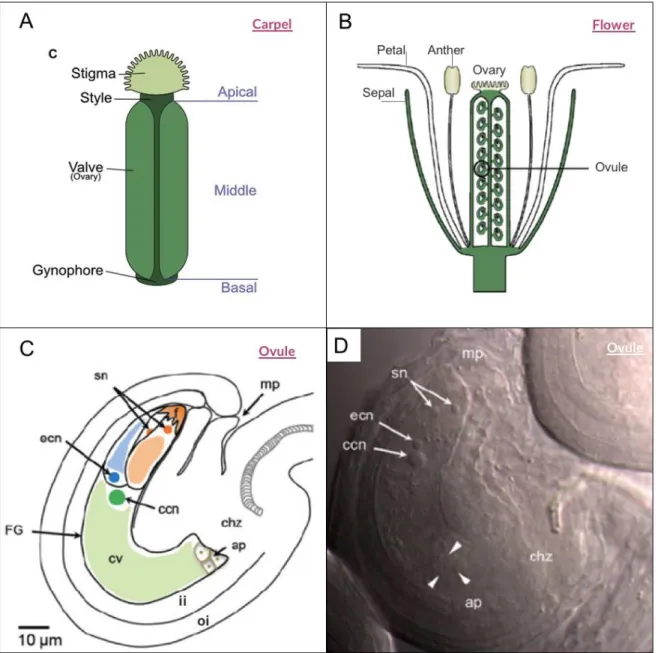
(31) 28. or genes related to ovule and seed development hesitating to establish direct cause-effect relationships. Additionally, transgenesis in grapevine is a difficult process and therefore, the generation of overexpressing or silencing lines in genes of interest is a difficult task (Parada et al., 2017). Thus, usually, the genes of interest are identified and subsequently evaluated in model plants such as Nicotiana benthamiana or Arabidopsis thaliana (Hanania et al., 2007).. 3.1. Morphological characteristics of grapevine reproductive organs Morphologically, the ovary of grapevine flower has two locules, each containing two ovules generally, that will form up to four seeds (Figure 4. A). The anthesis or blooming, when the stamens are able to fertilize the female gametophyte, is a crucial stage because environmental and nutritional conditions at bloom time affect the success of fertilization and the resulting number of seeds per berry. After pollination and fertilization, each ovary develops to form a fleshy fruit called berry (Figure 4. C) (Boss et al., 2001; Poupin et al., 2011). The ovule of the grapevine is ‘anatropous,’ that is, inverted with the micropyle towards the pedicel and with the funiculus (ovule stalk) joined to the outer integument forming a raphe (Figure 4.B). There are two integuments and a well- developed nucellus. The outer integument is joined on one side to the raphe for most of this length but is free at the micropylar end (Mullins et al., 1992). Also, Vitis vinifera has an endostomal micropylar in which the integuments do not.
(32) 29. FIGURE 4. Flower, ovule and seed of grapevine. A) Diagram of a longitudinal section of a mature grapevine flower. B) Longitudinal illustration of an ovule of the grape cultivar Concord at full bloom. C) Diagram of a longitudinal berry of a stenospermocarpic variety D) Longitudinal section of a grapevine seed. A, B and D were adapted from Pratt et al, 1971 and C was adapted from Dokoozlian et al, 2000..
(33) 30. entirely cover the ovule unlike Arabidopsis and therefore, it is presumed Arabidopsis and therefore, it is presumed do not participate in pollen tube attraction (Lora et al., 2011).. Flower formation is a critical process for the development of the resulting berries (Palumbo et al., 2019). As mentioned above, the morphology of the flower and particularly of its ovules are fundamental for the formation of fruits with seeds (Kennedy, 2002). Particularly, abnormal ovules formed during flower development can conduce to seedlessness, through either the abnormal development of the nucellus or ovule integuments, or the degeneration of the egg in the embryo sac (Ebadi et al., 1996).. 3.2. Seed and berry development Grape berry development consists of two successive sigmoidal growth periods separated by a lag phase (Coombe, 1992; Coombe and McCarthy, 2000) (Figure 5). Berry development starts after fertilization, with a process known as fruit set in which the ovary changes from a stationary state and experiences an abrupt increase in size that occurs due to cell division and enlargement, leading to rapid pericarp growth. During this period, seed development plays an important role, mainly because seeds produce auxins, gibberellins (GAs) and cytokinins, which have multiple functions in grape berry development (Keller, 2010). Both processes, seed and berry development are coordinated, and the changes that seeds undergo have an impact on fruit ontogeny (Serrano et al., 2017). In fact, the seeds also have three stages of development (Ristic and Iland, 2005) (Figure 5)..
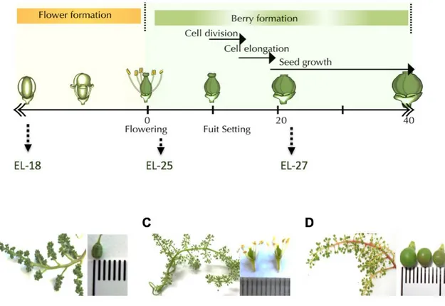
(34) 31. FIGURE 5. Flower, berry and seed development in Vitis vinifera. The upper panel shows the three stages of berry and seed development of grapevine. The low panel shows the flower development before bloom and the early stages of berry development. Figure adapted from Serrano and collaborators, 2017..
(35) 32. 3.2.1. Phase I. During this phase, the berry is formed, mainly by rapid cell division across the first weeks, and the total number of cells is established (Kennedy, 2002). At this period, seeds initiate their growth phase, mostly associated with cell division and differentiation (Ristic and Iland, 2005). Developing embryos produce phytohormones, which are released to the pericarp contributing to berry growth; therefore the pericarp growth correlates with the growth rate of developing seeds (Keller, 2010). Thus, this stage is characterized by a rapid increase in seed size, during which embryogenesis and endosperm growth occur. Towards the end of this stage, small embryos are formed (Conde et al., 2007; Coombe and McCarthy, 2000; Ristic and Iland, 2005; Staudt and Leidel, 1986).. 3.2.2. Phase II The second stage, also named ‘Lag Phase,’ is a short period where the berry growth ceases. The seeds undergo the Transition Phase, was there a small increase in dry weight together with a slight drop in water content occurs. The ‘notch’ or basal end of the seed expand, possibly to allow the accumulation of nutritional reserves. The end of this period coincides with the cessation of seed growth (Ristic and Iland, 2005).. 3.2.3. Phase III The berry faces a new period of growth, mainly due to volume increase. This stage is also called Ripening and starts with ‘veraison,’ a phenomenon in which the grape berry begins to gain color.
(36) 33. and ends around 120 days after flowering (Figure 5) (Costantini et al., 2008a; Kennedy, 2002; Kuhn et al., 2014).. During this period, seeds go through a phase of Drying and Maturation. At the beginning of this stage, the endosperm accumulates reserves until the seeds turn dormant (Keller, 2010; Serrano et al., 2017). Then, after 70 - 100 days post- flowering, the growth of the embryo becomes rapid and reaches its peak. Thereby, the maximum berry weight coincides with the maximum seed dryness and the maximum embryo length (Ebadi et al., 1996; Ristic and Iland, 2005; Staudt and Leidel, 1986). Moreover, during berry ripening the seeds are being prepared to be released into the environment, the drying process of the seed takes place, which is associated with seed coat impermeability and seed dormancy (Ristic and Iland, 2005).. The three phases of the development have been studied in seed varieties; however, naturally, some grapes do not contain seeds or in which these are not fully developed.. 3.3. Seedlessness phenotype. Fruit and seed development are processes that occur in a synchronized manner orchestrated by phytohormones and signals produced by developing embryos, after ovule fertilization (Pandolfini, 2009). In some extraordinary cases, fruit growth occurs independently of the ovule’s fertilization and seed development, as is observed in certain seedless mutant plants belonging to different species (Varoquaux et al., 2000). Seedlessness phenotype is characterized by fruits completely devoid of seeds, fruits containing a minimal number of seeds or presenting traces of aborted seeds (Pandolfini, 2009). A seedless.
(37) 34. fruit can be obtained by parthenocarpy, when fruits are developed without fertilization or by stenospermocarpy, if the seeds are aborted after fertilization, leaving seed traces (Varoquaux et al., 2000).. For fruit consumption, seedlessness has become an important characteristic since in some seedless plants it has been reported better quality and flavor, increase in shelf life, less browning, among other features of interest (Grossniklaus and Schneitz, 1998; Pandolfini, 2009). However, obtaining seedless fruits is difficult at the physiological level, as the developing seeds play a role in the fruit formation during the first days, influencing cell division rate of the surrounding tissues (Gillaspy et al., 1993), and accordingly, the number of seeds affects the final size of the mature fruit (Varoquaux et al., 2000).. In Vitis vinifera, seedless grape varieties have arisen spontaneously in nature and have been preserved over the years through asexual propagation. Seedless berries develop naturally via parthenocarpy or stenospermocarpy, which generate berries without or with rudimentary seeds, respectively (Varoquaux et al., 2000). Interestingly, the frequency of grape’s seedlessness phenotype is variable and does not relate to standard genetic ratios (Cain et al., 1983). Despite the interest generated by this phenotype, there is limited knowledge about genetic mechanisms involved in grapevine seedlessness.. 3.3.1. Parthenocarpy In parthenocarpic fruits, the stimulus of pollination is sufficient to trigger fruit set (Dokoozlian, 2000). Since the ovary is able to enlarge and form a berry without ovule fertilization, there is.
(38) 35. no seed in the fruit (Varoquaux et al., 2000). Until now, few parthenocarpic grape cultivars have been, and they are mostly used as raisins. Therefore, there are few studies focused on this process. In one of these, two somatic variants -with and without seeds- were compared with transcriptomics. The investigators found about 2000 genes differentially expressed in preanthesis flowers, many of them associated with reproductive development were downregulated in seedless grapevine variant (Royo et al., 2016; Vargas et al., 2007). Also, 14 single-nucleotide polymorphisms (SNP) were identified between both genotypes, which could explain the parthenocarpy phenotype (Royo et al., 2016). However, there are no functional studies of the candidate genes, and more studies are needed to characterize the contribution of these genes on the parthenocarpic phenotype.. 3.3.2. Stenospermocarpy During stenospermocarpy, pollination and fertilization occur normally, but a few weeks later, the embryo, the endosperm or both abort and the berries that have been generated possess only seed traces (Varoquaux et al., 2000). It has been demonstrated that stenospermocarpy occurs in several seedless varieties, and is stable and unaffected by environmental factors (Zhang et al., 2013). However, little is known about the molecular mechanisms that underlie stenospermocarpy in grapes. The most accepted hypothesis proposes the existence of a dominant regulator gene called Seed Development Inhibitor (SDI), which could control three other recessive genes (Bouquet and Danglot, 1996). Different studies based on quantitative trait locus (QTL) analysis have reported a main QTL in linkage group 18 (LG18) (Cabezas et al., 2006; Costantini et al., 2008b; Doligez et al., 2002; Mejía et al., 2007), which could explain.
(39) 36. between 50 and 70% of the seedlessness phenotype in grapes; LG18 could be considered as the SDI locus trait. In this context, VvAGL11 (a MADS- BOX transcription factor, homolog to STK, a D-MADS box gene) was in silico mapped to the SDI locus, and it has been proposed as the main functional candidate gene for seedlessness in grapes (Mejía et al., 2011). In fact, it was demonstrated that the silencing of a VvAGL11 homologous gene in tomato (Solanum lycopersicum L. cv Micro-Tom) generates fruits with few or rudimentary seeds (Ocarez and Mejía, 2016) and the ectopic expression of VvAGL11 in stk mutants which have smaller siliques with slow number of seeds, restored the wild type phenotype, confirming a role in seed development (Malabarba et al., 2017). Based on genome sequencing data, it is known that in the stenospermocarpic variety cv. Thompson Seedless, the VvAGL11 gene has an insertion in the 5’UTR, which could be the cause of the seedless phenotype (Di Genova et al., 2014). In addition, in cv. Sultanine Monococco, which is a seeded variety of Thompson, the VvAGL11 transcript level is higher in comparison with the seedless variety (Ocarez and Mejía, 2016) as occurs when comparing seeded cv. Chardonnay with seedless cv. Thompson (Malabarba et al., 2017), supporting the hypothesis that this gene is one of the central regulators of seed formation in grapes. However, this gene is incapable of explaining the seedlessness phenotype totally; thus there must be other genes involved that have not been identified (Mejía et al., 2011).. The use of somatic variants in combination with current transcriptomic technologies has helped to discover new genes playing important roles in seed abortion. As an example, Hanania and collaborators (2007, 2009) compared inflorescences from somatic variants with and without seeds and identify the ch-Cpn21, a gene that encodes for a chloroplastic chaperonin, which is repressed in developing flowers of cv. Thompson Seedless in comparison with the seeded somatic variant. Likewise, the silencing of this gene in tobacco plants (Nicotiana benthamiana).
(40) 37. and tomato induces seed abortion (Hanania et al., 2007). Also, Costenaro-da-Silva et al. (2010) and Nwafor et al. (2014), founded several genes have been associated with early stages of grape berry development. These include transcription factors, ribosomal proteins, genes related to hormone signaling, among others. Many of these genes have putative pleiotropic effects, so it is difficult to estimate their specific molecular contribution to the stenospermocarpy phenotype. Some of them could be involved in this process, but their functional characterization is needed to test this hypothesis (Costenaro-da-Silva et al., 2010; Nwafor et al., 2014).. Until now, it has been challenging to understand the genetic and molecular mechanisms involved in ovule and seed development in Vitis vinifera. It is believed that, as in other species, many genes participate in this process acting in reproductive organs during ovule development at flowers or seed development at fruit formation.. 4.. Expression profile to identify candidate genes involved in the formation of the seed and seedlessness in Vitis vinifera A previous study developed in our laboratory focused on analyzing the differential gene. expression between two phenotypes of segregating plants, seeded and seedless, as a tool to identify novel genes associated with the seedlessness’s phenotype. The plants were obtained from a cross between a seeded maternal line (cv. Moscatel rosada) and a seedless paternal line (cv. Ruby seedless). The study was performed using Affymetrix microarrays, comparing early developmental stages of berry: flower (bloom) and young berries (20 days post bloom). In this study, a total of 4077 differentially expressed genes were identified (Muñoz et al., 2012)..
(41) 38. From the analysis of Affymetrix performed in our laboratory, a series of filters were applied to choose candidate genes. To do this, of the 4077 differentially expressed genes, those with a contrasting expression pattern were selected between plants that generate seedbed berries and rudiments, that is, genes that present an induction or repression between the stages of flower and fruit set in a type of plant, but that remain constant in the other class. This contrast profile is similar to the expression pattern of genes previously associated with seedlessness in grapevine, such as S27A and CPN21 (Hanania et al., 2007, 2009). Approximately 400 genes were found in this condition. Subsequently, these were filtered according to their expression levels, considering only those whose expression change (logarithm in base 2) is higher than ±1, obtaining a list of 148 genes. Within this list, ten genes encoding for putative transcription factors or transcriptional regulators, which were considered in this analysis, due to their importance in the modulation and regulation of gene expression. Finally, genes belonging to families of transcription factors that have been described as important in plant development processes were selected, and genes whose transcripts are accumulated in reproductive tissues linked to the development of the seed in model plants were preferably considered.. Considering this analysis and literature, we select new candidate genes whose expression changes could be responsible for the seedlessness phenotype. Candidate genes are putative transcription factors such as VvAIL5, VvAGL6_1, VvAGL6_2, VvINO, and transcriptional co-repressors like VvLUG..
(42) 39. 4.1. VvAGL6. VvAGL6 encodes a putative MADS-box transcription factor. This gene family have a key role in flower and fruit development in different species (Boss et al., 2002; Roy Choudhury et al., 2012). Within this family, the AGAMOUS like 6 clade includes to AGL6 and AGL13 as members (Hsu et al., 2014) and both genes are located in a genetic block suggesting a duplication event (Schauer et al., 2009).. Studies based on the molecular characterization and mutant analysis in species like petunia (Petunia), rice (Oryza sativa) and maize (Zea mays) suggest a function as E class genes during flower development. However, in order to test their specific functions in flower and ovule development, Arabidopsis null mutants must be generated and characterized (Dreni and Zhang, 2016). The rice OsMADS6 is the most studied AGL6 gene in seed plants and has expression inside the carpel in different tissues as ovule integuments (Ohmori et al., 2009). Strong osmad6 mutant alleles show a partially lost in ovule’s identity within the carpels, leading a female sterility, which suggest a role in ovule development (Li et al., 2011). Also, over-expression of AGL6 homologues from orchid (Oncidium Gower Ramsey) and Hyacinthus orientalis L. in Arabidopsis, reveals a role in flowering timing and transition (Fan et al., 2007; Hsu et al., 2003).. Functional studies in Arabidopsis using over-expression or silencing lines of AGL6 suggest a role in flowering timing and formation of lateral organs in Arabidopsis (Koo et al., 2010; Yoo et al., 2011). Silencing lines 35S:AGL13 RNAi have half of the total ovules aborted at early stages, and pollen was also affected but viable (Hsu et al., 2014)..
(43) 40. In Arabidopsis, AGL6 is expressed in the endothelial layer of ovules, adjacent to the developing female gametophyte. Both AGL6 and AGL13 are co-expressed only in chalaza of the developing ovule (Schauer et al., 2009). AGL13 is also expressed in the tapetum, the innermost anther wall layer in developing shoot apical meristem (Schauer et al., 2009), floral meristem, pollen and ovules (Smyth et al., 1990) (Hsu et al., 2014).. Little is known about AGL6 homologous in Vitis vinifera. A first study identifies VvMADS3 as a homologous with a high expression in grapevine’s flowers, specifically in petals and carpels, and also in developing seeds, while it has no expression in vegetative tissues and berries. Also, this gene shared high similarity of sequence with both, AGL6 and AGL13, suggesting a possible role in floral and seed development (Boss et al., 2002). A recent study identifies two genes named as VviAGL6a (corresponds to VvMADs3) and VviAGL6b as homologs of AGL6 and AGL13. Transcripts of both genes are accumulated across flower development, suggesting a role during reproductive development (Palumbo et al., 2019). In this study, the genes will be named as VvAGL6_1 and VvAGL6_2.. 4.2. VvAIL5 Members of the AINTEGUMENTA-LIKE/PLETHORA (AIL/PLT) transcription factors have important roles in several plant development processes including flower development (Horstman et al., 2014; Krizek, 2015). AINTEGUMENTA (ANT), which belongs to the AP2/ ERF subfamily, is the most studied gene and is a key regulator of floral organs growth and a major ovule development regulator, since ant mutants have organs flowers with reduced size,.
(44) 41. ovule defects and female sterility (Baker et al., 1997; Elliott et al., 1996) and its ectopic expression causes an increase in size of organs and seeds (Mizukami, 2000).. On the other hand, phenotypes of the ectopic expression of AIL5 and ANT are similar, while ant 5 mutants lines have no observable defects on flower development (Nole-Wilson et al., 2005; Prasad et al., 2011). Otherwise, it has been demonstrated that AIL5 is involved in germination and seedling growth (Yamagishi et al., 2009; Yano et al., 2009), but it suggest a partially redundant role with other genes of this family (AIL7 and ANT) during floral development (Krizek, 2015).. There is no much information about AIL/PLT family in Vitis vinifera. Licausi and co-workers carried out a genomic and transcriptomic study of the putative members of the AP2 / ERF family and quantified the expression of VvAIL5, identifying a highest expression in leaf, inflorescence and stem, but not in fruit tissues (Licausi et al., 2010). Also, the analysis made using data from The Grapevine Expression Atlas shows transcript’s accumulation in reproductive tissues as carpel and developing seed, as well in vegetative tissue as root (Fasoli et al., 2012) (See Figure 6)..
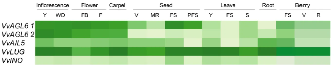
(45) 42. FIGURE 6. Profile expression of candidate genes in different tissues. These analysis was made in this study using data from the Transcriptomic Atlas of Grapevine (Fasoli et al., 2012). Y: young WD: well developed, FB: flowering begins (10% cap off), F: flowering (50% caps off), V: veraison, MR: mid ripening, R: Ripening, FS: fruit set, PFS: post fruit set, S: Senescing..
(46) 43. 4.3. VvLUG. LEUNIG (LUG) protein is similar in domain structure and biochemical function to the Groucho (Gro), Transducin-Like Enhancer of Split, and Tup1 family of corepressors in Drosophila, mammals, and yeast, respectively. These corepressors do not possess a DNA-binding domain and interact with DNA-bound transcription factors (Sitaraman et al., 2008).. LUG was firstly described it as a cadastral gene with important function in flower development, since leunig mutants’ flowers have sepals transformed toward stamens and carpels, and petals staminoids or absent, between other pleiotropic effects. Also, a role in flower development, as an A and C class gene during ABC(DE) was proposed (Liu and Meyerowitz, 1995).. LUG is expressed in almost all plant tissues, with strong expression across floral development, including placenta and ovules (Conner and Liu, 2000; Pfannebecker et al., 2016).. Regarding this function, LUG was described as a negative regulator of AGAMOUS (AG, a C class gene) during floral development, acting in a complex with a Q- rich protein (Franks et al., 2002; Sridhar et al., 2004). Furthermore, it has been established that LUG have functions related to floral development, and embryo formation (Sitaraman et al., 2008). It has also assigned a role in carpel development (Sitaraman et al., 2008) and recently, a physical interaction between LUG and INO has been probed, suggesting its coordinated participation during ovule’s integuments formation, possibly using a co-repressor as adapter (Simon et al., 2017a)..
(47) 44. VvLUG corresponds to putative homologous of LEUNIG (LUG) in grapevines. So far, there have been no studies characterizing this gene in this model. Previous analysis doing during this work using data from Transcriptomic Atlas of Grapes showed that it is expressed in many tissues, vegetative and reproductive, as occurs in LUG from Arabidopsis (Fasoli et al., 2012) (See Figure 6).. 4.4. VvINO. Villanueva and coworkers isolated the coding gene sequence (CDS) of INNER NO OUTER (INO) from Arabidopsis thaliana (Villanueva et al., 1999a). The CDS has 696 bp and encodes a 231 aminoacid protein that is a putative transcription factor belonging to the YABBY gene family, which is involved in the establishment of abaxial- adaxial polarity in lateral organs and asymmetric development (Bowman, 2000). Interestingly, INO is the only YABBY gene expressed in the ovule (Bowman and Smyth, 1999; Siegfried et al., 1999; Villanueva et al., 1999b).. The expression profile studied by in situ hybridization, shows that Arabidopsis INO mRNA is expressed on the abaxial zone of each ovule primordium, the area where the ovule’s integuments initiation occurs, specifically on a group of approximately 15 epidermal cells during early phase of ovule’s development, prior to visible integument initiation. During the next phases, when both integuments primordia are visible, INO mRNA is detected only in outer integument (abaxial side of ovule primordia). This transcript accumulation disappears during anthesis. Also,.
(48) 45. INO mRNA is present in young embryos prior to globular stage, but not in other floral structures (Villanueva et al., 1999a).. The expression pattern of INO orthologs has been conserved in some other species like Impatients (McAbee et al., 2005), Nymphacea (Yamada et al., 2003), Anonna (Lora et al., 2011) and Amborella (Arnault et al., 2018), supporting the hypothesis that INO might have a conserved role in the o.i. among angiosperms (Kelley and Gasser, 2009; Lora et al., 2011; Yamada et al., 2003). Also, unitegmic ovules (plants with i.i. and o.i. fusioned) express INO gene (Lora et al., 2015; Skinner et al., 2016).. Two strong INO Arabidopsis mutants have been studied, ino- 1 and ino- 2. The most studied, ino-1, has one nucleotide change (from G to A) in the fifth intron of a total of 6 introns, which creates a splicing acceptor site that results in a final transcript with 11 additional nucleotides. This generates a frameshift translation of the C- terminal region, near the YABBY domain, that cause an inactive protein (Villanueva et al., 1999a).. INO is a master regulator of o.i. formation required for both integument initiation and subsequent growth of this structure (Simon et al., 2017b; Villanueva et al., 1999a). Phenotypically, ino-1 completely lack of the o.i. and the gametophyte development is disrupted (Villanueva et al., 1999b). In consequence, ino-1 have female sterility. Moreover, in ino mutants there are no fruit development, because o.i. is involved in pollen tube guidance in Arabidopsis, so in these mutants pollen tubes rarely target the ovules during pollination (Herrero, 2000; Lora et al., 2018; Skinner and Gasser, 2009).. In addition, Tai seedless (Ts), a spontaneous mutant of Anonna squamosa with a deletion of the.
(49) 46. INO locus, generates unitegmic ovules, that no develops into seeds. Ts mutants have fruits completely developed, but without seeds(Lora et al., 2011). Unlike Arabidopsis, Anonna has an endostomal micropyle where the outer integument does not fully cover the inner integument and does not participate in the micropyle, and, presumably, in the pollen tube guidance. Hence, Ts mutant suffers the interruption of the reproductive program later than ino mutant, during the embryo and endosperm development and not during the embryo sac formation, which allows fruit development, despite disruption of seed development (Lora et al., 2011). Vitis vinifera has also an endostomal mycropile (Mullins et al., 1992) and therefore is likely that o.i. it does not have a role in the entry of pollen and subsequent fertilization.. The mechanisms that control INO expression are not completely known, however, there must be a precise spatio-temporal regulation. In fact, it has been proposed that several genes act by regulating their expression (Gaiser et al., 1995; Meister et al., 2002). This mechanism maintain the INO expression restricted to the gynobasal side of developing ovules, is required for initiation and asymmetric growth of outer integument (Meister et al., 2002).. Recently, Simon and coworkers, found a direct interaction between INO and co-repressors and co-activators. INO interacts with the co-repressors LEUNIG (LUG) and SEUSS (SEU) and with the co-activator protein ADA2b. Also, mutations in all this genes, generates a decrease in growth of the outer integument, suggesting a role in growth and extension of this integument (Simon et al., 2017b).. VvINO is the putative homologous of INO in grapevine and until now, it has not been studied. The analysis made using data from Fasoli and coworkers (shown in Figure 6) exhibit that VvINO.
(50) 47. has a very low expression profile in the vegetative and reproductive tissues analyzed, presumably because it is expressed in specific cells inside the ovule, as in Arabidopsis.. In summary, regarding grapevine, seedlessness is characterized by the absence of normal seeds in grape berries, an attractive phenotype, which has been poorly studied. The transcriptional analyses previously carried out in our laboratory allowed the selection of candidate genes with differential expression between the seeded and seedless phenotypes (Muñoz et al., 2012). Therefore, we asked whether any of these selected genes participate in the formation of the seed in grapevine..
(51) 48. CHAPTER 2. PROBLEM STATEMENT: HYPOTHESIS AND OBJECTIVES.
(52) 49. According to previously mentioned information in the general introduction, the biological question that directs this investigation is: Do any of the grapevine selected genes participate in the formation of the seed?. To address the biological question, the following hypothesis was proposed for this thesis work:. 1. Hypothesis The formation of the seed in the model plant Arabidopsis thaliana is affected by the expression of some of the genes of Vitis vinifera that are differentially expressed in seed and seedlessness grapevine: VvAGL6_1, VvAGL6_2, VvAIL5, VvLUG, and VvINO.. 2. Objectives The following main objective was proposed to answer the hypothesis:. Determine the expression profile of the genes; VvAGL6_1, VvAGL6_2, VvAIL5, VvLUG and VvINO; in seed and seedlessness grapevine and to evaluate if the expression of any of these affects the formation of the seed in Arabidopsis thaliana.. The general objective was divided into the following partial objectives:. 1. Correlate the seeded or seedlessness phenotype and the expression of the selected genes in the early stages of the development of the grape berries.. 2. To functionally evaluate the participation of one selected gene in the formation of the seeds in the model plant Arabidopsis thaliana..
(53) 50. CHAPTER 3 MATERIALS.
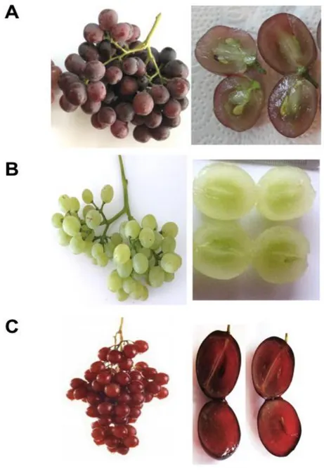
(54) 51. 1. Biological Material. 1.1. Plant material. 1.1.1. Vitis vinifera The grapevine plants used in this work were obtained from an experimental field, located in Miraflores, Curacaví, at the Metropolitan valley of Chile (33°24'01.0"S 71°03'17.6"W).. Samples of the following varieties were collected: Moscatel rosada, Red globe, Thompson seedless and Flame seedless. Also, in this study plants obtained from a segregating population from crosses between Moscatel rosada x Flame seedless were used.. 1.1.2. Arabidopsis thaliana Seeds of Columbia (Col-0) and Landsberg erecta (Ler-0) ecotypes were obtained from the seed’s collection of our laboratory. ino-1 (CS3881) mutant was obtained from the Arabidopsis Biological Resource Center (ABRC) catalog (The Ohio State University)..
(55) 52. 1.2. Bacterial strains. 1.2.1. Escherichia coli One Shot® TOP10 F–, mcrA, Δ(mrr-hsdRMS-mcrBC), Φ80lacZΔM15, ΔlacX74, recA1, araD139, Δ(ara leu)7697, galU, galK, rpsL, (strR), endA1, nupG. Competent cells by calcium chloride, used for transformation with plasmids. 1.2.2. Rhizobium radiobacter GV3101::pMP90 pMP90 (pTiC58ΔT-DNA), rifR, gmR. Electrocompetent cells, used for transformation with plasmids.. 1.3. Plasmids. pGEM®-T Easy by Promega was used to clone DNA fragments from Polymerase chain reactions (PCR). pENTRTM/SD/D-TOPO®, PK7FWG2 was used to generate constructions using Gateway® technology, as entry clone and donor receptor respectively (Table 1)..
(56) 53. TABLE 1. Vectors used in this work. Vector. Selection marker in plants -. Application. pGEMT-easy. Selection marker in bacteria Amp. pENTRTM/SD/D-TOPO®. Kan. -. Cloning. pK7FWG2. Spec. Kan. Over-expression in plants (35S promoter). Amp: ampicillin, Kan: kanamycin, Spec: spectinomycin.. Cloning.
(57) 54. 2. Culture media 2.1. Culture media for plants. Arabidopsis in vitro media culture (MS 0,5 X) includes 2,2 g/L of MS salts supplemented with vitamins (Duchefa Biochemie B.V.), 15 g/L of sucrose, 0,1 g of myo-inositol; 0,7% w/v of agaragar; pH 5,8 and antibiotics needed to make the selection for transgenic plants.. 2.2. Culture media for bacteria. The bacteria culture media used correspond to Luria-Bertani (LB) Broth medium from MO BIO Laboratories (Carlsbad, California, United States.) supplemented with antibiotics for selection according to the vector, as shown in Table 1. LB solid media (25 g/L of LB Broth) was further supplemented with Merck agar (15 g/L) (Darmstadt, Germany). The antibiotics used were gentamicin and rifampicin from PhytoTechnology Laboratories® (Shawnee Mission, Kansas, United States); ampicillin from Winkler (Santiago, Chile); kanamycin from United States Biological (Massachusetts, United States) and spectinomycin (Spec) from Duchefa Biochemie B.V. (Haarlem, Holland)..
(58) 55. 3. General reagents 3.1. Enzymes. The enzymes Taq DNA polymerase, T4 DNA ligase, SuperScript® II reverse transcriptase, Gateway® LR Clonase® II enzyme mix and Proteinase K were obtained from Invitrogen ™ (Carlsbad, California, United States). The enzyme deoxyribonuclease (DNase) TURBO DNAfree. TM. DNase was obtained from Ambion®). Restriction endonuclease DdeI from New. England BioLabs® (Ipswich, Massachusetts, United States) was also used.. 3.2. Commercial Kits. The following commercial kits were used: pGEM®-T Easy Vector System from Promega (Madison, Wisconsin, United States), FavorPrepTM Plasmid DNA Extraction Mini Kit from Favorgen (Ping-Tung, Taiwan), SensiMixTM SYBR® Hi-ROX Bioline Kit from Bioline (London, United Kingdom).. 3.3. General Reagents. The agarose was obtained from Bioline (London, United Kingdom). From Biotium (Hayward, California, United States) the GelRed® nucleic acid staining solution was obtained..
Figure


+7


Documento similar
No obstante, como esta enfermedad afecta a cada persona de manera diferente, no todas las opciones de cuidado y tratamiento pueden ser apropiadas para cada individuo.. La forma
The expansionary monetary policy measures have had a negative impact on net interest margins both via the reduction in interest rates and –less powerfully- the flattening of the
Jointly estimate this entry game with several outcome equations (fees/rates, credit limits) for bank accounts, credit cards and lines of credit. Use simulation methods to
In our sample, 2890 deals were issued by less reputable underwriters (i.e. a weighted syndication underwriting reputation share below the share of the 7 th largest underwriter
We demonstrated that, in addi- tion to its contribution to Mef2 transcriptional regulation of sarcomeric genes, CF2 is involved in the control of the fiber final size and in
To investigate the role of autophagy in otic neurogenesis, we studied the expression of autophagy genes in early stages of chicken (Gallus gallus) inner ear development and
In this work, we have addressed the expression of the FKTN (fukutin) and FKRP genes in the retina of mammals, and characterized the distribution pattern of their protein products
Analysis of the transposon insertion sites in the skin tumors identified 126 genes that were frequently mutated in different tumor samples and thus may have a role in human
