Glial connexin expression and function in the context of Alzheimer's disease
10
0
0
Texto completo
(2) A. Koulakoff et al. / Biochimica et Biophysica Acta 1818 (2012) 2048–2057. varying degrees of morphological and molecular remodeling, which leads to functional alterations that perturb their interactions with neurons [4, 5]. It is well established that glial cells exhibit diverse levels of connexin (Cx) expression [6] that are modified during reactive gliosis, although the consequences on the progression of the pathology remain to be elucidated in most cases [7]. Cxs are the molecular constituents of gap junctions, which are clusters of intercellular channels that allow the direct intercellular exchanges of ions and small molecules (e.g., IP3, ATP, glutamate, and energy metabolites) between neighboring cells [8]. These gap junction channels (GJCs) are made up by the head to head docking of two hemichannels (HCs) composed of hexamers of Cxs, each one located in opposing membranes of adjacent cells. In addition, it has been reported that in defined conditions, Cxs not engaged in GJCs can also operate as HCs mediating exchange between the cell cytoplasm and the extracellular medium [9]. For instance, inflammatory situations likely to occur in diverse pathologies trigger HC activation in glial cells in vitro and in vivo [10, 11]. HCs can also be made up of pannexins (Panxs), a family of three glycoproteins that share a similar transmembrane topology with Cxs but present only ~16% overall identity in amino acid sequence with Cxs [12, 13]. Although Panxs 1 and 2 are clearly detected in neurons within the brain [14, 15], up to now evidence of Panx expression in glial cells in vivo has only been reported in few cases [11, 16]. In this review, we will summarize the current knowledge on Cx expression and function in astrocytes and microglial cells, the two cell types involved in reactive gliosis. Special focus will be devoted to Alzheimer's disease (AD): after a short up to date review on reactive gliosis in this neurodegenerative pathology, we examined and discussed recent in vitro and in vivo data indicating that Cxs, and possibly Panxs, can contribute in the neurodegenerative process encountered during AD progression. 2. Connexin expression and function in glial cells In the brain, Cx expression is detected in all cell types with a specific distribution and chronology. The molecular identity of Cxs in the different glial cell types — astrocytes, oligodendrocytes and microglial cells – as well as in neurons – has been recently reviewed [6, 7, 17– 19]. The main features are that, among the diverse Cxs detected in the vertebrate brain (Cxs 26, 29, 30, 31, 32, 36, 40, 43, 45, 47 and 57) by different experimental approaches, each cell type is characterized by a distinct set of Cx expression and more than one Cx is expressed in a defined cell type. 2.1. Microglial cells Concerning microglial cells, Cx expression and function have been almost exclusively examined in cultured cells obtained from newborn mouse or rat cortex and in one case from embryonic human brain. In resting microglia, Cxs are either undetected or expressed at low levels. However, there is no consensus with respect to the type of Cx found, Cx43, Cx36 or Cx32, which may be due to culture conditions and/or sensitivity of the techniques used. Cx43 was the first to be detected in activated microglia, either in culture after tumor necrosis factor-αTNF-α) and interferon-γ treatment, or in vivo around stab-wounds where microglial cells exhibit a diffuse intracellular Cx43 immunoreactivity [20]. Also, exposure of microglia to staphylococcus aureus triggers Cx43 mRNA and protein expression associated with a low but significant gap junction coupling between neighboring cells, as assessed by dye diffusion assay [21]. Recently, the amyloid-β peptide (Aβ was shown to increase Cx43 expression in microglial cells in particular at the cell surface where it forms functional HCs that allow ATP and glutamate release to the extracellular milieu [22]. However, Cx43 has not been detected by immunoblotting in resting or activated (by lipopolysaccharide (LPS) treatment) microglia by others groups [23–26]. The presence of Cx36 mRNA and protein was reported in cultured microglial. 2049. cells from both mouse and human [25]. Due to its low level of expression, Cx36 could not be visualized by immunocytochemistry but was shown to form functional gap junctions between microglial cells as well as between microglia and neurons in co-cultures. Indeed, junctional currents measured in these cultures between pairs of cells (microglia/microglia and microglia/neurons) are characterized by a small unitary conductance (b20 pS) and low voltage sensitivity, properties typical of Cx36 GJCs. However, the incidence of such gap junction coupling is quite low (only one-third of microglial cell pairs are coupled) and while LPS treatment does not affect Cx36 expression or function, a mixed treatment with TNF-α and interferon-γ elicits a decrease in Cx36 protein level [25]. Finally, microglial cells were shown to express Cx32 that is up-regulated after LPS or TNF-α treatment as well as in microglial cells from Mecp2 null mice, a model of a neurodevelopmental disorder known as Rett syndrome, compared to wild-type mice microglia [27, 28]. In both cases, Cx32 operates as HCs allowing glutamate release with a deleterious effect on neurons. Altogether, these data indicate that microglial cells, at least in culture, can express low levels of Cxs and that, in most cases, Cx expression can be triggered by the activation of microglial cells by pro-inflammatory events. These Cxs form functional channels as demonstrated by HC activation and gap junction coupling between pairs of cells. Hence, Cx channels could contribute to the microglial response involved in pathological situations where inflammation takes place as discussed later. In addition, it was recently reported that Aβ treatment also induces the expression of Panx1 at the surface of cultured microglia in association with detectable HC activity [22]. However, the presence of microglial Cx and Panx expression in brain tissue remains to be determined. 2.2. Astrocytes Astrocytes are the cell population that exhibits the highest level of Cx expression in the brain, with Cx43 and Cx30 being the major astroglial Cxs in the adult. The molecular identity of astroglial Cxs was first established in cultured cells in which single channel activity between pairs of astrocytes is associated with the presence of gap junctions composed of Cx43 [29, 30]. During the following decades, new members of the Cx family were discovered leading to the detection of the mRNAs of Cxs 26, 30, 40, 43 and 45 and Cx46 in cultured astrocytes and the immunostaining for Cx40 and Cx45, although mostly detected at low levels with diffuse intracellular staining and few immunoreactive puncta present at membrane appositions [31]. Finally, Cx30 is detected in long-term cultures of rat astrocytes [32] while its expression, undetectable in mouse cortical astrocytes, is induced in subsets of cells in contact with neuronal cell soma in co-cultures [33]. Due to the prevalence and high level of Cx43 in cultured astrocytes, these cells have been extensively used as a model to analyze the mechanisms controlling Cx43 expression and function [34]. They provided information on the regulation of gap junction communication by neurotransmitters, growth factors, endogenous peptides and bioactive lipids through the activation of underlying intracellular signaling pathways [34], as well as on HC activation when triggered by metabolic inhibition and inflammatory treatments [35, 36]. In contrast to microglial cells, an abundant literature is available concerning the expression of astroglial Cxs in brain tissue. Although several Cx mRNAs are detected in hippocampal astrocytes by singlecell reverse transcription PCR [37], in situ hybridization for diverse Cxs argues for the presence of only Cx43 and Cx30 mRNAs in astrocytes [38]. Hence, although a minor expression of a third Cx, Cx26, cannot be excluded in sub-populations of astrocytes [39–41], the two prevalent astroglial Cxs are Cx30 and Cx43. This was further confirmed using mice double knock out (KO) for Cx30 and Cx43 in which gap junctional communication assessed by dye coupling in hippocampal astrocytes is totally abolished [42, 43]. Cx30 and Cx43 are widely but heterogeneously distributed in the brain, with quantitative differences among distinct cerebral areas, the main one.
(3) 2050. A. Koulakoff et al. / Biochimica et Biophysica Acta 1818 (2012) 2048–2057. concerning the white matter in which almost no Cx30 expression is detected [44]. More recently, the mapping of cre-mediated reporter activation for Cx30 and Cx43 has revealed their frequent co-expression in hippocampal astrocytes [45]. At the ultrastructural level, Cx30 and Cx43 are co-localized at gap junction plaques in brain and spinal cord as shown by TEM (transmission electron microscopy) immunocytochemistry and FRIL (freeze-fracture replica immunogold labeling) [46]. Hence, these two Cxs provide the structural basis for the organization of astrocyte networks that exhibit a certain degree of selectivity. This was recently demonstrated in the somatosensory cortex of whisker projection (barrel cortex) in which, due to the heterogeneity of Cx expression that is high within barrels but low between barrels, astrocyte coupling is oriented towards the center of the barrel while inter-barrel astrocytes are weakly coupled [47]. Such selective networks could coordinate the activity of local neuronal networks through the trafficking of neurotransmitters, e.g. glutamate or glutamine [7]. Also, by allowing the diffusion of energy metabolites (glucose and lactate), Cx30 and Cx43 contribute to metabolic networks that are able to feed distant neurons in case of high neuronal demand [43]. Interestingly, both Cxs are enriched in specialized processes of astrocytes that enwrap the walls of blood vessels, the endfeet at which level Cx immunoreactive puncta are of large size and exhibit a “honeycomb” organization delineating boundaries between endfeet. Such pattern of expression provides a perivascular route that facilitates trafficking between neighboring endfeet [43, 48]. Finally, Cx43 and Cx30 immunoreactivities decline with aging [49]. Such decrease in immunoreactivity is not correlated with a reduction in Cx43 protein level measured by Western blot analysis suggesting that a redistribution of this protein takes place with age and results in a small decrease in gap junction communication [49]. Although less information is available, there is now evidence for Panx1 expression detected by western blot and/or immunocytochemistry in cultured astrocytes [50–52]. In these cells, P2X7 receptor activation with BzATP induces ATP release from astrocytes via Panx1, but not Cx43 HCs [53]. In contrast, in cultured astrocytes submitted to hypoxia-reoxygenation or treated with pro-inflammatory cytokines or Aβ peptide, the HC activity is mediated by Cx43, but not Panx1 [22, 54, 55]. Interestingly, two recent studies, one in fibroblast growth factor (FGF)-treated cultures of spinal cord astrocytes [56] and the other in brain slices from mice harboring a brain abscess [11], indicate that both Cx43 and Panx1 participate in HC activity. Hence, the respective contribution of Panxs and Cxs in astrocyte HC function may depend on the conditions triggering their activation, an aspect that requires further investigation. In diverse pathological situations, the pattern of Cx expression and the extent of gap junction coupling are modified in astrocytes, as recently reviewed [7]. These changes depend on the type and severity of insult, the distance from the lesion site and the time post-injury. As a consequence, deleterious and protective effects have been reported, in particular after ischemia, stroke or trauma [7]. However, in most of these studies, the relative contribution of GJCs and HCs was not determined since the pharmacological agents used block both channel types [19]. In neurodegenerative diseases, changes in astroglial Cx expression have been observed but changes in their channel functions, HCs and/or GJCs, have been tested in only few of them (Table 1). Here, we present new insights that have been recently provided in the context of AD. 3. Alzheimer's disease and reactive gliosis Alzheimer's disease, discovered more than a century ago, is the most common neurodegenerative disease of the elderly [57–59]. This pathology is characterized by an abnormal accumulation of Aβ, generated by sequential proteolytic cleavage of the amyloid precursor protein (APP) by β- and γ-secretase [57]. Aβ oligomers aggregate in the brain parenchyma to form extracellular deposits called the amyloid plaques, which are a typical histopathological lesion of AD. Around blood vessels, these. Table 1 Pathology. Experimental models. Connexin expression. Gap junction coupling (GJC) Hemichannel activation (HCA). Alzheimer disease. In vivo. Human PM biopsies APP/PS1 mice. Cx43 IR at Aβ plaques [94] Cx43, Cx30 IR at Aβ plaques[95, 110]. PDAPP mice. NT. PDGFAPPSwInd NT In Aβ -treated No change Cx43[22, 26] vitro astrocytes Aβ-treated Cx43[22] microglia Aβ-treated Hip NT slices Parkinson disease Huntington disease Multiple sclerosis. MPTP mice Rotenone rat Post-mortem human biopsies EAE mouse EAE guinea pig. NT HCA astrocytes at plaques [110] GJC cortex [83] GJC no change Hip[83] GJC no change Hip[100] GJC no change [26] [100] GJC HCA[22]. HCA astrocytes and microglia [22] Cx43 striatum [123] GJC no change Cx43 basal ganglia [124] [123] Cx43 caudate nucleus,no NT change globus pallidus [124, 125] Cx43 in inflamed WM NT [126] Cx43 demyelinated WM NT [127] Cx43 remyelinating WM [127]. IR: immunoreactivity; NT: not tested; Hip: hippocampus; EAE: experimental autoimmune encephalomyelitis; MPTP: 1-methyl-4-phenyl-1,2,3,6-tetrahydropyridine; PM post-mortem; WM: white matter.. deposits constitute the vascular amyloid responsible for cerebral amyloid angiopathy [60]. Mutations responsible for the familial forms of AD lie within genes encoding APP and presenilins (PS), the catalytic subunits of the γ-secretase complex [57, 61]. Such mutations have been introduced in transgenic mice that exhibit several neuropathological features of AD including amyloid plaques, and are used as models to further explore the mechanisms coupling Aβ deposition to neural and vascular failure [62]. A consistent feature of Aβ accumulation is the strong reactive gliosis associated with amyloid plaques [63, 64]. 3.1. Microglial cells The intimate association of morphologically activated microglial cells with Aβ plaques is well established in both human AD brain and mouse models of the disease [65, 66]. Recently, the relationship between Aβ plaque formation and microglial dynamics has been examined using two-photon microscopy in AD mice and has shown that the recruitment of microglial cells occurs rapidly after plaque formation (within 1– 2 days) [67, 68]. However, in spite of an extensive research dedicated to these glial cells in AD, their role in the pathological process remains unclear [66]. Although microglia have the ability to phagocyte Aβ as amply demonstrated in vitro, they are inefficient to clear Aβ plaques in vivo, suggesting that the pathways involved in Aβ clearance are altered in AD [66, 69] Also, as central actors in neuroinflammation, microglial cells contribute to the inflammatory state observed in AD in which increased levels of a variety of pro-inflammatory cytokines are detected in brains from both AD patients and animal models [70]. However, accumulating evidence indicates that microglia exhibit multiple activation states, from an alternative phenotype characterized by an anti-inflammatory profile to a classical phenotype typified by the expression of pro-inflammatory cytokines [71]. In AD, recent data indicate.
(4) A. Koulakoff et al. / Biochimica et Biophysica Acta 1818 (2012) 2048–2057. that microglial responses are multifaceted, with differences during the progression of the disease and between sub-populations of microglial cells, as well as on their relation with Aβ plaques [71, 72]. Given such complexity, a better understanding of microglial function in the pathophysiology of AD requires further investigation.. 2051. subsequent activation of purinergic receptors on neighboring astrocytes [93]. Interestingly ICWs are also detected in vivo in APP/PS1 mice: these waves are initiated in astrocytes adjacent to cortical plaques and propagate over distances as long as 200 μm [89]. Consequently, one may wonder whether such waves, which are not detected in control age-matched mice, are underlain by changes in Cx expression (see below).. 3.2. Astrocytes The role played by reactive astrocytes in the alteration of synaptic function [73] and in neuronal toxicity [67] associated with Aβ plaques has only started to be considered. Although the pathologic potential of astrocytes in dementia was initially suggested in 1910 by Alois Alzheimer [74], the exact role of astrocytes in AD is only recently emerging. Both aggregated Aβ peptides and core of Aβ plaques from AD patients stimulate astrogliosis [63], a process observed in both AD patients and murine models, in which reactive astrocytes are characterized by an increased expression of GFAP and S100β proteins [64, 75]. Such reactive astrocytes are found close to neuritic plaques, even just at the outer edge of Aβ plaques [63, 76] with processes infiltrating the core of the plaques. In the hippocampus of a triple transgenic model of AD, APP/PS1/tau, the hypertrophy of reactive astrocytes encircling neuritic plaques contrasts with the atrophy of astrocytes located at a distance from them [77]. In addition to these phenotypic changes, several properties of reactive astrocytes are modified and some examples are presented below. First, the glutamatergic function of astrocytes is impaired in AD. Indeed, a reduced expression of EAAT2, the main astroglial glutamate transporter in the adult, and a deficient glutamate transport are observed in human post mortem biopsies from AD patients [78–80], suggesting that astrocytes can lose their role in glutamate clearance which may lead to neurotoxic effects. Similar decreases in glutamate transporter expression and function occur in Aβ-treated cultured astrocytes in which the mechanisms underlying such inhibition are partly mediated by altered MAP kinase signaling pathways triggered by oxidative stress [81]. However, the expression of glutamate transporters seems to be highly variable between AD individuals [82] and in murine models since increases in glutamate transporter currents are also detected in the reactive astrocytes of aged mice over-expressing APP [83]. Such variability may be due to the heterogeneity of reactive astrocytes that may be at different stages of the astrogliosis process [5, 80]. Second, in vitro studies show that the exposure of astrocytes to aggregated forms of Aβ modifies their metabolic status by increasing all main glucose metabolic pathways (glycolysis and lactate release, tricarboxylic acid cycle, pentose phosphate pathway and glycogen storage) [84], induces mitochondrial depolarization, activates NADPH oxidase and increases oxidative stress [84–86]. All of these events result in impaired neuronal viability. Finally, abnormalities in astrocyte Ca 2+ signaling have been detected both in vitro and in vivo in transgenic mouse models of AD, indicating that this key astroglial signaling pathway is altered. Indeed, increased Ca2+ signals are measured in cultured astrocytes treated with Aβ, probably forming channels permeable to Ca 2+ at the cell membrane [87, 88]. These Ca 2+ signals cause an increase in reactive oxygen species as well as a depletion in glutathione precursor supply from astrocytes to neurons whose consequence is an oxidative stress that leads to neuronal death. These observations obtained in culture are in line with recent in vivo observations made in a double transgenic APP/PS1 mouse whose astrocytes have a higher basal intracellular Ca 2+ level compared to wild-type mice [89]. Interestingly, two-photon in vivo imaging performed in AD mouse models has shown that increased spontaneous Ca2+ signaling in astrocytes affects microvascular circulation at early stages of the disease [90], a feature that is reminiscent of the cerebrovascular dysregulation observed in AD patients [91]. In addition, cultured astrocytes treated with Aβ exhibit intercellular Ca 2+ waves (ICWs) [92]. The propagation of ICWs involves two pathways in which astroglial Cxs acting as GJCs and/or HCs can be involved: i) a direct intercellular communication through GJCs and ii) a process mediated by ATP release (via HC or Ca2+-dependent exocytosis) and. 4. Changes in glial connexin expression in Alzheimer's disease and murine models The first evidence for Cx changes in AD was reported by Nagy and colleagues (1996) who observed increased immunoreactivity for Cx43 at Aβ plaque levels in post-mortem human brains from AD patients [94]. At the ultrastructural level, this increase in Cx43 immunostaining occurs at gap junctions between astrocytic processes adjacent to dystrophic neuronal processes present in plaque areas [94]. We have also observed in brain sections from AD patients such enrichment in Cx43 puncta in the intermingled strongly GFAP+ astrocyte processes that infiltrate amyloid plaques (Fig. 1A). This feature is also observed for Cx30, although to a lesser extent (Fig. 1B). Moreover, such elevated Cx immunoreactivity is associated with plaques exhibiting bulb-like structures identified by their content in phosphorylated tau, which is characteristic of damaged neurites (Fig. 2A). To investigate in more detail the pattern of Cx expression in the context of AD, APP/PS1 transgenic mice that develop an amyloid pathology have recently been used as experimental models [95]. In mice older than 4 months, hotspots of Cx43 and Cx30 immunoreactivity are visible in the hippocampus and the cortex at Aβ plaque levels. At high magnification, bright and large Cx immunoreactive puncta are concentrated in astrocyte processes that penetrate the core of the plaques, often encircled by neuritic dystrophies (Fig. 2B), as observed in human AD brains. In contrast, they are not detected in microglial cells that coexist with reactive astrocytes at the periphery of Aβ deposits [95]. A semi-quantitative analysis showed that such increases in Cx immunoreactivity occurs in a large majority of plaques. Nevertheless, depletion in astroglial Cxs is also observed in a small proportion of plaques (≤15%) and tends to diminish in older mice exhibiting a higher Aβ loading. This feature suggests that a decreased Cx expression could preferentially occur in “young”, recently formed plaques. Since Aβ plaques accumulate as the disease progresses, the relative proportion of young plaques will be lower at advanced stages of the amyloid pathology. In agreement with this hypothesis, human AD sections from post-mortem brain biopsies show no evidence for decreased Cx expression. The elevated Cx immunoreactivity detected in vivo in association with most Aβ plaques where activated microglial cells are engaged in a multistep inflammatory process is in apparent contradiction with in vitro observations showing single application of Cx43 expression is strongly decreased in astrocytes treated with pro-inflammatory cytokines (interleukin1-β [IL-1β] and TNF-α), an effect potentiated by Aβ [26]. Several hypotheses can be proposed to explain such discrepancy. First, the changes in Cx expression in culture occur after short-term treatments (24–48 h), a timing obviously different from the in vivo situation in which the chronic situation may lead to a desensitization of cytokine receptors, uncoupling internal signaling pathways involved in the regulation of Cx expression and their channel functions. For instance, receptor desensitization has been proposed for the TNF-α signaling pathway. Interestingly, an intracellular mediator of TNF receptor1, the protein DENN/MADD is down-regulated both in the hippocampus of AD patients and in murine models of AD [96, 97]. Second, although global inflammation develops in vivo as the pathology proceeds, the local inflammatory status around the plaques is much more complex. The phenotype of microglial cells can change as amyloid pathology progresses with an age-dependent switch from an “alternative” anti-inflammatory phenotype to a “classic” pro-inflammatory phenotype [69, 72]. In the first step of the amyloid pathology, activated microglial are restricted to Aβ plaques and characterized by YM-1.
(5) 2052. A. Koulakoff et al. / Biochimica et Biophysica Acta 1818 (2012) 2048–2057. Fig. 1. Increased immunoreactivity of Cx43 and Cx30 in reactive astrocytes at Aβ plaques in human AD brains. High magnification confocal images of triple immunostained sections of temporal cortex obtained post-mortem from AD patients showing Aβ deposits (blue, A1, B1), immunoreactive puncta of Cx43 (A2) or Cx30 (B2) in green and GFAP labeled astrocytes (red, A3, B3). Overlay images illustrate the enrichment in Cx immunoreactive dots at plaque level in (A4, B4). Scale bar: 50 μm.. mRNA expression, a marker of the alternative phenotype. Later on, while activated microglial cells show a widespread distribution in the brain parenchyma and produce increasing levels of TNF-α and IL-1β [69], surprisingly, microglial cells contacting the plaques remain YM-1 positive and are devoid of TNF-α [72]. Together these data indicate that global inflammation develops as the pathology proceeds but that the inflammatory status at plaque level is likely different. Also, it cannot be excluded in situ that other secretion products originated from microglia and/or astrocytes can counteract the effect of pro-inflammatory cytokines and Aβ. Hence, to understand the possible contribution of inflammation in the expression of Cxs, and perhaps Panxs, at amyloid plaque levels, it will be important to directly examine by in situ hybridization and/or immunohistochemistry the expression of a panel of markers of inflammation to determine the local inflammatory status of Aβ plaques and whether it is involved in the changes observed in Cx expression in glia. Finally, the expression of Cxs in astrocytes is under the control of neurons [33, 98], thus again in vitro situations that do not take into account this partnership could introduce a bias in reproducing pathological situation and lead to misinterpretations.. 5. Changes in connexin channel functions in Alzheimer's disease animal models and amyloid-β treated cultures Once at the membrane, Cxs can function as GJCs or HCs and recent evidence indicates that in glia both functions are modified in Aβ-treated cultures as well as in AD models. 5.1. Gap junction communication Concerning gap junction communication, the first evidence of a potential effect of Aβ comes from the observation that this peptide increases the propagation of ICWs in PC12 cells [99] and in astrocytes [92], a process in which Cxs are known to be involved [93]. Then, the effect of Aβ on gap junction communication was directly investigated by measuring dye coupling in cultured astrocytes. Several isoforms of aggregated Aβ (1–40, 1–42, 25–35) that per se do not affect dye coupling, potentiate the inhibition induced by inflammatory cytokines IL-1β and TNF-α [26]. However, the aggregation status of Aβ seems important since low concentrations of monomerized. Fig. 2. Elevated astrocyte Cx43 immunoreactivity associated with neuritic dystrophies in brain sections from AD patients and APP/PS1 mice. High magnification confocal images of triple immunostained cortical sections of human AD brains in the upper part (A1–4) and APP/PS1 mice in the lower part (B1–4). Representative images showing GFAP stained astrocytes (red A1, B1) with an increased Cx43 immunoreactivity (green A2, B2) in areas exhibiting bulb-like structures (arrows) characteristic of dystrophic neurites stained in blue with anti-Tau antibodies (A3, B3). Merge images are shown in (A4, B4). Scale bar: 50 μm..
(6) A. Koulakoff et al. / Biochimica et Biophysica Acta 1818 (2012) 2048–2057. Aβ1–40 impair gap junction communication in cultured rat astrocytes [100]. Indeed, several astrocyte properties are differentially affected by oligomeric versus fibrillar Aβ, including their ability to release pro-inflammatory cytokines and their metabolic status [84, 101]. Such observations in cultured astrocytes contrast with those obtained in adult brain slices from AD models. The extent of dye coupling in astrocytic networks in acute cortical brain slices of a mouse overexpressing APP is increased compared with age-matched control mice [83]. Such increase is likely dependent on the brain area since astrocyte gap junction communication is similar in hippocampal slices of wild-type mice and of AD models (PD-APP, PDGF-APPSwInd) in which Aβ deposits are present [83, 100]. These results indicate that the ability of astrocytes to send signals to distant sites via gap junction-mediated astroglial networks is maintained or enhanced in vivo in pathological conditions, but the nature of biological signals trafficking through this network remains to be identified. Since ICW propagation depends on gap junction communication in the cortex but not the hippocampus [102], ICWs observed recently in vivo in the cortex of the APP/PS1 mouse model [89] are likely to represent the first example of such intercellular trafficking.. 2053. Fig. 3. Aβ peptide triggers hemichannel activation in cultured glial cells leading to neuronal death. Aggregated Aβ peptide treatment of microglial cells (M) or astrocytes (A) triggers activation of HCs composed of Cx43 in reactive astrocytes (RA) and of Cx43 and Panx1 in activated microglia (MA) allowing for glutamate and ATP release. Both gliotransmitters can bind on neuronal (N) NMDA and purinergic receptors, respectively, leading to an increase in intracellular Ca2+ resulting in neuronal degeneration (DN). ATP can also activate Panx1 HCs generating Ca2+ influx and neuronal damage.. 5.2. Hemichannels To establish whether the changes in expression of astroglial Cxs have also an impact on their HC function, in vitro cultures are useful models for determining the respective contribution of Cxs and Panxs to HC activity by using pharmacological approaches and single channel patch-clamp recordings. Recently, a detailed in vitro analysis of the effect of aggregated Aβ25–35 on HC activity in the three cell types that interact at amyloid plaques (i.e. microglia, astrocytes and neurons) has been performed [22]. By monitoring the uptake of ethidium bromide (Et) as an index of activity, HC activation is detected in these three cell types, with microglia being the most sensitive to the Aβ treatment, followed by astrocytes and then neurons. The pharmacological profile of these activations indicates that Cx43 and Panx1 HCs are involved in microglia, while only Cx43 HCs are activated in astrocytes. These observations were confirmed by using glial cultures from Cx43 KO mice. In neurons, a Cx HC activity that involves Cx36, is associated to a Panx1 HC component. Accordingly, single channel events with unitary conductances characteristic for Panx1, Cx43 and Cx36 HCs were respectively recorded in microglia, astrocytes and neurons after treatment with Aβ25–35. These currents are recorded at negative holding potential (−60 mV) and in the presence of external divalent cations suggesting that such HC openings may occur in pathophysiological situation. The consequences of Aβ25–35 induced-HC activation were further analyzed at two levels: i) the molecular nature of molecules released and ii) their impact on neuronal fate. There is already evidence that HC activation can lead to “gliotransmitter” release in both types of glia [27, 28, 103, 104]. This was confirmed in microglial cells and astrocytes treated by Aβ25–35, which release glutamate and ATP via activated HCs [22]. Moreover, both gliotransmitters present in conditioned medium (CM) harvested from either microglia or astrocytes treated with Aβ25–35 were shown to trigger HC opening in neurons with effects on neuronal survival. Hence, in culture models, Aβ peptide generates HC activation, in addition to activation of NMDA and P2X receptors, in glia that release ATP and glutamate, which in turn activate HC opening in neurons leading to neuronal death (Fig. 3). Acute brain slices from adult mouse represent a more integrated system to test HC activity and perform pharmacological treatments. Shortterm treatments of acute hippocampal slices with Aβ25–35 result in an Et uptake in microglia identified after post-fixation by isolectin B4 labeling, in astrocytes visualized in hGFAP-eGFP mice as well as in pyramidal neurons [22]. In microglia and astrocytes, this membrane “permeabilization” is mediated by Cx43 HCs as indicated by their pharmacological profile and confirmed by the lack of Aβ25–35-induced Et uptake in a Cx43fl/flcreGFAP mouse. In pyramidal neurons, Et uptake is essentially. due to Panx1 HCs whose activity augments in the presence of the Aβ peptide. Like in cultures, neuronal death detected in Aβ-treated slices is prevented either by blocking HCs in microglia, astrocytes and neurons or by using NMDA or P2X receptor antagonists. These results confirm that short-term treatments with the Aβ peptide induce a cascade of HC activation in glia and neurons leading to neuronal death [22]. Interestingly, a novel putative HC blocker (INI-0602) able to cross the blood brain barrier was recently shown to inhibit in vivo the LPS-induced glutamate release from microglia and to improve memory deficits in APP/PS1 mice [105]. The authors argued that these effects are mediated by the pharmacological blockade of microglial Cx32 HCs. However, the possible involvement of other HC types or even other channels cannot be ruled out in this study since the specificity of INI-0602 requires further demonstration, using for example in vivo experiments with Cx32−/− microglia or knockdown of Cx32. Finally, the next step will be to analyze in slices from AD model mice (APP/PS1) the functional state of glial HCs using patch-clamp and membrane permeability assays to fully determine the HC involvement in this pathology. 6. Conclusions and perspectives A common feature of neurodegenerative diseases is the phenotypic transformation of glial cells that are engaged in the activation process resulting in reactive gliosis. In most of these pathologies, in situ studies have established that this glial response is associated with a change in Cx expression (Table 1). This is the case for AD in which functional approaches, based on the use of AD animal models or cell cultures treated with Aβ peptide, have recently contributed to give some insight on the consequences of these modifications in the pathological process. In normal situation, gap junction communication is high between astrocytes while it is still questioned in microglia (Fig. 4A). So far, available data in AD mouse models are concordant and indicate an enhancement, or at least maintenance of intercellular communication in astroglial networks, compared to control mice. Hence, such pathways, which allow the diffusion of a variety of substances to distant sites, would be still working (Fig. 4B). The nature of signaling molecules trafficking within these networks can be multiple and consequently may have various impacts on several functions. For instance, ICWs propagating between cortical astrocytes may have direct consequences on neuroglial interactions, which could modify the activity of neurons and/or their survival. This could operate through Ca2+-dependent mechanisms that are involved for instance in gliotransmission [106], K+ homeostasis [93], intercellular metabolic wave propagation [107], as well as blood flow control at the gliovascular interface [108]. Interestingly, cerebral blood flow is reduced.
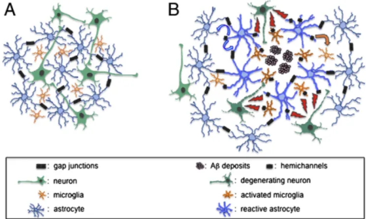
(7) 2054. A. Koulakoff et al. / Biochimica et Biophysica Acta 1818 (2012) 2048–2057. Fig. 4. Changes in connexin channel-mediated neuroglial interaction at amyloid plaques. (A) In normal situation with healthy neurons (green), astrocytes (blue) are highly coupled by gap junction channels (double black symbols), while hemichannels are virtually absent in astrocytes and microglial cells (orange). (B) In AD models characterized by amyloid plaques (violet aggregates), neighboring astrocytes are reactive (thick blue) and microglia are activated (thick orange). While gap junction communication is maintained in astrocytes, hemichannels (black symbols) are open in glial cells next to the Aβ deposits. This activation allows the release of active compounds (red lightnings), in particular glutamate and ATP, which can lead to neuronal damage. These gliotransmitters can also act on astrocytes (blue arrows) and microglia (orange arrows).. in AD, which may impair the proper delivery of nutrients to neurons. In this context, the preservation of gap junction communication within astroglial networks associated to an increase in glucose metabolism, recently demonstrated in astrocytes treated with Aβ [84], could represent a neuroprotective compensatory mechanism allowing the intercellular diffusion of energy metabolites to sustain neuronal activity and survival. Also, the diffusion of toxic substances, e.g. glutamate, within groups of communicating astrocytes, could contribute to their dissipation and thus result in an attenuation of neurotoxicity. However, addressing the issue of a beneficial or deleterious role of glial gap junctions is a complex problem since many available pharmacological tools have a limited specificity and block GJCs as well as HCs [19]. Obviously, mice KO for Cx genes do not represent good tools to solve this question since both channel functions are affected in these mice [19]. An interesting possibility would be to find mutations in Cx43 that block GJC formation without affecting the HC activity. Potential targets could be found in the extracellular Cx43 domains that are involved in the docking of HCs to form a GJC. Such dominant negative mutations have been described for other Cx genes, e.g. Cx50 [109]. Mice engineered to express these mutations could be crossed with APP/PS1 mice to determine their consequence on neuronal properties and survival as well as on the progression of the amyloid pathology. On the other hand, activation of glial HCs might provide a pathway for ATP and glutamate release and potentially other molecules that can have deleterious effects on neurons, as illustrated in Figs. 3 and 4B. Whether this process demonstrated in culture and in acute slices after short-term exogenous application of Aβ peptide [22] takes place in vivo remains to be investigated. Preliminary results indicate that HCs are activated in reactive astrocytes surrounding Aβ plaques in APP/PS1 mice [110]. Glutamate released through HCs in microglia and/or astrocytes at plaque levels could locally enhance intracellular [Ca 2+]i in neurites by acting on NMDA receptors and thus could lead to neuritic dystrophies underlying neuronal dysfunction in AD. Indeed, in APP/PS1 mice, the presence of Aβ plaques causes a Ca 2+ overload in axon and dendrites, further resulting in spinodendritic Ca 2+-dependent decompartmentalization and neuritic dystrophy [73]. Alternatively, ATP could represent another glial signal released through HCs during AD progression. ATP can act on microglia, astrocytes or neurons. For instance, it is now well established that the mobility of microglial cells depends on an ATP signal that contributes to. their rapid migration towards an injured area [111]. Recently, it was reported that ATP is required for the migration and accumulation of microglial cells after a nerve crush in the leech and is released through glial innexin HCs [112]. Thus, the ATP release via activated HCs in reactive astrocytes could contribute to the recruitment of microglia around Aβ plaques. Accordingly, microglia from ex vivo retinal explants exhibit a reduced process motility after treatment with probenecid, a specific Panx HC blocker, suggesting that Panx1 HCs could play a relevant role in this process [113]. In astrocytes, ATP can act in a paracrine manner on P2Y2 receptors and thus, contributes to the propagation of ICWs [114]. Accordingly, ATP release via HCs from reactive glial cells could be involved in the propagation of ICWs initiated close to the plaques in APP/PS1 mice [89]. Also, ATP acting on P2X7 receptors could amplify in an autocrine and/or paracrine manner the opening of Panx1 HCs in astrocytes and trigger the inflammasome with subsequent release of IL1β [115]. By the same mechanism, it has been recently proposed in neurons that ATP opens Panx1 HCs allowing the entry of Ca 2+ that leads to neuronal death [22]. Finally, since ATP modulates synaptic activity and is involved in synaptic depression after its conversion to adenosine [116, 117], its release through glial HCs could participate in synaptic impairment that characterizes AD. However, its conversion to adenosine could also exert a beneficial role, as shown in the response to ischemia after preconditioning [118]. In order to better define the role of glial Cxs and Panxs in AD, several lines of research can be defined for the future. One would be to develop new tools that selectively target the different glial and neuronal Cxs and Panxs, but also discriminate between their two channel functions, HC versus GJC. Interestingly, HCs appear more implicated in neurotoxic processes than GJC [54, 119], a feature that should improve the chance of success for pharmacological intervention. Indeed, while GJCs have their two ends within the cytoplasm, HCs offer a direct extracellular access that should facilitate the screening of drugs that block their activity. Reactive gliosis is a hallmark in AD and other neurodegenerative diseases, and focusing on glial targets is emerging as an alternative therapeutic approach in this field [120–122]. Since expression and function of glial Cxs (this remains to be addressed for Panxs) are modified in AD, these proteins should be considered as potential targets for the design of future therapeutic tools. The identification of molecular mechanisms as well as cellular interactions in which Cx channels are involved in during AD progression could then be used as a basis to address the question of the role of glial cells in other neurodegenerative diseases where it is already known that the expression of glial Cxs is modified (Table 1). Acknowledgments The authors would like to thank the CRPCEN, the FRC and France Alzheimer, ATC71 and ECOS-CONICYT C10S01 for their financial support. In addition, the authors would like to thank the GIE Neuro-CEB, Hopital de la Salpêtrière, Paris, for providing them with brain samples from AD patients and Dr A. Di Nardo for critical comments on the manuscript. References [1] D. Attwell, A.M. Buchan, S. Charpak, M. Lauritzen, B.A. Macvicar, E.A. Newman, Glial and neuronal control of brain blood flow, Nature 468 (2010) 232–243. [2] E. Sykova, C. Nicholson, Diffusion in brain extracellular space, Physiol. Rev. 88 (2008) 1277–1340. [3] M.M. Halassa, P.G. Haydon, Integrated brain circuits: astrocytic networks modulate neuronal activity and behavior, Annu. Rev. Physiol. 72 (2010) 335–355. [4] J.L. Ridet, S.K. Malhotra, A. Privat, F.H. Gage, Reactive astrocytes: cellular and molecular cues to biological function, Trends Neurosci. 20 (1997) 570–577. [5] M.V. Sofroniew, Molecular dissection of reactive astrogliosis and glial scar formation, Trends Neurosci. 32 (2009) 638–647. [6] M. Theis, G. Sohl, J. Eiberger, K. Willecke, Emerging complexities in identity and function of glial connexins, Trends Neurosci. 28 (2005) 188–195. [7] C. Giaume, A. Koulakoff, L. Roux, D. Holcman, N. Rouach, Astroglial networks: a step further in neuroglial and gliovascular interactions, Nat. Rev. Neurosci. 11 (2010) 87–99..
(8) A. Koulakoff et al. / Biochimica et Biophysica Acta 1818 (2012) 2048–2057 [8] R. Bruzzone, T.W. White, D.L. Paul, Connections with connexins: the molecular basis of direct intercellular signaling, Eur. J. Biochem. 238 (1996) 1–27. [9] D.C. Spray, Z.C. Ye, B.R. Ransom, Functional connexin “hemichannels”: a critical appraisal, Glia 54 (2006) 758–773. [10] J.A. Orellana, P.J. Sáez, K.F. Shoji, K.A. Schalper, N. Palacios-Prado, V. Velarde, C. Giaume, M.V. Bennett, J.C. Sáez, Modulation of brain hemichannels and gap junction channels by pro-inflammatory agents and their possible role in neurodegeneration, Antioxid. Redox Signal. 11 (2009) 369–399. [11] N. Karpuk, M. Burkovetskaya, T. Fritz, A. Angle, T. Kielian, Neuroinflammation leads to region-dependent alterations in astrocyte gap junction communication and hemichannel activity, J. Neurosci. 31 (2011) 414–425. [12] Y. Panchin, I. Kelmanson, M. Matz, K. Lukyanov, N. Usman, S. Lukyanov, A ubiquitous family of putative gap junction molecules, Curr. Biol. 10 (2000) R473–R474. [13] E. Scemes, S.O. Suadicani, G. Dahl, D.C. Spray, Connexin and pannexin mediated cell–cell communication, Neuron Glia Biol. 3 (2007) 199–208. [14] A. Ray, G. Zoidl, S. Weickert, P. Wahle, R. Dermietzel, Site-specific and developmental expression of pannexin1 in the mouse nervous system, Eur. J. Neurosci. 21 (2005) 3277–3290. [15] A. Vogt, S.G. Hormuzdi, H. Monyer, Pannexin1 and Pannexin2 expression in the developing and mature rat brain, Brain Res. Mol. Brain Res. 141 (2005) 113–120. [16] A. Zappala, G. Li Volti, M.F. Serapide, R. Pellitteri, M. Falchi, F. La Delia, V. Cicirata, F. Cicirata, Expression of pannexin2 protein in healthy and ischemized brain of adult rats, Neuroscience 148 (2007) 653–667. [17] J.I. Nagy, F.E. Dudek, J.E. Rash, Update on connexins and gap junctions in neurons and glia in the mammalian nervous system, Brain Res. Brain Res. Rev. 47 (2004) 191–215. [18] G. Sohl, S. Maxeiner, K. Willecke, Expression and functions of neuronal gap junctions, Nat. Rev. Neurosci. 6 (2005) 191–200. [19] C. Giaume, M. Theis, Pharmacological and genetic approaches to study connexinmediated channels in glial cells of the central nervous system, Brain Res. Rev. 63 (2010) 160–176. [20] E.A. Eugenín, D. Eckardt, M. Theis, K. Willecke, M.V. Bennett, J.C. Sáez, Microglia at brain stab wounds express connexin 43 and in vitro form functional gap junctions after treatment with interferon-gamma and tumor necrosis factor-alpha, Proc. Natl. Acad. Sci. U. S. A. 98 (2001) 4190–4195. [21] S. Garg, M. Md Syed, T. Kielian, Staphylococcus aureus-derived peptidoglycan induces Cx43 expression and functional gap junction intercellular communication in microglia, J. Neurochem. 95 (2005) 475–483. [22] J.A. Orellana, K.F. Shoji, V. Abudara, P. Ezan, E. Amigou, P.J. Sáez, J.X. Jiang, C.C. Naus, J.C. Sáez, C. Giaume, Amyloid beta-induced death in neurons involves glial and neuronal hemichannels, J. Neurosci. 31 (2010) 4962–4977. [23] N. Rouach, C.F. Calvo, J. Glowinski, C. Giaume, Brain macrophages inhibit gap junctional communication and downregulate connexin 43 expression in cultured astrocytes, Eur. J. Neurosci. 15 (2002) 403–407. [24] P.M. Faustmann, C.G. Haase, S. Romberg, D. Hinkerohe, D. Szlachta, D. Smikalla, D. Krause, R. Dermietzel, Microglia activation influences dye coupling and Cx43 expression of the astrocytic network, Glia 42 (2003) 101–108. [25] K. Dobrenis, H.Y. Chang, M.H. Pina-Benabou, A. Woodroffe, S.C. Lee, R. Rozental, D.C. Spray, E. Scemes, Human and mouse microglia express connexin36, and functional gap junctions are formed between rodent microglia and neurons, J. Neurosci. Res. 82 (2005) 306–315. [26] W. Même, C.F. Calvo, N. Froger, P. Ezan, E. Amigou, A. Koulakoff, C. Giaume, Proinflammatory cytokines released from microglia inhibit gap junctions in astrocytes: potentiation by beta-amyloid, FASEB J. 20 (2006) 494–496. [27] H. Takeuchi, S. Jin, J. Wang, G. Zhang, J. Kawanokuchi, R. Kuno, Y. Sonobe, T. Mizuno, A. Suzumura, Tumor necrosis factor-alpha induces neurotoxicity via glutamate release from hemichannels of activated microglia in an autocrine manner, J. Biol. Chem. 281 (2006) 21362–21368. [28] I. Maezawa, L.W. Jin, Rett syndrome microglia damage dendrites and synapses by the elevated release of glutamate, J. Neurosci. 30 (2010) 5346–5356. [29] C. Giaume, C. Fromaget, A. el Aoumari, J. Cordier, J. Glowinski, D. Gros, Gap junctions in cultured astrocytes: single-channel currents and characterization of channel-forming protein, Neuron 6 (1991) 133–143. [30] R. Dermietzel, E.L. Hertberg, J.A. Kessler, D.C. Spray, Gap junctions between cultured astrocytes: immunocytochemical, molecular, and electrophysiological analysis, J. Neurosci. 11 (1991) 1421–1432. [31] R. Dermietzel, Y. Gao, E. Scemes, D. Vieira, M. Urban, M. Kremer, M.V. Bennett, D.C. Spray, Connexin43 null mice reveal that astrocytes express multiple connexins, Brain Res. Brain Res. Rev. 32 (2000) 45–56. [32] P. Kunzelmann, W. Schroder, O. Traub, C. Steinhauser, R. Dermietzel, K. Willecke, Late onset and increasing expression of the gap junction protein connexin30 in adult murine brain and long-term cultured astrocytes, Glia 25 (1999) 111–119. [33] A. Koulakoff, P. Ezan, C. Giaume, Neurons control the expression of connexin 30 and connexin 43 in mouse cortical astrocytes, Glia 56 (2008) 1299–1311. [34] N. Rouach, E. Avignone, W. Même, A. Koulakoff, L. Venance, F. Blomstrand, C. Giaume, Gap junctions and connexin expression in the normal and pathological central nervous system, Biol. Cell 94 (2002) 457–475. [35] J.E. Contreras, H.A. Sánchez, E.A. Eugenín, D. Speidel, M. Theis, K. Willecke, F.F. Bukauskas, M.V. Bennett, J.C. Sáez, Metabolic inhibition induces opening of unapposed connexin 43 gap junction hemichannels and reduces gap junctional communication in cortical astrocytes in culture, Proc. Natl. Acad. Sci. U. S. A. 99 (2002) 495–500. [36] M.A. Retamal, N. Froger, N. Palacios-Prado, P. Ezan, P.J. Sáez, J.C. Sáez, C. Giaume, Cx43 hemichannels and gap junction channels in astrocytes are regulated oppositely by proinflammatory cytokines released from activated microglia, J. Neurosci. 27 (2007) 13781–13792.. 2055. [37] F. Blomstrand, L. Venance, A.L. Siren, P. Ezan, E. Hanse, J. Glowinski, H. Ehrenreich, C. Giaume, Endothelins regulate astrocyte gap junctions in rat hippocampal slices, Eur. J. Neurosci. 19 (2004) 1005–1015. [38] D.F. Condorelli, A. Trovato-Salinaro, G. Mudo, M.B. Mirone, N. Belluardo, Cellular expression of connexins in the rat brain: neuronal localization, effects of kainate-induced seizures and expression in apoptotic neuronal cells, Eur. J. Neurosci. 18 (2003) 1807–1827. [39] F. Mercier, G.I. Hatton, Connexin 26 and basic fibroblast growth factor are expressed primarily in the subpial and subependymal layers in adult brain parenchyma: roles in stem cell proliferation and morphological plasticity? J. Comp. Neurol. 431 (2001) 88–104. [40] J.I. Nagy, X. Li, J. Rempel, G. Stelmack, D. Patel, W.A. Staines, T. Yasumura, J.E. Rash, Connexin26 in adult rodent central nervous system: demonstration at astrocytic gap junctions and colocalization with connexin30 and connexin43, J. Comp. Neurol. 441 (2001) 302–323. [41] J.I. Nagy, B.D. Lynn, O. Tress, K. Willecke, J.E. Rash, Connexin26 expression in brain parenchymal cells demonstrated by targeted connexin ablation in transgenic mice, Eur. J. Neurosci. 34 (2011) 263–271. [42] A. Wallraff, R. Kohling, U. Heinemann, M. Theis, K. Willecke, C. Steinhauser, The impact of astrocytic gap junctional coupling on potassium buffering in the hippocampus, J. Neurosci. 26 (2006) 5438–5447. [43] N. Rouach, A. Koulakoff, V. Abudara, K. Willecke, C. Giaume, Astroglial metabolic networks sustain hippocampal synaptic transmission, Science 322 (2008) 1551–1555. [44] J.I. Nagy, D. Patel, P.A. Ochalski, G.L. Stelmack, Connexin30 in rodent, cat and human brain: selective expression in gray matter astrocytes, co-localization with connexin43 at gap junctions and late developmental appearance, Neuroscience 88 (1999) 447–468. [45] D. Gosejacob, P. Dublin, P. Bedner, K. Huttmann, J. Zhang, O. Tress, K. Willecke, F. Pfrieger, C. Steinhauser, M. Theis, Role of astroglial connexin30 in hippocampal gap junction coupling, Glia 59 (2011) 511–519. [46] J.E. Rash, T. Yasumura, F.E. Dudek, J.I. Nagy, Cell-specific expression of connexins and evidence of restricted gap junctional coupling between glial cells and between neurons, J. Neurosci. 21 (2001) 1983–2000. [47] V. Houades, A. Koulakoff, P. Ezan, I. Seif, C. Giaume, Gap junction-mediated astrocytic networks in the mouse barrel cortex, J. Neurosci. 28 (2008) 5207–5217. [48] M. Simard, G. Arcuino, T. Takano, Q.S. Liu, M. Nedergaard, Signaling at the gliovascular interface, J. Neurosci. 23 (2003) 9254–9262. [49] M.L. Cotrina, Q. Gao, J.H. Lin, M. Nedergaard, Expression and function of astrocytic gap junctions in aging, Brain Res. 901 (2001) 55–61. [50] Y. Huang, J.B. Grinspan, C.K. Abrams, S.S. Scherer, Pannexin1 is expressed by neurons and glia but does not form functional gap junctions, Glia 55 (2007) 46–56. [51] F. Bianco, A. Colombo, L. Saglietti, D. Lecca, M.P. Abbracchio, M. Matteoli, C. Verderio, Different properties of P2X(7) receptor in hippocampal and cortical astrocytes, Purinergic Signal. 5 (2009) 233–240. [52] S. Iwabuchi, K. Kawahara, Functional significance of the negative-feedback regulation of ATP release via pannexin-1 hemichannels under ischemic stress in astrocytes, Neurochem. Int. 58 (2011) 376–384. [53] R. Iglesias, G. Dahl, F. Qiu, D.C. Spray, E. Scemes, Pannexin 1: the molecular substrate of astrocyte “hemichannels”, J. Neurosci. 29 (2009) 7092–7097. [54] N. Froger, J.A. Orellana, C.F. Calvo, E. Amigou, M.G. Kozoriz, C.C. Naus, J.C. Sáez, C. Giaume, Inhibition of cytokine-induced connexin43 hemichannel activity in astrocytes is neuroprotective, Mol. Cell. Neurosci. 45 (2010) 37–46. [55] J.A. Orellana, D.E. Hernández, P. Ezan, V. Velarde, M.V. Bennett, C. Giaume, J.C. Sáez, Hypoxia in high glucose followed by reoxygenation in normal glucose reduces the viability of cortical astrocytes through increased permeability of connexin 43 hemichannels, Glia 58 (2010) 329–343. [56] J.M. Garré, M.A. Retamal, P. Cassina, L. Barbeito, F.F. Bukauskas, J.C. Sáez, M.V. Bennett, V. Abudara, FGF-1 induces ATP release from spinal astrocytes in culture and opens pannexin and connexin hemichannels, Proc. Natl. Acad. Sci. U. S. A. 107 (2010) 22659–22664. [57] D.J. Selkoe, Alzheimer's disease: genes, proteins, and therapy, Physiol. Rev. 81 (2001) 741–766. [58] M. Goedert, M.G. Spillantini, A century of Alzheimer's disease, Science 314 (2006) 777–781. [59] C. Duyckaerts, B. Delatour, M.C. Potier, Classification and basic pathology of Alzheimer disease, Acta Neuropathol. 118 (2009) 5–36. [60] C. Iadecola, Neurovascular regulation in the normal brain and in Alzheimer's disease, Nat. Rev. Neurosci. 5 (2004) 347–360. [61] A.L. Parks, D. Curtis, Presenilin diversifies its portfolio, Trends Genet. 23 (2007) 140–150. [62] J. Gotz, L.M. Ittner, Animal models of Alzheimer's disease and frontotemporal dementia, Nat. Rev. Neurosci. 9 (2008) 532–544. [63] R.G. Nagele, J. Wegiel, V. Venkataraman, H. Imaki, K.C. Wang, Contribution of glial cells to the development of amyloid plaques in Alzheimer's disease, Neurobiol. Aging 25 (2004) 663–674. [64] M.T. Heneka, J.J. Rodriguez, A. Verkhratsky, Neuroglia in neurodegeneration, Brain Res. Rev. 63 45 (2010) 37–46. 189–211. [65] H. Akiyama, S. Barger, S. Barnum, B. Bradt, J. Bauer, G.M. Cole, N.R. Cooper, P. Eikelenboom, M. Emmerling, B.L. Fiebich, C.E. Finch, S. Frautschy, W.S. Griffin, H. Hampel, M. Hull, G. Landreth, L. Lue, R. Mrak, I.R. Mackenzie, P.L. McGeer, M.K. O'Banion, J. Pachter, G. Pasinetti, C. Plata-Salaman, J. Rogers, R. Rydel, Y. Shen, W. Streit, R. Strohmeyer, I. Tooyoma, F.L. Van Muiswinkel, R. Veerhuis, D. Walker, S. Webster, B. Wegrzyniak, G. Wenk, T. Wyss-Coray, Inflammation and Alzheimer's disease, Neurobiol. Aging 21 (2000) 383–421..
(9) 2056. A. Koulakoff et al. / Biochimica et Biophysica Acta 1818 (2012) 2048–2057. [66] S. Mandrekar-Colucci, G.E. Landreth, Microglia and inflammation in Alzheimer's disease, CNS Neurol. Disord. Drug Targets 9 45 (2010) 37–46. 156–167. [67] M. Meyer-Luehmann, T.L. Spires-Jones, C. Prada, M. Garcia-Alloza, A. de Calignon, A. Rozkalne, J. Koenigsknecht-Talboo, D.M. Holtzman, B.J. Bacskai, B.T. Hyman, Rapid appearance and local toxicity of amyloid-beta plaques in a mouse model of Alzheimer's disease, Nature 451 (2008) 720–724. [68] T. Bolmont, F. Haiss, D. Eicke, R. Radde, C.A. Mathis, W.E. Klunk, S. Kohsaka, M. Jucker, M.E. Calhoun, Dynamics of the microglial/amyloid interaction indicate a role in plaque maintenance, J. Neurosci. 28 (2008) 4283–4292. [69] S.E. Hickman, E.K. Allison, J. El Khoury, Microglial dysfunction and defective beta-amyloid clearance pathways in aging Alzheimer's disease mice, J. Neurosci. 28 (2008) 8354–8360. [70] T. Wyss-Coray, Inflammation in Alzheimer disease: driving force, bystander or beneficial response? Nat. Med. 12 (2006) 1005–1015. [71] B. Cameron, G.E. Landreth, Inflammation, microglia, and Alzheimer's disease, Neurobiol. Dis. 37 45 (2010) 37–46. 503–509. [72] S. Jimenez, D. Baglietto-Vargas, C. Caballero, I. Moreno-Gonzalez, M. Torres, R. Sanchez-Varo, D. Ruano, M. Vizuete, A. Gutierrez, J. Vitorica, Inflammatory response in the hippocampus of PS1M146L/APP751SL mouse model of Alzheimer's disease: age-dependent switch in the microglial phenotype from alternative to classic, J. Neurosci. 28 (2008) 11650–11661. [73] K.V. Kuchibhotla, S.T. Goldman, C.R. Lattarulo, H.Y. Wu, B.T. Hyman, B.J. Bacskai, Abeta plaques lead to aberrant regulation of calcium homeostasis in vivo resulting in structural and functional disruption of neuronal networks, Neuron 59 (2008) 214–225. [74] A. Alzheimer, Beiträge zur Kenntnis der pathologischen Neuroglia und ihrer Beziehungen zu den Abbauvorgängen im Nervengewebe, in: F. Nissl, A. Alzheimer (Eds.), Histologische und Histopathologische Arbeiten uber die Grosshirnrinde mit besonderer Berucksichtigung der pathologischen Anatomie der GeisteskrankheitenJena, Verlag von Gustav Fischer, 1910, pp. 401–562. [75] A. Verkhratsky, M. Olabarria, H.N. Noristani, C.Y. Yeh, J.J. Rodriguez, Astrocytes in Alzheimer's disease, Neurotherapeutics 745 (2010) 37–46. 399–412. [76] H.M. Wisniewski, J. Wegiel, Spatial relationships between astrocytes and classical plaque components, Neurobiol. Aging 12 (1991) 593–600. [77] M. Olabarria, H.N. Noristani, A. Verkhratsky, J.J. Rodriguez, Concomitant astroglial atrophy and astrogliosis in a triple transgenic animal model of Alzheimer's disease, Glia 58 45 (2010) 37–46. 831–838. [78] S. Li, M. Mallory, M. Alford, S. Tanaka, E. Masliah, Glutamate transporter alterations in Alzheimer disease are possibly associated with abnormal APP expression, J. Neuropathol. Exp. Neurol. 56 (1997) 901–911. [79] C.P. Jacob, E. Koutsilieri, J. Bartl, E. Neuen-Jacob, T. Arzberger, N. Zander, R. Ravid, W. Roggendorf, P. Riederer, E. Grunblatt, Alterations in expression of glutamatergic transporters and receptors in sporadic Alzheimer's disease, J. Alzheimers Dis. 11 (2007) 97–116. [80] J.E. Simpson, P.G. Ince, G. Lace, G. Forster, P.J. Shaw, F. Matthews, G. Savva, C. Brayne, S.B. Wharton, Astrocyte phenotype in relation to Alzheimer-type pathology in the ageing brain, Neurobiol. Aging 31 45 (2010) 37–46. 578–590. [81] M. Matos, E. Augusto, C.R. Oliveira, P. Agostinho, Amyloid-beta peptide decreases glutamate uptake in cultured astrocytes: involvement of oxidative stress and mitogen-activated protein kinase cascades, Neuroscience 156 (2008) 898–910. [82] H. Beckstrom, L. Julsrud, O. Haugeto, D. Dewar, D.I. Graham, K.P. Lehre, J. StormMathisen, N.C. Danbolt, Interindividual differences in the levels of the glutamate transporters GLAST and GLT, but no clear correlation with Alzheimer's disease, J. Neurosci. Res. 55 (1999) 218–229. [83] O. Peters, C.G. Schipke, A. Philipps, B. Haas, U. Pannasch, L.P. Wang, B. Benedetti, A.E. Kingston, H. Kettenmann, Astrocyte function is modified by Alzheimer's disease-like pathology in aged mice, J. Alzheimers Dis. 18 (2009) 177–189. [84] I. Allaman, M. Gavillet, M. Belanger, T. Laroche, D. Viertl, H.A. Lashuel, P.J. Magistretti, Amyloid-beta aggregates cause alterations of astrocytic metabolic phenotype: impact on neuronal viability, J. Neurosci. 30 45 (2010) 37–46. 3326–3338. [85] A.Y. Abramov, L. Canevari, M.R. Duchen, Beta-amyloid peptides induce mitochondrial dysfunction and oxidative stress in astrocytes and death of neurons through activation of NADPH oxidase, J. Neurosci. 24 (2004) 565–575. [86] A.Y. Abramov, M.R. Duchen, The role of an astrocytic NADPH oxidase in the neurotoxicity of amyloid beta peptides, Philos. Trans. R. Soc. Lond. B. Biol. Sci. 360 (2005) 2309–2314. [87] A.Y. Abramov, L. Canevari, M.R. Duchen, Changes in intracellular calcium and glutathione in astrocytes as the primary mechanism of amyloid neurotoxicity, J. Neurosci. 23 (2003) 5088–5095. [88] O. Simakova, N.J. Arispe, Early and late cytotoxic effects of external application of the Alzheimer's Abeta result from the initial formation and function of Abeta ion channels, Biochemistry 45 (2006) 5907–5915. [89] K.V. Kuchibhotla, C.R. Lattarulo, B.T. Hyman, B.J. Bacskai, Synchronous hyperactivity and intercellular calcium waves in astrocytes in Alzheimer mice, Science 323 (2009) 1211–1215. [90] T. Takano, X. Han, R. Deane, B. Zlokovic, M. Nedergaard, Two-photon imaging of astrocytic Ca2+ signaling and the microvasculature in experimental mice models of Alzheimer's disease, Ann. N. Y. Acad. Sci. 1097 (2007) 40–50. [91] C. Iadecola, V. Hachinski, G.A. Rosenberg, Vascular cognitive impairment: introduction, Stroke 41 (45) (2010) 37–46 S127-128. [92] N.J. Haughey, M.P. Mattson, Alzheimer's amyloid beta-peptide enhances ATP/gap junction-mediated calcium-wave propagation in astrocytes, Neuromolecular Med. 3 (2003) 173–180. [93] E. Scemes, C. Giaume, Astrocyte calcium waves: what they are and what they do, Glia 54 (2006) 716–725.. [94] J.I. Nagy, W. Li, E.L. Hertzberg, C.A. Marotta, Elevated connexin43 immunoreactivity at sites of amyloid plaques in Alzheimer's disease, Brain Res. 717 (1996) 173–178. [95] X. Mei, P. Ezan, C. Giaume, A. Koulakoff, Astroglial connexin immunoreactivity is specifically altered at beta-amyloid plaques in beta-amyloid precursor protein/presenilin1 mice, Neuroscience 171 45 (2010) 37–46. 92–105. [96] K. Del Villar, C.A. Miller, Down-regulation of DENN/MADD, a TNF receptor binding protein, correlates with neuronal cell death in Alzheimer's disease brain and hippocampal neurons, Proc. Natl. Acad. Sci. U. S. A. 101 (2004) 4210–4215. [97] D. Rossi, L. Brambilla, C.F. Valori, A. Crugnola, G. Giaccone, R. Capobianco, M. Mangieri, A.E. Kingston, A. Bloc, P. Bezzi, A. Volterra, Defective tumor necrosis factor-alpha-dependent control of astrocyte glutamate release in a transgenic mouse model of Alzheimer disease, J. Biol. Chem. 280 (2005) 42088–42096. [98] N. Rouach, J. Glowinski, C. Giaume, Activity-dependent neuronal control of gap-junctional communication in astrocytes, J. Cell Biol. 149 (2000) 1513–1526. [99] J.I. Nagy, M.Z. Hossain, E.L. Hertzberg, C.A. Marotta, Induction of connexin43 and gap junctional communication in PC12 cells overexpressing the carboxy terminal region of amyloid precursor protein, J. Neurosci. Res. 44 (1996) 124–132. [100] N.F. Cruz, K.K. Ball, G.A. Dienel, Astrocytic gap junctional communication is reduced in amyloid-beta-treated cultured astrocytes, but not in Alzheimer's disease transgenic mice, ASN Neuro 2 (45) (2010) 37–46 e00041. [101] J.A. White, A.M. Manelli, K.H. Holmberg, L.J. Van Eldik, M.J. Ladu, Differential effects of oligomeric and fibrillar amyloid-beta 1–42 on astrocyte-mediated inflammation, Neurobiol. Dis. 18 (2005) 459–465. [102] B. Haas, C.G. Schipke, O. Peters, G. Sohl, K. Willecke, H. Kettenmann, Activitydependent ATP-waves in the mouse neocortex are independent from astrocytic calcium waves, Cereb. Cortex 16 (2006) 237–246. [103] Z.C. Ye, M.S. Wyeth, S. Baltan-Tekkok, B.R. Ransom, Functional hemichannels in astrocytes: a novel mechanism of glutamate release, J. Neurosci. 23 (2003) 3588–3596. [104] N. Kang, J. Xu, Q. Xu, M. Nedergaard, J. Kang, Astrocytic glutamate release-induced transient depolarization and epileptiform discharges in hippocampal CA1 pyramidal neurons, J. Neurophysiol. 94 (2005) 4121–4130. [105] H. Takeuchi, H. Mizoguchi, Y. Doi, S. Jin, M. Noda, J. Liang, H. Li, Y. Zhou, R. Mori, S. Yasuoka, E. Li, B. Parajuli, J. Kawanokuchi, Y. Sonobe, J. Sato, K. Yamanaka, G. Sobue, T. Mizuno, A. Suzumura, Blockade of gap junction hemichannel suppresses disease progression in mouse models of amyotrophic lateral sclerosis and Alzheimer's disease, PLoS One 6 (45) (2011) 37–46 e21108. [106] P.G. Haydon, G. Carmignoto, Astrocyte control of synaptic transmission and neurovascular coupling, Physiol. Rev. 86 (2006) 1009–1031. [107] Y. Bernardinelli, P.J. Magistretti, J.Y. Chatton, Astrocytes generate Na+-mediated metabolic waves, Proc. Natl. Acad. Sci. U. S. A. 101 (2004) 14937–14942. [108] S.J. Mulligan, B.A. MacVicar, Calcium transients in astrocyte endfeet cause cerebrovascular constrictions, Nature 431 (2004) 195–199. [109] E.A. Banks, M.M. Toloue, Q. Shi, Z.J. Zhou, J. Liu, B.J. Nicholson, J.X. Jiang, Connexin mutation that causes dominant congenital cataracts inhibits gap junctions, but not hemichannels, in a dominant negative manner, J. Cell Sci. 122 (2009) 378–388. [110] X. Mei, C. Giaume, A. Koulakoff, Changes in connexin expression and hemichannel activity in reactive astrocytes of APP/PS1 mice, FENS Abstr. 5 (2010) 078-63. [111] D. Davalos, J. Grutzendler, G. Yang, J.V. Kim, Y. Zuo, S. Jung, D.R. Littman, M.L. Dustin, W.B. Gan, ATP mediates rapid microglial response to local brain injury in vivo, Nat. Neurosci. 8 (2005) 752–758. [112] S.E. Samuels, J.B. Lipitz, G. Dahl, K.J. Muller, Neuroglial ATP release through innexin channels controls microglial cell movement to a nerve injury, J. Gen. Physiol. 136 45 (2010) 37–46. 425–442. [113] A.M. Fontainhas, M. Wang, K.J. Liang, S. Chen, P. Mettu, M. Damani, R.N. Fariss, W. Li, W.T. Wong, Microglial morphology and dynamic behavior is regulated by ionotropic glutamatergic and GABAergic neurotransmission, PLoS One 645 (2011) 37–46 e15973. [114] M. Nedergaard, B. Ransom, S.A. Goldman, New roles for astrocytes: redefining the functional architecture of the brain, Trends Neurosci. 26 (2003) 523–530. [115] W.R. Silverman, J.P. de Rivero Vaccari, S. Locovei, F. Qiu, S.K. Carlsson, E. Scemes, R.W. Keane, G. Dahl, The pannexin 1 channel activates the inflammasome in neurons and astrocytes, J. Biol. Chem. 284 (2009) 18143–18151. [116] G. Perea, M. Navarrete, A. Araque, Tripartite synapses: astrocytes process and control synaptic information, Trends Neurosci. 32 (2009) 421–431. [117] O. Pascual, K.B. Casper, C. Kubera, J. Zhang, R. Revilla-Sanchez, J.Y. Sul, H. Takano, S.J. Moss, K. McCarthy, P.G. Haydon, Astrocytic purinergic signaling coordinates synaptic networks, Science 310 (2005) 113–116. [118] J.H. Lin, N. Lou, N. Kang, T. Takano, F. Hu, X. Han, Q. Xu, D. Lovatt, A. Torres, K. Willecke, J. Yang, J. Kang, M. Nedergaard, A central role of connexin 43 in hypoxic preconditioning, J. Neurosci. 28 (2008) 681–695. [119] J.A. Orellana, N. Froger, P. Ezan, J.X. Jiang, M.V. Bennett, C.C. Naus, C. Giaume, J.C. Sáez, ATP and glutamate released via astroglial connexin 43 hemichannels mediate neuronal death through activation of pannexin 1 hemichannels, J. Neurochem. 118 (2011) 826–840. [120] N.J. Maragakis, J.D. Rothstein, Mechanisms of disease: astrocytes in neurodegenerative disease, Nat. Clin. Pract. Neurol. 2 (2006) 679–689. [121] D.A. Rempe, M. Nedergaard, Targeting glia for treatment of neurological disease, Neurotherapeutics 7 (2010) 335–337. [122] C. Escartin, G. Bonvento, Targeted activation of astrocytes: a potential neuroprotective strategy, Mol. Neurobiol. 38 (2008) 231–241. [123] M. Rufer, S.B. Wirth, A. Hofer, R. Dermietzel, A. Pastor, H. Kettenmann, K. Unsicker, Regulation of connexin-43, GFAP, and FGF-2 is not accompanied by.
(10) A. Koulakoff et al. / Biochimica et Biophysica Acta 1818 (2012) 2048–2057 changes in astroglial coupling in MPTP-lesioned, FGF-2-treated parkinsonian mice, J. Neurosci. Res. 46 (1996) 606–617. [124] A. Kawasaki, T. Hayashi, K. Nakachi, J.E. Trosko, K. Sugihara, Y. Kotake, S. Ohta, Modulation of connexin 43 in rotenone-induced model of Parkinson's disease, Neuroscience 160 (2009) 61–68. [125] J.C. Vis, L.F. Nicholson, R.L. Faull, W.H. Evans, N.J. Severs, C.R. Green, Connexin expression in Huntington's diseased human brain, Cell Biol. Int. 22 (1998) 837–847.. 2057. [126] E. Brand-Schieber, P. Werner, D.A. Iacobas, S. Iacobas, M. Beelitz, S.L. Lowery, D.C. Spray, E. Scemes, Connexin43, the major gap junction protein of astrocytes, is down-regulated in inflamed white matter in an animal model of multiple sclerosis, J. Neurosci. Res. 80 (2005) 798–808. [127] W.A. Roscoe, E. Messersmith, A. Meyer-Franke, B. Wipke, S.J. Karlik, Connexin 43 gap junction proteins are up-regulated in remyelinating spinal cord, J. Neurosci. Res. 85 (2007) 945–953..
(11)
Figure

Documento similar
At more advanced stages of limb development Id2 is expressed in the undifferentiated subectodermal and interdigital mesenchyme and exhibits specific domains of expression in the
The expansionary monetary policy measures have had a negative impact on net interest margins both via the reduction in interest rates and –less powerfully- the flattening of the
Jointly estimate this entry game with several outcome equations (fees/rates, credit limits) for bank accounts, credit cards and lines of credit. Use simulation methods to
In our sample, 2890 deals were issued by less reputable underwriters (i.e. a weighted syndication underwriting reputation share below the share of the 7 th largest underwriter
Incidence of dementia and probable Alzheimer´s disease in a general population: The Framingham study.. Hendiré H., Ogunniyi A.,
The tentative spin and parity assignment to the observed levels in 44 S is based on the comparison between the calcu- lated and experimental (i) excitation energies, (ii)
The histopathological diagnoses were classified in 10 categories, as follow: inflammatory/reactive lesions, pigmented/melanocytic lesions, benign bone lesions,
Altogether, the results presented in this Thesis on the regulation of the expression and the pathological role of the splicing variants sst5TMD4 and In1-ghrelin in the context of
