Laparoscopic video analysis for training a n d image-guided surgery
P A T R I C I A S Á N C H E Z - G O N Z Á L E Z1'2, A L I C I A M . C A N O1, I G N A C I O O R O P E S A1'2, F R A N C I S C O M . S Á N C H E Z - M A R G A L L O3, F R A N C I S C O D E L P O Z O1'2, P A B L O L A M A T A1, E N R I Q U E J. G Ó M E Z1'2
Bio engineering and Telemedicine Centre (GBT), Technical University of Madrid (UPM), Madrid, Spain, Networking Research Center on Bio engineering, Biomaterials and Nanomedicine (CIBER-BBN), Madrid, Spain, and Minimally Invasive Surgery Centre Jesús Usón (CCMIJU). Cáceres, Spain
Abstract
Automatic analysis of Minimally Invasive Surgical video has the potential to drive new solutions for alleviating needs of safe and reproducible training programs, objective and transparent evaluation systems and navigation tools to assist surgeons and improve patient safety. Surgical video is an always available source of information, which can be used without any additional intrusive hardware in the operating room. This paper is focused on surgical video analysis methods and techniques. It describes authors' contributions in two key aspects, the 3D reconstruction of the surgical field and the segmentation and tracking of tools and organs based on laparoscopic video images. Results are given to illustrate the potential of this field of research, like the calculi of the 3D position and orientation of a tool from its 2D image, or the translation of a preoperative resection plan into a hepatectomy surgical procedure using the shading information of the image. Research efforts are required to further develop these technologies in order to harness all the valuable information available in any video-based surgery.
tool tracking, organ reconstruction Key words: MIS, video image analysis, 3D reconstruction,
I n t r o d u c t i o n
Minimally Invasive Surgery (MIS) is b e c o m i n g a gold-standard choice for efficient, low-risk surgical procedures for several reasons. It is a safe, feasible, and patient-friendly way of performing a surgical intervention. Moreover, it satisfies patients' desire for faster recoveries and smaller injuries (aesthetic reasons), and shortens the hospitalization period. As a result, M I S is employed across various medical specialties and is replacing traditional surgical p r o c e -dures as swiftly as technology and h u m a n adoption of change allow. Nevertheless, M I S presents i m p o r t a n t limitations for surgeons due to its new interaction paradigm c o m p a r e d to traditional open surgery. I n M I S surgeons m u s t navigate the anatomical land-scape without the usual sensory clues. T h e deploy-m e n t of this surgery is based on an indirect manipulation of specialized surgical tools i n t r o d u c e d
into the abdominal cavity t h r o u g h small incisions. Surgeons follow the operation t h r o u g h an indirect visualization of the surgical field captured by an e n d o s c o p e . T h e motivation of the analysis of M I S video is the development of solutions to tackle two basic clinical needs: Better training a n d objective evaluation of M I S skills, and an e n h a n c e d perception and interaction in the surgical field for creation of real-time navigation systems.
Acquisition of all the necessary surgical skills requires a serious endeavour. Surgeons m u s t follow long traineeship periods in order to b e c o m e proficient in these techniques. Evaluation of surgical skills is n e e d e d for monitoring residents' learning and for a transition towards a quality-based surgical culture. C u r r e n t evaluation m e t h o d s , based on the Halsted tradition, are in general subject to bias and halo effects. Steps have b e e n taken to deal with these issues, such as the development of standardised
questionnaires as objective structured assessment of technical skill (OSATS) and their MIS counterpart, global operative assessment of laparoscopic skills (GOALS) (1). Nevertheless, there is a shared opinion that new training and evaluation programs are required (2) in order to obtain skills outside the operating room (OR) and certify progress accord-ingly. These programs have in common the need for an objective evaluation of the surgical skills, regardless of the methodology and systems employed. Those new assessment methodologies should be transparent for the trainee, not requiring direct super-vision and providing immediate feedback.
There are two main research issues related to providing an objective evaluation of surgical skills: The definition of meaningful evaluation metrics and the development and adoption of non-intrusive acquisition systems. The first one has generated an interesting debate over the last few years on which metrics discriminate surgical performance better (3). Two main categories are proposed (4): Efficiency metrics based on physical data such as movements and forces applied, and quality metrics concerned with the overall performance and the errors incurred. Acquisition systems have evolved in parallel following two different courses: Sensor-based systems and virtual simulation. Sensor-Sensor-based systems are employed in controlled environments such as box trainers to capture physical information regarding the task at hand. This information is usually based on the forces applied (5) and/or the kinematics involved (6), which can be employed for speed and acceleration calculi, optimal path and economy of movement determination, depth per-ception and movement sequences analysis, and repetitions or idle states detection. Virtual reality simulators are also an always available training workbench for the resident and surgeon, which can not only emulate the laparoscopic interaction, but also act as a virtual instructor that guides the trainee and provides him with constructive feedback (7).
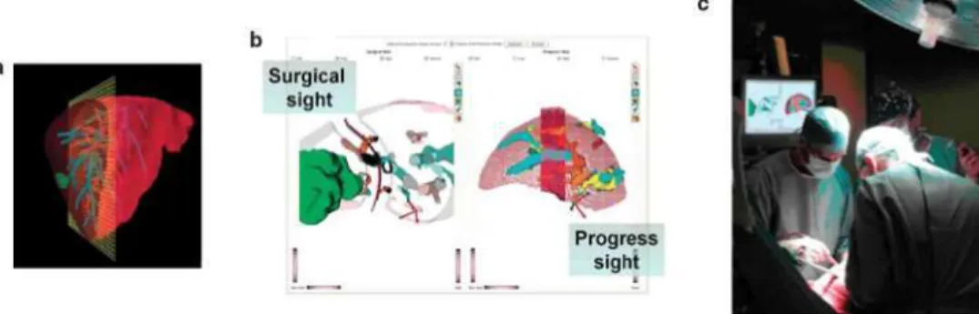
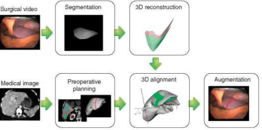
On the other hand, surgical navigation systems are providing new possibilities for MIS. These systems allow to transfer preoperative data, images and deci-sions to the OR (Figure la and b), and give the surgeon guidance during the procedure (Figure lc). Traditionally, the surgeon had to translate all this information mentally to the real scenario in front of him, with the difficulties this entails, most notably disorientation with respect to the medical image stud-ies. Image-guided surgery (IGS) systems have the potential to compensate these limitations. However, difficulties arise in the case of soft-tissue surgeries, such as those of the abdominal cavities, where there are technical challenges to cope with such as tissue deformation, shifting and other topological changes caused by the pneumoperitoneum, respiration, heart rate or tissue manipulation (8). Thus, organ tracking presents even greater difficulties due to said phenom-ena, and the use of fiducial markers and optical or electromagnetic systems to track organ deformations is not a feasible solution in many cases. Usually, location and tracking of organ position and deforma-tion are addressed by the use of intra-operative med-ical imaging modalities (most notably US, C T and MR) (9) and some specialised hardware based on laser or structured light technology (10,11). Tools' tracking is also a critical issue in any IGS solution, since optical and electromagnetic systems are the most widely used in the OR. However, they both add extra instrumentation in the OR, which can disturb surgeons during interventions (12).
Automatic analysis of surgical video sequences has applications in surgical training, evaluation and nav-igation systems (13). It provides useful information about the position of instruments and organs, surgical manoeuvres, measurements of distances or even an approximate 3D reconstruction of the surgical scene. Laparoscopic video images are an always available source of information and can be used without adding extra technological components in the OR. Moreover, with the development of high definition endoscopes
b
Surgical
sight
! LL'I
pfe^^i
Progresssight
and monitors, this information is sharper and cleaner than ever, and its analysis is surely to benefit from these trends. Following sections illustrate the potential of harnessing laparoscopic video images in order to improve surgical training, evaluation and guidance.
Laparoscopic video analysis: Literature overview
In order to increase patient safety during laparoscopic surgery, training systems can include overlays with tool tracking information, positions of anatomical structures and directions to follow in the surgical scenario. Immediate visual feedback can be obtained from training systems to help surgeons during their formation, monitoring and evaluating the entire pro-cess to minimize potential hazards, identify problem areas and find solutions. In training systems, cognitive skills training can benefit from video analysis by verting surgical videos into multimedia didactic con-tents adding texts, images, 3D virtual animations, etc. For these applications, segmentation and tracking are required to identify organs, regions of interest or areas where special attention should be paid during the surgical intervention, and warn about the proximity of delicate regions.
Multiple semiautomatic segmentation methods can be found in the literature, based e.g. on morphological border detection combined with growing region techniques (14), or in a combination of a growing segmentation and image enhancement steps based on the Bezier model (15). Moreover, some automatic segmentation approximations can be found, such as an algorithm based on a vague initial graph-based segmentation and a posterior merging multi-region stage, where a modification of the Fischer criteria is used (16).
Tool segmentation and tracking constitute another important contribution of video analysis. Keeping surgical tools centred in the field of view of the image is most important in MIS in order to reduce risks of damaging non-visible anatomical structures. Segmen-tation has been addressed using several approaches, using colour either on red, green, and blue (RGB) (17,18) or hue, saturation value (HSV) (19), space, geometrical properties such as border detection (20), or a combination of both (21).
Acquisition of the 3D shape of the surgical sce-nario has a wide variety of applications, especially in the field of minimally invasive surgery. There is a wide range of computer vision techniques for this purpose, and shading, stereoscopy and motion are the three main physical cues studied so far in a MIS
video. Shading refers to the light intensity reflected by a physical object dependent on the geometrical relation between its surface and the source of light. Shape from shading reconstruction techniques exploit this physical property of the behaviour of light in the surgical scene (22). Shape from stereo vision refers to the ability to acquire information on the 3D structure and the relative distances on the scene from two or more intensity images taken from different viewpoints (23,24). However, they are not always feasible in MIS applications because stereo-scopic endoscopes are not generally available in hospitals. Structure from motion techniques obtain depth information through movement analysis (25-27). These algorithms usually compute local frame-to-frame motion feature matching and are refined in a global optimization moving backward and forward through the whole sequence (called bundle adjustment). State-of-the-art technology solves this problem in a scenario with static objects or rigid movements. In MIS these solutions have therefore limited application because movements of anatomical structures are non-rigid. An interesting approximation uses simulataneous localization and mapping (SLAM) technology (28) which extracts in real-time not only the 3D position of some key landmarks, but also the 3D trajectory of a mono-cular camera moving rapidly through a previously unknown scene.
As briefly described here, there have been recent advances in video image processing and computer vision algorithms in order to enrich information pro-vided by laparoscopic surgical videos. In this paper we focus on two main applications of video analysis: Non-intrusive tracking of tools and organs, and 3D reconstruction of the surgical scene.
Non-intrusive tracking of organs and tools
Organs'' tracking
T h e main p r o b l e m to be solved w h e n tracking a structure in video sequences is the identification of the 2 D position and area of said structure. This is also a key issue in object recognition, scene and image u n d e r s t a n d i n g . Segmentation algorithms are thus n e e d e d not only for organ tracking, b u t also as a preliminary step for a 3 D reconstruction of shape or for a d d i n g or enhancing information present in the surgical scene. T h e s e algorithms exploit different image properties or organs a n d anatomical structures, such as colour, textures, edges or shading.
Characteristics of the segmentation algorithm developed for laparoscopic images will be constrained by its potential application. T h u s , a m a n u a l or semiautomatic algorithm will be sufficient for a u g m e n t a -tion of videos for surgical training. However, for I G S applications the segmentation process needs to be automatic and real-time. Laparoscopic images have b o t h small and large anatomical structures, which adds an additional level of difficulty for an automatic algorithm. It has to be n o t e d that a segmentation of structures is mostly limited to those which are clearly discernible in the image by the algorithm, as e.g. vessels by use of a contrast agent (8). T h e segmen-tation algorithm used for laparoscopic images has to be able to detect any region that has a slightly different colour c o m p a r e d to its neighbouring regions, because small differences in colour or texture m a y indicate different or special regions that should be taken into account w h e n performing a surgical technique. L a p -aroscopic sequences have features that make segmen-tation quite challenging, like the lack of sharp b o r d e r s , specularities, interreflections and colour variability due to clinical condition.
I n contrast with traditional approaches to the p r o b -lem, we propose in this p a p e r a m e t h o d which exploits shading as an additional segmentation criterion. A split and merge technique is chosen as the evolution strategy in the m e t h o d . T h e m e t h o d is based on a simplified illumination model with light source and
camera systems located in the same spatial point (30). A n illumination equation enables the definition of an optimal feature to distinguish structures, which is the gradient of the logarithm of image intensity. T h e logarithm simplifies the segmentation process w h e n constructing the gradient image by making an h o m o g -enisation in the region of a structure. T h e gradient of the logarithm reaches its m a x i m u m in the edge of a region, which is suitable for a combination of water-shed segmentation (split step) a n d a merging step with Hotelling's T - s q u a r e (or alternatively Mahalanobis distance) as the similarity metric. A n exemplary result of this m e t h o d is illustrated in Figure 2, where 2a shows the original image and 2b the segmented image. N o t e h o w the m e t h o d is able to segment b o t h large organs, i.e. liver or spleen, a n d anatomical details such as vessels.
Tools' tracking
A u t o m a t i c video analysis provides different solutions to detect the 2 D or 3 D position, and even orientation, of surgical instruments during a surgical process. S o m e applications will only require to follow a char-acteristic 2 D point of the tool, as for example its tip. F o r I G S applications, real-time, accurate and robust tracking of tools during the entire surgical p r o c e d u r e is required. Nevertheless, these conditions can be relaxed in an off-line analysis of a video sequence for objective evaluation. Video-based tool tracking m u s t cope with partial occlusions, the insertion and removal of different tools, a n d with image quality degradation caused by gas and smoke. O u r approach solves the p r o b l e m of calculating the 3 D position and orientation of a tool with only the 2 D information extracted from each frame of a video sequence. T w o pre-processing steps are n e e d e d , distortion correction and segmentation, before the application of the m a t h -ematical solution for it.
E n d o s c o p i c images are affected by image distor-tions, mainly geometrical deformations as vignetting
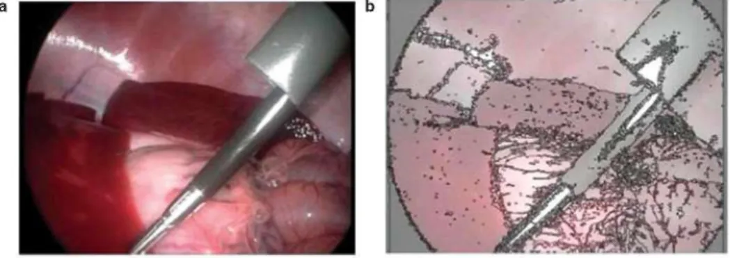
and barrel distortion (also named eye globe distor-tion), which cause straight lines to appear in the image as curves ones. A barrel distortion correction method uses a calibration procedure in order to define a rectification mapping function. The idea is to capture an image of a regular pattern drawn in a board in order to characterise the distortion. The rectification geometrical transformation is built finding the defor-mation between the captured image and its homolo-gous corrected one. This is illustrated in Figure 3 (3a represents the original frame and 3b the result of the distortion correction process) (31).
A segmentation algorithm is then used to detect surgical tools present in the surgical image. The algorithm exploits two key tools characteristics:
• surgical tools exhibit rigid motion;
• tools edges can be approximated by straight lines.
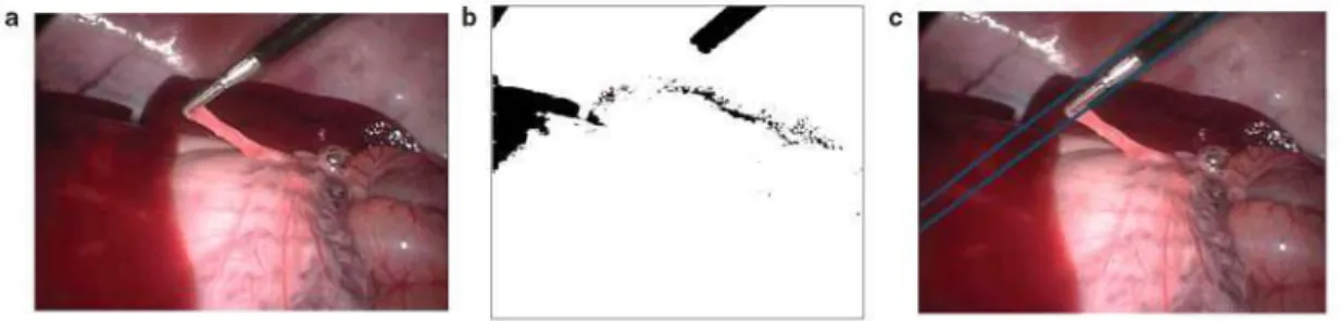
The algorithm takes two steps (Figure 4 - 4a is the original image), the detection of regions of interest (ROIs) (Figure 4b), and the detection of edges of tools (Figure 4c). ROIs are defined as the area which includes the tool black axis. An analysis of both colour components and geometrical properties such as ori-entation and size are used for this ROIs identification. The result is a simplified image where anatomical structures are deleted, as well as the metallic tool tip (Figure 4b). The detection of edges is achieved by combination of a laplacian gradient filter and Hough or Radon transforms. Finally, knowing the tool's cylindrical geometrical dimensions and its 2D projection as denoted by the detected borders, real 3D pose of tools is calculated. The mathematical equa-tion for this calculus is a descripequa-tion of the geometrical relation between the tools, trocars and the optical centre of the camera, and its complete description and explanation can be found in (31). Tool tip local-ization Root Mean Square error (in Z coordinates) has been characterised as 9.28 mmRMS, and the performance is dependent on the quality of the image of the tools (32). This tracking performance is good enough for gesture analysis and objective evaluation
of surgical manoeuvres. However, IGS applications require higher accuracy.
3D reconstruction of a surgical field from shading
A 3D reconstruction allows visualizing anatomy from different angles, which can enhance the appreciation of the surgical scene. Characteristics of the recon-struction of a 3D model from laparoscopic images will be strongly affected by its application, and its desired accuracy, robustness, velocity or computing cost will be constrained by this issue. For example, on a training scenario, where videos are pre-recorded and there is no real time limitation, conditions will be more lenient than on an IGS one. The handling of laparoscopic image data in IGS requires real-time algorithms and also careful synchronization; in particular between simultaneously acquired data from heterogeneous sources (the registration problem). Accuracy and robustness needed in this case scenario are extremely important due to risks carried out when operating a patient in the OR. Moreover, accurate methods have to be developed to register image space to physical or patient space based on video processing. We here address the reconstruction problem using shading information. As explained before, shape from shading (SfS) techniques amount to 3D reconstruct-ing a surface from an image captured by the endo-scope, given surface, light source and camera parameters. Although shading can be in a first analysis a poor approach, there are several reasons for using it:
(1) It is an interesting new approach, and
(2) although it implies some difficulties in laparo-scopic scenarios light source is controlled and fixed in its optical centre.
Two methods have been implemented for calculat-ing a depth map of the surgical scene, assumcalculat-ing a lambertian light interaction model with the surface. It is important to realise that by using shading informa-tion alone it is not possible to determine the absolute depth of the whole surgical scene. This limitation
Figure 4. Process to calculate the 3D position and orientation of a surgical tool in a laparoscopic photogram: (a) Original frame, (b) result of ROI identification and (c) detection of edges.
leads to a scale ambiguity problem. Knowing this computing the image intensity gradient in a given limitation, the purpose of the analysis of one frame direction. The 3D reconstruction is then calculated should be to achieve the relative depth map associated adding all the differential increments of depth and to the laparoscopic image, which is the objective of the dividing the gradient by the image intensity. An first method. The second method described here exemplary result of this method is shown in Figure 5, corrects for scale uncertainty by employing pre- illustrating the original image captured by the endo-operative knowledge of the shape under study. scope during the surgical intervention (Figure 5a) and
The first method proposes an algorithm based on the depth map associated to it, as well as the 3D a simplified illumination model of the surgical reconstruction combining scene and tool reconstruc-scene (29). The relative depth map is obtained by tion. Results in Figure 5b show how shines present in
the image limit the method resolution. Thus, to cancel their effects a pre-processing step is required. However, since the scope of this paper is to show the virtues of the 3D reconstruction method, its influence is omitted on the results given in Figure 5b. Their effects have been corrected in Figure 5c.
The second method uses a propagation SfS tech-nique initialized from the closest point to the cam-era (33). Its development is specifically motivated by augmentation of laparoscopic hepatectomy and aims at integrating visualization of major vessels and tar-geted tumours as well as preoperative resection planning information into the endoscopic images (Figure 6). It takes into account the assumption that the closest point in the image is the brightest, which is valid for the geometry of the liver. It then performs an iterative 3D reconstruction and 3D reg-istration via a modified ICP (Iterative Closest Point algorithm) to compensate for unknown scale and a correction of an unknown albedo and changing light-ing conditions.
An important limitation of shading information is the presence of noise sources, such as reflections or spec-ularities. These reflections originate in surrounding organs, and are quite unpredictable and variable due to movements caused by respiration, cardiac motion, and mechanical shift accompanying the surgical inter-vention. Our current work is focussed on correcting linearities in the acquisition of images and non-homogeneities in the radiance of the source of light.
Discussion
Video image analysis and computer vision algorithms have had a great impact in the last decades applied on
fields such as surveillance, control process or the entertainment business. Thus, it is our belief that such a potential source of information will prove invaluable in minimally invasive surgery, where it is an ever-present resource. In itself, video information is quite intuitive to the human eye. Still, there are several aspects which can be difficult to process men-tally and which a computer can handle much better, such as an automatic extraction of the 3D orientation of a surgical tool.
Video image analysis can tackle some human per-ceptual limitations and improve surgeons' awareness. But, unlike the human eye and brain, no algorithm is completely general, i.e. able to perform its intended function given any possible video input. Thus, it is critically dependent on certain unique assumptions about the real-world video scene it is expected to analyze. Without these assumptions, methods can produce ambiguous results. Thus, a relevant stage in the solution development process is modelling the surgical scene and the imaging system. The final application clearly conditions requirements.
Surgical video analysis provides an alternative sens-ing strategy to track tools and organs. An important advantage over sensor-based systems, optical or elec-tromagnetic (EM) tracking technologies, is that this solution does not require any additional hardware, and, compared to EM sensing, is immune to back-ground EM noise. Sensors also deteriorate ergonom-ics of surgical instruments and thus change the way the surgeon holds them or experiences their form and weight. Video analysis is already the technology used in some hybrid surgical simulators for tool tracking (34).
Video-based tracking of surgical tools allows the registration of objective surgical evaluation metrics
Surgical video
not only on controlled settings such as box trainers, but also in the OR. The information about kinemat-ics, regarding their position and trajectories, is avail-able straightaway, and this serves as the grounds to extract higher levels of evaluation with activity recog-nition algorithms (35). Useful information can then be extracted about the surgical steps taken, mainly related to the order of performance, repetitions and idle states. Although such analyses can be done man-ually (36), this is a tedious, time-consuming task. Automation of this task is achieved by manoeuvre video-based recognition systems. Moreover, a 3D reconstruction of the surgical scenario would allow to measure distances between instruments and targets or landmarks previously established, which can also be used for evaluation purposes.
In the field of IGS, endoscopic video analysis can be a valuable information source, as a means of tracking both organs and laparoscopic tools. Tracking systems are critical for computer-assisted image-guided inter-ventions: They provide the necessary real-time infor-mation on the localization of equipment (mainly surgical tools) and patients. Some efforts have been done using video in image guided applications for hepatic (37) and urologic (38) laparoscopic surgeries. Nevertheless, a video-based system is limited by image quality and field of view, and in this field of IGS this technology will probably complement, and not replace, other solutions.
Another relevant application of video analysis is superimposing pre-operatory information, like virtual anatomical reconstructions extracted from medical images, over surgical images, offering a view in trans-parency of the patient on the video images (39). One important strength of a video-based solution is its potentially higher accuracy, based on the fact that the alignment between the real and virtual anatomy of the patient is calculated from the same image where the information is superimposed. Some authors sug-gest that image-guided systems will be simplified if automated image registration is developed, which will need a real-time video analysis tool (40). However, there are important challenges to be tackled such as adaptation to the variability of quality and appearance of images, strongly affected by surgical actions and also by inter-patient biological variability.
There are also some limitations to be considered regarding the information extracted from a video sequence. Information at any given moment will be constrained by the camera's field of view. Indeed, any possible real-time adjustments regarding the tool and organ position and orientation will be limited by the field of view of the endoscope. Also, robust solutions are needed for tracking the camera shifting due to intra-operative movements and field of view
adjustments. This issue has not been tackled in this paper, but camera tracking solutions like the Mono-S I A M (28) are necessary in order to obtain fully-equipped navigation systems.
Our long-term vision is to achieve robust and instantaneous automatic video analysis algorithms, and there are important challenges in front of the scientific community. Thus, in order to achieve a quick and effective 3D reconstruction, an optimal combination of shading, motion and texture should be found, exploiting all cues present in endoscopic video images. Moreover, an important issue is the design of a complete tracking system, fusing endo-scope positioning system with a robust organ and tool tracking solution. Furthermore, video-based solu-tions can be combined with other hardware-based algorithms for precision increase.
Nowadays, the more challenging aspects are con-centrated in IGS applications, where accuracy and real-time needs are crucial issues. On the other hand, these constraints are laxed for evaluation purposes, where data analysis can be carried out offline and precision is not so critical. To date, real-time condi-tions for IGS applicacondi-tions have not been achieved. Surgical evaluation applications need complete and accurate task analysis of all operating techniques. It is not an easy task because it requires breaking down a surgical procedure into sub-tasks thanks to a rigorous observation process with expert surgeons.
Conclusion
Automatic analysis of MIS video has the potential to drive new solutions for alleviating needs of safe and reproducible training programs, objective and trans-parent evaluation systems and navigation systems to assist surgeons and improve patient safety. This source of information is always available and can be used without any additional intrusive hardware. This paper has shown new methods for tracking and 3D reconstruction, and has illustrated some of the current challenges to be solved in order to harness all its potential.
Declaration of interest: The authors report no
conflicts of interest. The authors alone are responsible for the content and writing of the paper.
References
1. Moorthy K, Munz Y, Sarker SK, Darzi A. Objective assess-ment of technical skills in surgery. BMJ: British Medical Journal 2003;327:1032.
3. Satava RM, Cuschieri A, Hamdorf J. Metrics for objective assessment. Surg.Endose. 2003;17:220-6.
4. Fried GM, Feldman LS. Objective Assessment of Technical Performance. World journal of surgery 2008;32:156-60. 5. Rosen J, Brown JD, Chang L, Barreca M, Sinanan M,
Hannaford B. The BlueDragon-a system for measuring the kinematics and the dynamics of minimally invasive surgical tools in-vivo. Proceedings- IEEE Int Conf on Robotics and Automation 2002:1876-81.
6. Chmarra MK. TrEndo Tracking System. Motion Analysis in Minimally Invasive Surgery. PhD. 2009.
7. Lamata P, Gómez EJ, Bello F, Kneebone RL, Aggarwal R, Lamata F. Conceptual framework for laparoscopic VR simu-lators. IEEE Comput Graph Appl. 2006;26:69-79.
8. Baumhauer M, Feuerstein M, Meinzer HP, Rassweiler J. Navigation in Endoscopic Soft Tissue Surgery - Perspectives and Limitations. Journal of Endourology 2008;22: 751-66.
9. Marvik R, Lango T, Tanguen GA, Iindseth F, Yavuz Y, Nagelhus Hemes TA. Image-guided laparoscopic surgery. Review and current status. Minerva Chir. 2005;60: 305-25.
10. Cash DM, Miga MI, Glasgow SC, Dawant BM, Clements LW, Cao Z, et al. Concepts and preliminary data toward the realization of image-guided liver surgery. J Gastrointest Surg. 2007;11:844-59.
11. Ackerman J, Keller K, Fuchs H. Surface Reconstruction of Abdominal Organs Using Laparoscopic Structured Light for Augmented Reality Applications. Proceedings of SPIE 2002; 4661:39-46.
12. Lamata P, Lamata F, Sojar V, Makowski P, Massoptier L, Casciaro S, et al. Use of the Resection Map system as guidance during hepatectomy. Surg Endose. 2010 Feb 23. [Epub ahead of print].
13. Sánchez-González P, Oropesa I, Cano AM, Gaya F, Lamata P, Sánchez-Margallo FM, et al. Endoscopic video images analysis for surgical training and image-guided sur-gery, World Congress on Medical Physics and Biomedical Engineering (WC2009); IFMBE Proceedings 2009:25:251-4. 14. Hsiao YT, Chuang CL, Jiang JA, Chien CC. Robust multiple objects tracking using image segmentation and trajectory estimation scheme in video frames. Image and Vision Com-puting 2006;24:1123-36.
15. Li Y, Lu D, Lu X, Liu J. Interactive color image segmentation by region growing combined with Image enhancement based on bezier model. Proceedings of the Third International Conference on Image and Graphics (ICIG'04) 2004:96-9. 16. Shu Y, Bilodeau GA, Cheriet F. Segmentation of
Laparo-scopic Images: Integrating Graph-Based Segmentation and Multistage Region Merging. Proceedings of the Second Cana-dian Conference on Computer and Robot Vision (CRV05) 2005:429-36.
17. Voros S, Long JA, Cinquin P. Automatic localization of laparoscopic instruments for the visual servoing of an endo-scopic camera holder. Med. Image Comput. Comput. Assist. Interv. 2006;9:535-42.
18. Casals A, Amat J, Laporte E. Automatic guidance of an assistant robot in laparoscopic surgery. Proceedings 1996 IEEE International Conference on Robotics and Auto-mation 1996;1:895-900.
19. Krupa A, Gangloff J, Doignon C, de Mathelin M, Morel G, Leroy J, et al. Autonomous 3-D positioning of surgical instru-ments in robotized laparoscopic surgery using visual servoing. IEEE Transactions On Robotics And Automation - Special Issue On Medical Robotics 2003;19:842-53.
20. Tonet O, Ramesh T U , Megali G, Dario P. Tracking endo-scopic instruments without localizer: image analysis-based approach. Stud. Health Technol Inform. 2006;119:544-9. 21. McKenna SJ, Nait Charif H, Frank T. Towards Video
Under-standing of Laparoscopic Surgery: Instrument Tracking. Conf. Image and Vision Computing New Zealand 2005: 317-21.
22. Zhang R, Tsai PS, Cryer JE, Shah M. Shape from Shading: A Survey. IEEE Transactions on pattern analysis and machine intelligence 1999;21:690-706.
23. Samaras D, Metaxas D, Fua P, Leclerc YG. Variable Albedo Surface Reconstruction from Stereo and Shape from Shading. IEEE Computer Society Conference on Computer Vision and Pattern Recognition (CVPR00) - 2000;1:1480.
24. Banks J, Bennamoun M, Kubik K, Corke P. An accurate and reliable stereo matching algorithm incorporating the rank constraint. Symposium on Intelligent robotic systemsl999; 23-32.
25. Newman PM. On the structure and solution of the simulta-neous localization and map building problem. Ph.D. disser-tation, University of Sydney, 1999.
26. Jebara T, Azarbayejani A, Pentland A. 3D Structure from 2D Motion. IEEE Signal Processing Magazine 1999;16:66-84. 27. Dellaert F, Seitz S, Thorpe C, Thrun S. Structure from
Motion without Correspondence. IEEE Computer Society Conference on Computer Vision and Pattern Recognition (CVPR00), 2000.
28. Davison AJ, Reid I, Molton N , Stasse O. Real-time single camera SLAM. IEEE Trans. Pattern Anal.Mach. Intell. 2007; 29:1052-67.
29. Chmarra MK, Grimbergen CA, Dankelman J. Systems for tracking minimally invasive surgical instruments. Min In vas Ther and Allied Technol. 2007;16:328-40.
30. Sánchez-González P, Gaya F, Cano AM, Gómez EJ. Segmen-tation and 3D reconstruction approaches for the design of laparoscopic augmented reality environments. Lecture Notes in Computer Science (LNCS) 2008:127-34.
31. Cano AM, Vara I, Sánchez-González P, Gómez EJ. Laparo-scopic image analysis for automatic tracking of surgical tools. Proceedings of Computer Assisted Radiology and Surgery (CARS 2008),2008;3:S279.
32. Cano AM, Gaya F, Lamata P, Sánchez-González P, Gómez EJ. Laparoscopic tool tracking method for augmented reality surgical applications, Lecture Notes in Computer Sci-ence LNCS 2008;5104:191-6.
33. Lamata P, Morvan T, Reimers M, Samset E, Declerck J. Addressing Shading-Based Laparoscopic Regis-tration. World Congress on Medical Physics and Biomedical Engineering 2009:189-92.
34. ProMIS simulator: http://www.haptica.com/promis.htm. Last access: 15th April.
35. Estebanez B, Jimenez G, Muñoz V, Garcia-Morales I, Bauzano E, Molina J. Minimally Invasive Surgery Maneuver Recognition Based on Surgeon's Model. 2009 IEEE/RSJ International Conference on Intelligent Robots and Systems, 2009.
36. Rosen J, Brown JD, Chang L, Sinanan MN, Hannaford B. Generalized approach for modeling minimally invasive surgery as a stochastic process using a discrete Markov model. Bio-medical Engineering, IEEE Transactions 2006:53:399-413. 37. Herline A, Stefansic JD, Debelak J, Galloway RL,
Improve Surgical Accuracy during Laparoscopic Partial Nephrectomy? Preliminary In Vitro and In Vivo Results. European Urology 2009;56:332-8.
39. Soler L, Ayache N , Nicolau S, Pennec X, Forest C, Delingette H, et al.. Virtual Reality, Augmented Reality and Robotics in surgical procedures of the liver. Perspectives in
Image-guided Surgery. Proceedings of the Scientific Work-shop on Medical Robotics, Navigation and Visualization (MRNV) 2004:476-84.