Facultad de Odontología Vol. 18, No. 3 July-September 2014
pp 175-179
Revista Odontológica Mexicana
ORIGINAL RESEARCH
www.medigraphic.org.mx
* Orthodontics Department Graduate, Graduate and Research School, National School of Dentistry, National University of Mexi-co (UNAM).
§ Orthodontics Department Professor, Graduate and Research School, National School of Dentistry, National University of Mexi-co (UNAM).
II Professor, Graduate and Research School, National School of Dentistry, National University of Mexico (UNAM).
This article can be read in its full version in the following page: http://www.medigraphic.com/facultadodontologiaunam ABSTRACT
At the Graduate and Research School of the National School of Dentistry, National University of Mexico (UNAM) we developed several analytic and descriptive methods. A statistical study of skeletal classifi cation was undertaken with a sample of 428 patients subjected to orthodontic treatment. Age range of selected patients was 8-40 years. Data were collected according to gender, age and skeletal malocclusion in order to assess the epidemiological panorama. After statistical analysis, it was found among other data, that 53.3% of the sample was in skeletal class I, 64.7% were female and 52.08% was found to be in the 13 to 19 year age range.
Key words: Prevalence, malocclusion, epidemiology, skeletal class. Palabra clave: Prevalencia, maloclusiones, epidemiología, clase esqueletal.
RESUMEN
En la División de Estudios de Postgrado e Investigación de la Uni-versidad Nacional Autónoma de México, en el Departamento de Ortodoncia, nosotros desarrollamos diferentes métodos analíticos y descriptivos, donde se realizó estudio estadístico de la clasifi cación esqueletal con una muestra de 428 pacientes que recibieron trata-miento de ortodoncia. Se seleccionaron personas entre 8 y 40 años de edad. Se capturaron datos de acuerdo a sexo, edad y maloclu-sión esqueletal para conocer el panorama epidemiológico. Después del análisis estadístico encontramos que el 53.3% de la muestra se encontraban en clase I esqueletal, que el 64.7% eran del sexo femenino y que el 52.08% se encontraba en el rango de edad de 13 a 19 años; además de otros datos.
Prevalence of malocclusions at the Orthodontics Department
of the Graduate School, National School of Dentistry, National
University of Mexico (UNAM)
Prevalencia de las maloclusiones en el Departamento de Ortodoncia de la
División de Estudios de Postgrado e Investigación de la Facultad de Odontología
de la Universidad Nacional Autónoma de México
Sergio Tokunaga C,* Mario Katagiri K,§ Haroldo Elorza PTII
INTRODUCTION
In recent years, many efforts have been devoted to issue a correct diagnosis. In the decade of the 50’s, the onset of cephalometric X-rays and the development of different descriptive and analytical methods conferred a new dimension to orthodontic diagnosis. There were limitations of study model classification according to Angle’s original concepts.1 The relationship between
upper and lower jaw plays an important role in the positioning of the molars.2,3 This relationship can only be
radiographically determined, and study models can only give an approximate idea of the position of the mandible.4
BACKGROUND
The main purpose of cephalometry was to research growth patterns and the maxillofacial system. Cephalometries have assumed an invaluable position
in the assessment of dento-facial proportions and clarifi cation of the anatomical bases of malocclusion.5
The importance of differential diagnosis among skeletal class I, II and III malocclusions will mainly be the manner in which the malocclusion is remedied with different techniques which can vary from the mesial or distal sliding of teeth as a whole, use of traction masks or extra-oral devices, and even resorting, or not, to conduct extractions.
www.medigraphic.org.mx
The introduction of Down’s analysis encouraged several researchers and clinical operators to develop their own analyses. As a result, countless cephalometric brands appeared in the market for skull analysis; they elicited great amounts of useful measurements. Nevertheless, Dr Steiner selected those he considered most important, he created his own analysis whereby he obtained the greatest amounts of clinical information performing a minimum amount of measurements.6 This analysis
was doubtlessly the most commonly used to assess anterior-posterior discrepancies of the maxillary-mandibular binomial, the ANB (point «A», maxillary, point «B» mandibular and point «N» cranial nasion).
The term «anomaly» can be limited or inadequate, but it represents a valid term for the clinical operator who tries to achieve a differential diagnosis of the patients he is going to treat. Anomaly can be described as the deviation with respect to the individual normality. Every individual is different from the others, with a morphogenetic pattern which is normal for him, nevertheless it can present differences in position, volume and shape of the components of the masticatory apparatus. When the term anomaly is thus understood, it facilitates its application in the diagnosis, since it separates what is considered normal from abnormal.7
Genetic aspects of occlusion are related to growth patterns of upper and lower jaws, their dental arches vary among each other mainly in anterior-posterior direction, as a consequence of growth vectors established by the genetic pattern.8
Information provided by the Instituto Nacional de Estadística, Geografía e Informática (National Institute of Geographic Statistics) (INEGI)9 reveals that in the
last 30 years, population under 14 years of age has noticeably increased, and therefore, the epidemiologic panorama shows that in the near future illnesses of childhood and of senior citizens will experience a noticeable change in distribution of general population.
Alterations in occlusion during puberty are very noticeable, to this we can add the fact of decrease in birth rates and stability of mortality rates, it is then feasible to understand that malocclusions, will, per force, suffer a re-distribution in cases presently treated by the clinical operator.
Growth and development play an important role in orthodontic treatment,10-13 as well as tooth loss, bone
metabolism14 and periodontal disease.15-17 All the
aforementioned factors represent new challenges for the ingenuity and skill of the clinical operator.
A modern country can ascertain the frequency of malocclusion problems suffered by its population.
Dental professionals can become aware of the scope of those problems and thus satisfy the requirements of those affl icted by them. Epidemiologists who gather information on the frequency of malocclusion, obtain data on the prevalence and severity of malocclusions. They furthermore cooperate with dentists, so that gathered information will become pertinent to those who treat patients.
HOW TO APPROACH THE PROBLEM
What amount of skeletal class I, II and II are treated at the Orthodontics Department of the Graduate School, National School of Dentistry, National University of Mexico (UNAM)?
VINDICATION
The Orthodontics Department, Graduate School, National School of Dentistry, National University of Mexico, (UNAM) treats a great number of patients for their different malocclusions, but up to the present date, their epidemiological panorama is as yet unknown. We actually ignore the amount of skeletal class I, II and III malocclusions we are treating. We equally ignore what incidence and prevalence we have periodically. This information is necessary, since, in a more objective manner, if we ascertain what type of skeletal malocclusions we are treating, it would be possible, for instance, to improve the techniques and treatment philosophies which are taught at the UNAM. We would then be able to guide, according to population requirements, the possibility of undertaking surgical or orthopedic treatments which will contribute to solve skeletal problems as well as to diversify treatments in the clinic.
STUDY TYPE
Descriptive, cross-sectioned and retrospective study.
OBJECTIVES
1. To ascertain the number of patients treated in the service.
2. To ascertain the amount of skeletal I, II and III malocclusions which are treated at the Orthodontics Department of the Graduate School, National School of Dentistry, National University of Mexico. 3. To quantify skeletal malocclusions present
according to gender and age.
www.medigraphic.org.mx
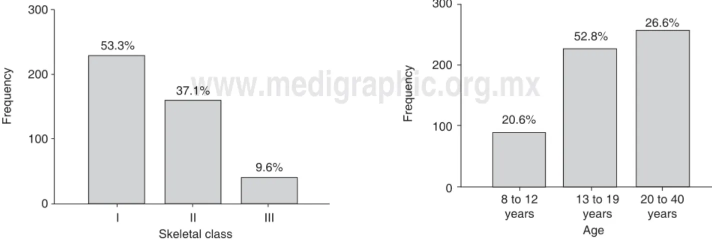
Figure 1. Skeletal class frequency.
Frequency I 53.3% II 37.1% Skeletal class III 9.6% 300 200 100 0
Figure 2. Frequency according to gender.
Frequency 35.3% M Gender 64.7% F 300 200 100 0
Figure 3. Frequency according to age.
Frequency Age 20 to 40 years 26.6% 13 to 19 years 52.8% 8 to 12 years 20.6% 300 200 100 0 METHOD
Files were selected from patients treated at the Orthodontics Department of the Graduate School, from 1998-2004.
To obtain measurement of SNA, SNB and ANB angles (described by Steiner) of initial cephalometric X-rays (S0 sella, N = nasion, A = point Aβ maxillary, and B= point B mandibular) and verify the fi le; Rank taken was: Skeletal class I 2o ± 2o. Skeletal class II 3o
or more and Skeletal class III -1o or more.
The sample was made up according to gender, age and skeletal malocclusion.
STATISTICAL ANALYSIS
Data were captured according to gender, age and skeletal malocclusion, in a calculus sheet of the statistics program SPSS for windows 11.0. Data analysis revealed the following results.
RESULTS
The present study was composed of a 428 patient sample. Age range was 8 to 40 years. Mean age was 16.85 years.
According to the frequency of skeletal class table, there were 228 patients in class I, which represented 53.3% of the total sample, 159 patients in class II, which represented 37.1% and only 41 patients in class III which represented 9.6% (Figure 1).
According to the frequency of gender table, there were 64.7% females (277) and 35.3% males (151)
(Figure 2).
In the table which depicts frequency according to age groups, we fi nd, in group 1, age ranges 8 to 12
years (88 patients), represents 20.6%. In group 2, age ranges 13 to 19 years (226 patients) represents 52.8%, and group 3, age ranges 20 to 40 years (114 patients) represents 26.6% (Figure 3).
When considering skeletal class and gender, we find that out of 228 (53.3%) patients in class I, 151 (35.3%) were female and 77 (18%) were male. Out of the 159 (37.1%) patients in class II, 102 (23.8%) were female and 57 (13.3%) were male. Out of the 41 (9.6) patients in class III, 24 (5.6%) were female and 17 (4.0%) were male (Table I).
Square χ2 was calculated. χ2 = 0.936, p = 0.626.
This indicates that there is no association between gender and skeletal class.
When considering age and gender, it was found that out of the 277 (64.7%) female patients, 52 (12.1%) were in the 8-12 year age range, 146 (34.1%) were in the 13 to 19 year age range, and 79 (18.5%)
www.medigraphic.org.mx
Este documento es elaborado por Medigraphic
Table I. Association between gender and skeletal class.
Skeletal class-gender Gender F M Total Skel. I Count 151 77 228 Class % of total 35.3% 18.0% 53.3% II Count 102 57 159 % of total 23.8% 13.3% 37.1% III Count 24 17 41 % of total 5.6% 4.0% 9.6% Total Count 277 151 428 % of total 64.7% 35.3% 100.0%
Table II. Association between gender and age.
Sexo - edad 8 to 12 13 to 19 20 to 40 Total Gender F Count 62 146 79 277 % of total 12.1% 34.1% 18.5% 64.7% M Count 36 80 35 151 % of total 8.4% 18.7% 8.2% 35.3% Total Count 88 226 114 428 % of total 20.6% 52.8% 26.6% 100.0%
Table III. Association between skeletal class and age.
Clase esqueletal - edad
8 to 12 13 to 19 20 to 40 Total Skel. I Count 38 124 66 228 Class % of total 8.9% 29.0% 15.4% 53.3% II Count 40 78 41 159 % of total 9.3% 18.2% 9.6% 37.1% III Count 10 24 7 41 % of total 2.3% 5.6% 1.6% 9.6% Total Count 88 226 114 428 % of total 20.6% 52.6% 26.6% 100.0% were in the 20 to 40 year age range. Out of the 151
(35.3%) male patients, 36 (8.4%) were in the 8 to 12 year age range, 80 (18.7%) were in the 13 to 19 years age range and 35 (8.2%) were in the 20 to 40 years age range (Table II).
When calculating square χ2, it was found that
square χ2 = 2.269, p = 0.322. This revealed there
was no association between age and gender (Table
III).
When calculating square χ2 it was found that:
square χ2 = 6.302, p = 0.178. This revealed the fact
there was no association between skeletal class and age.
When considering skeletal class and age, we found that out of the 228 (53.3%) patients in skeletal class I, 38 (8.9) were in the 8 to 12 year old range, 124 (29.0%) were in the 13 to 19 year age range and 66 (15.4%) were in the 20 to 40 years age range. Out of the 159 (37.1%) patients in skeletal class II, 40 (9.3%) were in the 8 to 12 years age range, 78 (18.2%) were in the 13 to 19 years age range and 41 (9.6%) were in the 20 to 40 years age range. Out of the 41 (9.6%) patients in skeletal class III, 10 (2.3%) were in the 8 to 12 years age range, 24 (5.6%) were in the 13 to 19 years age range and 7 (1.6%) were in the 20 to 40 years age range (Figure 4).
DISCUSSION
When conducting epidemiological research on malocclusion, it is necessary to somehow characterize the population which is being assessed.18 The population can be satisfactorily
classifi ed according to several methods. Alongside with a strong connotation with orthodontic diagnosis,
several researchers in this area concur in asserting that different cephalometric measurements can refer morphological variations. Out of the aforementioned, angles SNA, SNB, and ANB (Riedel 1952) are those which stand out most.
In the realm of orthodontic literature, many studies propose cephalometric norms to endorse harmony between facial profile and skeletal class in several population groups in the world. We could mention some authors who, in addition to the aforementioned angles, chose to endorse their populations with other analyses. Such is the case of Dr Björk, or Dr Tweed, and more recently, Drs Perez and Rosales, who in 1990 stated that through observation of cephalometric measurements it was possible to clarify anatomical bases of malocclusion in different populations, as well as the need to characterize individuals, from an anthropological-physical point of view and thus be able to analyze them from a cephalometric perspective.19-22
An article where research was conducted on facial prognathism was cephalometrically endorsed with the use of ANB angle and similar work where there could be variations in the racial groups factor as well as the age factor.23-26
www.medigraphic.org.mx
Figure 4. Number of patients, age and skeletal class.
Number of patients Skeletal class Age 8 to 12 13 to 19 20 to 40 I II III 140 120 100 80 60 40 20 0 CONCLUSIONS
After statistical analysis we found that maximum percentages were as follows: 53.3% of the sample was in skeletal class I, 64.7% were female and 52.08% were in the age range of 13 to 19 years. Minimum percentages were as follows: 9.6% were in skeletal class III, 35.3% were male and 20.6% were in the 8-12 years age range. This leads us to infer with respect to statistical probabilities of different clinical pictures which we might encounter and show the importance of acquiring knowledge on the growth and development of facial structures, in order to use them at developmental early stages and thus correct skeletal discrepancies, since the patients included in the 8-12 years age group were susceptible to orthopedic skeletal changes and they represented 20.6% of the sample. In a stricter sense, when speaking of anterior-posterior discrepancy of the upper jaws, 45% of all cases should be surgically treated. Moreover, 26.6% of the sample corresponds to age range 20-40 years; this corresponds to adulthood, and represents a challenge to orthodontic techniques. This is due to the different complications inherent to age such as periodontal disease, bone metabolism, loss of teeth, etc.
REFERENCES
1. Jacobson A. Radiographic cephalometry. Ed. Quintessence Publishing Co. Inc. 1995.
2. Alves CR, Noriega E. Actualizaciones en Ortodoncia y Ortopedia Funcional de los Maxilares. San Paulo Brasil; Artes médicas: 2002.
3. Rakosi T. Orthodontics, diagnosis color atlas of dental medicine, New York, U.S. 1993.
4. Graber T, Vanarsdall R Jr. Orthodontics. Current principles and techniques. Third edition. Ed. Mosby. 2000.
5. Mayoral HG. Ficción y realidad en ortodoncia. Ed. AMOLCA, Caracas, 1997.
6. Zamora MOC. Compendio de cefalometría, análisis, clínica y práctica. Ed. AMOLCA Colombia 2004.
7. Echarri LP. Diagnóstico en ortodoncia estudio multidisciplinario. Barcelona: Nexus, 2002.
8. Quiroz AO. Manual de ortopedia funcional de los maxilares y ortodoncia interceptiva. Caracas, Venezuela. 1995.
9. INEGI XII. 2000, pp. 166-177.
10. Bishara SE. Ortodoncia. Ed. Mc Graw Hill. 2003.
11. Goldsman S. The variations in skeletal and denture patterns in excellent adult facial types. Angle Orthod. 1959; 29: 63-92. 12. Shields TE, Little RM, Chapko MK. Stability and relapse of
mandibular anterior alignment: a cephalometric appraisal of fi rst-premolar-extraction cases treated by traditional edgewise orthodontics. Am J Orthod. 1985; 87 (1): 27-38.
13. Harris EF et al. Effects of patient age on postorthodontic stability in class II, division 1 malocclusions. Am J Orthod Dentofacial Orthop. 1994; 105: 25-34.
14. Chavanaz M. Screening and medical evaluations of adult, absolute and relative contraindications for invasive dental procedures. Indiana Dent Assoc J. 1999; 78 (3): 10-17.
15. Swanson WD, Riedel RA, D’Anna JA. Posretention study: insidence and stability of rotated teeth in humans. Angle Orthod. 1975; 45 (3): 198-203.
16. Wennström JL et al. Periodontal tissue response to orthodontic movement of teeth with infrabony pockets. Am J Orthod. 1993; 103: 313-319.
17. Artun J, Urbye KS. The effect of orthodontic treatment on periodontal bone support in patients with advanced loss of marginal periodontium. Am J Orthod. 1988; 93: 143-148. 18. Canales FH, Alvarado EL, Pineda EB. Metodología de la
investigación. Manual para el desarrollo de personal de salud. México: Organización Mundial de la Salud, 1992.
19. Bindari EL, Hammad A. Primary health care reviews, guidelines and methods. World Health Organización. Geneve, 1992, pp. 3-11.
20. Little RM. The irregularity index: a quantitative score of mandibular anterior alignment. Am J Orthod. 1975; 75: 554-563. 21. Ackerman JL, Proffi t WR. The characteristics of malocclusion:
approach to classifi cation and diagnosis. Am J Ortod. 1969; 56: 443-454.
22. Harris EF, Johnson MG. Heritability of craniometric and occlusal variables: a longitudinal sib analysis. Am J Orthod. 1991; 99: 258-268.
23. Bjoerk A, Krebs A Solow B. A method for epidemiological registration of malocclusion. Acta Odont Scand. 1964; 22: 27-41. 24. Galvão CAAN. O ângulo ANB em várias populações do mundo.
Rev Odont UNESP. 1984; 13: 163-74.
25. Civolani MI. Padrões Cefalométricos de Tweed, Steiner, Wylie e Downs, aplicados a Indivíduos brasileiros com “Oclusão normal” Piracicaba, Faculdade de Odontologia. UNICAMP, 1977 (Tese Mestrado) Volume 27 - N l - Janeiro/Fevereiro/Março/Abril – 1994.
26. Jacobson A. Prognathism in the South African Negro. J Dent Ass S Afr. 1976; 31: 613-619.
Mailing address: Haroldo Elorza PT