www.medigraphic.org.mx
Correction of an skeletal anterior open bite
with mini-screws and a modifi ed bite block
Corrección de una mordida abierta anterior esquelética
mediante miniimplantes y un bite block modifi cado
Ángel Eduardo Miranda Salguero,* Alfredo Sánchez Valverde§* Resident of the Orthodontics Specialty. § Professor of the Orthodontics Specialty.
Division of Post-Graduate Studies and Research Division of the Faculty of Dentistry, National Autonomous University of Mexico.
© 2017 Universidad Nacional Autónoma de México, [Facultad de Odontología]. This is an open access article under the CC BY-NC-ND license (http://creativecommons.org/licenses/by-nc-nd/4.0/).
This article can be read in its full version in the following page: http://www.medigraphic.com/ortodoncia
RESUMEN
La mordida abierta esquelética anterior puede ser tratada con mi-niimplantes, ya que proveen un anclaje absoluto para corregir me-diante la intrusión de los molares maxilares con un adecuado con-trol, en el presente caso se ayudó con un bite block modifi cado. Se presenta el caso de un paciente de 15 años de edad, dolicofacial, con una maloclusión clase II subdivisión 1, mordida abierta anterior de -4.5 mm e incompetencia labial. Etiología: por una altura facial anterior superior disminuida. El objetivo del tratamiento fue conse-guir una adecuada sobremordida anterior disminuyendo la altura maxilar dentoalveolar posterior. La cirugía ortognática se le indicó pero fue rechazada. Por lo tanto se realizó una intrusión molar su-perior con dos miniimplantes colocados en el paladar más un bite
block modifi cado activado con cadenas elásticas. Resultados: La
intrusión molar superior fue de -2 mm, la sobremordida anterior cambió a +2 mm, hubo autorotación mandibular y se mejoró el per-fi l facial. Conclusiones: La mordida abierta anterior fue corregida con un adecuado control de la intrusión evitando alguna inclinación bucal de los molares.
Key words: Open bite, mini-screws, molar intrusion, bite-block.
Palabras clave: Mordida abierta, miniimplantes, intrusión molar, bite-block. ABSTRACT
Skeletal anterior open bite may be treated with mini-screws since they provide an absolute anchorage to correct it through maxillary molar intrusion. With an adequate control a bite block was used in this case to help correct the malocclusion. Case report: A 15 year-old male dolichofacial patient with an Angle class II division 1 malocclusion, a -4.5 mm anterior open bite and incompetent lips is hereby presented. Etiology: reduced anterior upper facial height. The treatment goal was to obtain a normal anterior overbite decreasing the posterior maxillary dentoalveolar height. Orthognathic surgery was indicated but the patient refused it. So the treatment consisted in two mini-screws implanted on the palatal side and a modified fi xed bite block, activated with elastomeric chains. Results: A molar intrusion of 2 mm was achieved; the anterior overbite changed to +2 mm, a mandibular counterclockwise rotation took place and the facial profi le was improved. Conclusions: The anterior open bite was corrected with a good control during molar intrusion and without buccal tipping.
Vol. 5, No. 2 April-June 2017 pp 102-110
Revista Mexicana de Ortodoncia
CASE REPORT
BACKGROUND INFORMATION
Open bite is a malocclusion of the vertical plane, due to a lack of anterior contact. It may be of skeletal or dental origin.1 The malocclusion is attributed to
a multifactorial etiology since it may be genetic, anatomical and environmental as well as due to the development of pernicious oral habits,2,3 The
prevalence of anterior open bite is 3.5% (8 to 17 years of age).4 In Mexican population, at early ages, the
open bite is related to habits in 96.6% of the cases.5
The open bite patient is characterized by adenoid facies or long face syndrome with lip incompetence caused by incisor proclination and lack of overbite, which when combined with habits cause gingival infl ammation.6 The retruded position of the mandible
decreases the mentocervical distance and shortens
the projection of the chin.2 This mandibular position
may also decrease the airways.
To differentiate if it is a skeletal or dental open bite cephalometric analysis are used. A dental open bite is characterized by incisors in infraocclusion, while
www.medigraphic.org.mx
plane respectively.
Among the skeletal cephalometric data, the inclination of the Mandibular Plane will be found increased as well as a downward and rearward inclination of the mandibular ramus, a short mandibular ramus, a more marked antegonial notch and a shortened posterior facial height, which increases the mandibular angle and the anterior facial height.2 If the problem is in the maxilla an increased
inclination of the palatal plane will be present due to excess of posterior growth or to poor anterior growth of the maxilla.10 In addition to the vertical analysis of
the anterior and posterior facial height, the height of the dentoaveolar processes must be measured as indicated by Sean Biggerstaff et al.11 When a vertical
problem is evident in a skeletal open bite, Sassuoni described that the occlusal, palatal and mandibular planes make a closer convergence near the posterior portion of the face.12
Several therapies have been described to correct the skeletal or dental open bite such as functional education of the tongue; extraction of fi rst premolars, second premolars or fi rst molars; high-pull headgear, chin cup, elastics, multi-loop arch wires (MEAW), bite blocks, tongue cribs and functional appliances. In the skeletal open bite orthognathic surgery will always be the standard treatment.8,13-17
In the last decade molar intrusion through the use of mini-implants has been described to correct the skeletal open bite thus causing a decrease in incisor extrusions. It has also been described to provide favorable cosmetic results such as a forward rotation of the mandible and a reduction in anterior facial height. It has even been used as an alternative for patients who have not agreed to orthognathic surgery.18,19
There are different ways to perform molar intrusion with mini-implants, whether placed palatally, labially or in both sides as well as with the help of other appliances.17,18,20-23
In Graber’s book it is stated that molar intrusion with mini-implants must be designed to provide three-dimensional control of the tooth regarding rotation,
Diagnosis and etiology
A male patient of 14 years and 9 months of age attended the Orthodontics Clinic of the Division of Post-Graduate Studies and Research, UNAM, with the following reason for consultation: «My upper teeth are too forward». Regarding pathological data, the patient mentioned being under treatment for rhinitis. Oral habits: Mouth breathing. The facial photographs showed a convex profi le, lip incompetence and poor projection of the chin; the smile photograph revealed an anterior open bite (Figure 1). Upon intraoral examination the patient presented a right molar class II malocclusion, bilateral canine class II, bilateral posterior cross bite (-2 mm), anterior open bite (-4.5 mm) and spaces between the incisors (Figures 2
and 3). In the panoramic radiograph no caries may
be observed, there are good root inclinations and third molars are under development. The analysis of the lateral head fi lm identifi ed a skeletal class II due to mandibular retro position (ANB: 6o, Convexity
of Ricketts: 5 mm), vertical growth: (SN-PM: 41o,
Facial Axis: 83o), dental biproclination (1U-SN: 110o,
IMPA: 101o). As etiology it was determined that a
poor anterior maxillary growth and an increased posterior maxillary height (N-ENA: 52 mm palatal plane: -7o) was the cause of the malocclusion (Figure
4). Through the use of CT cone-beam it may be
observed that there is no damage to the cortical bone
(Figure 5).
Treatment goals
Facial: to improve the profile, decrease lip incompetence and improve incisor exposure during smile. Skeletal: to correct the skeletal open bite, favor a CCW mandibular rotation and control the vertical dimension. Dental: coordinate arches, achieve molar and canine class I, retrocline the incisors, match midlines, and obtain occlusal contact. Functional: to improve respiratory function through open bite closure.
www.medigraphic.org.mx
Treatment alternatives
1. Orthognathic surgery: LeFort I surgery for maxillary impaction. The patient did not accept this treatment alternative.
2. Extraction of four fi rst premolars: This would allow the correction but could affect the facial profi le in the future, as well as increase the vertical dimension. 3. Intrusion of upper molars through the use of
mini-implants: molar intrusion has been reported as the most stable option when it comes to performing a camouflage treatment. To achieve this, it was proposed the placement of 2 mini-implants on the palate between the first and second molars, combined with a device similar to a bite block cemented into the upper molars, which would be activated by elastomeric chains, from the
mini-implants to the device. This would allow an intrusion without causing undesirable inclination movements.
Treatment progress
Fixed appliances: 0.022” × 0.028” slot Edgewise brackets and tubes.
1. Alignment and Leveling Phase: 0.014”, 0.018” and 0.018” × 0.025” NiTi arch wires.
2. Work phase: Placement of 2 mini-implants (1.8 × 9 mm) on the palate between the first and second molars; the bite block was cemented with glass ionomer. The bite block was modified by adding two hooks between fi rst and second upper molar palatally, activation was made with closed elastomeric chains, 3 Oz of force on each side in
Figure 2.
Initial intraoral photographs.
Figure 1.
www.medigraphic.org.mx
ideal bends and 60 progressive lingual torque. • Interconsultation with Periodontics: Phase
I (hygiene and control) to treat a moderate plaque- related gingivitis; support and education
RESULTS
A good alignment and a proper occlusal settlement were achieved. Retraction of the upper and lower
Figure 3.
Initial study models.
Figure 4.
Initial lateral head fi lm, panoramic radiograph and CBCT images.
www.medigraphic.org.mx
incisors was performed thus obtaining a normal overbite and a Class I molar, premolar and canine relationship. A good arch form and an adequate gingival aesthetics were also obtained. Molar intrusion was -2 mm, the overbite changed to 2 mm thus favoring a mandibular rotation. The facial profi le improved, the lip incompetence was eliminated and a better chin projection was obtained. The anterior facial height decreased by 2 mm, in addition to a better incisor exposure at smile. Radiographically, an adequate root parallelism may be observed as well as
an increase of the airways at pharyngeal level (Figures
7 to 10 and Table I).
DISCUSSION
In the case hereby presented the goals were to correct an anterior open bite caused by a skeletal problem. According to the analysis of Sean Biggerstaff et al.,11 it was found that the malocclusion was caused
due to a growth defi ciency of the anterior portion of the maxilla, in contrast to the inclination of the Ricketts
Figure 5. Initial CBCT images.
Figure 6.
T r e a t m e n t p r o g r e s s : m i n i -implants in the palate, between the fi rst and second molars and modifi ed bite-block activated with elastomeric chains.
www.medigraphic.org.mx
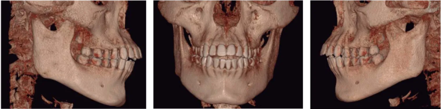
Figure 8. F i n a l l a t e r a l h e a d f i l m a n d ortopantomography. Figure 7. F i n a l f a c i a l a n d i n t r a o r a l photographs.www.medigraphic.org.mx
Este documento es elaborado por Medigraphic
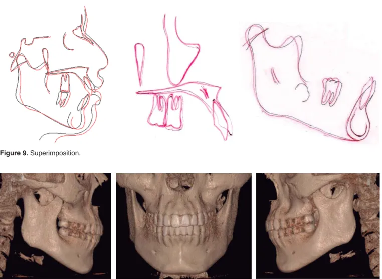
Figure 9. Superimposition.
Figure 10. Final CBCT images.
space of 4-5 mm ideal for mini-implant placement. In this case, a similar placement site was chosen.24
P o s t e r i o r i n t r u s i o n w i t h m i n i - i m p l a n t s t o correct an open bite is well described by several authors.1,14-25 Cifter et al using a finite element model
for transpalatal (Goshgarian) arch and mini-implants found that there is more intrusion of the buccal roots than the palatal roots.21 When mini-implants
are placed only on the palatal side some auxiliary appliance should always be placed to avoid an undesirable lingual crown torque as described by Buschanget and Xun et al.23,22
The applied force in this case was 3 Oz per side by means of elastomeric chains to avoid root resorption. Some cases using NiTi spring coils of 150 g per each side have been described.1,22,26 Greater intrusion
forces have been presented in cases from Everandi et al with the use of zygomatic mini-plates combining the use of 200 g springs.20
The results in this clinical case show that an intrusion of 2 mm in the fi rst molar and of 1mm in the second molar are similar to that reported in the studies of Scheffler et al, who determined that for a 2 mm intrusion there will be a 4 mm overbite.10 Alsafadi et al
and Scheffeler et al. indicated that when performing this movement, passive extrusion of the lower molars must be controlled. This may be avoided with the use of mini-implants for intrusion or only as an indirect anchorage and the use of acrylic plates.27 The modifi ed bite block
cemented to the fi rst and second molars helped crown-root three-dimensional control during intrusion and prevented lower molar extrusion. Other authors such as Bushang et al. analyzed intrusion performed only with palatal mini-implants using coil springs attached to a palatal expander and the results indicated that the molar intrusion was controlled properly.22
One advantage was that there were interproximal spaces in both the upper and lower arches, which
www.medigraphic.org.mx
favored the retroclination to consume those spaces (retroclination or retraction of the teeth), thus decreasing the open bite. It also favored lip closure and improved the aesthetics considerably. It should be noted how Daguchi et al. indicated that molar intrusion combined with retraction of the anterior segment, favors signifi cant aesthetic changes due to a CCW mandibular rotation and improves the stability of the case in contrast to performing only incisor and canine extrusion.25,27
Several authors such as Scheffer et al., Härt et al, Kuroda et al, Buschang et al., Xun et al. and Alsafadi et al. stated that the effects of intruding the upper posterior teeth are favorable causing a mandibular rotation of 1o to 4o, hence improving chin projection,
decreasing the facial height and the mandibular angle. Similar results were also achieved in the present case but not signifi cantly.18,19,22,23,26,27
CONCLUSIONS
• Upper molar intrusion mechanics is described as being more stable than extrusion of the anterior teeth for open bite closure.
• The use of two mini-implants on the palate combined with a modifi ed bite block provided excellent three-dimensional control avoiding any unwanted upper molar tipping during intrusion.
REFERENCES
1. Park YC, Lee HA, Choi NC, Kim DH. Open bite correction by intrusion of posterior teeth with miniscrews. Angle Orthod. 2008; 78 (4): 699-710. doi: 10.2319/0003-3219(2008)078[0699:OBCBI O]2.0.CO;2.
nocivos en una muestra de mexicanos. Rev Mex Ortod. 2014; 2 (4): 220-227. doi: 10.1016/S2395-9215(16)30038-1.
6. Proffi t W, Fields H, Sarver D. Contemporary orthodontics. 2007. doi: 10.1038/sj.bdj.2012.829.
7. Graber TM, Vanarsdall RL VK. Orthodontics. Current Principles & Techniques. 4th ed.; 2005.
8. Kim YH. Anterior openbite and its treatment with multiloop edgewise archwire. Angle Orthod. 1987; 57 (4): 290-321. doi: 10.1043/0003-3219(1987)057<0290:AOAITW>2.0.CO;2. 9. Janson G, Laranjeira V, Rizzo M, Garib D. Posterior tooth
angulations in patients with anterior open bite and normal occlusion. Am J Orthod Dentofac Orthop. 2016; 150 (1): 71-77. doi:10.1016/j.ajodo.2015.12.016.
10. Ricketts RM. Perspectives in the clinical application of cephalometrics. Angle Orthod. 1981; 51 (2): 115-150. doi:10.1043/0003-3219.
11. Biggerstaff RH, Allen RC, Tuncay OC, Berkowitz J. A vertical cephalometric analysis of the human craniofacial complex.
Am J Orthod. 1977; 72 (4): 397-405. doi:
10.1016/0002-9416(77)90352-9.
12. Sassouni V. The class II syndrome: differential diagnosis and treatment. Angle Orthod. 1970; 40 (4): 334-341. doi: 10.1043/0003-3219(1970)040<0334:TCISDD>2.0.CO;2. 13. Uribe FA, Chandhoke TK, Nanda R. Individualized orthodontic
diagnosis. Second Edi. Elsevier Inc.; 2014. doi:
10.1016/B978-1-4557-5085-6.00001-1.
14. Gurton AU, Akin E, Karacay S. Initial intrusion of the molars in the treatment of anterior open bite malocclusions in growing patients. Angle Orthod. 2004; 74 (4): 454-464. doi:10.1043/0003-3219(2004)074<0454:IIOTMI>2.0.CO;2.
15. Iscan HN, Sarisoy L. Comparison of the effects of passive posterior bite-blocks with different construction bites on the craniofacial and dentoalveolar structures. Am J Orthod
Dentofacial Orthop. 1997; 112 (2): 171-178.
doi:10.1016/S0889-5406(97)70243-9.
16. Erverdi N, Usumez S, Solak A, Koldas T. Noncompliance open-bite treatment with zygomatic anchorage. Angle Orthod. 2007; 77 (6): 986-990. doi:10.2319/101206-422.1.
17. Kuo CC, Chen YJ, Lai EHH, Yao CCJ, Chang JZC. Long-term stability of an adult class III open-bite malocclusion treated with multiloop edgewise archwire. J Dent Sci. 2009; 4 (3): 149-158. doi:10.1016/S1991-7902(09)60020-9.
18. Hart TR, Cousley RRJ, Fishman LS, Tallents RH. Dentoskeletal changes following mini-implant molar intrusion in anterior open bite patients. Angle Orthod. 2015; 85 (6): 941-948. doi: 10.2319/090514-625.1.
19. Kuroda S, Sakai Y, Tamamura N, Deguchi T, Takano-Yamamoto T. Treatment of severe anterior open bite with skeletal anchorage in adults: Comparison with orthognathic surgery outcomes. Am J
Orthod Dentofac Orthop. 2007; 132 (5): 599-605. doi:10.1016/j.
ajodo.2005.11.046.
20. Erverdi N, Tosun T, Keles A. A new anchorage site for the treatment of anterior open bite: zygomatic anchorage. Case
Convexity 0-2 mm 7 mm 5 mm Maxillary depth 90o 94o 92o FH_MP 26o 28.8o 26.8o SN-MP 32o 41o 39o Facial depth 87o 88o 89o Facial axis 90o 82o 83o AFH 112 mm 129 mm 127 mm Upper lip 1-4 mm +1 mm -1 Lower lip 0-2 mm +4 mm +1
www.medigraphic.org.mx
report. World J Orthod. 2002; 43 (3): 147-153. doi:10.1067/ mod.2002.128731.
21. Çifter M, Saraç M. Maxillary posterior intrusion mechanics with mini-implant anchorage evaluated with the fi nite element method. Am J Orthod Dentofacial Orthop. 2011; 140 (5): 233-241. doi: 10.1016/j.ajodo.2011.06.019.
22. Buschang PH, Carrillo R, Rossouw PE. Orthopedic correction of growing hyperdivergent, retrognathic patients with miniscrew implants. J Oral Maxillofac Surg. 2011; 69 (3): 754-762. doi: 10.1016/j.joms.2010.11.013.
23. Xun C, Zeng X, Wang X. Microscrew anchorage in skeletal anterior open-bite treatment. Angle Orthod. 2007; 77 (1): 47-56. doi:10.2319/010906-14R.1.
24. Ludwig B, Glasl B, Kinzinger GS, Lietz T, Lisson JA. Anatomical guidelines for miniscrew insertion: vestibular interradicular sites.
J Clin Orthod. 2011; 45 (3): 165-173.
25. Deguchi T, Kurosaka H, Oikawa H, Kuroda S, Takahashi I, Yamashiro T et al. Comparison of orthodontic treatment outcomes in adults with skeletal open bite between conventional
edgewise treatment and implant-anchored orthodontics. Am
J Orthod Dentofac Orthop. 2011; 139 (4 Suppl): S60-S68.
doi:10.1016/j.ajodo.2009.04.029.
26. Scheffl er NR, Proffi t WR, Phillips C. Outcomes and stability in patients with anterior open bite and long anterior face height treated with temporary anchorage devices and a maxillary intrusion splint. Am J Orthod Dentofac Orthop. 2014; 146 (5): 594-602. doi:10.1016/j.ajodo.2014.07.020.
27. Alsafadi AS, Alabdullah MM, Saltaji H, Abdo A, Youssef M. Effect of molar intrusion with temporary anchorage devices in patients with anterior open bite: a systematic review. Prog Orthod. 2016; 17 (1): 9. doi:10.1186/s40510-016-0122-4.
Mailing address:
Ángel Eduardo Miranda Salguero