Avances en Química, 9(3), 125-129 (2014)
Nota técnicaSynthesis of silver nanoparticles using chitosan as a coating agent
by sonochemical method
José Vega-Baudrit*, Ricardo Alvarado-Meza, Federico Solera-Jiménez
National Nanotechnology Laboratory (LANOTEC), National High Technology Center (CeNAT), San José, Costa Rica
Recibido: 22/09/2014 Revisado: 29/10/2014 Aceptado: 12/10/2014
---
Resumen
Se diseñó y evaluó un método de síntesis sonoquímica de nanopartículas de plata recubiertas con quitosano. En este acercamiento, el quitosano fue previamente tratado con hidróxido de sodio con el fin de neutrailizar su carga y facilitar la unión y reducción de los iones plata. La caracterización UV-vis y mediante Microscopía de Fuerza Atómica (AFM) mostraron que nanopartículas esféricas de tamaños de aproximadamente 10 nm de diámetro fueron obtenidas. Análisis de Dispersión Dinámica de Luz (DLS) determinaron una distribución de tamaños con un índice de dispersión de 0.532 indicando la presencia de múltiples grupos de distintos tamaños, sin embargo, el (98 ± 1) % de las nanopartículas mostraba un diámetro inferior a los 20 nm representando aproximadamente el (87 ± 5) % del volumen total de partículas en disolución. Se concluye que, a pesar de la larga distribución de tamaño obtenida, el método evaluado corresponde a una manera simple y factible para optimizar la obtención de nanopartículas de plata mediante un procedimiento rápido y económico.
Palabras claves: Sonoquímica; ultrasonido; nanopartículas de plata; quitosano; química verde.. Abstract
A sonochemical synthesis method for silver nanoparticles coated with chitosan was designed and evaluated. In this approach, chitosan was previously treated with sodium hydroxide in order to neutralize its charge and facilitate binding and reduction of silver ions. UV-Vis and Atomic Force Microscopy (AFM) characterization showed that spherical nanoparticles were obtained of approximately 10 nm in diameter. Dynamic Light Scattering (DLS) analysis for size dispersion shows a polydispersion index of 0.532 indicating the presence of several nanoparticle groups of different sizes. The DLS results show that (98 ± 1) % of the nanoparticles have a diameter ≤ 20 nm and that these particles represent the (87 ± 5) % of the total volume of particles in the solution. We conclude that, despite the relatively large size dispersion obtained, the evaluated method corresponds to a simple and feasible to optimize way for the obtaining of silver nanoparticles through a quick and inexpensive synthesis.
Keywords: Sonochemistry; ultrasonic; silver nanoparticles; chitosan; green chemistry.
Introduction
Exploration of efficient silver nanoparticle synthesis methods has received particular attention due their unique optical, electrical, chemical, catalytic and antimicrobial properties which make them a promissory material for its use in nanotechnology, medicine, chemical catalysis, biomaterials and environmental fields1.
Several researches have been conducted in order to evaluate the efficiency of different synthesis methods. These approaches include physical and chemical synthesis or a combination of both. Among the physical methods, the most used are evaporation-condensation and laser ablation while in the chemical methods, chemical reduction to form colloidal dispersions of silver nanoparticles has been of the most popular2.
In the latter approach, the reduction of silver ions (Ag+) in
aqueous solutions has been achieved using several reducing agents such as borohydride3-7, elemental hydrogen8,9 and
chitosan10-14, resulting in silver nanoparticles of several
nanometers in diameter.
Furthermore, to prevent agglomeration of the nanoparticles without interfering with their activity different stabilizer agents have been employed such as polyvinylpyrrolidone (PVP)15, polyethylene glycol (PEG)16, polymethacrylic acid
(PMMA)17, polyacrylamide, guar gum18, gelatin19and
chitosan10-14.
regeneration (especially of skin and bones), non-toxic, inexpensive, biocompatible and biodegradable12, 14, 20. These
features, among others, make it very attractive for its use as a reducing and stabilizing agent allowing efficient synthesis of silver nanoparticles11, 14. Since the chitosan has already
antimicrobial properties it is expected that the interaction of these silver nanoparticles could result in a synergy of their antiseptic properties for potential biomedical applications. In recent years, sonochemical synthesis of several nano-materials has been made as a top-down physicochemical synthesis method. Sonochemistry is based on conversion of an electrical frequency (50 Hz) through a piezoelectric transducer to mechanical energy at high frequency (20 kHz)1,19. The ultrasonic vibrations generated pass through the
liquid causing an alternating compression and relaxation. Those changes in pressure cause the formation of steam micro-bubbles (micro-cavitations) which expand during the decompression step and violently implode during the compression, generating an enormous amount of shock-waves1.
This article evaluates a sonochemical procedure for silver nanoparticles synthesis from silver nitrate (AgNO3), using
chitosan as a stabilizing and reducing agent. The chitosan was alkali treated with sodium hydroxide in order to deprotonate it and thus topromote effective reduction and coating of silver nanoparticles.
Experimental
A 0.4 % (w/v) chitosan solution was prepared in 0.2 % (v/v) acetic acid. Sodium hydroxide (1 M) was added to 10 mL of the chitosan solution until a pH of 11.7 was obtained, following, the solution was left at room
temperature for 10 min. 1 mL of AgNO3 solution (Lot.
027-A-2011 Quimar S.A.) was added and immediately miliQ water was poured to reach 20 mL of total volume. The mixture was transferred to a 25 mL Pyrex beaker and placed on the ultrasonic processor (GEX130 Cole-Parmer, 20 kHz) with a 6 mm, in diameter, titanium probe. To avoid excessive evaporation, the beaker was covered with aluminum foil and a hole approximately 3 mm bigger than the diameter of the probe was made at the center. The mixture was sonicated for 10 minutes (45 s pulse on, 30 s pulse off and 100 % amplitude). At the end of the sonication program, acetic acid (99 %) was added until a pH of 4 was obtained. The experiment was carried out two more times and a “blank” solution was prepared under the same procedure omitting the addition of AgNO3.
A sample of the nanoparticle solution was diluted with distilled water (1:10 sample:water) and analyzed by spectrophotometry UV-vis (JascoV630). Additionally, the viscosity of the “blank” solution was measured using a Brookfield LV viscometer with YULA-15 spindle at 25.0 ºC
and 60 rpm. The refractive index of the “blank” solution was determined using a Brix refractometer at 25.0 ºC. The size distribution (by volume and quantity of nanoparticles) was obtained using a Zeta sizer Nano ZS90 assuming a sphere-like morphology, the viscosity and refractive index of the dispersant were substituted by the data of the “blank” solution. The average Dynamic Light Scattering (DLS) result from the three repetitions was analyzed. An Atomic Force Microscopy (AFM, Asylum Research) was used.
Results and discussion
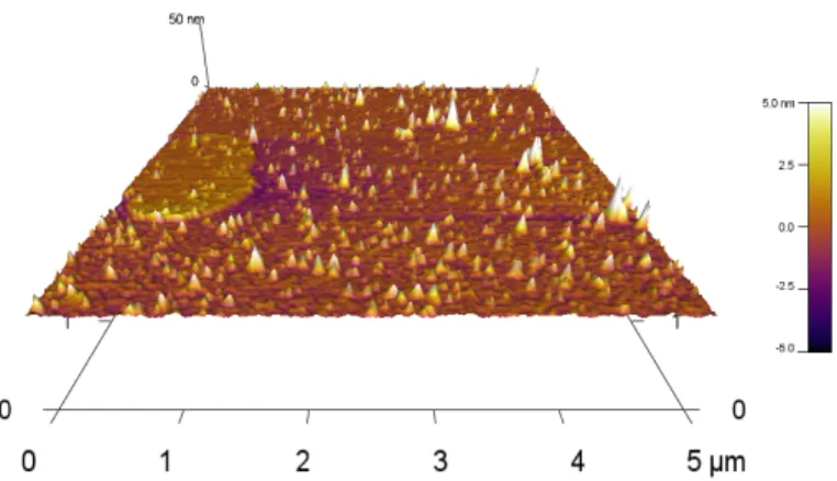
The nanoparticle solution showed a unique and relatively broad absorption peak at 414 nm (Fig 1) while the AFM showed the presence of nanoparticles with less than 10 nm of height (Fig 2). The broad absorption peak is a sign of a large size distribution of the particles which was consistent with the data observed in the AFM image.
Fig. 1: UV-vis absorption spectra of silver nanoparticles. Solid line corresponds to nanoparticle solution while segmented line corresponds to "blank" solution.
Fig. 2: 3D image of silver nanoparticles observed by atomic force microscopy. Particles of different sizes were observed in the AFM analysis.
approximation21, the intensity is proportional to the sixth
power of the particle’s diameter. Thus, to avoid signal concealment of the smaller particles, a size distribution analysis based on percentage of total volume and total quantity of nanoparticles by size range was made.
The different size distributions showed that (98±1) % of the nanoparticles had a diameter equal or inferior to 20 nm and that these nanoparticles represented (87±5) % of the total particle volume in the solution (Fig. 3).
The proposed method uses alkali treated chitosan as an electron donor for the silver ions since, once its charge is neutralized, it can be used as a weak reducing agent12,22.
When the solution of AgNO3 is mixed with the alkali
treated chitosan a brown color is observed due the formation of Ag2O, probably from its reaction with NaOH
producing AgOH which is an intermediate and yields Ag2O23. However, after several minutes of sonication the
solution became yellowish. This phenomenon could be explained considering that the combined effect of high ultrasonic irradiation could degrade large chitosan chains24, 25. Thus, the reduction of the Ag+ and even of Ag
2O could
be ascribed principally to the products of chitosan hydrolysis26. These products consists mainly of chitosan
oligomers with higher acetylation degree, derivatives of
glucosamine acids and formate ion26. In this regard,
several researches propose that the glucosamine moieties activate the reducing function of chitosan and thus, are the ones involved in the reduction of silver ions 12, 26, 27.
The UV-vis results show a typical silver absorption peak at 414 nm which is in the reported range of silver and silver
oxide nanoparticles3,6,13,14,19. The number of surface
plasmon resonance (SPR) peaks is dependent of the symmetry of the nanoparticles6, 28. Since just one peak was
observed in the UV-vis spectra, spherical silver nanoparticles were synthesized. This fact validates the size distribution generated by DLS since those results are calculated based on the movement of sphere-like particles. Another important fact is the high polydispersity index showed by DLS analysis. This could be explained based on the growing mechanism of the nanoparticles. The ultrasonic vibration alternatively compresses and decompresses the liquid causing the formation and violent implosion of micro-cavities which generate millions of shock waves in the fluid1. After the reduction of the silver
ions, the atoms form small nuclei that grow by molecular additions and by collisions between them.
During the process the temperature can be regulated in order to control the nuclei growth1, 29. However, in the
proposed methodology the temperature was not regulated reaching final temperatures (after sonication) around 80 ºC. As the temperature increases the cavitation process gets easier, due the rise in the vapor pressure, supporting excessive formation and implosion of micro-cavities which could promote a non-uniform nuclei growth which could explain the high polydispersion index observed. Nevertheless, 98% of the particles had a diameter inferior to 20 nm without temperature control which makes it a versatile and easy method for their obtainment. To reduce polydispersion a systematic approach evaluating different chitosan:Ag+ ratios could be used in the future.
A drawback of this approach is the fact that the chitosan is only soluble on water at pH < 6 due to the protonation of amine groups20. To avoid nanoparticle sedimentation due to
insolubility of chitosan, concentrated acetic acid was added to acidify the solution until a pH of 4.
Since aqueous solutions have great cohesive energy, the fact that neither a brown nor black precipitate (characteristic of Ag2O) was observed after several weeks from its synthesis
indicates that there was a successful stabilization of the nanoparticles by the chitosan. This fact contrasts with the findings that degradation of chitosan macromolecule decreases its efficiency as a stabilizer26. One possible
explanation may be that chitosan suffered just partial degradation allowing for mid-size chitosan macromolecules to act as stabilizers indicating that a balance between reducing activity and stabilizing efficiency was found. Nevertheless, this hypothesis should be further explored by determining size distribution of chitosan chains under different sonochemical synthesis conditions.
Conclusions
A simple, fast and relatively cheap method for the synthesis of silver nanoparticles was developed. The DLS analysis showed that more than 98% of the nanoparticles had a diameter inferior to 20 nm but there was a high polydispersion index indicating that the method needs to be optimized. This study demonstrated that the chitosan treated with sodium hydroxide can be an effective reducing and stabilizing agent for silver nanoparticles synthesized by the sonochemical method.
Acknowledgements
We want to thank to POLIUNA at Universidad Nacional in Costa Rica for providing the chitosan used in this research.
References
1. VS Manoiu, A Aloman. Obtaining silver nanoparticles by sonochemical methods. U.P.B. Buletin Scientific. Series B, 72, 179-186 (2010).
2. KMM Abou El-Nour, AA Eftaiha, A Al-Warthan, RAA Ammar. Synthesis and applications of silver nanoparticles, Arabian Journal of Chemistry, 3, 135-140 (2010).
3. JS Kim, E Kuk, KN Yu, JH Kim, SJ Park, HJ Lee, SH Kim, YK Park, YH Park, CY Hwang, YK Kim, YS Lee, DH Jeong, MH Cho. Antimicrobial effects of silver nanoparticles, Nanomedicine, 3, 95-101 (2007).
4. CN Lok, CM Ho, R Chen, QY He, WY Yu, H Sun, PK Tam, JF Chiu, CM Che. Silver nanoparticles: partial oxidation and antibacterial activities, Journal of Biological Inorganic Chemistry, 12, 527-534 (2007).
5. RJ Pinto, SC Fernandes, CS Freire, P Sadocco, J Causio, CP Neto, T Trindade. Antibacterial activity of optically transparent nanocomposite films based on chitosan or its derivatives and
silver nanoparticles. Carbohydrate Research, 348, 77-83 (2012).
6. S Pal, YK Tak, JM Song. Does the antibacterial activity of silver nanoparticles depend on the shape of the nanoparticle? A study of the Gram-negative bacterium Escherichia coli. Applied and Environmental Microbiology, 73, 1712-1720 (2012).
7. CS Costa, JV Ronconi, JF Daufenbach, CL Goncalves, GT Rezin, EL Streck, MM Paula. In vitro effects of silver nanoparticles on the mitochondrial respiratory chain, Molecular and Cellular Biochemistry, 342, 51-56 (2010). 8. MH Majles Ara, Z Dehghani, R Sahraei, G Nabiyouni.
Non-linear optical properties of silver nanoparticles prepared by hydrogen reduction method- Optics Communications, 283, 1650-1653 (2010).
9. KD Bhatte, KM Deshmukh, YP Patil, DN Sawant, SI Fujita, M Arai, BM Bhanage. Synthesis of powdered silver nanoparticles using hydrogen in aqueous medium. Particuology, 10, 140-143 (2012).
10. FM Reicha, A Sarhan, MI Abdel-Hamid, IM El-Sherbiny. Preparation of silver nanoparticles in the presence of chitosan by electrochemical method, Carbohydrate Polymers, 89, 236-244 (2012).
11. HV Tran, LD Tran, CT Ba, HD Vu, TN Nguyen, DG Pham, PX Nguyen. Synthesis, characterization, antibacterial and anti-proliferative activities of monodisperse chitosan- based silver nanoparticles, Colloids and Surfaces A: Physico-chemical and Engineering Aspects, 360, 32-40 (2010).
12. YK Twu, YW Chen, CM Shih. Preparation of silver nanoparticles using chitosan suspensions. Powder Technology, 185, 251-257 (2008).
13. D Wei, W Qian. Facile synthesis of Ag and Au nanoparticles utilizing chitosan as a mediator agent, Colloids and Surfaces. B, Biointerfaces, 62, 136-142 (2008).
14. D Wei W Sun, W Qian, Y Ye, X Ma. The synthesis of chitosan-based silver nanoparticles and their antibacterial activity. Carbohydrate Research, 344, 2375–2382 (2009). 15. H Wang, X Qiao, J Chen, X Wang, S Ding. Mechanisms of
PVP in the preparation of silver nanoparticles. Materials Chemistry and Physics, 94 (2-3), 449–453 (2005).
16. C Luo, Y Zhang, X Zeng, Y Zeng, Y Wang. The role of poly (ethylene glycol) in the formation of silver nanoparticles. Journal of Colloid and Interface Science, 288, 444-448 (2005).
17. M Torres-Cisneros, N Yanagihara, B Gonzalez-Rolon, MA Meneses-Nava, OG Ibarra-Manzano, DA May-Arrioja, J Sánchez-Mondragón, E Aguilera-Gómez, LA Aguilera-Cortés. Synthesis and nonlinear optical behavior of Ag nanoparticles in PMMA, Microelectronics Journal, 40, 621-623 (2009). 18. ES Abdel-Halim, MH El-Rafie, SS Al-Deyab.
Hakimi. Green synthesis of colloidal silver nanoparticles by sonochemical method. Materials Letters, 66, 117-120 (2012). 20. MC Rodriguez-Arguelles, C Sieiro, R Cao, L Nasi. Chitosan
and silver nanoparticles as pudding with raisins with anti-microbial properties. Journal of Colloid and Interface Science, 364, 80-84 (2011).
21. F Ross Hallett. Scattering and Particle Sizing Applications. In Encyclopedia of Spectroscopy and Spectrometry (Second Edition), J.C. Lindon, Editor. Academic Press: Oxford. 2488-2494.(1999)
22. P Sanpui, A Murugadoss, PV Prasad, SS Ghosh, A Chattopadhyay. The antibacterial properties of a novel chitosan-Ag-nanoparticle composite. International Journal of Food Microbiology, 124, 142-146 (2008).
23. E Wiberg, N Wiberg. Silver. In: Inorganic Chemistry. 2001, Academic Press. 1265-1275.(2001)
24. MR Kasaai, J Arul, G Charlet. Fragmentation of chitosan by ultrasonic irradiation. Ultrasonics Sonochemistry, 15, 1001-1008 (2008).
25. E Savitri, SR Juliastuti, A Handaratri, Sumarno, A Roesyadi. Degradation of chitosan by sonication in very-low-concentration acetic acid. Polymer Degradation and Stability, 110, 344-352 (2014).
26. A Pestov, A Nazirov, E Modin, A Mironenko, S Bratskaya. Mechanism of Au(III) reduction by chitosan: Comprehensive study with 13C and 1H NMR analysis of chitosan degradation products. Carbohydrate Polymers, 117, 70-77 (2015).
27. KDN Vo, E Guillon, L Dupont, C Kowandy, X Coqueret. Influence of Au(III) Interactions with Chitosan on Gold Nanoparticle Formation. The Journal of Physical Chemistry C, 118, 4465-4474 (2014).
28. IO Sosa, C Noguez, R Barrera. Optical properties of metal nanoparticles with arbitrary shapes. The Journal of Physical Chemistry B, 107, 6269-6275 (2003).