Role of cerebral dopamine neurotrophic factor in endoplasmic reticulum stress
118
0
0
Texto completo
(2) 2. DEDICATORY. This thesis is dedicated to my wonderful parents, Jorge Arancibia and Isabel Radich, my brother Jorge, my sister Isabel, my nephew Dante and my beloved Rafaella.. Whose selfless love, support, faith, and trust in me have brought me here. They have always allowed me to pursue my wildest dreams, still believing that I could achieve what I wanted.. Your encouragement and unwavering love have given me the greatest motivation to continue and achieve this degree. I hope you feel proud of me. For this, and for all the countless things they have given me, thank you, thank you very much..
(3) 3. EPIGRAPH. “Don't wait until everything is just right. It will never be perfect. There will always be challenges, obstacles, and less than perfect conditions. So what. Get started now. With each step you take, you will grow stronger and stronger, more and more skilled, more and more self-confident and more and more successful.”. -Mark Victor Hansen.
(4) 4. ACKNOWLEDGEMENTS This project would not have been possible without the always present hand of my tutors Dr. María Estela Andrés and Dr. Pedro Zamorano. I consider myself fortunate to have been able to work with them. They are not only excellent scientists but also extraordinary people. Their vision was crucial for all the work I have done. In addition to being exceptional scientists, they are genuinely interested and concerned about their students. These are just some of the traits that I have admired and learned from them, and I know that these skills will be useful to me, no matter where I go.. I am also sincerely grateful to my doctoral thesis committee, formed by Dr. Jorge Campusano, Dr. María Paz Marzolo, and Dr. Soledad Matus. I was fortunate to have such a combination of good mentors in one place, where each meeting with my committee was fruitful. They always gave me a critical vision after my presentations, spending time, and showing a genuine interest in my work.. I must thank once again my family, to whom this thesis is dedicated. They always have had confidence in me and my abilities, they care for me, they encourage me, and they support me in everything I could ask. I want to thank my brother and sister, who have always cared for me, they have supported me tirelessly throughout all these years, through all the ups and downs of my academic and personal life. They have been the walls that have always contained me.. I must thank my beloved, Rafaella, for always putting a smile on my face and showing me the right side of things when I was discouraged. I cannot thank her enough for being the yin of my yang, often ungovernable, for the happiness, joy, and laughter that she brought to my life, completing it.. Finally, grateful for lab members Sebastián, Montserrat, Marcela, Verónica, Gianluca, Fabián, Carlos, Daniel, and all my friends not mentioned here, for being part of my life and being in the good and the bad situations with me. Many, many people have contributed to this thesis, some small and some big, but all-important. Thanks to all my family and friends, in Santiago, Antofagasta, Calama, and elsewhere, for the encouragement and support during these years..
(5) 5. TABLE OF CONTENTS DEDICATORY ...................................................................................................................... 2 EPIGRAPH ............................................................................................................................. 3 ACKNOWLEDGEMENTS .................................................................................................... 4 TABLE OF CONTENTS........................................................................................................ 5 TABLE OF FIGURES ............................................................................................................ 7 ABBREVIATIONS.................................................................................................................9 RESUMEN…………………………………………………………………………………10 ABSTRACT.......................................................................................................................... 11 1. Introduction ....................................................................................................................... 12 1.1. Neurotrophic factors................................................................................................... 12 1.2. Cerebral Dopamine Neurotrophic Factor CDNF)…………………………………...14 1.3. Role of CDNF in neurodegenerative diseases ........................................................... 17 1.4. Endoplasmic reticulum and Unfolded Protein Response ........................................... 18 1.5. Unfolded protein response and Neurodegenerative Diseases .................................... 21 1.6. CDNF: Modulator of the Unfolded Protein Response? ............................................. 21 2. Hypothesis and Objectives………………………………………………………………22 3. First Part Doctoral Thesis ................................................................................................. 25 INTRODUCTION ........................................................................................................... 33 MATERIALS AND METHODS..................................................................................... 34 Plasmids………………………...………………………………….………………34 Antibodies…………………………………………………...…..…………………34 Cell culture, transfection and thapsigargin treatment………………………...……34 Primary culture of rat hippocampal neurons…………………………....……….…34 MTS assay………………………………………………………….……........……34 RNA extraction and RT-qPCR……………………………………..…...………….34 Immunoblotting……………………………….………………………...…...….….34 Immunofluorescence imaging and quantification of fluorescence intensity…...…..35 Statistical analysis………………………….……………………....….……..…….35 Ethical statement………….………………..………………………....……….…...35 RESULTS…………..…………………………………………………………...……....35 CDNF protects HEK293-T cells against TG-induced cell death………..……..…..35 CDNF promotes an adaptive UPR………………………..……..…….…….…..…35 CDNF attenuates ER stress-induced apoptosis………………………...…...…...…38 CDNF induces an early adaptive UPR and blocks the terminal UPR in neurons.....38.
(6) 6. Secreted CDNF fails to protect cells against TG-induced ER stress…….......….....39 DISCUSSION………………….………………………………………………...……..41 REFERENCES…………….………………………………………………..………......43 SUPPLEMENTARY FIGURES…………………………………………..….…..….....44 4. Second Part Doctoral Thesis……...………………………………………………..…….51 INTRODUCTION ............................................................................................................ 56 MATERIALS AND METHODS................................................................................. ….58 DNA manipulation and Lentiviral plasmid construction………………………......58 Antibodies……………………………………………….………………...…..…...58 Cell culture and transfection………………………………….…….………….......59 Lentivirus production and transduction…………………………….….…..……....59 Rat hippocampal neurons culture and lentiviral transduction……..………….…....60 Induction assay with blue light (465 nm) …….…………………….…...60 Immunoblotting…….……………………………………………………………....60 Immunofluorescence…………………………………………………………….....61 Imaging and quantification of fluorescence intensity…………………...………....61 Statistical analysis……………….…………………………………………....…....62 RESULTS………………..…………………………………………….…………...…...62 Construction of GAVPO and reporter lentiviral vectors……………...……...….....62 Expression and induction of LightOn lentiviral vectors by blue light………...........63 Inducing the expression of CDNF by blue ligh………………………………...….65 Expression of LightOn components by lentiviral particles in neurons…………….66 DISCUSSION.......................................................................................................................66 REFERENCES ..................................................................................................................... 70 FIGURES .............................................................................................................................. 73 SUPPLEMENTARY FIGURES……………………………...……………….…………...77 Lentiviral optogenetic vector OPTO-CDNF…………….…………………………………82 5. General Discussion………..……………………………………………………...….…..85 Molecular mechanism of the cytoprotective role of CDNF is associated with ER stress and UPR pathways………………………………………………………..…85 Role of CDNF on UPR…………………………………………………………….90 Therapeutic challenges for Parkinson`s disease: Prover CDNF delivery………….96 6. Summary…...……………...………………………………………………..………….103 7. Conclusion……………………………………………………………………………...104 7. General References……………………………………...……………………………...105.
(7) 7. TABLE OF FIGURES Introduction Figure i. Mode of action of CDNF is drastically different from other known neurotrophic factors such as BDNF, GDNF or NRTN .......................................................................... …13 Figure ii. Hypothetical modes of CDNF and MANF action……………………………….16 Figure iii. The Unfolded Protein Response is mediated by three receptors present in the Endoplasmic Reticulum ........................................................................................................ 20 Figure 1. CDNF is widely expressed in different cell lines………………………………...27 Figure 2. Establishing a model for the study of ER stress………………………………….28 Figure 3. Transfecting FUG-CDNF-W plasmid in HEK293-T cells induces a five-fold increase of CDNF……………………………………………………………………….….30 Figure 4. CDNF localizes in the ER……………………………………………..................31 1. 2. First Scientific Article Fig. 1. CDNF protects HEK293-T cells against TG-induced cell death…………………...36 Fig. 2. CDNF increases BiP protein and mRNA levels……………………………………36 Fig. 3. CDNF increases the splicing of XBP1 and ATF6 protein levels. ............................. 37 Fig. 4. CDNF increases the expression of ATF4 ................................................................ 38 Fig. 5. CDNF attenuates TG-induced expression of CHOP and active caspase-3 in HEK293T cells………………………………………………………………………………………39 Fig. 6. CDNF triggers adaptive UPR in hippocampal neurons ............................................ 40 Fig. 7. CDNF attenuates TG-induced expression of CHOP in hippocampal neurons. .... 41 Fig. 8. Secreted CDNF fails to protect cells against TG-induced ER stress and induce BiP expression. ............................................................................................................................ 42 Supplementary Figure 1. CDNF does not change PDI in HEK293-T cells ......................... 44 Supplementary Figure 2. Expression of mRFP-KDEL in the ER of HEK293-T cells does not induce UPR markers ............................................................................................................. 45 Supplementary Figure 3. Transfection of FUG-CDNF-W increases CDNF in cultured hippocampal neurons ............................................................................................................ 46 Supplementary Figure 4. CDNF does not change PDI in hippocampal neurons ................. 47.
(8) 8. Supplementary Figure 5. The expression and secretion of CDNF do not change during a TGinduced ER stress. ................................................................................................................. 48 Supplementary Figure 6. Characterization of the CDNF mutant lacking the QTEL sequence ............................................................................................................................................ ..49 Figure 5. Non-canonical retention sequence QTEL is necessary to induce the expression of proteins related to the UPR ................................................................................................... 50 Second Scientific Article Figure 1. Schematic drawing of the blue-light inducible lentiviral expression vectors……73 Figure 2. Expression of lentiviral plasmids modulated by blue light ................................... 74 Figure 3. Expression of cerebral dopamine neurotrophic factor (CDNF) modulated by blue light as proof of concept ....................................................................................................... 75 Figure 4. Transduction of primary hippocampal neurons with lentiviral particles .............. 76 Supplementary Figure 1. Characterization of the expression of LightOn lentiviral vectors 77 Supplementary Figure 2. Expression of different versions of UAS light-responsive promoter in no-GAVPO expressing cells ............................................................................................. 78 Supplementary Figure 3. Co-transfection of the F-UAS(L)-mCherry-W ∆CMV lightresponsive and the FUG-1D/2A-HA-GAVPO-W vectors.................................................... 79 Supplementary Figure 4. Expression of mCherry and CDNF dependent on GAVPO…….80 Supplementary Figure 5. Home-made blue light-modulated expression system used in the cell culture incubator. ........................................................................................................... 81 Figure 5. Schematic drawing of the blue-light inducible lentiviral vector OPTO-CDNF…82 Figure 6. OPTO-CDNF vector express HA-GAVPO and CDNF correctly…………...…..83 Figure 7. Expression of CDNF regulated by blue light can induce UPR markers such as BiP, ATF4, ATF6, and XBP1………………………………………………………....................84.
(9) 9. ABBREVIATIONS. 6-OHDA: 6-hydroxydopamine ATF4: Activating transcription factor 4 ATF6: Activating transcription factor 6 BBB: Blood-Brain Barrier CDNF: Cerebral Dopamine Neurotrophic Factor DA: Dopamine eIF2α: Initiating factor of eukaryotic translation 2α ER: Endoplasmic reticulum GRP78: Chaperone regulated by glucose-78 HEK293-T: Human Embryonic Kidney 293 T IF: Immunofluorescence IRE1: Enzyme that requires inositol-1 MANF: Mesencephalic astrocyte-derived Neurotrophic Factor PERK: Pancreatic endoplasmic reticulum kinase ROS: Reactive oxygen species UPR: Unfolded Protein Response WB: Western Blot XBP1: Box binding protein X.
(10) 10. RESUMEN. El factor neurotrófico cerebral dopaminérgico (CDNF) es un péptido de 187 aminoácidos localizado en el retículo endoplasmático de numerosos tipos celulares y conservado evolutivamente. CDNF ejerce un efecto protector sobre las neuronas en modelos celulares y animales de diversas enfermedades neurodegenerativas, pero se desconoce el mecanismo molecular de este efecto. Muchas enfermedades neurodegenerativas se han relacionado con una desregulación de la proteostasis en el retículo endoplásmico. La desregulación de la proteostasis produce estrés reticular lo que gatilla inicialmente la respuesta adaptativa a proteínas mal plegadas y, si el estrés persiste en el tiempo, se inducen vías de señalización relacionadas a la muerte celular o apoptosis. Varios estudios han revelado que una respuesta adaptativa resuelve el estrés de retículo endoplasmático atenuando la síntesis de proteínas, induciendo la expresión de chaperonas y metabolizando las proteínas mal plegadas. En esta tesis, investigamos si CDNF regula la proteostasis del retículo endoplásmico. Con este fin, se determinó la función de CDNF en un modelo de estrés de retículo endoplásmico inducido por Tapsigargina, en células HEK293-T y neuronas hipocampales en cultivo. Se observó que la expresión de CDNF aumentó significativamente la viabilidad de células HEK293-T expuestas a Tapsigargina. Este efecto, correlacionó con aumento de los niveles de proteínas protectoras de la respuesta a proteína mal plegada temprana tales como BiP, ATF4, ATF6 y XBP-1, tanto en las células HEK293-T como en neuronas. Además, la expresión de CDNF redujo los niveles de las proteínas pro-apoptóticas CHOP y caspasa-3 activa. Se determinó que CDNF protege a las células desde el retículo endoplásmico, dado que una versión mutante de CDNF carente de la secuencia de retención en retículo endoplásmico, y por lo tanto secretada, no protegió a las células frente al estrés inducido por Tapsigargina. En conclusión, CDNF regula la proteostasis en retículo endoplásmico induciendo la respuesta a proteína mal plegada adaptativa e inhibiendo las vías pro-apoptóticas activadas por el estrés de retículo endoplásmico. Finalmente, en la última parte de esta tesis se diseñó y construyó un dispositivo molecular de expresión regulada de CDNF, que combina métodos optogenéticos con una plataforma lentiviral..
(11) 11. ABSTRACT The cerebral dopamine neurotrophic factor (CDNF) is a peptide of 187 amino acids located in the endoplasmic reticulum of numerous cell types and evolutionarily conserved. CDNF exerts a protective effect on neurons in cellular and animal models of various neurodegenerative diseases, but the molecular mechanism of this effect is unknown. Many neurodegenerative diseases are associated with the deregulation of proteostasis in the endoplasmic reticulum. Deregulation of proteostasis produces reticular stress, which initially triggers the response to misfolded proteins and, if stress persists over time, signaling pathways related to cell death or apoptosis are induced. Several studies have revealed that an adaptive response resolves the endoplasmic reticulum stress by attenuating protein synthesis, inducing the expression of chaperones, and metabolizing misfolded proteins. In this thesis, it was investigated whether CDNF regulates proteostasis of the endoplasmic reticulum. To this end, the function of CDNF was determined in an endoplasmic reticulum stress model induced by Thapsigargin, in HEK293-T cells and hippocampal neurons in culture. It was observed that the induced expression of CDNF significantly increased the viability of HEK293-T cells exposed to Thapsigargin. This effect correlated with the increase in protective protein levels of the early misfolded protein response such as BiP, ATF4, ATF6, and XBP-1, both in HEK293-T cells and in neurons. Also, the induced-expression of CDNF reduced the levels of pro-apoptotic proteins CHOP and active caspase-3. It was determined that CDNF protects cells from the endoplasmic reticulum, since a mutant version of CDNF, lacking the endoplasmic reticulum retention sequence, and therefore secreted, did not protect the cells against stress-induced by Thapsigargin. In conclusion, CDNF regulates proteostasis in the endoplasmic reticulum by inducing the response to adaptive misfolded protein and inhibiting pro-apoptotic pathways activated by endoplasmic reticulum stress. Finally, in the last part of this thesis, a molecular device for the regulated expression of CDNF that combines optogenetic methods with a lentiviral platform was designed and constructed..
(12) 12. 1. INTRODUCTION 1.1 Neurotrophic factors Neurotrophic factors are essential peptides for growth, maturation, and survival of neurons. To date, many neurotrophic factors have been discovered and it has been recognized that they have several biological effects, among them: brain-derived neurotrophic factor (BDNF), neurotrophin-3 (NT-3) and NT-4; neurotrophic factor derived from the glial cell line (GDNF) and its family of ligands, such as neurturin, and neurotrophic cytokines. Along with these, cerebral dopamine neurotrophic factor (CDNF) and mesencephalic astrocyte-derived neurotrophic factor (MANF) have been discovered and characterized as a new family of neurotrophic factors (Figure i) (Lindholm et al. 2007; Petrova et al. 2003).. Preclinical data using several models of neurodegenerative disorders show alterations in the levels, and even in the function of neurotrophic factors. Therefore, neurotrophic factors have received increasing attention for their therapeutic potential, especially for Alzheimer's and Parkinson's diseases (Rangasamy et al. 2010). Neurodegenerative diseases are complex, and their etiology is diverse. Current treatments aim to alleviate the symptoms, instead of preventing the loss of synapses and neuronal death. Therefore, neurogenic agents, including neurotrophic factors, represent a new therapeutic strategy to address the degenerative process itself..
(13) 13. Figure i. The mode of action of CDNF is drastically different from other known. 3. neurotrophic factors such as BDNF, GDNF, or NRTN. Neuronal survival-promoting actions of BDNF, GDNF, and NRTN are mediated by transmembrane tyrosine kinase receptors leading to the activation of PI3K/Akt, PLCc, and MAP kinases pathways. On the other hand, different from other neurotrophic factors, CDNF is retained in the endoplasmic reticulum (Voutilainen, Arumäe, Airavaara, & Saarma, 2015)..
(14) 14. 1.2 Cerebral Dopamine Neurotrophic Factor (CDNF) A few years ago, a new class of neurotrophic factors was discovered, mesencephalic astrocyte-derived neurotrophic factor (MANF) (Petrova et al. 2003) and cerebral dopamine neurotrophic factor (CDNF) (Lindholm et al. 2007). They form a distinct family that does not share structural similarities with other families of neurotrophic factors (Figure 1). MANF was initially purified from conditioned media from an immortalized ventral mesencephalic astrocytic cell line (Petrova et al. 2003) and its sequence determined by a combination of proteomics and bioinformatics technologies. CDNF was discovered by Lindholm and colleagues in 2007 using a bioinformatics approach and confirmed by biochemical and molecular studies (Lindholm et al., 2007). MANF and CDNF proteins function as secreted neurotrophic factors, but some of their activities might occur while they are resident in the endoplasmic reticulum (ER) lumen due to a non-canonical ER retention signal. CDNF is an 18 kDa peptide of 187 aminoacids, of which the N-terminal 26 amino-acids are a signal peptide that is cleaved at the ER. This neurotrophic factor is expressed in both neuronal and non-neuronal tissues, and although databases report a splicing variant for CDNF, no experimental confirmation is available. The structural analysis of the mature peptide shows two featured domains, a saposin-like domain localized in the N-terminal and a disulfide bridge formed by two cysteine residues localized in the C-terminal (Apostolou et al., 2008; Parkash et al., 2009). Besides, the C-terminal domain has a non-canonical KDEL retention sequence (QTEL in rat, KTEL in human), suggesting that CDNF could be an ERresident protein (Lindahl and Lindholm, 2017). However, many studies in cell lines have shown that CDNF could be secreted to the extracellular medium (Latge et al. 2015; Mei and.
(15) 15. Niu 2014; Norisada et al., 2016; Sun et al. 2011). This dual localization of CDNF has led to propose that the neuroprotector effect could be exerted either by secreted CDNF or by direct effect in the ER.. Regarding the mode of action of CDNF, as shown in Figure ii, the N-term saposin motif of this protein could confer the capacity to interact with lipids in the plasma membrane, but this idea has not been demonstrated to date. On the other hand, the C-terminal contains a disulfide bond between conserved cysteines in a CXXC motif. The CXXC motif is a consensus sequence of proteins of the thiol-protein oxidoreductase superfamily, other members of which include thioredoxins, glutaredoxins, and peroxiredoxins. Common to this enzyme superfamily is that all members are involved in disulfide-mediated redox reactions and glutathione metabolism in which the CXXC domain takes center stage. However, no enzymatic oxidoreductase activity has been detected for CDNF (Figure ii)..
(16) 16. Figure ii. Proposed modes of action of CDNF. (A) Changes in pH or lipid binding may. 4. induce the dimerization of CDNF. (B) The C-terminal domain of CDNF may facilitate the formation of disulfide bridges on secretory proteins in the ER. (C) CDNF may reduce disulfide bonds on a transmembrane receptor protein on the cell surface. (D) CDNF may interact with the cell surface membrane and subsequently activate a transmembrane receptor protein. (Adapted from Lindholm & Saarma, 2010)..
(17) 17. 1.3 Role of CDNF in neurodegenerative diseases CDNF is significantly expressed in neurons of the cortex, the hippocampus, and the striatum (Lindholm et al., 2007). However, CDNF is not detectable in positive tyrosine hydroxylase cells, a protein present in dopamine neurons. Despite not being expressed in dopamine neurons, the infusion of CDNF in the striatum and substantia nigra has neuroprotective and, more importantly, neurorestorative effects in animals treated with 6-OHDA and MPTP toxins (Airavaara et al., 2012; Lindholm et al., 2007; Voutilainen et al., 2011). Intriguingly, the neuroprotective effect was also observed when neurons of the substantia nigra were transduced with an adeno-associated virus or lentivirus encoding CDNF, supporting the idea that it could protect the cells from intracellular compartments (Bäck et al. 2013; CorderoLlana et al. 2015; Wang et al. 2017). Another study showed that CDNF and MANF together produced synergistic neuroprotection and neurorestoration effects on the nigrostriatal system (Cordero-Llana et al., 2015). It should be noted that the neuroprotective effect of CDNF is not only circumscribed to dopamine neurons, but also hippocampal neurons were protected in an animal model of Alzheimer's disease (Kemppainen et al., 2015). Finally and, in accordance with the above, other study showed that CDNF alleviates synaptotoxicity induced by β-amyloid (Zhou et al., 2016), suggesting that CDNF could be of importance in others neurodegenerative diseases.. In this context, several studies have suggested that a common component among neurodegenerative disorders such as Parkinson's and Alzheimer's diseases is the aggregation of misfolded proteins. Regardless of the cause, the aggregation of proteins would cause the.
(18) 18. death of specific populations of neurons due to an alteration of the proteostasis of the ER. Supporting this idea, it was reported that the treatment with 6-OHDA causes ER stress in dopamine neurons (Holtz et al., 2006; Ryu et al., 2002; Ryu et al., 2005), and in PC-12 cells (Elmazoglu et al., 2017). Similarly, Alzheimer's disease has been related to a disturbance in the homeostasis of the ER (Hoozemans et al., 2005; Hoozemans et al., 2012). Taken together, the data discussed above suggest that the protection and restorative effect exerted by CDNF could be due to its role in relieving ER stress.. 1.4 Endoplasmic Reticulum and the Unfolded Protein Response. The ER is an essential organelle for the functioning of the cell since it participates in the folding and processing of secretory and membrane proteins. Therefore, ER is pivotal for cellular proteostasis, which refers to the balance between synthesis, folding, and protein degradation. Alterations of ER homeostasis results in the accumulation of misfolded proteins, a condition called ER stress. In turn, ER stress activates several signaling pathways known as "unfolded protein response" (UPR) (Ron & Walter, 2007).. The UPR is mediated by three receptors present in the ER membrane that act as ER stress sensors: Enzyme requiring inositol-1 (IRE1), pancreatic ER kinase (PERK), and activator transcription factor-6 (ATF6) (Walter and Ron 2011). It has been proposed that ER stress triggers the dissociation of these three receptors from the chaperone protein regulated by glucose-78 (GRP78/BiP), which binds to hydrophobic residues of misfolded proteins (Figure 3) (Reviewed in Ron & Walter, 2007). However, it has been reported recently that UPR can.
(19) 19. be activated of a BiP-independent manner, which misfolded proteins during ER stress can bind to PERK and IRE1, promoting their oligomerization and activation (Adams et al. 2019; Wang et al. 2018).. Essentially, the progression of the UPR comprises two main phases: the adaptive UPR and the terminal UPR that play a restorative and apoptotic role, respectively (Rutkowski & Kaufman, 2007; Tabas & Ron, 2011; Erguler, Pieri, & Deltas, 2013). In the adaptive phase, UPR signaling pathways are vital to restoring the ER proteostasis. During this phase, both PERK and IRE1 are activated by oligomerization and phosphorylation (Hamanaka et al., 2005; Ron & Hubbard, 2008). At the same time, ATF6 is cleaved in the Golgi apparatus, and its N-terminal domain translocates to the nucleus as well as the spliced form of the X-box binding protein (XBP1), which is subjected to an IRE1 ribonuclease activity. In the nucleus, both ATF6 and XBP1, which act as transcription factors, increase the expression of chaperones and genes related to degradation mediated by the ER (ERAD) (Chen et al., 2002).. In parallel, global protein translation is attenuated by the PERK-eIF2a-ATF4 pathway, thus reducing the overload of the ER, while the rate of degradation of accumulated proteins is markedly increased. Finally, if the ER stress persists, the UPR activates the signaling pathways PERK-eIF2a-ATF4-CHOP and IRE1-TRAF2-ASK1-JNK, which lead to mechanisms of programmed cell death or apoptosis (Reviewed in Chan et al. 2015)..
(20) 20. Figure iii. The Unfolded Protein Response is mediated by three receptors present in the. 5. ER. Three membrane proteins are the ER stress sensors: Enzyme requiring inositol-1 (IRE1), pancreatic ER kinase (PERK), and activating transcription factor-6 (ATF6). During ER stress, these receptors dissociate from the protein chaperone regulated by glucose-78 (BiP/GRP78), which joins to a hydrophobic place of misfolded proteins (Adapted from Hetz and Mollereau, 2014)..
(21) 21. 1.5 Unfolded Protein Response and Neurodegenerative Diseases Activation of UPR signaling pathways has been observed in various diseases, such as diabetes, inflammation, and neurodegenerative diseases (Lin, Walter, & Yen, 2008; Scheper & Hoozemans, 2015; Wang & Kaufman, 2012). One of the characteristics observed in these diseases is the presence of aggregated or misfolded proteins. Among them are the aggregates of α-synuclein in Parkinson´s disease, pTau, and beta-amyloid in Alzheimer´s disease, and Huntingtin in Huntington’s disease. In recent years, there has been growing evidence that alterations in proteostasis in the ER contribute to the pathological process (Brown & Naidoo, 2012; Matus, Glimcher, & Hetz, 2011). Interestingly, post-mortem brain samples of patients suffering Alzheimer’s and Parkinson’s diseases show strongly increased UPR markers such as phospho-PERK and phospho-eIF2α, a feature also observed in animal models of these neurodegenerative diseases (Hartmann, 2004; Hoozemans et al., 2005; Hoozemans et al., 2007).. 1.6 CDNF: Modulator of the Unfolded Protein Response? As discussed in previous paragraphs, CDNF has a protective and restorative effect on DA neurons that degenerate due to the application of 6-OHDA and MPP in preclinical models of Parkinson’s disease or neuronal cultures. Both 6-OHDA and MPP have been strongly associated with ER stress (Ryu et al. 2002), and the inhibition of ER stress could exert neuroprotection in these models (Cai et al., 2016). The non-conventional neurotrophic factor CDNF is an ER-resident protein, and it is unclear whether it can be secreted during ER stress. Thus, as an ER-resident protein, it could have a role inside the ER, possibly regulating the unfolded protein response pathways..
(22) 22. 2. HYPOTHESIS AND OBJECTIVES The molecular mechanism of action of CDNF remains unresolved. Our preliminary data show that overexpressing CDNF increases the expression of BiP (Figure 1), a protein that has been reported that is involved in the modulation of the UPR. Thus, the neuroprotection observed by CDNF in different cellular and animal models could be mediated by inducing a preemptive activation of the UPR.. The maintenance of proteostasis in the ER is fundamental for the proper functioning of the secretory capacities of cells that produce hormones and neurotransmitters, and like-wise essential in cells with high structural complexity as observed in neurons (Walter & Ron 2011). Aberrant activation of the UPR signaling pathways has been found in various diseases such as diabetes, inflammation, and neurodegenerative diseases. As a consequence, a disturbance in the ER proteostasis could lead to cell death. It is the mechanism that has been proposed as the main pathway leading to the loss of neurons in neurodegenerative diseases (Hoozemans et al., 2005; Hoozemans et al., 2007).. Neurons as secretory cells of high structural complexity are characterized by a constant vesicle production and protein demand to maintain its function and structure. Therefore, the overload in the ER is critical and makes the neurons more susceptible to a disturbed UPR. The protective and restorative effect of CDNF on dopaminergic neurons in animal models of Parkinson’s disease locates this neurotrophic factor among those with high therapeutic potential (Lindholm et al., 2007; Airavaara et al. 2012). However, the cellular and molecular.
(23) 23. mechanisms of neuroprotection and restoration exerted by CDNF have not been elucidated, waiting to be uncovered to assess its full therapeutic potential. In summary, given the protective effect of CDNF against neuronal degeneration and the emerging evidence pointing to presumed participation in the UPR during ER stress, the following hypothesis is presented:. "CDNF exerts a cytoprotective effect by restoring ER proteostasis through the activation of the misfolded protein response (UPR) pathways.". Main Objective To determine whether CDNF exerts a cytoprotective effect by activation of the misfolded protein response pathways (UPR) during the induction of a pharmacological ER stress by either an extracellular or intracellular action.. Specific objectives 1. To determine if CDNF exerts a cytoprotective effect by modulating the UPR. 1.1 To evaluate whether the gain of function of CDNF exerts a cytoprotective effect in HEK293-T cells and neurons through the induction of UPR pathways, during a pharmacological ER stress condition.. 1.2 To evaluate whether the gain of function of CDNF exerts a cytoprotective effect in HEK293-T cells and neurons, through the attenuation of the apoptosis pathways induced by ER stress..
(24) 24. 2. To determine whether the cytoprotective effect of CDNF is intra or extracellular.. 2.1 To evaluate the cytoprotective effect of a CDNF mutant lacking the non-canonical ER retention sequence QTEL (CDNF-ΔQTEL).. 2.2 To evaluate whether the gain of function of CDNF-ΔQTEL exerts a cytoprotective effect in HEK293T cells through the induction of UPR pathways, during a pharmacological ER stress condition.. 2.3 To evaluate if the gain of function of CDNF-ΔQTEL exerts a cytoprotective effect in HEK293T cells, through the attenuation of apoptosis pathways induced by ER stress.. 3. To generate a lentiviral platform to assess the delivery of CDNF in a spatially and temporally dosed manner with potential therapeutic application.. 3.1 To construct a lentiviral vector that encodes CDNF and whose expression is regulated through an optogenetic system.. 3.2 To determine the light conditions that allow controlled expression of CDNF in neurons.. 3.3 To assess the activation of the UPR pathways in cells expressing CDNF regulated by blue-light..
(25) 25. FIRST PART DOCTORAL THESIS. CDNF is part of a group of unconventional neurotrophic factors with cytoprotective and regenerative effects in neurons of animal models of different diseases (Bäck et al. 2013; Cordero-Llana et al. 2015; Glembotski et al. 2012; Lindahl, Saarma, and Lindholm 2017; Lindholm et al. 2007; Mätlik et al. 2017; Voutilainen et al. 2009). The exact function of MANF and CDNF in the nervous system is not yet understood, but their mechanism of action differs from traditional neurotrophic factors, which exert their actions through binding and activation of cell trans-membrane receptors (Airaksinen and Saarma 2002). Thus, the molecular mechanism behind the cellular protective effects of CDNF in animal models of different diseases is still not understood, but it has been suggested to be related to modulation of ER stress.. In the first part of this thesis, we explored the possibility of whether CDNF, given its intracellular location, could be regulating the survival pathways that modulate ER homeostasis. Among the many survival pathways that have been reported so far, we decided to evaluate the UPR pathway signaling, due to its location in the ER and its association with many neurodegenerative diseases. In this context, this first part of our study focuses on 1) To evaluate whether CDNF protects against ER stress in a thapsigargin-induced cellular model of ER stress. 2) To determine if CDNF modulates the adaptive response of the three main UPR pathways, and finally 3) To evaluate whether CDNF modulates cell death pathways associated with ER stress..
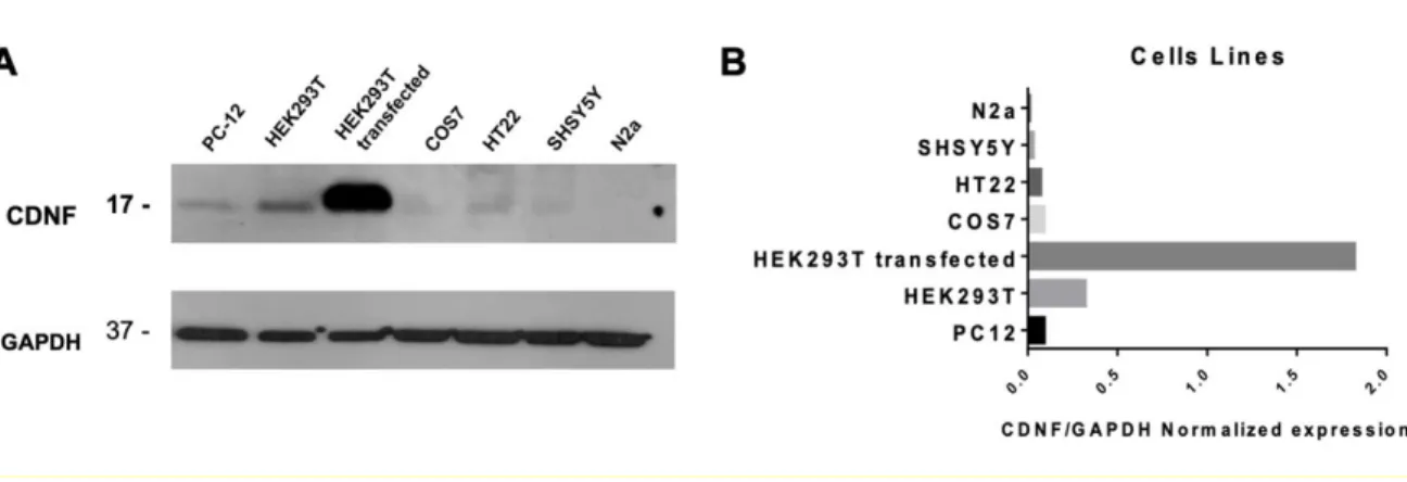
(26) 26. Establishing a cellular model for the study of ER stress. Although most studies with CDNF show a protective role in dopaminergic neurons, CDNF is expressed in non-neural tissues, and its cytoprotective activity extends beyond the dopaminergic system (Latge et al. 2015; Liu et al. 2018; Mei and Niu 2014). To characterize the expression of CDNF, we performed western blot analysis in different cell lines. The results show that CDNF protein levels vary among the different cell lines, observing a considerable endogenous expression in rat PC-12, human HEK293-T, and mouse HT22 cell lines (Figure 1). Thus, CDNF is expressed not only in neurons but also in a wide range of cell lines. In the present study, we choose HEK293-T due to this cell line is efficiently transfected.. To study the potential protective effect of CDNF against ER stress, we established in HEK293-T cells an ER stress model induced by Thapsigargin (Tg), an inhibitor of an ER calcium transporter (Chen et al. 2000). Tg causes ER stress and in the long-term, triggers cell death by apoptotic mechanisms (Fribley et al. 2009). The treatment of HEK293-T cells with Tg increased the expression of BiP and XBP1 and CHOP (Figure 2), demonstrating that the Tg treatment induces ER stress..
(27) 27. Figure 1. CDNF is expressed in different cell lines. A) Whole-cell extracts of rat PC-12, human HEK293-T, human SHSY5Y, monkey COS7, mouse HT22, and N2a were fractionated on SDS-PAGE and analyzed by Western blot assay to detect the CDNF peptide. The whole extract of HEK293-T cells transfected with FU-CDNF-W plasmid was used as a positive control, and GAPDH was used as load control. B) Protein levels of CDNF were quantified by densitometry using the ImageJ program..
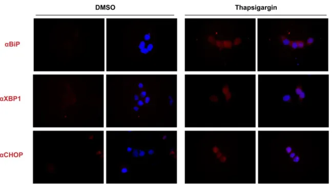
(28) 28. Figure 2. Establishing a model of ER stress using Thapsigargin (Tg). HEK293-T cells were treated with DMSO (A) or 1 uM of Tg (B) for 24h. Then, cells were fixed, and indirect immunofluorescence was performed to detect the UPR markers BiP, XBP1, and CHOP..
(29) 29. Establishing a CDNF gain-of-function model in HEK293-T cells. Considering the distinctive localization of CDNF in the ER, we hypothesized that its protective effect emerges from its subcellular location as a key regulator of the UPR. To test this hypothesis, first, we expressed rat CDNF (87% homology to human CDNF) using a bicistronic plasmid that encodes GFP as a reporter of transfection and the complete rat CDNF cDNA sequence, including its signal peptide (Figure 3A). Transfection of FUG-CDNF-W plasmid in HEK293-T cells resulted in a 5- fold increase of CDNF protein levels, and a similar increase of its mRNA (Figure 3B, C) compared to control transfected cells (mock).. Overexpressed CDNF showed a cytoplasmic reticular and perinuclear localization (Figure 3D), indicative of ER localization. To demonstrate that expressed CDNF localizes at the ER, the plasmid FU-CDNF-W was co-transfected with the FU-mRFP-KDEL-W plasmid, an ER marker. With the same purpose, we performed immunofluorescence for PDI, an ER endogenous marker, in cells transfected with FU-CDNF-W. As shown in Figure 4, the staining of CDNF distinctly overlaps with the fluorescence of mRFP-KDEL and PDI, demonstrating that the expressed CDNF specifically localizes in the ER compartment..
(30) 30. Figure 3. Transfecting FUG-CDNF-W plasmid in HEK293-T cells induces a five-fold. 6. increase of CDNF (A) Scheme of parental bicistronic FUG-W (mock) and derived FUGCDNF-W (CDNF) plasmids. (B-D) HEK293-T cells were transiently transfected with Mock or CDNF plasmids. Twenty-four hours later, cells were harvested to quantify the expression of CDNF by RT-qPCR (B), Western blots (C), and immunofluorescence (D). GAPDH was used as a reference gene in Western blots and RT-qPCR assays to normalize CDNF expression. Data are expressed as the mean of two independent experiments, performed each by duplicate. A.U.= arbitrary units. Scale bar: 20 µm..
(31) 31. Figure 4. CDNF localizes in the ER. HEK293-T cells were co-transfected with the FU-. 7. CDNF-W plasmid, which encodes only full-length rat CDNF, and FU-mRFP- KDEL-W (A), that encodes the monomeric red fluorescent protein fused to a signal peptide and containing the ER retention signal KDEL to mark the ER. CDNF immunofluorescence shows a distribution pattern that coincides with the mRFP-KDEL signal (red). Also, PDI immunofluorescence was performed, as an endogenous ER marker, indicating that CDNF (green) localizes in the ER (B). Scale bar: 10 µm..
(32) 32. First Scientific Article: Published CDNF induces the adaptive Unfolded Protein Response and Attenuates Endoplasmic Reticulum Stress-Induced Cell Death This article encompasses the execution of the following objectives: 1. To determine if CDNF exerts a cytoprotective effect by modulating the UPR. 1.1. To evaluate whether the gain of function of CDNF exerts a cytoprotective effect in HEK293-T cells and neurons through the induction of UPR pathways, during a pharmacological ER stress condition. 1.2. To evaluate whether the gain of function of CDNF exerts a cytoprotective effect in HEK293-T cells and neurons, through the attenuation of the apoptosis pathways induced by ER stress.. 2. To determine whether the cytoprotective effect of CDNF is intra or extracellular. 2.1. To evaluate the cytoprotective effect of a CDNF mutant lacking the non-canonical ER retention sequence QTEL (CDNF-ΔQTEL). 2.2. To evaluate whether the gain of function of CDNF-ΔQTEL exerts a cytoprotective effect in HEK293T cells through the induction of UPR pathways, during a pharmacological ER stress condition. 2.3. To evaluate if the gain of function of CDNF-ΔQTEL exerts a cytoprotective effect in HEK293T cells, through the attenuation of apoptosis pathways induced by ER stress..
(33) 33. Contents lists available at ScienceDirect. BBA - Molecular Cell Research journal homepage: www.elsevier.com/locate/bbamcr. CDNF induces the adaptive unfolded protein response and attenuates endoplasmic reticulum stress-induced cell death Duxan Arancibiaa, Pedro Zamoranob*, María Estela Andrésa*, a b. ARTICLEINFO Keywords: CDNF UPR ER stress Thapsigargin. DEPARTMENT of CELLULAR AND MOLECULAR Biology, FACULTY of BIOLOGICAL Sciences, PontifiCIA UNIVERSIDAD CATÓLICA de Chile, Chile. DEPARTAMENTO Biomédico, FACULTAD de CIENCIAS de LA SALUD, UNIVERSIDAD de ANTOFAGASTA, Chile. ABSTRACT The Cerebral Dopamine Neurotrophic Factor (CDNF) is a neurotrophic factor that has a protective effect in cell and animal models of several neurodegenerative diseases. The molecular mechanism of the protective effect of CDNF is unclear. Many neurodegenerative diseases have been related to a proteostasis dysregulation in the endoplasmic reticulum (ER). A failure of proteostasis produces ER stress, triggering the unfolded protein re- sponse (UPR) and, in the long-term, induces cell death. An adaptive UPR solves ER stress by attenuating protein synthesis, inducing chaperones expression, and degradation of misfolded proteins. Since CDNF is an ER resident protein, we investigated whether the role of CDNF is to regulate ER proteostasis. To this end, we determined the effect of CDNF in thapsigargin-induced ER stress in HEK293-T cells and cultured hippocampal neurons. Our results show that CDNF improved the viability of HEK293-T cells exposed to thapsigargin. CDNF increased levels of protective proteins of the early UPR, such as BiP, ATF4, ATF6, and XBP-1 in both HEK293-T cells and neurons. Conversely, expression of CDNF attenuated ER stress-induced apoptotic proteins, CHOP and cleaved caspase-3 in HEK293-T cells and neurons. A mutant CDNF lacking the ER retention sequence failed to protect against ER stress. In conclusion, CDNF regulates proteostasis in the ER by inducing the adaptive UPR response and in- hibiting apoptotic pathways triggered by ER stress. We propose that neuroprotection induced by CDNF is mediated by regulating ER proteostasis.. 1. Introduction The endoplasmic reticulum (ER) is an organelle that participates in the synthesis, folding and sorting of many proteins [1]. The equilibrium among these processes is called proteostasis of the ER and its disturbance is produced by an accumulation of misfolded proteins. A disruptive proteostasis triggers the unfolded protein response (UPR) [2,3], an evolutionary conserved mechanism in which the Binding Immunoglobulin Protein (BiP, also called GRP78) acts as a master regulator binding to the misfolded proteins in the ER. During this process, BiP detaches from the UPR sensors localized in the ER membrane resulting in the activation of three associated signaling pathways: protein kinase R-like endoplasmic reticulum kinase (PERK), activated transcription factor 6 (ATF6), and inositol-requiring enzyme 1 (IRE1). Autophosphorylation of PERK leads to the eIF2a phosphorylation [4] that attenuate protein translation, but selectively increases the. expression of proteins involved in oxidative stress, protein folding, and apoptosis. Thus, PERK mediates pro-survival and pro-death signaling [5]. ATF6 is cleaved at Golgi and the N-terminal domain translocate to the nucleus to induce expression/transcription of chaperones and ERassociated degradation proteins [6,7]. The activation of IRE1 induces the cytoplasmic splicing of XBP1 transcript and the protein translocate to the nucleus to induce ER stress-related gene expression [8]. A dysregulated UPR has been associated with many pathologic states such as cancer, diabetes and neurodegenerative diseases [9,10]. Many studies have found a causal link between the progress of neurodegenerative diseases and an alteration in the proteostasis in the ER, characterized by elevated UPR markers associated with cell death [11– 13]. Particularly, in cellular and animal models of Parkinson dis- ease, the treatment with neurotoxic agents such as 6-hydroxydopamine (6OHDA), 1-methyl-4-phenyl-1,2,3,6-tetrahydropyridine (MPTP), or αsynuclein induces ER stress and pro-apoptotic related proteins. ⁎ Correspondence to: P. Zamorano, Departamento Biomédico, Facultad de Ciencias de la Salud, Universidad de Antofagasta, Avenida Universidad de Antofagasta, Antofagasta, Chile. ⁎⁎ Correspondence to: M. E. Andrés, Department of Cellular and Molecular Biology, Faculty of Biological Sciences, Pontificia Universidad Católica de Chile, Avenida del Libertador Bernardo O'Higgins 340, Postal Code: 8331150 Santiago, Chile..
(34) 34 [14–16] supporting the idea that this neurodegenerative disease is related to a disturbed ER proteostasis. The Cerebral Dopamine Neurotrophic Factor (CDNF) has emerged as a potential treatment against Parkinson's disease and other neurodegenerative disorders due to its neuroprotective and restorative effects of neurons in in vitro and in vivo studies [17,18]. The application of CDNF to the extracellular milieu protected cultured mesencephalic neurons against α-synuclein-induced toxicity [19]. Likewise, overexpression of CDNF protected PC-12 cells against 6-OHDA-induced cell death [20], and alleviated Aβ-induced synaptotoxicity of hippocampal neurons [21]. In vivo studies have shown that a direct injection of CDNF into the striatum [22] or transduction with adeno-associated viruses encoding CDNF protected midbrain dopamine neurons against 6-OHDA neurotoxic effects [23–25]. CDNF is a non-conventional neurotrophic factor, which along with its homologue, the mesencephalic astrocyte-derived neurotrophic factor, MANF [26–28], reside in the ER, but can also be secreted to the extracellular medium [22,29,30]. Molecular studies showed that CDNF is retained in the ER through a non-canonical QTEL retention signal [28,30]. In spite that the three-dimensional structure of CDNF has been revealed [19], the molecular mechanism and the cell compartment from which CDNF exerts a cytoprotective effect are unknown. Con- sidering that CDNF is an ER resident protein that protects against da- mages associated to ER stress, we tested the hypothesis that CDNF protects cells and neurons against ER stress by modulating the UPR. We determined the role of CDNF in the UPR using HEK293-T cells treated with thapsigargin (TG), a well-established ER stressor and in cultured hippocampal neurons. Our data show that increasing CDNF levels in the ER of HEK293-T cells and hippocampal neurons triggers an early adaptive UPR and blocks the increase of pro-apoptotic proteins, indicating that the protective effect of CDNF is due to a key regulatory role in ER proteostasis. A mutant CDNF lacking the ER retention sequence failed to protect against ER stress and inducing BiP, indicating that CDNF must be localized in the ER to induce UPR and protect cells against TG-induced ER stress. 2. Material and methods 2.1. PLASMIDS FUG-W is a lentiviral plasmid that encodes green fluorescent protein (GFP, G in the plasmids) under the human ubiquitin promoter [31]. The bicistronic lentiviral plasmid FUG-CDNF-W [32] encodes separately GFP and rat full-length CDNF, including its signal peptide. CDNF was cloned downstream from the 1D2A sequence that allow cutting CDNF protein [33]. A mutated version of CDNF lacking QTEL sequence in the C-terminal was cloned in FUG-W plasmid. To analyze the localization of overexpressed CDNF, we co-transfected FU-CDNF-W (without EGFP) along with pFUmRFP-KDEL-W plasmid, which encodes the monomeric red fluorescent protein (mRFP) fused to an ER peptide signal and to the canonical ER retention sequence KDEL. All plasmids were verified by sequencing. 2.2. Antibodies The following primary antibodies were used: goat polyclonal antiCDNF (1:1000, R&D Systems), rabbit polyclonal anti-BiP (1:1000, Santa Cruz Biotechnology), rabbit polyclonal anti-XBP1 (1:1000, Santa Cruz Biotechnology), rabbit polyclonal anti-PDI (1:1000, Santa Cruz Biotechnology), rabbit polyclonal anti-ATF6 (1:500, Abexxa Biologics), rabbit polyclonal anti-ATF4 (1:500, Abexxa Biologics), rabbit polyclonal anti-CHOP (1:500, Abexxa Biologics), mouse monoclonal antiactive caspase-3 (1:100, Abexxa Biologics), mouse monoclonal antiGAPDH (1:10,000, Millipore) and Alexa Fluor 568 Phalloidin (Thermo Fisher Scientific). The secondary antibodies for immunofluorescence were Alexa Fluor® 488, Alexa Fluor® 568 anti-mouse, anti-rabbit and. anti-goat (1:500, Invitrogen, USA). For immunoblotting, horseradish peroxidase-conjugated antibodies were used to detect goat, mouse, and rabbit primary antibodies (1:5000, Invitrogen, USA). 2.3. Cell culture, TRANSFECTION AND THAPSIGARGIN TREATMENT HEK293-T cells were cultured in DMEM (Dulbecco's modified Eagle's medium; Gibco), supplemented with 10% (v/v) fetal bovine serum (Gibco), 100 units/ml penicillin (Gibco) and 100 μg/ml streptomycin (Gibco), and maintained at 37 °C in an atmosphere of 95% air and 5% CO2. Cells were transfected with Calfectin agent following manufacturer's recommendations (Calbiotech). Twenty-four hours after transfection the cells were treated in free-serum medium with thapsigargin (Sigma-Aldrich) or dimethyl sulfoxide DMSO (Sigma-Aldrich) during times and at concentrations indicated in the figures. 2.4. PRIMARY culture of RAT HIPPOCAMPAL neurons Pregnant Sprague-Dawley rats were obtained from Pontificia Universidad Católica de Chile. Hippocampi were dissected from E18 embryos and neurons were prepared using a modified Banker's culture protocol [34]. Cells were dissociated with trypsin and plated at 2.5 × 10⁵ cells/cm2 on poly-D-lysine (Sigma-Aldrich) coated coverslips in 24-well cell-culture dishes. Neuronal cultures were maintained in Neurobasal media supplemented with B27 (Gibco, USA), GlutaMAX-I (Gibco), 100 U/ml Ampicillin and 100 μg/ml Streptomycin (Gibco, USA) at 5% CO2 and at 37 °C. Hippocampal neurons were transfected with Lipofectamine 2000 following manufacturer's recommendations (Invitrogen). The cultures were stained with MAP2, showing over 90% of MAP2 positive cells. 2.5. MTS ASSAY Cells were seeded in 96-well plates at a density of 2 × 10⁴ cells/ml and incubated overnight. Then, cells were transfected with FUG-W and FUG-CDNF-W plasmids. Twenty-four hours after transfection, the media was changed for fresh media containing different amounts of TG, and cells were incubated for different times. At the end of the incubation period, 20 μl of MTS was added to each well and the plates were incubated for 3 h at 37 °C. Cell viability was evaluated by measuring the mitochondrial-dependent conversion of the yellow tetrazolium salt of MTS to purple formazan crystals by metabolic active cells. The optical density at 570 nm (proportional to the number of live cells) was assessed with an Epoch (Biotek). 2.6. RNA EXTRACTION AND RT-qPCR Total RNA was extracted using Trizol reagent (Ambion) according to the manufacturer's instructions. cDNA was synthesized from 1 μg of total RNA by RT-PCR using ImProm-II RT (Promega). Quantitative realtime PCR was performed using Evagreen qPCR Mix plus (Solis Byodine) in a LightCycler thermocycler (Roche), using specific primers for UPR markers described in [35]. Primers efficiency was calculated from a standard curve of increasing concentrations of cDNA targets (Supplementary Table 1). 2.7. Immunoblotting Whole cell extracts were obtained by homogenization in RIPA buffer (Millipore) containing inhibitors (1 mM PMSF, 7 μg/ml Pepstatin, 5–10 μg/ml Leupeptin and 10 μg/ml Aprotinin) and sonication. Protein concentrations were quantified by the BCA method (Thermo scientific). Samples were heated in Laemmli's loading buffer at 95 °C for 5 min, fractionated by SDS-PAGE, and transferred to a nitrocellulose membrane (Hybond, Amersham Biosciences). Membranes were blocked with Tris-buffered-saline (TBS) pH 7.4, containing 5% non-fat milk, and.
(35) 35 0.1% Tween-20. After incubation with the appropriate primary and secondary antibodies western blots were revealed by chemiluminescence (ECL, Amersham, USA). 2.8. Immunofluorescence IMAGING AND QUANTIfiCATION of fluorescence intensity Cells were fixed with 4% paraformaldehyde for 10 min at room temperature. Then, cells were permeabilized in 0.05% Triton X-100 in PBS and incubated with blocking solution (2% glycine, 2% BSA, 5% FBS, 50 mM NH4Cl in PBS pH 7.4) for 1 h. After incubation with ap- propriate primary and secondary antibodies, coverslips were mounted with Vectashield/DAPI solution (Vector). Images were acquired with an Olympus DS-Fi2 epifluorescence microscope furnished with 40× and 100× Olympus UplanFI oil immersion objective and equipped with a Nikon DS-fi2 camera operated with the standard QC capture software (Q-Imaging). Quantification was performed with ImageJ software (NIH, Baltimore, MD) using the corrected total cell fluorescence (CTCF) method. 2.9. STATISTICAL ANALYSIS Statistical analyses were performed by One-way or two-way ANOVA followed by Bonferroni post-test, using GraphPad Prism 6 software (GraphPad Software, Inc., San Diego, CA). Data are reported as mean ± SEM. Values of P < 0.05 were considered significant. 2.10. ETHICAL STATEMENT All experimental procedures (protocol number 161213007) were approved by the Ethic Committee of Pontificia Universidad Católica de Chile (CA 1492008598726). Efforts were made to minimize the number of animals used and their suffering. 3. Results 3.1. CDNF protects HEK293-T cells AGAINST TG-induced cell DEATH Considering the distinctive localization of CDNF in the ER, we hypothesized that its protective effect emerges from its subcellular location as a key regulator of the UPR. To test this hypothesis, we expressed rat CDNF (87% homology to human CDNF) using a bicistronic plasmid that encodes GFP as a reporter of transfection and the complete rat CDNF cDNA sequence including its signal peptide (Suppl. Fig. 1A). Transfection of FUG-CDNF-W plasmid in HEK293-T cells resulted in a 5fold increase of CDNF protein levels and a similar increase in mRNA (Suppl. Fig. 1B, C) compared to control transfected cells (mock). Overexpressed CDNF showed a cytoplasmic reticular and perinuclear localization (Suppl. Fig. 1D), indicative of ER localization. To further demonstrate that expressed CDNF is localized at the ER, the plasmid FU-CDNF-W was co-transfected in COS-7 cells with FU-mRFP-KDEL-W, an ER-directed marker. As shown in Suppl. Fig. 2, staining of CDNF distinctly overlaps with the fluorescence of mRFP-KDEL, demonstrating that expressed CDNF specifically localizes in the ER. To determine the mechanism by which CDNF protects cells, we modeled an altered ER proteostasis using HEK293-T cells exposed to TG, a potent inhibitor of the ER calcium transporter SERCA [36], which causes ER stress and in the long-term triggers cell death by apoptotic mechanisms [37]. Cell viability of HEK293-T cells cultured with increasing concentrations of TG (0, 0.25, 0.5, 1, 5, 10 μM) was assessed at 24 h by the MTS assay. A significant decrease in cell viability was evident at 1 μM TG and nearly two-thirds of cells died after 24 h of culture with 10 μM TG compared to untreated cells (Mock, Fig. 1A). These results confirm that HEK293-T cells are sensitive to TG that induced a dose-dependent cell death. As expected, CDNF induced a significant increase in HEK293-T viability (Fig. 1A). Interestingly, the. protection induced by CDNF was lost when the cells were exposed to 10 μM TG during 24 h, indicative of a limit to the protective role of CDNF when ER-stress is excessive. To further characterize CDNF protective effect against ER-stress, a time-course study was carried out by treating HEK293-T cells with 2 μM TG at different times (0, 3, 6, 9, 12, 24 h). Cell viability decreased after 9 h of treatment, reaching sig- nificance at 12 and 24 h (Fig. 1B). Expression of CDNF induced a sig- nificant protection against TG-induced HEK293-T cell death at 12 and 24 h (Fig. 1B). Altogether, these results show that expression of CDNF in the ER exerts a cytoprotective effect against TG-induced cell death. 3.2. CDNF promotes AN ADAPTIVE UPR Considering that CDNF protected the cells from TG-induced cell death and UPR is a conserved protective mechanism that emerges during ER stress, we tested the hypothesis that the protective effect of CDNF is due to a modulation of UPR. For this purpose, we assessed the expression of BiP, XBP1 and ATF6. Similar to a previous report [38], we observed a significant increase of BiP protein levels in HEK293-T cells treated with 1 μM TG at 24 h (Fig. 2A and B), confirming that TG induces ER stress and triggers the UPR. Expression of CDNF per se also increased BiP levels in HEK293-T cells (Fig. 2A and B), as compared with cells transfected with the control plasmid FUG-W (Mock). The time-course analysis of BiP mRNA expression and protein levels showed that TG induced a progressive increase in BiP that reach 4-fold increment at 12 h of exposure compared to untreated cells (Fig. 2C and D). Interestingly, expression of CDNF induced significantly higher levels of BiP mRNA and protein above TG-induced levels, until reaching a plateau at 12 h of treatment. These data indicate that CDNF regulates the expression of BiP, suggesting that CDNF is an inductor of an early UPR. The fact that CDNF increased BiP prompted us to identify the UPR signaling pathway stimulated by CDNF. BiP activates three pathways: IRE1, ATF6, and PERK [39–41]. Therefore, we studied XBP1, activated ATF6, and ATF4, downstream signals of IRE1, ATF6 and PERK pathways, respectively. Immunofluorescent assays show a significant increase in XBP1 (Fig. 3A, B) and ATF6 (Fig. 3C, D) in HEK293-T cells treated with 2 μM TG. Interestingly, CDNF induced a significant increase in basal levels of XBP1 (Fig. 3A, B) and ATF6 (Fig. 3C, D) proteins. In addition, at all times tested, CDNF expression induced a further increase in XPB1 and ATF6 protein levels compared to cells exposed to TG 2 μM alone (Fig. 3E, F and G). XBP1 mRNA is spliced during the UPR in the cytoplasm of stressed cells by IRE1, and this spliced XBP1 (sXBP1) mRNA isoform is the one that is translated into the XBP1 protein [42]. Therefore, we studied the effect of expressing CDNF in XBP1 splicing, by RT-qPCR using primers specific to quantify the sXBP1 isoform and total mRNAs. Expression of CDNF did not change total levels of XBP1 mRNA, but significantly increased sXBP1 mRNA isoform (Fig. 3H and I). Conversely, TG treatment induced a big increment of total XBP1 mRNA expression along with an increase of sXBP1 mRNA and protein. These data suggest that the mechanism by which CDNF induces an increase of the XBP1 protein is by increasing the IRE1 signaling. Finally, we measured the levels of a chaperone that belongs to the Glucose-Regulated Proteins (GRP), GRP94, which increases during ER stress [43]. We observed that TG 2 μM induced a significant increase of GRP94 mRNA levels. However, expression of CDNF did not change GRP94 mRNA levels (Fig. 3J), indicating that CDNF effect is specific over some UPR signaling pathways. To rule out the possibility of an induction of UPR is due to increasing the load in protein levels in the ER, we carried out two experiments. First, we analyzed the expression of the Protein Disulfide Isomerase (PDI) an ER-resident chaperone, whose expression is induced by protein accumulation [44]. Immunofluorescence and Western blot assays show that PDI levels were not modified by CDNF expression (Suppl Fig. 3). Second, we studied the effect of expressing mRFP-KDEL using the same promoter used to induce CDNF expression. The expression of mRFP-KDEL in the ER did not change BiP and XBP1 levels in.
(36) 36. Fig. 1. CDNF protects HEK293-T cells against TG-induced cell death. HEK293-T cells were transfected with FUG-W (black bars) and FUG-CDNF-W (gray bars) plasmids. Twentyfour hours later, cells were treated with different concentrations of TG (A) or with 2 μM TG during different time-length (B). Cell viability was quantified by MTS assays. The data are presented as % of control (mock). Data correspond to the mean ± SEM of 3 independent experiments, each performed in duplicates. The statistical analysis was performed with two-way ANOVA and Bonferroni post hoc test, *p < 0.05 and ***p < 0.001. a, ***p < 0.01 compared to control without TG treatment. 8. Fig. 2. CDNF increases BiP protein and mRNA levels. HEK293-T cells were transfected with FUG-W (Mock) or FUG-CDNF-W (CDNF). Twenty-four hours after transfection, cells were treated with TG (1 μM) for additional 24 h for immunofluorescent assays to detect BiP (A, B), or the indicated times for RT-qPCR (C) and western-blots (D) to quantify BiP mRNA expression and protein levels. GAPDH was used to normalize western blot and RT-qPCR data. The bars represent the mean ± SEM of 3 independent experiments, each performed in duplicates. The statistical analysis was carried out with ANOVA and a post hoc Bonferroni test, *p < 0.05, *** p < 0.001 and **** p < 0.0001 Scale bar: 20 μm. 9.
(37) 37. 3 8 5 1. Fig. 3. CDNF increases splicing of XBP1 and ATF6 protein levels. HEK293-T cells were transfected with FUG-W (Mock) or FUG-CDNF-W (CDNF). Twenty-four hours after transfection, cells were treated with TG (1 μM) for 24 h to perform immunofluorescent assays to detect XBP1 (A, B) and ATF6 (C, D). Western-blots were carried out at indicated times after TG treatment to quantify XBP1 (E, F) and ATF6 (G) protein levels. Expression of unspliced (H) and spliced (I) XBP1mRNA, and GRP94 mRNA (J) were quantified by RT-qPCR at indicated times after TG treatment. GAPDH was used to normalize RT-qPCR and western blot data. Data correspond to the mean ± SEM of 3 independent experiments performed as duplicates. Statistical analysis was performed with ANOVA and a post hoc Bonferroni test, *p < 0.05, **p < 0.01, ***p < 0.001. Scale bar is 20 μm. 10.
(38) 38. Fig. 4. CDNF increases the expression of ATF4. HEK293-T cells were transfected with FUG-W (Mock) or FUG-CDNF-W (CDNF). Twenty-four hours after transfection, cells were treated with TG (1 μM) for 24 h to perform immunofluorescent assays to detect ATF4 (A, B). Expression of ATF4 mRNA (C) and protein levels (D) were assessed respectively by RT-qPCR and western blots at indicated times after TG treatment. GAPDH was used to normalize RT-qPCR and western blot data. Data correspond to the mean ± SEM of 3 independent experiments performed as duplicates. Statistical analysis was performed with ANOVA and a post hoc Bonferroni test, *p < 0.05, ***p < 0.001 and **** p < 0.0001. Scale bar: 20 μm. 11. HEK293-T cells (Suppl Fig. 4A and B). Altogether, the data indicate that CDNF triggers an early UPR response. 3.3. CDNF ATTENUATES ER stress-induced APOPTOSIS Cell fate during an ER stress is determined by the balance between the adaptive UPR and the terminal UPR [45]. Therefore, we evaluated whether CDNF prevents the induction of the terminal UPR during an ER stress. To this end, we choose ATF4 protein to evaluate the PERK/ ATF4/CHOP signaling pathway, which is activated by prolonged ER stress to induce apoptosis [46]. Since expression of CDNF reduced cell death induced by TG, we expected a decrease in the expression of ATF4. Surprisingly, HEK293-T cells expressing CDNF showed a significant increase of ATF4 (Fig. 4). RT-qPCR assays showed that CDNF induced the expression of ATF4 mRNA in the basal condition, and produced a significantly greater increase at 6 h after treatment with TG, but not at later tested times (Fig. 4C). This increase in ATF4 mRNA levels was reflected in increased protein levels of ATF4 up to 12 h of TG treatment (Fig. 4D). The intriguing previous result suggested that CDNF could counteract TG-induced cell death by blocking pro-apoptotic pathways. Thus, we evaluated the pro-apoptotic protein CCAAT-enhancer-binding protein homologous protein (CHOP) induced in terminal UPR and cleaved caspase-3, a recognized marker of apoptosis. As expected, TG increased. the expression of CHOP and cleaved caspase-3 in HEK293-T cells (Fig. 5). A significant increase in CHOP mRNA levels (Fig. 5C) and a concomitant increase in protein levels were observed in cells treated with TG (Fig. 5A, B, D). Similarly, increased levels of active caspase-3 were observed at 12 and 24 h (Fig. 5E). Consistent with a protective effect, the expression of CDNF counteracted the increase in CHOP and caspase-3 in HEK293-T cells induced by TG (Fig. 5). Interestingly, CDNF per se induced a low but significant increase of CHOP basal levels, but arrested further increases induced by TG. Taken together, the data suggest that CDNF protects the cells by inhibiting the pro-apoptotic mechanisms activated by the PERK pathway. 3.4. CDNF induces AN EARLY ADAPTIVE UPR AND blocks the TERMINAL UPR in HIPPOCAMPAL neurons CDNF has emerged as a potential therapeutic tool for clinical use given its protective and restorative effects of neurons in models of neurodegenerative diseases. Our previous results prompted us to study whether CDNF protection of neurons is due also to its capacity of inducing the adaptive UPR. To answer this question, we transfected hippocampal neurons with FUG-CDNF-W to express CDNF (Suppl Fig. 5) and tested the expression of UPR marker proteins by quantitative immunofluorescence. As shown in Fig. 8, the expression of CDNF triggered an increase in BiP, XBP1, and ATF6 protein levels, suggesting that.
Figure
Documento similar
(A) HIF1 binding locations common to at least two out of four different ChIP-chip studies in HeLa, HepG2, MCF-7 and U87 cells (left), mammalian sequence conservation of the HIF
Glial Cell Line-Derived Neurotrophic Factor Promotes the Arborization of Cultured Striatal Neurons Through the p42/p44 Mitogen Activated Protein Kinase Pathway..
Inoue, Y., et al., Role of hepatocyte nuclear factor 4alpha in control of blood coagulation factor gene expression.. Safdar, H., et al., Modulation of mouse
At the same time, ZEB1 can switch from a transcriptional repressor to an activator by binding to p300/CBP suggesting another potential mechanism by which ZEB1 regulates
In addition, accumulating evidence in human lesion patients on the Val66Met BDNF polymorphism and cognitive function indicates that the Met allele exerts a protective effect
Our preliminary data on patient-derived MES GBM cells, in which we observed a downregulation of the MES signature upon FOSL1 knockdown (Figure R.20A-B), is quite convincing that
En este sentido, es interesante mencionar que la expresión de TCFL5 aparece en las fases finales de diferenciación a línea germinal coincidente con una disminución de los genes
In situ hybridization in late third instar wing discs of genes which expression levels change comparing 756-Gal4/UAS-sal vs 756-Gal4/UAS-GFP (UAS) or wild type vs Df(2L) 32FP5
