MDMA ("ecstasy") impairs learning in the Morris Water Maze and reduces hippocampal LTP in young rats
5
0
0
Texto completo
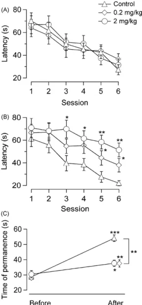
(2) 376. A. Arias-Cavieres et al. / Neuroscience Letters 469 (2010) 375–379. All animals’ care and procedures described below were approved by the Committee for Animal Care and Experimental Use of the University of Santiago de Chile and by the Chilean Council for Science and Technology Research (CONICYT). Male Sprague-Dawley rats, 21 days old (40–50 g), were housed two per cage with food and water available ad libitum under a 12:12 h light–dark cycle. For two days they were acclimatized to the holding room, maintained at a temperature of 22–23 ◦ C. MDMA (racemic, hydrochloride) or vehicle (isotonic saline) was administered 1 h before conducting experiments. Drug or saline was administered subcutaneously in the dorsum. The animals received 2 mg/kg MDMA (n = 22), 0.2 mg/kg MDMA (n = 22) or saline (n = 24), respectively in the training sessions (the twelve sessions of Fig. 1). No treatment was administered before the first two sessions and the last one that were run without platform. Behavioral training and testing were conducted in a circular pool (diameter 180 cm, depth 60 cm) painted white and, half-way filled with water that was rendered opaque with white latex paint and maintained at a temperature of 24 ± 2 ◦ C. Four geometrical figures in black and white (approx. 20 cm wide) attached to the upper rim of the pool and situated at an angle of 90◦ to each other, provided the optical cues, thus forming four quadrants. A white curtain surrounded the pool to obscure external cues. Behavioral data were recorded and analyzed using ANY-maze video tracking software (Stoelting Co., IL, USA). In the first 2 sessions (a session consisted in only 1 trial) the rats were allowed to swim for 2 min in the pool. In the subsequent 6 sessions a circular white platform (12 cm wide) was present, 1 cm above water level at a distance of 20 cm from the pool’s edge in the middle of a quadrant whose position was changed arbitrarily from session to session. During the following 6 sessions the escape platform was rendered invisible by placing it 2 cm below the water surface without changing its position. Finally one more session was performed with the platform removed. Subjects were allowed to swim until they had placed all four paws on the platform, or until 120 s had elapsed. Animals that did not get to the platform in two minutes were placed on it. The animals were left on the platform for 30 s after the end of each trial. For all groups, experimental sessions were run twice daily beginning at 10 a.m. and 15 p.m. for 6 days. Animal movements were monitored with a video camera mounted above the pool. The day after the last session in the maze, the rats were sacrificed by decapitation under halothane anesthesia. Hippocampi were dissected out, slices of 400 m thickness were cut, maintained and electrophysiological recordings obtained as described before [19].. Field excitatory postsynaptic potential (fEPSP) responses in the CA1 region were evoked delivering 0.2 ms electric pulses of 15–300 A with a bipolar electrode stimulating the Schaeffer collaterals and recorded in the stratum radiatum. The initial slope of the fEPSP was used as a measure of the evoked population excitatory synaptic response. A base line was established with test pulses adjusted to evoke 50% of the maximal response. Applying the same stimulus intensity, LTP was induced by application of theta burst stimulation (TBS): 10 trains, each with 10 bursts at 5 Hz, each burst consisting of 4 pulses at 100 Hz. Averaged slopes recorded for 20 min before TBS were compared to data obtained between 40 and 60 min after TBS. Paired-pulse (PP) stimulation was used varying stimulus intervals between 20 and 2560 ms, doubling the times between pulses after each stimulation, as previously described [19]. This protocol was applied 20 min prior to the TBS application and 30 min after. No MDMA or other drugs were applied in the slice experiments. In a random fashion, in each rat, one of the hippocampi was chosen to prepare slices for the electrophysiological recordings, while the other one served to determine tissue serotonin content. The tissue was stored at −80 ◦ C until analysis. Then it was suspended in HClO4 (0.1 M), sonicated and centrifuged (10,000 × g at 4 ◦ C) for 5 min. Fifty microliters of the supernatant was injected into a HPLC with electrochemical detection to quantify serotonin, as previously described [16]. Statistical evaluation: Results of the MWM test were analyzed using the general linear model ANOVA (fixed factor) for repeated (intragroup) and independent (intergroup) testing as appropriate, followed by the Newman–Keuls comparison test, as described by Vorhees and Williams [25]. The Greenhouse-Geisser correction was used when the variance–covariance matrices were significantly non-spherical. The same procedure was applied comparing the HPLC results. The LTP data of the different groups were evaluated using the Mann–Whitney U-test, while the Wilcoxon signed rank test was applied for comparing values before and after TBS in the same group. All animals increased their weight normally, without detectable differences between the groups (data not shown). In the MWM experiments differences in swimming speed between groups as well as sessions were far from being significant implying that, MDMA did not influence locomotor activity (Fig. 1). With the platform visible (cued trials), only a session effect was detected (F1,404 = 10.42, p < .001) as in all groups the latencies to reach the visible escape platform decreased significantly and with a similar time course during the 6 sessions (Fig. 2A). There were. Fig. 1. MDMA does not influence swimming speed. There are no significant differences either between groups or between sessions (control, 0.2 mg/kg and 2 mg/kg; n = 22 for both treated groups and n = 24 for controls; error bars: S.E.M.)..
(3) A. Arias-Cavieres et al. / Neuroscience Letters 469 (2010) 375–379. 377. no significant differences between the groups. Treatment did not affect acclimation to the task. When visuo-spatial learning was tested, with the platform rendered invisible, there were significant effects of drug treatment (F4,992 = 13.82, p < .001) and session (F1,035 = 6.61, p < .001), whereas no significant interaction of drug treatment × session was detected. Thus, both MDMA-treated groups were slower in learning the task, compared to the controls (Fig. 2B). Significant differences in the mean latency to reach the platform were observed comparing the group treated with 2 mg/kg MDMA with the control group, beginning with the 3rd session. In the 0.2 mg/kg group significant differences were found only in the 5th and 6th sessions. The differences in the average latencies comparing the group treated with 2 mg/kg with the 0.2 mg/kg group never reached significance even though the 0.2 mg/kg values (3rd through 6th session) were consis-. Fig. 3. MDMA reduced LTP in the CA1 region of the hippocampus in a dosedependent manner. (A) All groups display significant potentiation after TBS, the increase being significantly different between the groups (p < 0.01 throughout). Inset: traces for each condition at 40 min post TBS (thin) compared to recordings before TBS (thick) (n = 7 from 7 slices for all groups). (B) Paired-pulse facilitation remained unchanged across all ISI before and after TBS (left: control; middle: 0.2 mg/kg; right: 2 mg/kg). Inset: traces for each condition; 20 ms interval; open symbols: before TBS; closed symbols: after TBS.. Fig. 2. MDMA slows learning in the MWM. (A) Cued trials: average times to reach the visible platform diminished in a similar way for animals with and without treatment. Average times for the first session compared with the last session were significantly different for all groups. (B) Training trials: visuo-spatial learning with the invisible platform was dose-dependently slower when rats had received MDMA. However, all groups showed a significant learning effect that is also seen in (C) where average times are shown that animals stayed before and after training in the quadrant where the hidden platform was situated during training trials. (n = 22 for both treated groups and n = 24 for controls; * p < 0.05; ** p < 0.01; *** p < 0.001; error bars: S.E.M.).. tently shorter than those for the 2 mg/kg group. These results are compatible with a dose-dependent effect. On the day after the last training session the time spent in the quadrant where the platform had been was recorded. As shown in Fig. 2C, all groups remained significantly longer in that quadrant as compared to the time spent there before training. However, controls were found significantly more time in this quadrant than the treated animals (p < 0.01). These data demonstrate that all groups did learn, but in the treated groups learning was less efficient. fEPSPs were recorded from hippocampal slices of the same rats used in the MWM behavioral studies, 24 h after the last session. As shown in Fig. 3, LTP was significantly reduced in the slices obtained from MDMA-treated rats as compared to those from control rats. Interestingly, this reduction was also dose-dependent. On the other hand, no difference was found in the responses to PPF protocols between treated rats and controls before and after TBS (Fig. 3B), suggesting that the observed change in LTP was postsynaptic. In order to exclude possible neurotoxic effects of MDMA, we measured serotonin levels in the hippocampus. No significant differences among the three experimental groups were detected (in ng/g of tissue: control (n = 13) 506.5 ± 127.3; 0.2 mg/kg (n = 11) 617.3 ± 134.8; 2 mg/kg (n = 12) 536.5 ± 187.1; means ± SEM; p > 0.05). These figures are close to those reported previously [3,10,14]. The main result of the present study is that MDMA, administered in a non-neurotoxic dose regime [13] to young rats for a week, impairs visuo-spatial learning and, in the same animals, reduces LTP in the hippocampal CA1 region. The observations are congruent as LTP has been shown to occur together with behavioral manifestations of learning [29]. Further, the hippocampus plays a major role in visuo-spatial learning as shown by Morris et al. [12]. There-.
(4) 378. A. Arias-Cavieres et al. / Neuroscience Letters 469 (2010) 375–379. fore, our results are compatible with the idea that MDMA-induced learning impairment be caused by an impairment of LTP induction. During our experiments animals were 23–31 days old. While it is difficult to identify an equivalent age in humans and, although the brain is still developing, all classical neurotransmitter receptors are present and active at this age [9]. The age resembles human childhood and adolescence more than those in previous studies that used either adult or very young animals at the moment of MDMA administration [1,22,25–28]. To estimate the relevance of the doses applied in the present study to doses taken by humans, we may use the formula proposed by Mordenti and Chappell [11]: Dhuman = Danimal (Whuman /Wanimal )0.7 . In our case the dose of 2 mg/kg and an animal weighing 70 g, compared to a 70 kg human, would yield approximately: 142 g (absolute single dose given) × (70,000 g/70 g)0.7 = 17.6 mg, or, a relative dose of about 0.25 mg/kg, and the lower dose would even correspond to 0.025 mg/kg. Taking into account that this dose was administered twice daily over 6 days, this amounts to 3/0.3 mg/kg or an absolute intake of 211/21 mg. The higher dose regimen might therefore resemble that of a “novice” to “moderate” human user [15,21], with the difference that the human subject will have taken this dose in a single session of several hours. Only a “heavy” user would have taken MDMA on several occasions during the week, possibly exceeding one gram in this period. The lower dose regimen is far below anything but an averaged “weekly” dose of a hypothetical occasional user taking one tablet a month. Earlier studies assessing the effects of MDMA on learning in rats with the MWM were performed using higher doses (≥10 mg/kg), reporting significant reductions in serotonin levels [1,21]. This implies toxic effects at serotonergic terminals [3]. In contrast, we believe that the doses applied in the present study did not reach toxic levels as evidenced by the serotonin contents remaining unchanged and within the normal range. Further, in the present experiments no abnormalities in locomotor activity (Fig. 1) or weight gain were detected. Together with the dose the timing of MDMA application seems to be a critical factor. It makes a difference whether the drug is actually present in the brain or effects caused by a pretreatment of several days are being investigated. In the slice experiments the differences seem to be clear cut. Acutely applied, MDMA furthers LTP (EC50 = 12.5 M; [20]), whereas in the present experiments a pretreatment of the whole animal over days caused LTP reduction without MDMA present in the tissue (the last injection was given about 44 h before the animal was sacrificed for tissue preparation and recordings). In the MWM experiments it is not possible to separate effects of acute and sustained treatment and neither it is clear whether acquisition, recall, or both are affected and resulted in the impaired performance observed. Still, the LTP results imply that the impairment of visuo-spatial learning observed is due to postsynaptic plasticity mechanisms and not to other factors like a change in attention as in the case of methylphenidate [32]. Interestingly, for the latter substance, as with the case of acute MDMA treatment, an increase of LTP has been reported [5]. In mice, acute MDMA administration (3 mg/kg) led to an improved performance in an avoidance task that was not attributable to enhanced locomotor activity. However, a pretreatment over several days impaired the acquisition performance tested two days later, particularly with higher doses [24]. Interestingly, recall was impaired even at a dose of 1 mg/kg. These effects did not last longer than a few days. These and other findings [18] together with the present results underline the complex action of MDMA on learning—being advantageous when the substance is present, but impeding learning after pretreatment. Our LTP data, showing LTP increase as acute. effect [20], but decrease after pretreatment, are in line with this view. A pretreatment with repetitive administration of relatively high doses may cause long-term or permanent damage (especially in very young individuals) as shown by the group of Vorhees [1,25–28], whereas lower doses may induce only transitory learning impairment [24]. Of the three hypothetical ways by which MDMA affects learning, namely a toxic, long-lasting effect, a short-term effect of treatment over several days and the immediate effect of the substance being actually present, the underlying pharmacological and molecular mechanisms still remain unclear. However, Gudelsky and Yamamoto [8], employing in vivo microdialysis, found that MDMA increases the release of dopamine, serotonin and acetylcholine in the hippocampus and various other brain areas implicated in learning and memory (see also [6,23]). Moreover, they demonstrate that the dopamine release is modulated by serotonin receptor activation (5-HT2A/C or 5-HT2B/C ). Consistent with their finding, we have presented preliminary evidence that the MDMA-induced increase of LTP seen with acute application may be mediated by 5-HT2A/2C and D1 receptors [20]. Thus, a next step, we suggest, should be a further elucidation of the mechanisms that produce MDMA-induced learning impairment through long-term and medium-term changes. In summary, three important conclusions can be drawn from the results of this study. First, low, presumably non-neurotoxic doses of MDMA, can produce impairment in visuo-spatial learning. Second, that impairment is correlated with a reduction in LTP. Third, MDMA pretreatment at low doses over several days, produces postsynaptic changes involved in LTP induction. Acknowledgements The authors wish to thank Dr. Bruce Cassels and Dr. Claudio Acuña-Castillo for critically reading the manuscript. We are also indebted to Dr. Fernando Ortiz for his experienced help in the revision of the statistical analysis. Financial support from FONDECYT to BM and MLZ (grant no. 1080652) and from DICYT to MLZ and BM (grant no. 020993Z) is gratefully acknowledged. References [1] J. Able, G. Gudelsky, C. Vorhees, M.T. Williams, 3,4-Methylenedioxymethamphetamine in adult rats produces deficits in path integration and special reference memory, Biol. Psychiatry 59 (2006) 1219–1226. [2] ACMD Advisory Council on the Misuse of Drugs, MDMA (‘ecstasy’): a review of its harms and classification under the Misuse of Drugs Act 1971, 2009. http://drugs.homeoffice.gov.uk/publication-search/acmd/mdma-report. [3] G. Battaglia, S. Yeh, E. O’Shearn, M. Molliver, M. Kuhar, E. Souza, 3,4Methylenedioxymethamphetamine and 3,4-methylenedioxyamphetamine destroy serotonin terminals in rat brain: quantification of neurodegeneration by measurement of [3 H]paroxetine-labeled serotonin uptake sites, J. Pharmacol. Exp. Therap. 242 (1987) 911–916. [4] J.P. Capela, H. Carmo, F. Remião, M.L. Bastos, A. Meisel, F. Carvalho, Molecular and cellular mechanisms of ecstasy-induced neurotoxicity: an overview, Mol. Neurobiol. 39 (2009) 210–227. [5] E.J. Dommett, E.L. Henderson, M.S. Westwell, S.A. Greenfield, Methylphenidate amplifies long-term plasticity in the hippocampus via noradrenergic mechanisms, Learn. Memory 15 (2008) 580–586. [6] E. Escubedo, J. Camarasa, C. Chipana, S. García-Ratés, D. Pubill, Involvement of nicotinic receptors in methamphetamine- and MDMA-induced neurotoxicity: pharmacological implications, Int. Rev. Neurobiol. 88 (2009) 121–166. [7] R. Green, A. Mechan, M. Elliot, E. O’Shea, M.I. Colado, The pharmacology and clinical pharmacology of 3,4-methylenedioxymethamphetamine (MDMA, “Ecstasy”), Pharmacol. Rev. 55 (2003) 463–508. [8] G.A. Gudelsky, B.K. Yamamoto, Actions of 3,4methylenedioxymethamphetamine (MDMA) on cerebral dopaminergic, serotonergic and cholinergic neurons, Pharmacol. Biochem. Behav. 90 (2008) 198–207. [9] E. Herlenius, H. Lagercrantz, Development of neurotransmitter systems during critical periods, Exp. Neurol. 190 (2004) S8–S21. [10] M. Higuchi, Y. Suzuki, Y. Yatani, Y. Kitagawa, K. Nagayasu, H. Shirakawa, T. Nakagawa, S. Kaneko, Augmentation of serotonin release by sustained exposure to MDMA and methamphetamine in rat organotypic mesencephalic slice.
(5) A. Arias-Cavieres et al. / Neuroscience Letters 469 (2010) 375–379. [11]. [12] [13]. [14]. [15]. [16]. [17]. [18] [19]. [20]. [21]. cultures containing raphe serotonergic neurons, J. Neurochem. 106 (2008) 2410–2420. J. Mordenti, W. Chappell, The use of interspecies scaling in toxicokinetics, in: A. Yacobi, J. Kelly, V. Batra (Eds.), Toxicokinetics in New Drug Development, Pergamon Press, New York, 1989, pp. 42–96. R.G.M. Morris, P. Garrund, J.N.P. Rawlins, J.O. O’Keefe, Place navigation impaired in rats with hippocampal lesions, Nature 297 (1982) 681–683. National Drug Intelligence Center, National Drug Threat Assessment 2009, Document ID: 2008-Q0317-005, 2009. http://www.justice.gov/ndic/pubs31/31379/. E. O’Shea, R. Granados, B. Esteban, M. Colado, A. Green, The relationship between the degree of neurodegeneration of rat brain 5-HT nerve terminals and the dose and frequency of administration of MDMA (‘ecstasy’), Neuropharmacology 37 (1998) 919–926. A.C. Parrott, Chronic tolerance to recreational MDMA (3,4methylenedioxymethamphetamine) or ecstasy, J. Psychopharmacol. 19 (2005) 71–83. M. Reyes-Parada, M. Scorza, R. Silveira, F. Dajas, G. Costa, K. Tipton, B. Cassels, Monoamine oxidase inhibitory effects of some 4-p-aminophenethylamine derivatives, Biochem. Pharmacol. 47 (1994) 1365–1371. G. Rogers, J. Elston, R. Garside, The harmful health effects of recreational ecstasy: a systematic review of observational evidence, Health Technol. Assess. 13 (2009) 1–338. A.G. Romano, J.A. Harvey, MDMA enhances associative and non-associative learning in the rabbit, Pharmacol. Biochem. Behav. 47 (1993) 289–293. C. Rozas, H. Frank, A.J. Heynen, B. Morales, M.F. Bear, A. Kirkwood, Developmental inhibitory gate controls the relay of activity to the superficial layers of the visual cortex, J. Neurosci. 21 (2001) 6791–6801. C. Rozas, F. Pancetti, M. Reyes-Parada, B. Cassels, B. Morales, MDMA facilitate the long-term potentiation but not long-term depression in CA1 of hippocampus, in: Annual Meeting of the Society for Neuroscience, Washington D.C., USA, 2008 (abstract P737). A.B. Scholey, A.C. Parrott, T. Buchanan, T.M. Heffernan, J. Ling, J. Rodgers, Increased intensity of ecstasy and polydrug usage in the more experienced recreational ecstasy/MDMA users, Addict. Behav. 29 (2004) 743–752.. 379. [22] J. Sprague, A. Preston, M. Leifheit, B. Woodside, Hippocampal serotonergic damage induced by MDMA (ecstasy): effects on spatial learning, Physiol. Behav. 79 (2003) 281–287. [23] G.E. Torres, R.R. Gainetdinov, M.G. Caron, Plasma membrane monoamine transporters: structure, regulation and function, Nat. Rev. Neurosci. 4 (2003) 13–25. [24] J.M. Trigo, A. Cabrero-Castel, F. Berrendero, R. Maldonado, P. Robledo, MDMA modifies active avoidance learning and recall in mice, Psychopharmacology 197 (2008) 391–400. [25] C. Vorhees, T. Reed, M. Skelton, M. Williams, Exposure to 3,4 methylenedioxymethamphetamine (MDMA) on postnatal days 11–20 induces reference but not working memory deficits in Morris water maze in rats: implications of prior learning, Int. J. Dev. Neurosci. 22 (2004) 247–259. [26] C. Vorhees, M. Williams, Morris water maze: procedures for assessing spatial and related forms of learning and memory, Nat. Protoc. 1 (2006) 848–858. [27] C. Vorhees, T. Schaefer, M. Williams, Developmental effects of ±3,4 methylenedioxymethamphetamine on spatial versus path integration learning: effects of dose distribution, Synapse 61 (2007) 488–499. [28] C. Vorhees, T. Schaefer, M. Skelton, C. Grace, N. Herring, M. Williams, (+/−) 3,4 methylenedioxymethamphetamine (MDMA) dose-dependently impairs spatial learning in the Morris water maze after exposure of rats to different five-day intervals from birth to postnatal day twenty, Dev. Neurosci. -Basel 31 (2009) 107–120. [29] J. Whitlock, A. Heynen, M. Shuler, M. Bear, Learning induces long-term potentiation in the hippocampus, Science 313 (2006) 1093–1097. [30] P.J. Winsauer, M.S. Quinton, J.R. Porter, C.B. Corll, J.M. Moerschbaecher, M.S. Delatte, S.T. Leonard, S.B. Stroble, Effects of MDMA administration on scopolamine-induced disruptions of learning and performance in rats, Pharmacol. Biochem. Behav. 79 (2004) 459–472. [31] K.K. Zakzanis, Z. Campbell, D. Jovanovski, The neuropsychology of ecstasy (MDMA) use: a quantitative review, Hum. Psychopharmacol. 22 (2007) 427–435. [32] M.L. Zeise, S. Espinoza, A. González, F.S. Cerda, J. Nacarate, C.G. Yáñez, B. Morales, Methylphenidate improves cue navigation in the Morris water maze in rats, NeuroReport 18 (2007) 1059–1062..
(6)
Figure

Documento similar