Accessing new biomedical applications by combining genetic design and chemical modification of elastin like recombinamers
293
0
0
Texto completo
(2)
(3) Für Jalah Für Lian.
(4)
(5) AGRADECIMIENTOS ACKNOWLEDGEMENTS.
(6)
(7) Agradecimientos – Acknowledgements. Agradecimientos – Acknowledgements En las siguientes páginas me gustaría conmemorar a las personas que me han acompañado y siempre me han apoyado en esta fase emocionante e intensiva de mi vida para obtener el grado de doctorado. Este capítulo de mi vida no solo me ha educado científicamente, sino que también me ha desarrollado personalmente. Si mi vida fuera una novela, esta última etapa llenaría las paginas de más de más de un capítulo, pudiendo rellenar una pagina con cada uno de vosotros. A veces unas palabras son capaces de trasmitir un mensaje más grande, como el amor, el respeto o la amistad, que me conectan con todos de usted. En primer lugar, quisiera dar las gracias a mi director Carlos. Gracias por acogerme en el grupo, por guiarme y por darme todos los medios para poder llevar a cabo este trabajo y por tu tiempo y tus consejos indispensables. Quiero mostrar igualmente mi gratitud a Matilde y Israel, que siempre han dedicado su tiempo y su predisposición cuando lo necesitaria. Gracias a los seniors de grupo, Javi, Merche, Alessandra y Luis, por vuestra amabilidad y por vuestras ganas de ayudar. Muchas gracias a mis compañeros de despacho BIOGEL, Soraya y Filippo vosotros habéis llenado el espacio con alegría, y me habéis dado ganas de volver a trabajar no solo en momentos buenos, pero también en temporadas criticas . Tanto a mis “compañeros de doctorado y sobre mesa” (Doriana, Tatjana, Sofia, Ito, Sergio, Arturo, Juan), como a las nuevas incorporaciones Miguel, Marcos, Fernando. Me habéis apoyado muchísimo para llegar a Valladolid y habéis apoyado aprender las técnicas del grupo. Muchísimos gracias.. III.
(8) ACCESSING NEW BIOMEDICAL APPLICATIONS BY COMBINING GENETIC DESIGN AND CHEMICAL MODIFICATION OF ELASTIN-LIKE RECOMBINAMERS. Rocío, muchas gracias por enseñarme los secretos de la purificación y por tu paciencia. Gracias Irene y Alicia por proporcionarme cientos de veces vuestro ánimo. Todos vosotros impregnáis esta tesis. Solo espero haber sido capaz en este tiempo de devolveros, aunque solo sea una pequeña parte de todo el afecto que me habéis dado durante estos años. This work as being part of an incredible international project was further supported by a phenomenal group of people. I am very proud that I can consider you as my friends and that we have shared such an incredible time together at so many interesting and wonderful places and that you have shared your research with me. This interdisciplinary input really leveraged my knowledge. First, I have to name Laura, Professor Möller and Elisabeth, who did an incredible job in managing and realizing this wonderful project. Further, I like to thank Alan and Paul for their input on the mechanical characterization, especially during my stay in the university of Nijmegen (the Netherlands) and the support of my project colleagues Paula and Max (Kai-zengh Li-u) it was so inspiring to work on your side. My journey also brought me to Aachen were I want to thank my resident colleagues Arturo, Luis, Marcel and Sitara and their co-workers Christopher and Yashoda for their help in the lab and for their inspiring thoughts. To complete the Biogel group, a special thanks goes to those who I unfortunately could not work with for a longer period, but not least we had an incredible time in countless meetings and I think we managed to get to know each other pretty well. Thank you Melanie, Daria, Jenny, Dominik, and Nestor, and of course Delphine who was a short member, but remains part of our group. IV.
(9) Agradecimientos – Acknowledgements. Abschließend möchte ich noch meiner Familie Danken ohne deren Unterstüzung ich diese Aufgabe in der Form nicht hätte realisieren können. Als erstes Danke Ich meiner Ehefrau Jalah für Ihre Unterstüzung und Ihre Stärke in einer Phase in der Ich gerne mehr für Sie getan hätte als es mir durch die räumliche Trennung möglich war. Ich Danke Dir, vielen Dank das du so wundervoll bist und unserem Sohn eine so tolle Mutter bist, auch wenn ich nicht immer da sein konnte. Weiterhin gilt ein Dank meinem Bruder Nico und meiner Mutter Andrea und meiner Großmutter Eva, für Ihre immer währende Unterstüzung. Ein Dank auch an meinen Vater Piet und seine Frau Anke, ich bin froh das ich in dieser prägenden Phase auf euch zählen konnte.. Leander. V.
(10) ACCESSING NEW BIOMEDICAL APPLICATIONS BY COMBINING GENETIC DESIGN AND CHEMICAL MODIFICATION OF ELASTIN-LIKE RECOMBINAMERS.
(11) ABSTRACT.
(12)
(13) Abstract. Abstract The diversity of nature including biopolymers, organisms, or active molecules and the fact that these natural systems can be transformed to be used for other purposes, enables a myriad of promising starting points for new developments. Here, Elastin-like recombinamers (ELRs) as a class of intrinsically disordered recombinant proteins that exhibit features like a lower. critical. solution. temperature. (LCST),. thermosensitive. hydrophobicity, self-assembly, together with the possibility to introduce other peptide-based functionalities, stand out. The present work coveres the steps of the value chain of ELRs from nucleotide fragments, over DNA transfection, protein production, purification, chemical modification and application of the final protein. Including the utilization of related methods ranging from genetic engineering, microbiology, physico-chemical characterization and modification to in-vitro cell culture and manipulation. One part of the tesis is focused on the generation of new proteolytically cleavable ELRs modified with methacrylamide groups as a substrate for a new zymographic technique enabling the detection of proteases. The present method uses the advantage of the spatial design of the ELR for the detection of proteases in a zymography-like manner. As a prerequisite, the ELR should consist of at least three domains, a degradable group (peptidase target), a mechanism of detection, and a stable cross-link to the acrylamide matrix. For better integrity with the matrix an additional block, which is not a target to most of the peptidases was implemented, as a spacer, to augment the molecular weight and to minimize sterical hindrance between cross-links and degradable groups. The presented method has the potential as a protease screening, which gains significance directly proportional to the. IX.
(14) ACCESSING NEW BIOMEDICAL APPLICATIONS BY COMBINING GENETIC DESIGN AND CHEMICAL MODIFICATION OF ELASTIN-LIKE RECOMBINAMERS. number of protease targets (ELRs) used. The presented in-well zymography (IWZ) method has a high throughput potential, therefore, further substrates are currently developed to generate a library of substrates that allow discovering new cleavage patterns of proteases, and might have relevance to detect abnormalities in clinical samples.. In the following, the low complexity of these polypeptides is used as a model for the investigation of hydrophobic contributions to self-assembly. In this work, an upper critical solution temperature (UCST)-type phase separation was obtained upon adding hydrophobic cholesteryl side-chains, which leads to the formation of stable hydrogels. Circular dichroism, dynamic light scattering, and viscosity measurements indicate reversible UCST gelation of a pseudo-zwitterionic species, with retention of the ELR secondary structure. Furthermore, the gelation process could be associated with micelle-derived nanofiber formation to yield a porous or laminar structure in a process that is either time- or concentration-dependent. Herein we present a model system that sheds light on the diversity of effects resulting from the incorporation of cholesteryl side-chains into proteins by relating structural changes to macroscopic observations.. In addition, a cellular coating based on hydrophobic interactions of an elastin-like recombinamer (ELR) with the cell membrane is presented. It is well documented that biophysical properties such as net charge, hydrophobicity, and protein-driven cell-ligand (integrin binding) interactions influence the interaction of polymers, proteins or peptides with model membranes and biological cells. Most studies to enhance membranesubstrate interactions have focused on the introduction of positively charged. X.
(15) Abstract. groups to foster electrostatic interactions with the negatively charged membrane. Herein we present an antagonistic approach based on ELRs with varying amounts of hydrophobic cholesteryl groups (ELRCTAs). The ability of membranes to stabilize cholesteryl groups is hypothesized to assist the coordination of hydrophobic ELRs with the membrane. The main objective was to generate a defined cellular coating of a recombinant protein that allows for total sequence control and less host, or batch-to-batch, variation as a substitute for existing coatings like alginate, polyelectrolytes, collagens and fibronectin. We used an in vitro cell-binding assay to quantify cellmembrane interactions, with the substrates showing enhanced cellular recognition and matrix distribution with an increasing number of cholesteryl groups incorporated. These novel materials and the versatile nature of their protein sequence have great potential as cellular markers, drug carriers, or hydrophobic cell-binding domains.. XI.
(16)
(17) RESUMEN.
(18)
(19) Resumen/Summary. Resumen/Summary Introducción La adquisición de un conocimiento profundo de biomateriales requiere comprender su papel en diversas aplicaciones, tanto en ingeniería de tejidos como en nanotecnología. Dentro de las distintas estrategias destinadas al desarrollo de aplicaciones biomédicas, cabe destacar el uso de materiales que mimetizan las propiedades y características propias que se encuentran en materiales naturales1. En primer lugar, los materiales empleados para aplicaciones médicas deben de cumplir una serie de requisitos como la biocompatibilidad, pero además deben aportar funciones específicas para su uso, como estabilidad mecánica, o la posibilidad de ajustar la degradación del propio material. Los primeros materiales empleados en biomedicina fueron distintos metales o cerámicas, ya conocido por las antiguas civilizaciones2. Tras esto, con el avance de la química macromolecular aparecieron los polímeros sintéticos como el ácido poli(láctico-co-glicólico) (PLGA) o el ácido poliláctico (PLLA). En las últimas décadas el desarrollo de la ingeniería genética y de la tecnología del ADN recombinante ha permitido generar nuevos materiales proteicos, los cuales representan un gran paso en la fabricación de materiales biomiméticos3,4. De este modo, se abría un extenso abanico de posibilidades para el uso del biomaterial más abundante, y probablemente más complejo que podemos encontrar en la naturaleza, las proteínas. En los últimos años, la ciencia ha hecho un gran avance en la elucidación estructural de proteínas, y sigue avanzando en el estudio de las relaciones entre estructura y función3,4. Este. XV.
(20) ACCESSING NEW BIOMEDICAL APPLICATIONS BY COMBINING GENETIC DESIGN AND CHEMICAL MODIFICATION OF ELASTIN-LIKE RECOMBINAMERS. conocimiento permite no sólo modificar la secuencia de un material tan complejo como las proteínas, e incluso mejorarlas. Dentro de esta clase de biomateriales recombinantes, encontramos la familia de proteínas inspirada en la secuencia de aminoácidos de la tropoelastina natural, que es el monómero soluble de la elastina5–11. De forma simplificada nos referimos a esta familia de proteínas es como recombinámeros tipo elastina (“elastin-like recombinamers”, ELRs, en inglés). Las ventajas en el uso de la tropoelastina como “proteína madre”, son las funcionalidades ésta como biocompatibilidad, elasticidad y comportamiento termo-sensible que conduce a procesos de autoensamblado12. Además, la secuencia subyacente de amino ácidos a estas propiedades es bastante simple desde el punto de vista de la complejidad de las proteínas. Dicha secuencia está basada en la repetición del pentapéptido L-Valina-L-Prolina-Glicina-X-Glicina (VPGXG), en el que X (denominado aminoácido invitado) puede ser cualquier aminoácido, excepto L-Prolina7,13– 17. . Dependiendo del número de repeticiones y de la composición del amino. acido invitado, X, se pueden crear una gran variedad de ELRs. Debido a su carácter recombinante, es posible fusionar secuencias de ADN que codifican distintos péptidos y proteínas. Así se puede combinar bloques de pentapéptidos con distintos aminoácidos invitados, en el lugar X, en la misma molécula del ELR. Además, es posible añadir secuencias peptídicas como secuencias biodegradables18,19, o con dominio de adhesión celular20,21 para generar materiales ‘a la carta’ de la aplicación deseada.. Objetivos El objetivo de esta tesis es demostrar que la versatilidad de estos recombinámeros se puede aumentar meidante modificación química y. XVI.
(21) Resumen/Summary. genética para la configurar la degradación, el autoensamblado y la interacción con células de los ELRs. El trabajo desarrollado en esta tesis aborda todo el proceso de diseño, producción, purificación, caracterización y aplicación directa de los nuevos ELRs. Para ello, se han utilizado una amplia variedad técnicas de ingeniería genética, microbiología, física, química junto con los correspondientes cultivos celulares. A. La tecnología del ADN recombinante permite un control total sobre el diseño de ELR, y de este modo la inserción de distintas secuencias biofuncionales, como secuencias sensibles a proteasas. Mediante el control de la disposición espacial de este tipo de secuencias proteolíticas queremos demostrar la biodegradación especifica de ELRs. Además, la capacidad. de. biodegradación. selectiva. será. aplicada. para. la. biofabricación de sustratos para detección zimográfica. B. Debido a la degradabbilidad controlable de los ELRs, su uso será estudiado como sustrato selectivo para la identificación de enzimas proteolíticas. Por lo tanto, siguiendo el estudio de la aplicación de ELRs para técnicas zimográficas diseñaremos un nuevo método de detección de proteasas con potencial para sistemas de inspección avanzados de alto rendimiento. C. Se ha demostrado que los biomateriales modificados con colesterol exhiben fuertes interacciones intermoleculares. Así, aplicaremos estas interacciones en un sistema de ELRs para generar fuerzas intermoleculares que desencadenen el autoensamblado de los ELR. D. Además, gracias a la capacidad de interacción de los grupos colesterol con membranas lipídicas, estudiaremos la capacidad de ELRs ricos en colesterol para mejorar la interacción de los éstos con ciertos tipos. XVII.
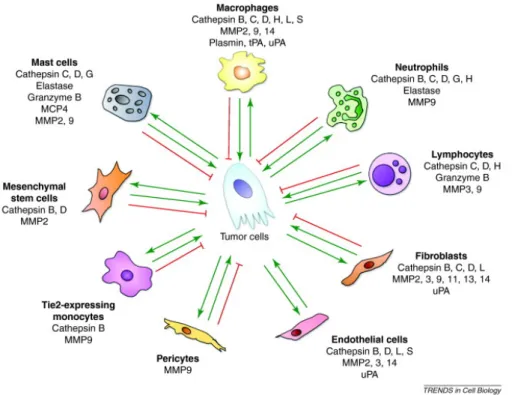
(22) ACCESSING NEW BIOMEDICAL APPLICATIONS BY COMBINING GENETIC DESIGN AND CHEMICAL MODIFICATION OF ELASTIN-LIKE RECOMBINAMERS. celulares implicados en la captación de lípidos, para aumentar el recubrimiento de células vivas con proteínas ELR.. Metodología del Diseño de ELRs con grupos proteolíticos (A y B) Con el objetivo de utilizar ELRs como sustrato de zimografía de geles (IGZ) más específicas, se pueden fusionar secuencias de degradación enzimática, por ejemplo la secuencia que contiene el hexapéptido L-Valina-Glicina-LValina-L-Alanina-L-Prolina-Glicina (VGVAPG), y cuya L-Alanina con especificad a la enzima elastasa, aumentando la biodegradabilidad de los ELRs, que es de gran importancia en el remodelado del tejido22. Además concentraciones altas en proteasas también son típicamente efectos secundarios de cáncer promovando la infiltración de tejido23. Pese a que la degradación enzimática es más estudiada en el campo de la regeneración tisular, la degradación de sustratos basados en proteínas también puede ser utilizada para desarrollar técnicas de detección de proteasas, enzimas capaces de degradar proteínas. Una de las técnicas más conocido es la zimografía de gelatina24. El método funciona como una electroforesis de poliacrilamida en que la gelatina se añade al gel, si la muestra electroforética en ese gel contiene una enzima con actividad proteolítica, la gelatina será degradada en el lugar que corresponde al peso molecular de esta proteasa. En el caso de la gelatina este método está bien establecida por los metalloproteasas de la matriz MMP2 y MMP925. Sin embargo, aparte de gelatina, otras proteínas naturales pueden utilizarse como substrato de la zimografía, y de este modo aumentar la variedad de secuencias degradables presentadas a las enzimas. Por otro, lado esa variedad en su secuencia depende de muchos factores, como el organismo. XVIII.
(23) Resumen/Summary. de origen o el proceso de purificación. Así, aunque de la gelatina se conocen muchos elementos estructurales, se desconoce su secuencia completa. Por lo que, los resultados pueden ser confusos, e inclusos erróneos debido a que no se conozca la secuencia proteolítica que está siendo degradada por la enzima. En esta tesis utilizamos ELRs como substrato para zimografia que presenten únicamente un grupo proteolítico, y que tengan una secuencia conocida, para investigar la degradación con una selección de MMPs 1 a 20. Esto facilita el uso de dicho ELRs en un nuevo método de zimografia dentro de una placa (in-well zymography (IWZ).. Resultados (A y B) Los resultados mostraron que las diferencias en las concentraciones de peptidasas se pueden medir con zimografía utilizando ELRs como sustrato. Además, el uso de ELRs permitió la detección de proteasas de degradación inespecífica tipo serina, las cuales son indetectables en sustratos clásicos como la gelatina. Más concretamente, los resultados revelaron que, a diferencia de métodos zimográficos clásicos, la ELR-IGZ permite la detección de las enzimas metaloproteasas MMP2 y MMP9. La sensibilidad hacia MMP9 fue casi diez veces mayor con ELREla + FN que en gelatina, mientras que el control negativo ELR sin ninguna región proteolítica no fue degradado. El cambio a IWZ reduce la complejidad en comparación con IGZ mediante la eliminación de la separación electroforética y la desnaturalización de la muestra. La sensibilidad se limitó a un 10% respecto al control no degradado. Se pudo detectar patrones distintos de degradación de cada MMP y se redujo la cantidad necesaria para la cuantificación de 50-500 ng (en IGZ) a 10 ng por muestra.. XIX.
(24) ACCESSING NEW BIOMEDICAL APPLICATIONS BY COMBINING GENETIC DESIGN AND CHEMICAL MODIFICATION OF ELASTIN-LIKE RECOMBINAMERS. Los resultados permitieron clasificar las enzimas proteasas en cuatro grupos diferentes respecto a los sustratos empleados: 1) sin degradación (MMP 2, 3, 10, 14, 16, 19 y 20); 2) mediada por FN (MMP1, 13, 16); 3) mediada por ELA (MMP7); 4) no específica (MMP 8,9, 12, 17).. Conclusiones (A y B) Se demostró que la producción de sustratos para IGZ basados en ELRs y secuencias proteolíticas. Los cuales permiten la incrustación en matrices de gel, y la detección con CBB. Empleando dos grupos de degradación diferente, se revelaron diferencias en la especifidad frente proteasas tipo serina y distintas MMPs. Cabe mencionar que el sustrato ELR-Control, al carecer de secuencias específicas de degradación, no fue digerido por ninguna de las MMPs seleccionadas, pero sí se detectó su ligera degradación en presencia de tripsina. La degradación del control pudo deberse a la presencia de lisinas que son son un punto de corte preferente para las proteasas tipo serina, además las lisinas, debido a su cadena lateral catiónica, son sensibles a la tinción con CBB. Por otro lado, presentamos un nuevo método de detección de proteasas mediante la adaptación de la IGZ en una zimografía de placa de pocillo (IWZ), lo que permite una lectura rápida y cuantitativa. Este nuevo método proporciona una técnica de detección capaz de determinar diferencias en patrones de degradación de metaloproteasas (MMP1-20) con sólo dos secuencias de degradación. El control positivo y negativo sirvió como un circuito de retroalimentación adicional en términos del patrón derivado. Complementariamente, este método de cuantificación permitiría seguir la cinética de degradación de cada una de las enzimas sobre los sustratos específicos. La especificidad de los sustratos basados en ELRs aporta varios. XX.
(25) Resumen/Summary. avances a las técnicas zimográficas basadas en gel, por ejemplo gracias al control de los motivos de degradación se pueden elaborar dispositivos de reconocimiento de enzimas proteolíticas basado en un sistema binario.. Metodología de la Modificación de ELRs con grupos hidrofóbicos (C) El interés por los biomateriales autoensamblados ha aumentado considerablemente en las últimas décadas debido a su compleja disposición espacial y a que puedan controlar la exposición de distintas funcionalidades. Las implicaciones directas de la estructura en el autoensamblaje en polímeros de bloque anfifílicos y proteínas, junto a la posibilidad de generar interacciones hidrofóbicas26,27 e hidrofílicas28,29, ha permitido cierto grado de control sobre estructuras jerárquicas, como micelas esféricas o cilíndricas, vesículas, membranas bimoleculares e hidrogeles30–33. Además, propiedades tales como la respuesta frente a estímulos como cambios de temperatura27,34 o pH35, han dado como resultado la aplicabilidad de estos materiales en la ingeniería de tejidos36, sistemas para la administración de fármacos35–38, dispersantes de polímeros39, agentes gelificantes físicos40 o biosensores41–43. La capacidad de respuesta térmica es la característica más investigada y mejor controlada para los copolímeros de bloque27,34 y, como tal, ha ganado un mayor interés en el diseño de proteínas recombinantes de vanguardia, especialmente a la luz de los hallazgos de Urry con respecto a la LCST de péptidos miméticos de elastina44–47. Además, debido a que los ELRs permiten distintos diseños modulares combinando bloque análogos con dominios de diferente hidrofilicidad, se han estudiado como modelos para comprender el plegamiento, el autoensamblaje y la función de otras proteínas naturales más complejas48–50.. XXI.
(26) ACCESSING NEW BIOMEDICAL APPLICATIONS BY COMBINING GENETIC DESIGN AND CHEMICAL MODIFICATION OF ELASTIN-LIKE RECOMBINAMERS. Aunque se espera que el comportamiento de separación de fases del tipo de temperatura de solución crítica superior (UCST) en el agua51–54 contribuya a la creación de materiales poliméricos intrínsecamente ordenados55–59, también se han observado ejemplos de proteínas intrínsecamente desordenados que exhiben separación de fases de tipo UCST55–59. Como la abductina y la resilina, que exhiben una UCST y LCST60,61, sin embargo, hasta la fecha no se ha observado una UCST tras la modificación química de las proteínas recombinantes, especialmente las ELRs. En este sentido, hemos aumentado la interacción hidrófoba de las proteínas mediante la adición de cadenas laterales de colesterol, que se sabe que fomenta las interacciones intermoleculares en los polímeros, aumentando el orden en la proteína y hemos investigado cómo se ve afectada la estructura secundaria de la proteína. Para una mejor comprensión. de. los. determinantes. de. la. gelificación. inducida. hidrofóbicamente, los ELR resultantes de la modificación con grupos colesterol (CTA) se caracterizaron en detalle; concentración crítica de micelas, ángulo de contacto, dispersión dinámica de la luz, dicroísmo circular, imágenes SEM y TEM. Se descubrió que los CTA resultantes (CTAx (x = número de grupos de colesterol)) constituyen un grupo de materiales novedoso y peculiar, los cuales presentan dos Tts y gelificación fría a temperaturas inferiores a UCST.. Resultados (C) Debido a la naturaleza intrínsicamente desordenada de los ELRs, la formación de estructuras cristalinas para su análisis mediante espectroscopía de rayos X no es posible. Además su alto peso molecular dificulta enormemente su evaluación mediante espectroscopía de RMN, de este modo, se estudió mediante CD la estructura secundaria de los distintos. XXII.
(27) Resumen/Summary. ELRs modificados con grupos colesterol. La similitudes estructurales observadas entre los estados del CTA5 por encima de la LCST y por debajo de la UCST confirmaron la hipótesis de que el colesterol contribuye al autoensamblado de los ELRs por debajo de la temperatura de transición. De este modo, demostramos por primera vez que el comportamiento UCST puede ser inducido por modificaciones de la cadena lateral en ELRs. Para la caracterización de la gelificación fría (característica del los ELRCTAx), se investigaron soluciones de mayor concentración utilizando técnicas de reología oscilatoria y de flujo. En general, no se observó gelificación en ausencia de grupos laterales colerterol. Comparando el módulo de almacenamiento, se observó que éste aumentó en función del número de grupos colesterol (31 Pa para ELRCTA1 y 60 Pa ELRCTA5). De los resultados obtenidos se puede derivar el comportamiento de fase de ELRCTA5 con dos transiciones alrededor de 10 y 20 °C con una región intermedia de solubilidad, caracterizada por la ausencia de estructuras secundarias paralela y antiparalela (relajada).. Metodologia Interacción celular de colesteryl ELRs (D) La bicapa de membrana es una barrera física que define la frontera de las células y divide el interior en distintos compartimentos con funciones especializadas. La naturaleza anfifílica de la membrana conduce a una superficie externa cargada negativamente y a un núcleo hidrófobo dentro de la membrana, que sirve como anclaje para proteínas de membrana y transmembrana62. En consecuencia, las células tienen la capacidad de unirse a sustratos mediante la unión de integrinas, interacciones electrostáticas, enlaces de hidrógeno o interacciones hidrofóbicas63. Además, la membrana contiene una variedad de compuestos y proteínas que son cruciales para la. XXIII.
(28) ACCESSING NEW BIOMEDICAL APPLICATIONS BY COMBINING GENETIC DESIGN AND CHEMICAL MODIFICATION OF ELASTIN-LIKE RECOMBINAMERS. función celular. Una de las moléculas más abundantes en la membrana es el colesterol, que se encuentra dentro de la membrana debido a su hidrofobicidad. El colesterol representa hasta el 20% de la masa de la membrana63 y es responsable de la integridad de membrana además de participar en procesos de señalización celular. El colesterol, entre los muchos componentes lípidos de las membranas celulares de los mamíferos, es un regulador clave de la fluidez de la membrana y contribuye a la formación de caveolas y al mantenimiento del microdominio de las caveolas64,65. Las caveolas son dominios especializados de la membrana plasmática que se encuentran en la mayoría de los tipos de células66. Sin embargo, son más abundantes en células como adipocitos, células endoteliales, fibroblastos y células musculares67. Además de la influencia de la fluidez de la membrana, el colesterol es importante para la estructura y la función celular, es esencial en la locomoción y sirve como precursor metabólico. para. varias. moléculas. de. señalización,. incluidos. los. oxiesteroles68, los esteroides68 y los ácidos biliares65,69,70. La mayoría de los sustratos o hidrogeles se centran en motivos de unión a integrinas, en particular el motivo de RGD, para generar unión celular o la infiltración. Sin embargo, los materiales sintéticos con residuos de RGD unidos covalentemente luchan por cumplir con los resultados de los materiales de proteínas ECM nativas71. Otra investigación para la fabricación de construcciones de tejido 3D se ha centrado en la ingeniería de la lámina celular62, los liposomas magnéticos72, las perlas celulares73 y las células que contienen capas de gel74–76. En particular, la creación de nanodominios y microdominios específicos para distintos tipos celulares o específicos de tejido utilizando interacciones biológicas, como avidina-biotina77–80, anticuerpo-antígeno81,82, lectina-polisacáridos83,84 o proteínas ECM como fibronectina, colágeno y / o gelatina74,75,85–89, ha sido prometedora y permite la "manipulación jerárquica de células" in vitro85,88,90,91.. XXIV.
(29) Resumen/Summary. Seleccionamos grupos colesterol como anclajes hidrófobicos debido que simulan la situación intramembranosa y facilitan la movilidad y distribución de las proteínas en la membrana. Para una mejor interacción del material de colesterol con la membrana, se estudió en tipos celulares ricos en caveolina como son las células derivadas del endotelio de la vena umbilical humana (HUVEC) y las células del músculo liso aórtico humano (HASMC), que están involucradas en la absorción de lípidos del fluido sanguíneo y en procesos tales como estenosis. Para ello, se modificaron varios ELRs con grupos colesterol y se estudió el potencial de estos grupos hidrófobos como anclajes intramembranosos para recubrimientos celulares. En este sentido, hemos desarrollado un recubrimiento celular basado en interacciones hidrófobas con la membrana celular. Para eludir las variaciones de proteínas naturales como variaciones específicas al organismos de origen, se utiliza un recombinante similar a la elastina análogo de ECM como proteína modelo para el recubrimiento inductivo de tejido. El objetivo principal es la creación de un recubrimiento proteico para células basado en una monocapa bien distribuida.. Resultados (D) Los diferentes sustratos se probaron para determinar su efecto sobre la proliferación celular durante 7 días usando el ensayo AlamarBlue® a concentraciones de 375 y 750 nM. Los HUVEC no mostraron diferencias significativas en la proliferación en todas las condiciones, mientras que para los HASMC, el tratamiento con una alta concentración de los ELRs ELRCTA0 y ELRCTA1 condujo a la disminución de la proliferación con el tiempo. Se observó que el número de eventos de células fluorescentes aumentó de ELRCTA0 a ELRCTA5, del 10% al 80% (HUVEC), o del 20% al 85% (HASMC),. XXV.
(30) ACCESSING NEW BIOMEDICAL APPLICATIONS BY COMBINING GENETIC DESIGN AND CHEMICAL MODIFICATION OF ELASTIN-LIKE RECOMBINAMERS. respectivamente. Las células HASMC interactúaron en mayor medida con ELRCTA0 a ELRCTA3 , aumentando de un 20% a 60% respectivamente, mientras que la eficiencia de recubrimiento de estos ELRs permaneció por debajo del 30% para las células HUVEC. Además, las imágenes confocales de células tripsinizadas después de la incubación revelaron un cambio en la distribución de los ELRs en la membrana celular en función del contenido en colesterol, variando desde partículas de tamaño micrométrico aglomeradas (sin colesterol) hasta puntos submicrométricos altamente dispersados (con colesterol).. Conclusiones (C y D) Hemos. demostrado. que. las. propiedades. novedosas,. como. el. autoensamblaje y la gelación termorreversible, se pueden controlar mediante la modificación selectiva de IDPs poliméricas, lo que resulta en materiales altamente innovadores que pueden exhibir un comportamiento UCST y LCST, donde el plegamiento y la formación de estructuras secundarias ordenadas se ve favorecida tanto por encima de la LCST como por debajo de la UCST. En base a nuestros resultados, los grupos de colesterol parecen actuar como mediadores estructurales, en este caso como estabilizadores de micelas en condiciones impulsadas por la entropía por encima de la LCST o como conectores de micelas expuestas en condiciones impulsadas por la entalpía por debajo de la UCST. En ambos casos, los grupos de colesterol aceleran la organización estructural de los ELR con el tiempo. El hecho de que se obtengan estructuras secundarias similares con grupos de colesterol orientados hacia adentro o hacia afuera (por debajo o por encima de la UCST y LCST, respectivamente) podría deberse al esqueleto del ELR subyacente, que gobierna los motivos estructurales a los que se puede acceder.. XXVI.
(31) Resumen/Summary. Hasta donde sabemos, los restos hidrofóbicos que funcionan como anclajes de membrana celular para proteínas aún no se han investigado. Dado que no se observó ningún efecto citotóxico en los tipos celulares seleccionados a concentraciones bajas, con un resultado > 60% de células marcadas, el presente sistema puede conducir a una mejor comprensión de las interacciones hidrofóbicas membrana-sustrato y al desarrollo de nuevos recubrimientos celulares impulsados por la hidrofobicidad, proporcionando una alternativa a los recubrimientos actuales. Además, estos nuevos materiales y su versatilidad en términos de secuencia tienen un alto potencial como marcadores celulares, portadores de fármacos o dominios de unión celular hidrófobos.. XXVII.
(32)
(33) INDEX.
(34)
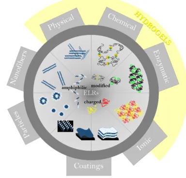
(35) Table of Content. Table of Content Agradecimientos – Acknowledgements ................................................................................... III Abstract ............................................................................................................................................IX Resumen/Summary ...................................................................................................................... XV Introducción ............................................................................................................................. XV Objetivos .................................................................................................................................. XVI Metodología del Diseño de ELRs con grupos proteolíticos (A y B) ....................... XVIII Resultados (A y B)................................................................................................................. XIX Conclusiones (A y B) ............................................................................................................. XX Metodología de la Modificación de ELRs con grupos hidrofóbicos (C) ................... XXI Resultados (C)....................................................................................................................... XXII Metodologia Interacción celular de colesteryl ELRs (D) ........................................... XXIII Resultados (D) ...................................................................................................................... XXV Conclusiones (C y D) .........................................................................................................XXVI Table of Content.............................................................................................................................. 3 Introduction ........................................................................................................................................... 9 1 Introduction ................................................................................................................................ 11 1.1. Biomimetic Materials ...................................................................................................... 11. 1.1.1.. Natural Elastin......................................................................................................... 12. 1.1.2.. Elastin bio-inspired polypeptides (Elastin-like Recombinamers) ................. 14. 1.2. Biosynthesis of ELRs ....................................................................................................... 22. 1.2.1. Design of the codifying gene .................................................................................... 23. 1.2.2. Construction of the multi block ............................................................................. 24. 1.2.3. Gene expression and recombinant production .................................................. 28. 1.2.4. Purification ................................................................................................................... 29. 1.3. Protein - interactions ..................................................................................................... 31. 1.3.1. Physical ELR interactions .......................................................................................... 32. 1.3.2. Ionic interactions ........................................................................................................ 32. 1.3.3. Hydrophobic interaction of amphiphilic blocks and graft copolymers .......... 33. 1.3.4. Intermolecular interaction of protein secondary structures ........................... 33. 1.4 1.4.1 1.5. Chemical Modifications of ELRs .................................................................................. 36 Functionalization of ELRs and Covalent Cross-linked ELR Hydrogels .......... 36 ELR structures and applications................................................................................... 38. 1.5.1. Hydrogels ..................................................................................................................... 39. 1.5.2. ELRs as drug carriers ................................................................................................. 40. 3.
(36) ACCESSING NEW BIOMEDICAL APPLICATIONS BY COMBINING GENETIC DESIGN AND CHEMICAL MODIFICATION OF ELASTIN-LIKE RECOMBINAMERS. 1.6.1. Cholesterols role in cell membranes......................................................................41. 1.6.2. Cholesterol modified materials ................................................................................41. 1.7. Role of Proteases .............................................................................................................44. 1.7.1. Peptidase Classification ..............................................................................................44. 1.7.2 Role of Proteases in Matrix Remodeling and Related diseases............................46 1.7.2. Serine Proteases ..........................................................................................................49. 1.7.3. MMPs ..............................................................................................................................50. 1.7.4. Cleavable peptide bonds............................................................................................52. 1.7.5. Involvement of Peptidase in tissue regeneration .................................................54. 1.7.6. Disease related to peptidases...................................................................................58. 1.7.7. Zymographic Methods of Protease Detection .....................................................60. 2. Hypothesis..............................................................................................................................69. 3. Materials & Methods ............................................................................................................73. 3.1 Materials ....................................................................................................................................73 3.1.1 Chemical Reagents..........................................................................................................73 3.1.2 Glass ware & other materials.......................................................................................74 3.1.3. Molecular biology materials. .....................................................................................74. 3.1.4. Other reagents.............................................................................................................79. 3.1.5. Bacterial Strain .............................................................................................................79. 3.1.6. Bacterial culture media ..............................................................................................79. 3.1.7. Buffers ............................................................................................................................79. 3.1.8. Elastin-like recombinamers .......................................................................................80. 3.1.9. Cell lines and culture conditions .............................................................................82. 3.2. Methods..............................................................................................................................83. 3.2.1 DNA agarose gel electrophoresis...............................................................................83 3.2.2 Plasmid purification.........................................................................................................84. 4. 3.2.3. DNA digestion with restriction enzymes ..............................................................84. 3.2.4. DNA dephosphorylation ...........................................................................................84. 3.2.5. DNA separation and extraction from agarose gel ..............................................84. 3.2.6. Ligation of genes ..........................................................................................................85. 3.2.7. Cloning on the pDrive/ p7 vector ...........................................................................85. 3.2.8. Transformation of competent cells.........................................................................85. 3.2.9. Glycerol stock preparation .......................................................................................87. 3.2.10. Biopolymers expression and purification ..........................................................87. 3.2.11. SDS Polyacrylamide gel electrophoresis ...........................................................89. 3.2.12. Modification of ELRs ..............................................................................................91.
(37) Table of Content. 4. 3.2.13. Detection of Trypsin in ELR gels........................................................................ 93. 3.2.15. Optimization of screening parameters ............................................................. 95. 3.2.16. Screening of MMPs ................................................................................................ 95. 3.2.17. Critical Micelle Concentration ........................................................................... 96. 3.2.18. Dynamic light scattering ....................................................................................... 96. 3.2.19. Zeta potential.......................................................................................................... 97. 3.2.20. Circular Dichroism ................................................................................................ 97. 3.2.21. Viscosity measurements ....................................................................................... 98. 3.2.22. Rheological determination of low-temperature gelation ............................. 98. 3.2.23. Contact Angle ......................................................................................................... 99. 3.2.24. Transmission electron microscopy TEM .......................................................... 99. 3.2.25. Scanning electron microscopy SEM ................................................................. 100. 3.2.26. Spectrophotometric cloud point determination .......................................... 100. 3.2.27. Cell Culture........................................................................................................... 100. 3.2.28. FACS and Confocal Microscopy....................................................................... 101. Results and Discussion ..................................................................................................... 107. 4.1 CHAPTER 1: Development of a novel screening method for proteases based on proteolytically degradable ELR-acrylamide hydrogels ........................................................ 107 4.1.1 ELR IK-HEX gene Synthesis ....................................................................................... 111 ELRs expression and purification........................................................................................ 115 4.1.2 Methacrylation of ELRs ............................................................................................... 117 4.1.3 WORKING PRINCIPLE OF THE METHOD ........................................................ 120 4.1.4 ELR in gel zymography ................................................................................................ 122 4.1.5 ELR in-well zymography (IWZ) ................................................................................ 126 4.2 CHAPTER 2: Chemical Modification of ELRs with Cholesterol and Structural Mechanistics of Cholesteryl Group Mediated UCST Gelation........................................ 135 4.2.1 Chemical Modification of ELRs ................................................................................. 139 4.2.2 Characterization in solution ...................................................................................... 142 4.2.3 Characterization of gelation ...................................................................................... 152 4.2.4 Evolution of macrostructures below the UCST ................................................... 158 4.3 CHAPTER 3: ELR – Cell interaction triggered by hydrophobic modification....... 169 4.3.1 ELR Modification .......................................................................................................... 173 4.3.2 Characterization in solution ...................................................................................... 177 4.3.3 Cytotoxicity of ELR particles .................................................................................... 179 4.3.4 ELR – cell Interactions ................................................................................................ 181 5. Conclusions ......................................................................................................................... 201. 5.
(38) ACCESSING NEW BIOMEDICAL APPLICATIONS BY COMBINING GENETIC DESIGN AND CHEMICAL MODIFICATION OF ELASTIN-LIKE RECOMBINAMERS. 5.1 ELR Zymography.................................................................................................................. 201 5.2 Structural deductions of cholesterol modified ELRs ................................................... 202 5.3 Cellular interactions with Cholesteryl-ELRs ................................................................. 204 6. References ........................................................................................................................... 209. 7. Appendix.................................................................................................................................. III. 7.1 Supporting Information .......................................................................................................... III 7.1.1 ABBREVIATIONS ............................................................................................................ X 7.1.2 TABLE OF STANDARD AMINO ACID ABBREVIATIONS............................. XIII 7.1.3. List of Tables ............................................................................................................. XIV. 7.1.4. List of Figures ............................................................................................................. XV. 7.1.3 LIST OF ARTICLES AND PATENTS ORIGINATED FROM THE WORK OF THIS PhD THESIS ................................................................................................................ XXII. 6.
(39) Table of Content. 7.
(40)
(41) INTRODUCTION.
(42)
(43) 1 Introduction. 1 Introduction 1.1 Biomimetic Materials Building on the discoveries and experiences of our ancestors is one key driver of mankind’s progression and technical evolution, saving us from reinventing the wheel over and over again. Biomimicking pursues the same target to build on the results of several millions of years of evolution, which have forged nature close to perfection generating a diversity of biological systems with extraordinary properties92. The goal is not just to copy natural systems, but rather to deduce synergetic potential with known technologies to improve certain applications. The diversity of nature including biopolymers, organisms, or active molecules and the fact that these natural systems can be transformed to be used for other purposes, enables a myriad of promising starting points for new developments1.. Taking a closer look at biomimetic materials, one class of biopolymers stands out in terms of diversity, taylorability and applicability. These are protein based materials, where the properties are based on the arrangement of the amino acid residues3,4. Natural proteins consist of up to 20 different amino acids and an unspecified number of amino acids (n) which opens possible 20n arrangements for a distinct number of amino acids n. Since this number becomes rather big with protein size, a fundamental understanding of the biophysical properties imparted by certain peptide structures is crucial. The resulting structural complexity and limited computing power made it inevitable to deduce structural relations by manipulating single amino acids in known structural resolved natural amino acid sequences. The number of natural protein sources and their diversity already facilitated their use in. 11.
(44) ACCESSING NEW BIOMEDICAL APPLICATIONS BY COMBINING GENETIC DESIGN AND CHEMICAL MODIFICATION OF ELASTIN-LIKE RECOMBINAMERS. novel materials and applications. The advancements in exogenous protein expression, in the past decades enabled the creation of de novo proteins with new or enhanced properties, which were a priori designed to comprise specific functionalities3,4. The primary goal of protein polymer science is to alter protein sequences to obtain unique properties in a single molecule either in terms of structure or functionality which satisfies a need in biomedicine, or nanotechnology amongst others.. When considering the effect on structure of protein sequences alteration, covalent and noncovalent interactions, such as peptide and disulfide bonds, interactions or van der Waals forces, have to be taken into account12. Moreover, spatial arrangements can foster intermolecular interaction which can lead to aggregation or self-assembly events. In general, many structure function relation known in polymers can be translated to protein based materials, such as alternating- or block-copolymers by using as monomers, amino acids of differing hydrophilicities, which in turn result in comparable structural observations like micelles, vesicles, micro phase separation, or even. macroscopic. hydrogels93.. furthermore,. current. advances. in. biotechnology and recombinant protein expression facilitate the generation of desired protein motifs in a defined and accurate way towards an increasing understanding of protein structure and function relationship.. 1.1.1.. Natural Elastin. The prerequisite for materials to be used in biomedicine is biocompatibility and bioactivity, when desired and controlled, which make proteins a suitable candidate. Especially, in tissue engineering, extra cellular matrix (ECM) components are in focus of current research94. Here, elastin has to be. 12.
(45) Biomimetic Materials. highlighted. Elastin is a fibrous and insoluble protein that constitutes one of the most important structural and functional components of the ECM, allowing for high deformations without damage95. It is abundant in the lungs (3-7 %), skin (2-3 %), blood vessels (28-32 %) and elastic ligaments (50 %)96, where elastic properties are required. Elastin whose main function is to provide elasticity to organs and tissues, is an excellent example of how all the properties displayed by biological materials and systems are determined exclusively by the physicochemical properties of the monomers and their sequence97,98. Elastin possesses several extraordinary characteristics, such as an ability to undergo high deformation without breaking and subsequent recovery of the original conformation once the stress disappears99,100. This energy-conserving process allows for billions of relaxation-stretching cycles of the elastic fibers101. Several peptide sequences like VPGVG, VPGG, VGVAPG in elastin’s soluble precursor tropoelastin have been found to be responsible for the elastic behavior102, which have been used to circumvent the insolubility of native elastin. In the beginning elastin mimetic proteins/peptides, derived from chemical solid phase synthesis have been used, and since the late 90s are produced by genetic engineering strategies103–107. Investigation of short peptide sequences originating from tropoelastin by D. Urry further deduced a sub-structure of elastin, the socalled α-elastin, to be responsible for the thermal behavior of elastin. In aqueous solution, α-elastin is able to transition from a disordered state to an ordered state where the molecules are capable of aggregating, resulting in a sticky and dense separated phase as a result of a temperature increment7,13–17.. 13.
(46) ACCESSING NEW BIOMEDICAL APPLICATIONS BY COMBINING GENETIC DESIGN AND CHEMICAL MODIFICATION OF ELASTIN-LIKE RECOMBINAMERS. 1.1.2.. Elastin bio-inspired Recombinamers). polypeptides. (Elastin-like. The translation of this properties into novel proteins by conservation of a reduced sequence, a repetitive pentapeptide, which has been shown to be responsible for the elastic behavior in elastin, led to a new class of recombinant proteins called elastin-like proteins (ELPs) or recombinamers (ELRs)5–11. The term ELR will be used throughout this work to avoid confusion. The most used sequence is a pentapeptide, which is responsible for the beta helical shape, based on the discoveries by Urry et al.. Interestingly, the fourth position of the pentapeptide can be altered with any amino acid besides proline, without loss of beta helical secondary structure7,13–17. This led to the. class of pentapeptide of the structure. VPGXaaG (namely poly(Val-Pro-Gly-Val-Gly), where Xaa is any natural amino acid except proline).. In natural elastin, lysine residues act as intermolecular crosslinking nods by desomsin formation, which are in part responsible for mechanical stability and poor solubility108 and more efficient recombinant genetic engineering methodologies103,104,106,107. The spatial incorporation of lysine in the four positions of the elastin mimetic pentapeptides (VPGXG VPGKG VPGXG)n, permits spatially controlled chemical or enzymatic crosslinking106,109,110. Moreover, genetic engineering based strategies facilitate the alteration of peptide chain length, consensus repeat sequence, as well as the introduction of additional functional groups or oligopeptide units that modulate the biological, thermodynamic, and mechanical properties of the peptide polymer. The synthetic design adapted from the native sequence, and its repetition enhances the bioproducibility of the resulting protein. The genetic. 14.
(47) Biomimetic Materials. engineering further allows for the incorporation of other biological relevant moieties into the elastin-mimetic peptide. The appropriate choice of peptide sequence has led to the development of recombinant proteins that have been processed into elastomeric hydrogels, thin films104, thermoreversible gels111, lyotropic, or smectic mesophases112,113, and lamellar crystallites114. Moreover, the sequences of the obtained proteins are uniform and completely controlled. In summary, the approaches that have been described to date have provided important insights into the physiochemical behavior of bioelastic systems under a variety of environmental conditions and have led to novel processing strategies for creating elastomeric hydrogels for drug delivery and other applications. Whereas little work has been done on the use of elastin mimetic proteins for detection or sensing applications.. 1.1.2.1 Properties and Characteristics. All functional ELRs present reversible lower critical solution temperature (LCST) in aqueous solution with sharp responsiveness115, according to Urry’s model the polymer chains undergo a hydrophobic folding and conformational transition that leads to phase separation108,116. The underlying mechanism is the water ELR interaction which in Urry’s definition is characterized by highly ordered, hydrogen-bonded shells of water molecules, which surround nonpolar residues of the polymer in a clathratelike organization, which enables the “hydrophobic hydration” (Figure 1b). With increasing temperature, the mobility of water molecules is increased, which at the transition temperature (Tt) results in a disruption of the “hydrophobic hydration”, initiating folding of the polypeptide by hydrophobic forces. The molecular structure of ELR pentapeptides leads to. 15.
(48) ACCESSING NEW BIOMEDICAL APPLICATIONS BY COMBINING GENETIC DESIGN AND CHEMICAL MODIFICATION OF ELASTIN-LIKE RECOMBINAMERS. the formation of β-turns (Figure 1C) which in the polypeptide result in βspirals formed by consecutive β-turns117. As a consequence, the hydrophobic groups are exposed to the solvent phase leading to phase separation. Due to the hydrophilicity of the ELRs the segregated phase might still retain more than 60% of water by weight and has viscoelastic properties118.. Figure 1: a) Structural changes of VPGVG oligopeptide below and above their LCST. b) Water in the clathrate-type structured state. c) Type II β -turn in the VPGVG pentapeptides119.. In most proteins an increase in temperature causes loss of order (unfolding – denaturation), remarkably ELRs behave in a distinct way. Nonetheless, taking enthalpic and entropic events into account, this abnormality does not contradict the second law of thermodynamics. The loss of clathrate interactions, is compensated by approximately just one third with Van der Waals forces of the hydrophobic aggregation of the. 16.
(49) Biomimetic Materials. polypeptides119, therefore the folding is not driven by enthalpy. With respect to entropy, the order in the polypeptide is increased with folding, which as well is unfavorable. But considering the whole system, the rupture of water clathrate structures does increase the entropy of the whole system by the release of water molecules into a disordered “free” state. This process of folding and unfolding water is completely reversible in water.. 1.1.2.2 Factors to influence ELR properties. It has been proven that the amino acid sequence has a great influence on the LCST of ELRs120. Substitutions of the amino acid at the fourth position (Xaa) of the pentamer modify the LCST, to an extent that depends on the polarity of the amino acid side chain. The transition temperature of an ELR sequence based on (VPGXG)n can be controlled and adjusted to the desired applications by the selection of X (hydrophobic AA decrease, hydrophilic AA increase Tt)120, the segment length n (longer ELR sequences have lower Tt)121,122, concentration (higher concentrations of ELR decrease Tt)121, and by extrinsic factors like pH48, salt concentration, or solvents123–125.. Intrinsic Factors. Manipulation of the X position and the chain length have the biggest potential to tailor the Tt121,122. The substitution of the X position affects the Tt with respect to the hydrophobicity of the inserted AA (Figure 2)126. Trp > Tyr > Phe > Leu ̴ Ile ̴ Met > Val > Ala > Gly. 17.
(50) ACCESSING NEW BIOMEDICAL APPLICATIONS BY COMBINING GENETIC DESIGN AND CHEMICAL MODIFICATION OF ELASTIN-LIKE RECOMBINAMERS. Figure 2: Dependence of Tt on the guest residue in poly penta-peptide of the structure poly[fv (VPGVG), fx (VPGXG)]. fv and fx represent the mole fractions of the respective co-monomers (fv + fx = 1)127.. As a rule of thumb an increase in the polarity, causes an increase in Tt and a decrease in ΔHT. Incorporation of an acid or a basic amino-acid (acid: Glu, Asp; basic: Lys, Arg, Asn or Gln) are also pH sensitive, and the ionization degree of those amino-acids will have an important effect on the Tt as can be seen in Figure 2. In this sense, above its characteristic pKa (for acidic AA), or below its characteristic pkb(for basic AA), , the residue is charged, resulting in an increasing of the mean polarity, which leads to an increase in the Tt127.. The effect of chain length is of interest for very short oligopeptides, where the Tt decreases with increasing chain length, but can be neglected for polypeptides composed by more than 100 pentapeptides according to Meyer et al.128.. 18.
(51) Biomimetic Materials. Figure 3: Tt as a function of the number of pentapeptides for three different types of ELRs at 25µM in PBS. Reproduced from Meyer et. al.128.. It is worth to mention, that none of the three intrinsic factors, (concentration, guest residue and chain length) should be considered independently in order to alter Tt, since the conjunction of the three together will determine the final Tt.. Extrinsic Factors. The effect of different salt anions and their concentration on the Tt of ELRs has been extensively studied by Cho et al.49 as depicted in Figure 4. The effect of salt concentration on ELRs can be best explained with the Hofmeister series129,130, anions to the left of Cl- (kosmotropes) have a strong tendency to salt out polymers/ proteins from solution, as well as cations to the right of Na+. “Salting in” ions (chaotropes) provoke opposite effects.. 19.
(52) ACCESSING NEW BIOMEDICAL APPLICATIONS BY COMBINING GENETIC DESIGN AND CHEMICAL MODIFICATION OF ELASTIN-LIKE RECOMBINAMERS. Figure 4: Effect of Hofmeister anions on the transition temperature of ELRs. Figure adopted from 49 .. CO32- > SO42- > S3O32- > H2PO4- > F- > Cl- > Br- > NO3- > I- > SCNNH4+ > Cs+ > Rb+ > K+ > Na+ > Li+ > Ca2+ > Mg2+. It is believed, that kosmotropic anions polarize the water molecules which are involved in hydrogen bonding to the amide group of the ELR employed. The direct interaction between the anions and the water molecules, weakens ELR-water interaction, causing a decrease in Tt and solubility. Whereas, chaotropic anions directly interact with amide moieties by ion binding, causing a salting-in effect129,130. The most commonly used salt is NaCl which has been found to cause a significant concentration-dependent decrease in Tt and an increase in the transition enthalpy in ELR-water systems119. The effect on the thermal behavior of the ELR can be compared to an increase in the hydrophobicity. 20.
(53) Biomimetic Materials. of the recombinamer chain, fostering a better organization of the recombinamer in the folded state. Besides salt changes, pH changes can result in changes in the degree of ionization of the ELR, which is more pronounced when polar guest amino acids are present 48. Biotechnology provides us with a powerful set of tools that can be used to successfully control the physicochemical features of the amino acid side chains and their association131,132, or to include any protein based functionality like protease active sides, which become important when degradation of the scaffold has to be adjusted, e.g. to the growth rate of new tissue133.. 21.
(54) ACCESSING NEW BIOMEDICAL APPLICATIONS BY COMBINING GENETIC DESIGN AND CHEMICAL MODIFICATION OF ELASTIN-LIKE RECOMBINAMERS. 1.2. Biosynthesis of ELRs. Biosynthesis of proteins uses the natural route of protein production by inserting a DNA fragment specific for the protein of interest into the protein expression chain of an organism. The very susceptible process of protein expression requires very close control of DNA manipulation, which is obtained by recombinant DNA techniques2. The manipulation of DNA, insertion into an expression vehicle (vector), the insertion into a producing organism and the purification of the final protein is a time and resource intense process, nonetheless it is without alternative for the generation of larger amino acid sequences, where synthetic peptide production reaches its limit. Despite the time and effort required for the synthesis of genetically encoded polypeptides and the restriction to natural amino acids, the use of genetic engineering to obtain recombinant proteins has several advantages over their synthetic counterparts. Bioproduction provides a facile route for the design of novel protein polymers composed of repetitive amino acid sequences or peptide blocks whose structural complexity imparts distinct mechanical, chemical or biological properties2. The accentuate reduction on the production costs, the time reduction in large scale bio-production, the greater control over product sequence, size and uniformity, higher yields and the possibility of achieving structural complexity into an assortment of bio-inspired materials with distinct mechanical, chemical or biological properties, summarize some of them. Related to the establishment of this recombinant technology for the creation of protein-based materials, a new term, namely recombinamer134, has been implemented to evoke the oligomeric and recombinant nature of protein polymers produced by genetic engineering techniques. Following this new. 22.
(55) Biosynthesis of ELRs. terminology, from now onwards, recombinant protein polymers will be referred to as recombinamers in the present work. The biosynthesis of a recombinamer can be drafted in four main steps, namely 1) design of the codifying gene for the protein, 2) obtaining the monomeric gene and construction of the multi-block, 3) transformation of a bacterial strain for expression with the selected positive plasmid and 4) bio-production of the recombinant polypeptide and subsequent purification135.. 1.2.1 Design of the codifying gene At the gene design stage it proved necessary to try to overcome the contradiction that exists between the use of the most common and appropriate codons that identify highly repeated amino acids and the need not to overload or even impoverish the cell translation system to the point of collapse136,137. If heterologous prokaryotic systems are used, the problem arising from their limited use of the genetic code, which varies with species, must be taken into account. The problem arising from the creation of a gene formed by a long sequence that codes for multiple and/or small artificial fragments have also to be taken into account. Finally, the high recombination frequency commonly found when the exogenous DNA contains repeats of highly similar domains also has to be overcome138,139. This stage involves the use of automated DNA synthesizers together with recombinant DNA techniques. The production of polymers with multiple repeats necessarily involves the production of a gene that codes for them which, depending on the length of the sequence, may require the prior synthesis of a polynucleotide-based monomer that can be connected linearly in the correct direction135.. 23.
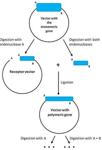
(56) ACCESSING NEW BIOMEDICAL APPLICATIONS BY COMBINING GENETIC DESIGN AND CHEMICAL MODIFICATION OF ELASTIN-LIKE RECOMBINAMERS. The production of sufficient amounts of the monomeric gene containing the correct sequence for producing the repeat nucleotide sequences that code for the various polypeptides required a culture of the appropriate clones and digestion of their plasmids. This allows for high yields of monomer required for the controlled ligation, concatenation or concatenamerisation reaction to obtain oligomerized genes in a simple manner140,141. Although this approach may appear to be wasteful in terms of time, in vivo synthesis guarantees the production of the correct sequence in large amounts and also offers additional advantages, such as control of the monomeric gene prior to oligomerisation and the ability to subsequently modify the sequence by either directed mutagenesis or by the creation of copolymers prior to oligomerisation.. 1.2.2 Construction of the multi block Polymerisation of the monomeric nucleotide sequences may be undertaken by, but is not limited to, concatenation, in other words random ligation of the monomeric blocks; the iterative/recursive method, a step-by-step technique for preparing oligomers from monomers; or by the Seamless cloning method, a definition that refers to the possibility to select a specific sequence that is translated into the desired amino acid at the cut-off point140,141.. 24.
(57) Biosynthesis of ELRs. Figure 5: Condensed summary of recombinant gene oligomerization in a vector with 2 restriction enzymes. Adapted from142.. Concatenation allows the synthesis of polymers from oligomers by the unidirectional, linear head-to-tail attachment of the DNA segments that make up the monomer, although such attachment is only possible if the ends of the segments are single-stranded, protuberant and cohesive amongst themselves but not with themselves. Thus, the single stranded head end of the monomer will be complementary to, and will only hybridise with, the tail end of another identical monomer. These ends cannot therefore be palindromic, as currently occurs when they are generated by the restriction endonucleases routinely used in genetic engineering, where the recognition and cleavage sites occur sequentially, but must be different143.. 25.
(58) ACCESSING NEW BIOMEDICAL APPLICATIONS BY COMBINING GENETIC DESIGN AND CHEMICAL MODIFICATION OF ELASTIN-LIKE RECOMBINAMERS. Various modifications of the concatenation technique have been developed considering the specific requirements. Monomers with cohesive, singlestranded ends were obtained by the hybridisation of oligomers, by the joining of short oligonucleotides (linkers) and also by digestion with specific endonucleases The iterative/recursive method is a step-by-step technique used to prepare oligomers from monomers that allow both, the addition sequence and the number of blocks to be joined during each growth stage to be controlled, thereby allowing ad hoc polymer production. This method requires the creation of sequences at the ends of the segment that code for the proteinbased monomer, which are recognised by two different endonucleases and whose cleavage produces complementary, but not palindromic, ends144. By this approach DNA monomers are oligomerized exclusively “head-to-tail” by enzymatic ligation, which ensures the translation of the sense strand into the polypeptide of interest and allows to fully control the polypeptide length (compare Figure 5). This monomeric sequence is cloned in a plasmid that serves as a gene amplification vector and provides both the following cloning vector when digested with a single enzyme and the clone insert when cleaved by two enzymes145. The Seamless cloning method removes the relationship between the design of the sequence for the insert that codes for the monomer and the sequences recognised by the endonucleases that generate it. This strategy is possible due to the existence of a limited number of type II restriction enzymes that recognise a specific, non-palindromic sequence that does not coincide with the cleavage site. This unique characteristic eliminates the drawbacks resulting from the need to include unusual sequences into the polymer in order to allow the generation of cohesive ends that allow. 26.
(59) Biosynthesis of ELRs. concatenation and also means that fragments that join in a unidirectional manner can be achieved in single digestion.. Type IIS endonucleases SapI and EarI. Sap1 and Ear1 are two type IIS endonucleases that recognize similar nonpalindromic sequences only varying by the fact that the Sap1 sequence is extended by one nucleotide (Figure 6).. Figure 6: Illustration of the recognition sequences of Ear1 and Sap1 type IIS endonucleases.. The fact that the Ear1 recognition sequence is fully presented in the Sap1 recognition site, implies that Ear1 is capable of digesting the Sap1 region, whereas Sap1 does not cut the Ear1 region when the preceding nucleotide differs from guanine. Additionally, the resulting asymetric ends with three nucleotides (N in Figure 6) generated with Sap1 or Ear1 digestion have the same size and orientation. The existence of a constant displacement between the recognized sequence and the exact cleavage place, which is characteristic for type IIS restriction endonucleases, allows for the insertion of nucleotides of any order143. This makes this pair of restriction enzymes an ideal candidate for the seamless cloning technique, since the additional base of the restriction site (“seam”) are not part of the extracted final gene (Figure 7)145.. 27.
Figure
![Figure 2: Dependence of Tt on the guest residue in poly penta-peptide of the structure poly[fv (VPGVG), fx (VPGXG)]](https://thumb-us.123doks.com/thumbv2/123dok_es/7213276.425506/50.892.145.723.201.448/figure-dependence-guest-residue-peptide-structure-vpgvg-vpgxg.webp)

+7


Documento similar