Review Article
Synaptic Wnt/GSK3
𝛽
Signaling Hub in Autism
Mario O. Caracci,
1,2Miguel E. Ávila,
1,2and Giancarlo V. De Ferrari
1,21Center for Biomedical Research, Faculty of Biological Sciences and Faculty of Medicine, Universidad Andres Bello,
P.O. Box 8370134, Santiago, Chile
2FONDAP Center for Genome Regulation, Santiago, Chile
Correspondence should be addressed to Giancarlo V. De Ferrari; [email protected]
Received 18 September 2015; Revised 29 November 2015; Accepted 30 November 2015
Academic Editor: Preston E. Garraghty
Copyright © 2016 Mario O. Caracci et al. This is an open access article distributed under the Creative Commons Attribution License, which permits unrestricted use, distribution, and reproduction in any medium, provided the original work is properly cited.
Hundreds of genes have been associated with autism spectrum disorders (ASDs) and the interaction of weak andde novovariants derive from distinct autistic phenotypes thus making up the “spectrum.” The convergence of these variants in networks of genes associated with synaptic function warrants the study of cell signaling pathways involved in the regulation of the synapse. The Wnt/𝛽 -catenin signaling pathway plays a central role in the development and regulation of the central nervous system and several genes belonging to the cascade have been genetically associated with ASDs. In the present paper, we review basic information regarding the role of Wnt/𝛽-catenin signaling in excitatory/inhibitory balance (E/I balance) through the regulation of pre- and postsynaptic compartments. Furthermore, we integrate information supporting the role of the glycogen synthase kinase 3𝛽(GSK3𝛽) in the onset/development of ASDs through direct modulation of Wnt/𝛽-catenin signaling. Finally, given GSK3𝛽activity as key modulator of synaptic plasticity, we explore the potential of this kinase as a therapeutic target for ASD.
1. Introduction
Autism spectrum disorders (ASDs) are highly heteroge-neous, pervasive developmental disorders characterized by impaired social communication skills, repetitive behaviors, and a restricted range of interests [1]. The wide range of phenotypical traits regarding comorbidities and various degrees of cognitive and language impairments makes up the “spectrum” and adds complexity to the determination of genetic markers associated with a distinct phenotype [2]. ASDs have a strong genetic component as ascertained by a 90% concordance among monozygotic twins [3]. Significant advancements have been made in identifying molecular mechanisms involved in ASDs by studying disorders with Mendelian inheritance patterns such as Tuberous Sclerosis
complex (TSC1andTSC2), Rett syndrome (MECP2),
ile X syndrome (FXS; which results from mutated
Frag-ile X mental retardation-1, FMR1), and Cowden syndrome
(PTEN), but, altogether, these disorders do not account for more than 10% of cases [4]. In the last few years, efforts have focused on understanding the genetic contribution of single nucleotide variants (SNVs) and copy number variants
(CNVs) in ASD [5, 6]. While genome wide association studies (GWAS) have identified over 100 genes associated with ASDs, most of the variants identified have a weak effect suggesting a
greater contribution for rare variants [7]. Rare variants andde
novooccurring SNVs and CNVs have a larger contribution to
the onset of ASD [6]. Indeed,de novoCNVs are significantly
enriched in individuals affected with the disorder and it is estimated that 8% of cases that carry these variants are likely
to be pathogenic [8, 9]. On the other hand, 9% ofde novo
SNVs in affected individuals are disruptive or frameshift mutations that generate nonconserved amino acid changes such as premature stop codons or alternative splice sites ultimately affecting the normal biological function of the resulting protein [10, 11]. Overall, it is estimated that these
deleteriousde novovariants affect ASD susceptibility in 10–
15% of probands [10, 11]. Nevertheless, exomic data suggests that no single gene could account for more than 1% of ASD cases, which makes it difficult to target a single protein to treat autistic behaviors. More recently, the integration of these genes into functional networks has allowed the identification of specific molecular pathways that could be disrupted in ASD [12, 13]. In this regard, recent exome sequencing studies Volume 2016, Article ID 9603751, 10 pages
in family trios identified that 39% of the more disruptive
de novo mutations are part of an interconnected network
of chromatin remodeling, synaptic plasticity, and Wnt/𝛽-catenin signaling genes [13–15].
Through the analysis of biochemical and pharmacological data, animal models of the disease, and genetic association studies, we predicted earlier that the onset/development of ASDs might involve the additive effect of genetic variants within Wnt/𝛽-catenin signaling components and/or genes coding for molecules that modulate its functional activity [16], and such hypothesis has received considerable attention recently [6, 17, 18]. Wnts are lipid modified secreted glyco-proteins that signal through three major pathways: the Planar
Cell Polarity (PCP), Wnt/Ca+2, and the canonical
Wnt/𝛽-catenin signaling pathway [19]. Wnt/𝛽-Wnt/𝛽-catenin signaling is the most well understood cascade and it starts via binding of the Wnt ligand to cell membrane receptors Frizzled (FZD), belonging to the 7-transmembrane domains family of proteins and to members of the low density lipoprotein receptor related proteins 5 and 6 (LRP5/6), which act as coreceptors [20]. Wnt binding to its membrane receptor activates intracellular signaling leading to the dissociation
of 𝛽-catenin from the degradation complex consisting of
Axin and adenomatous polyposis coli (APC) scaffolds [21], and the serine-threonine kinases casein kinase 1 (CK1) and
glycogen synthase kinase 3𝛽(GSK3𝛽) [22]. As a net result,
𝛽-catenin accumulates in the cytosol and translocates to the nucleus where it interacts with T-cell factor/lymphoid enhancing factor (TCF/LEF) transcription factors to activate transcription of target genes [23]. Conversely, in the absence of a Wnt ligand, Axin and APC facilitate CK1 and GSK3𝛽
sequential phosphorylation of 𝛽-catenin [22] targeting the
protein for ubiquitination by the 𝛽-transducing
repeat-containing protein (𝛽-TrCP) and subsequent proteasome degradation [24].
It is interesting to note that the tumor suppressor complex
formed by TSC1 and TSC2 interacts with the 𝛽-catenin
degradation complex and thus modulates the action of Wnt signaling [25, 26]. Other genetic elements associated with ASDs are the canonical Wnt2 ligand [27], the hepatocyte growth factor receptor (MET) [28, 29], which is a target gene of Wnt/𝛽-catenin signaling [30], and several genes
encod-ing for cadherins, includencod-ingCDH5,CDH8,CDH9,CDH10,
CDH13,CDH15,PCDH10,PCDH19,andPCDHb4[31], some
of which may interact with 𝛽-catenin in cell-cell adhesion
complexes. More recently, the chromo-helicase domain
pro-tein 8 (CHD8) [13, 14, 32], which inhibits𝛽-catenin through
direct binding [33], and DYRK1A that modulates Wnt sig-naling through interaction with the p120 catenin [34] have been found to be associated with ASDs. Interestingly, these genes harbor recurrent disruptive mutations and display a high correlation with head size abnormalities [14], which is a feature commonly observed during the first 2-3 years of
life of an ASD individual [35]. Finally, rarede novogenetic
variants in the𝛽-catenin (CTNNB1) gene itself have been
implicated in severe intellectual disability [36]. Therefore, the convergence of genetic markers in synaptic components opens a therapeutic window that aims not only to correct developmental brain abnormalities, but also to compensate
the inherent plasticity through modulation of the highly dynamic synapse. In the present paper, we review current knowledge of synaptic transmission leading to excitatory and inhibitory (E/I) imbalance commonly seen in ASD and how this phenomenon relates to dysfunction of the Wnt/𝛽-catenin pathway. Furthermore, we trace functional defects to
GSK3𝛽activity and explore its pharmacological regulation as
a potential therapeutic target for ASD, particularly in relation to synaptic plasticity.
2. Wnt/
𝛽
-Catenin Signaling and Synaptic
Transmission Defects in ASDs
The inherent ability of the brain to process information is accomplished by a highly sophisticated network that allows long-distance communication between cells and which is largely based on the E/I balance from neuronal connections. Genetic, functional, and structural information suggests that the E/I balance may underlie the symptomatology of ASDs [37–39]. This idea has been examined through optogenetic methods in the medial prefrontal cortex of mice, and it was found that the elevation, but not the reduction, of cellular E/I balance (i.e., increase in excitatory transmission) induced cel-lular defects in information processing, leading to behavioral and social deficits [39]. E/I balance anomalies have similarly been observed in several ASD animal models, including the neuroligin 3 (NLGN3) mutant mice, and the models for Rett, Fragile X, and Angelman syndromes (Rev. in [40]). In humans, one of the most relevant evidence associating the E/I balance with ASDs is its high comorbidity with epilepsy (30% comorbidity with ASDs) [41, 42]. Epileptic activity can be triggered by blocking synaptic inhibitory transmission or by activating excitatory transmission linking the E/I imbalance in the establishment of epileptiform seizures [43].
Wnt signaling has been widely acknowledged during pat-terning, development, and maturation of functional synapses within the CNS [16, 44–48]. Wnt1, Wnt3a, Wnt7a, and Wnt8 are ligands known to activate Wnt/𝛽-catenin signaling and are involved in brain development and synaptogenesis [49– 51]. Wnt7a and Wnt8a have also been shown to regulate excitatory synaptic formation [45, 52]. Furthermore, a recent study suggests that LRP6, Wnt/𝛽-catenin signaling corecep-tor, is critical for the development of functional synapses
in vivo [52], which further supports the involvement of
Wnt/𝛽-catenin signaling in synaptic development. Interest-ingly tetanic stimulation induces the release of the Wnt3a ligand from the postsynaptic terminal [53]. We demonstrated later that treatment with purified Wnt3a protein of cultured
hippocampal neurons enhanced a fast influx of Ca2+in the
Postsynaptic
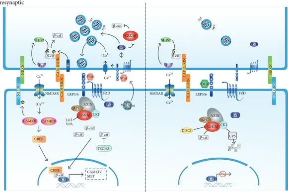
Figure 1: Wnt/𝛽-catenin signaling in ASDs. Wnt binding to FZD-LRP5/6 complex receptor at the membrane recruits the destruction complex and inhibits GSK3𝛽activity thus stabilizing𝛽-catenin in the cytoplasm and nucleus. Activation of the Wnt/𝛽-catenin pathway facilitates synaptic plasticity through the activation of voltage gated ion channels that allows activation of CAMK and CREB mediated transcription. Mutations in TSC associated with ASD prevent𝛽-catenin degradation which results in a gain of function of the Wnt pathway. In the presynaptic terminal cadherin mediated cell adhesion between synapses is weakened by phosphorylation of𝛽-catenin and synaptic vesicle clustering is enhanced through DVL1. Clustering is also dependent on NLGN/NRXN cell adhesion complexes. Both lithium (LiCl) and VPA activate Wnt/𝛽-catenin signaling through inhibition of GSK3𝛽activity. Conversely, in the absence of a Wnt ligand, activated GSK3𝛽targets
𝛽-catenin for proteosome-mediated degradation. Mutations associated with DISC1 fail to inhibit GSK3𝛽and thus activate Wnt/𝛽-catenin pathway. In the presynaptic side Wnt signaling buffering of synaptic vesicles is inhibited and adherens junctions mediated by cadherins are strengthened.
3. ASDs and Wnt Signaling at
the Presynaptic Terminal
At the presynaptic region, canonical Wnt signaling has a major role in clustering and recycling of synaptic vesicles (SVs). Conditioned media containing Wnt7a, and to a lesser extent Wnt3a, were found to enhance SVs recycling in primary cultures of rat hippocampal neurons [55]. Similarly, loss of Wnt7a function inhibits SVs clustering, an effect that is mimicked by loss of function of Dishevelled 1 (DVL1) signaling downstream of Wnt ligands [47]. Interestingly, Dvl1 knockout mice exhibit social interaction and sensorimotor abnormalities [56]. Moreover, the Wnt7a/Dvl1 double mutant mice show defects in spine morphogenesis and excitatory synaptic neurotransmission [45], which parallels behavioral abnormalities with a disrupted presynaptic assembly and E/I balance, as it is likely observed in ASDs.
Wnt/𝛽-catenin signaling also seems to trigger neu-rotransmitter release and SV trafficking by modulating the function of SVs-associated phosphoproteins, including membrane-trafficking proteins such as synapsin and synap-totagmin. While all three members of the synapsin (SYN) gene family (SYN1-3) [57] have been associated with ASDs
[58–60], it has been shown that canonical Wnt ligands such as Wnt7a and Wnt3a enhance the clustering [61] and phosphorylation [54] of Syn1 at the synaptic button prior to
neurotransmitter release. Likewise, SYN2 is predicted as a
Wnt/𝛽-catenin target gene [62] and is upregulated as a con-sequence of enhanced Wnt signaling activity in hippocampal neurons from APC conditional knockout mice that has impaired learning and memory and that displays ASD-like behaviors [63]. Finally, it was shown that the Wnt signaling component Dvl1 is involved in neurotransmitters release at the tip of neurites of differentiated neurons through direct binding to the presynaptic protein synaptotagmin I [64].
Other mechanisms modulating the activity of the presy-naptic terminal involve the function of cell adhesion proteins, most notably trans-synaptic cadherin interactions. It is widely accepted that cadherin-𝛽-catenin adhesion complexes have an essential function during the recruitment and clustering
of SVs to synapses [65–69]. Indeed, ablation of 𝛽-catenin
results in the mislocalization of SVs along the axon, while clustering of active zone proteins like Bassoon is unchanged
[68]. Tyrosine 654 phosphorylation of 𝛽-catenin weakens
activates the tyrosine phosphatase SHP-2 which removes 𝛽-catenin phosphorylation and strengthens cadherin mediated
adhesion [72]. Among other proteins modulating𝛽-catenin
dissociation from cell adhesion complexes that have been genetically linked with ASD is the MET receptor tyrosine
kinase [30], which phosphorylates Tyr142 in𝛽-catenin and
promotes its dissociation from cadherins [73], thus linking regulation of cell adhesion by catenins in the pathophysiology of ASDs. In sum, the data available indicates an essential role for Wnt/𝛽-catenin signaling in synaptic structure stabil-ity and function through modulating cell adhesion, vesicle
exocytosis, and clustering well beyond𝛽-catenin functioning
solely as a TCF/LEF transcriptional coactivator.
4. ASDs and Wnt Signaling at
the Postsynaptic Terminal
Experience driven plasticity is highly dependent on proper
synaptic transmission and is mainly modulated by Ca2+
related pathways. Canonical and noncanonical Wnt pathways
have been extensively related to Ca2+homeostasis and
signal-ing [47, 54, 74, 75]. Ligands such as Wnt3a [54], Wnt5a [75],
and Wnt7a [47] have all been shown to increase Ca2+influxes
in neurons. It is accepted that activation of L-type voltage
sensitive Ca2+ channels (L-VSCCs) or NMDA receptors
allows the entrance of Ca2+which in turn activate CAMKII
triggering actin cytoskeleton reorganization to regulate den-dritic growth [76]. In this regard, CAMKII and the Wnt target gene CAMKIV [77] activate transcription factors such as CREB to start activity dependent transcription to further promote synaptic development [78]. CAMKIV has been
asso-ciated with ASD [79] and additionally it mediates𝛽-catenin
dependent dendritic growth upon Ca2+influx [78, 80].
Activation of CAMKII and other kinases through
NMDAR-mediated Ca2+ influx is an event preceding the
establishment of long-term synaptic potentiation (LTP) that allows the recruitment of AMPARs at the postsynaptic ter-minal, which in turn enhances long lasting excitatory trans-mission [81]. Additionally, CAMKII robustly phosphorylates the cell adhesion neuroligin 1 (NLGN1) protein increasing its surface expression [82]. Notably, suppression of Wnt/𝛽-catenin signaling impairs LTP and conversely its activation facilitates it [53], and both enhanced and diminished LTP have been observed in animal models of ASD. For instance, given that enhanced LTP has been observed in TSC2 mutant
model [83] and thatTSC2missense mutations fail to inhibit
the Wnt pathway [26], it is likely that overactivation of the signaling cascade may enhance LTP in this specific model. In contrast, mutant models for Fragile X mental retardation-1 (FMRretardation-1), and also for the disrupted in schizophrenia retardation-1 (DISC1) genes, exhibit diminished capacity to establish LTP [84, 85]. Besides their putative role in schizophrenia, ASDs
and other neurological diseases [86–88], common DISC1
genetic variants, directly impact Wnt/𝛽-catenin signaling function (see below) [89]. Altogether, the data suggest that
the Wnt/𝛽-catenin pathway plays a central role in Ca2+
homeostasis at postsynaptic terminals, which is commonly disrupted in ASD. In addition, abnormal establishment of
LTP, phenomenon in which the signaling cascade plays an important role, has profound effects in activity driven plas-ticity affecting efficient synaptic transmission and disrupting the E/I balance.
LTP is the most well understood paradigm of activity driven plasticity and is considered to be one of the synaptic mechanisms underlying learning and memory [81]. In turn, several aspects of the ASD core symptomatology and the high comorbidity with intellectual disability disorder could be explained by defective memory mechanisms [90]. Indeed, diminished episodic memory has been reported for high functioning ASD individuals and is thought to impair the relational binding of elements comprising complex stimuli [91]. Therefore, rescuing defects in LTP that appears to be highly regulated by the Wnt/𝛽-catenin pathway specifically
through the modulation of GSK3𝛽 could improve core
ASD symptomatology and open a therapeutic window for the treatment of ASD through the fine-tuning of synaptic plasticity.
5. Synaptic Wnt/GSK3
𝛽
Signaling Hub in ASD
GSK3 is an evolutionary conserved serine/threonine kinase highly abundant in the brain. Two homologous isoforms,
GSK3𝛼 and GSK3𝛽, have been described in mammals and
are involved in multiple cellular processes including glycogen metabolism, gene transcription, microtubule stability, and
apoptosis [92]. GSK3𝛽is as a convergence point of major
prevalent neurological disorders, including Alzheimer’s dis-ease, schizophrenia, and bipolar disorder [93–95], and its activity is negatively regulated by Wnt signaling. As
men-tioned before, theDISC1gene has an essential role in
mod-ulating brain structure and function and when mutated leads
to neuropsychiatric behavior. DISC1 inhibits GSK3𝛽activity
by direct physical interaction resulting in reduced𝛽-catenin
phosphorylation and activation of Wnt/𝛽-catenin signaling cascade [96] and common genetic variants affecting the coding sequence of the gene were found to suppress Wnt/𝛽-catenin signaling activity [89]. Regarding ASDs,
hyperacti-vation of GSK3𝛽 has been documented in animal models
of FXS [97–99]. For instance, knock in mice expressing
constitutively active form of GSK3𝛽displays similar social
preference abnormalities as FMR1 KO mice [99].
Mouse models for Fragile X, Phellan-McDermid, and Angelman syndromes, as well as for Tuberous Sclerosis, all present an abnormal number of dendritic spines that suggest a dysregulation in synaptic turnover [100–102]. In this regard,
postnatal ablation of GSK3𝛽in mice forebrain has anxiolytic
and prosocial effects [103] and its overexpression accounts for spatial learning deficits in the Morris water maze paradigm
[104]. Interestingly, forebrain deletion of GSK3𝛽 leads to
reduced spine density where persistent spines are lost and newly formed spines are unstable [105]. These structural abnormalities are accompanied by a drop in AMPA depen-dent mEPSC and the effect is mimicked by the expression of
constitutively active𝛽-catenin [105]. Furthermore,
pharma-cological inhibition of GSK3𝛽 has been shown to increase
Conversely, activation of GSK3𝛽impairs the establishment of LTP [107] and high frequency stimulation inhibits GSK3𝛽
in a Ca2+dependent mechanism [108]. Given that increased
abnormal spine density is a pathological hallmark in ASD that may lead to brain hyperconnectivity underlying the basis for E/I balance, the data suggests that inhibition of the
Wnt/𝛽-catenin signaling through hyperactivation of GSK3𝛽might
help to explain transmission anomalies as it is observed in ASD.
6. Pharmacological Regulation of
GSK3
𝛽
in ASD
Due to its high heterogeneity, genetic factors cannot be held accountable for the entire spectrum of autism suggesting
a role for environmental factors in the onset of ASD. In
utero exposure to anticonvulsive medication is known to
cause neurodevelopmental abnormalities [109]. The most well studied anticonvulsive agent in these subjects is valproic
acid (valproate, VPA), a known inhibitor of GSK3𝛽[110] and
of histone deacetylase (HDAC) [111] activities. As an inhibitor
of GSK3𝛽, VPA induces the stabilization of 𝛽-catenin and
the activation of Wnt target genes, though the exact
mech-anism of GSK3𝛽 is not currently understood. Indeed, in
uteroexposure to VPA increases the incidence of autism in
the offspring [112, 113] and mice models, which have been prenatally exposed to VPA exhibiting ASD-like behaviors and morphological brain abnormalities [112, 114]. Currently, mice prenatally exposed to VPA (VPA mice) are widely used as animal models to understand the onset/development of ASDs [115]. This VPA mouse model results from intraperitoneal injection in embryonic stages E12–E17, which is a critical period in forebrain development, where dysregulation of Wnt signaling (different time points) induces morphological abnormalities in the brain [116].
While several molecular mechanisms regarding the onset of ASDs in VPA mice have been reported, the activation of Wnt/𝛽-catenin signaling is central through the regulation of GSK3𝛽. VPA mice exhibit elevated NMDA receptor levels and enhanced LTP [117] and inhibition of GABA transporter VGAT expression in cortical cultures [118], suggesting an important enhancement in excitatory neuro-transmission. Likewise, VPA mice induce demethylation of
WNT1and WNT2genes further enhancing Wnt/𝛽-catenin
signaling [119]. In this regard, sulindac treatment, an anti-inflammatory drug that downregulates Wnt/𝛽-catenin
sig-naling by enhancing GSK3𝛽expression in the prefrontal
cor-tex or the hippocampal region of VPA mice [120], improved repetitive stereotypic activity, learning and memory, as well as behavioral abnormalities [120, 121]. Interestingly, the VPA transcriptome revealed enhanced expression of multiple genes involved in Wnt/𝛽-catenin, neurotrophin, and LTP signaling, the same pathways which also appear enriched in the transcriptome of lithium [122], which mimics
Wnt/𝛽-catenin signaling by inhibiting GSK3𝛽 [123]. Nonetheless,
although prenatal treatment with VPA appears to enhance the expression of Wnt/𝛽-catenin signaling, most of the data
comes from in vitro cell cultures exposed to VPA and
not from in vivo studies using mice prenatally exposed to
the drug. In this context, it is interesting to note that chronic VPA treatment in mice has been shown to correct dendritic spine deficits and to improve novel object recognition [124]; thus, the postnatal basal activity of the Wnt/𝛽-catenin path-way is still unknown. In this context, it is interesting to note that ASD could result from a transient gain of function of the Wnt/𝛽-catenin pathway during embryonic development and a subsequent decline after birth.
Lithium has been widely used to manage mood disorders, such as bipolar disorders, and it is not uncommon for ASD children to feature symptoms within this spectrum such as euphoria, mania, or paranoia [125]. Few studies have documented the effects of lithium in ASDs but overall they show promising results as a therapeutic agent. For instance, lithium administration to 30 children and adolescents diag-nosed with ASD through DSM-IV-TR criteria improved the symptomatology on 43% of patients [125]. Likewise, chronic administration of lithium to neonatal rats who exhibit ASD-like behaviors abolished their symptoms and improved defects in neurogenesis and E/I balance [126]. Additionally, chronic lithium treatment reversed the increase in cerebral protein synthesis and ameliorates behavioral abnormalities commonly observed in FXS mice models [127], probably
through inhibitory GSK3𝛽phosphorylation (phosphor-Ser9
and phosphor-Ser21) [128]. Interestingly, pharmacological
inhibition of GSK3𝛽rescues LTP and hippocampal
neuroge-nesis defects in FMR1 knockout mice and improves cognitive
tasks [97, 103]. Furthermore, GSK3𝛽inhibition similarly
res-cues dendritic spines deficit observed in FXS mice suggesting that inhibition of this kinase and thus activation the Wnt/𝛽-catenin play a role in reactivating synaptic plasticity and these effects might play an important role in the behavioral and learning improvements observed.
Antagonists for metabotropic glutamate receptor (mGluRs) are up to date the most successful pharmacological mod-ulators improving ASD symptomatology probably through regulation of abnormal mRNA translation at synapses [129]. In this context, the use of MPEP (2-methyl-6-phenylethynyl-pyridine), mGluR5 antagonists, increases inhibitory GSK3𝛽
phosphorylation selectively in FMR1 knockout mice [130],
effect that is mimicked by chronic lithium treatment. More-over, this compound corrects dendritic spine deficits through upregulation of PSD-95 and learning impairment in FXS mice model [131], further ascribing a regulatory function directly at the synapses as the underlying mechanism for the therapeutic effect. Finally, MPEP treatment induces the expression of several pathways, including those governed by Wnt signaling in the frontal cortex of rats [132].
7. Concluding Remarks
signaling regulation of serine/threonine kinase GSK3𝛽has profound effects in activity dependent synaptic plasticity and thus in the regulation of the E/I balance. Through
dissecting Wnt/GSK3𝛽 activity and pharmacology in cells
and animal models of ASDs, it seems plausible that there may be differential effects driven by Wnt/𝛽-catenin signaling activity during the initial patterning of brain structures and later on when these structures have been established. Overall,
the therapeutic value of GSK3𝛽modulation that seems to
rescue synaptic plasticity events that could be disrupted in ASD brains warrants further basic and clinical investigation.
Conflict of Interests
The authors declare that there is no conflict of interests regarding the publication of this paper.
Authors’ Contribution
Mario O. Caracci and Miguel E. ´Avila contributed equally to
this work.
Acknowledgment
This work was supported by FONDECYT Regular 1140353 to Giancarlo V. De Ferrari.
References
[1] C. J. Newschaffer, L. A. Croen, J. Daniels et al., “The epidemi-ology of autism spectrum disorders,”Annual Review of Public Health, vol. 28, pp. 235–258, 2007.
[2] S. S. Jeste and D. H. Geschwind, “Disentangling the hetero-geneity of autism spectrum disorder through genetic findings,”
Nature Reviews Neurology, vol. 10, no. 2, pp. 74–81, 2014. [3] R. E. Rosenberg, J. K. Law, G. Yenokyan, J. McGready, W. E.
Kaufmann, and P. A. Law, “Characteristics and concordance of autism spectrum disorders among 277 twin pairs,”Archives of Pediatrics & Adolescent Medicine, vol. 163, no. 10, pp. 907–914, 2009.
[4] C. Betancur, “Etiological heterogeneity in autism spectrum disorders: more than 100 genetic and genomic disorders and still counting,”Brain Research, vol. 1380, pp. 42–77, 2011.
[5] B. S. Abrahams and D. H. Geschwind, “Advances in autism genetics: on the threshold of a new neurobiology,” Nature Reviews Genetics, vol. 9, no. 5, pp. 341–355, 2008.
[6] N. Krumm, B. J. O’Roak, J. Shendure, and E. E. Eichler, “A de novo convergence of autism genetics and molecular neuroscience,”Trends in Neurosciences, vol. 37, no. 2, pp. 95–105, 2014.
[7] R. Anney, L. Klei, D. Pinto et al., “Individual common variants exert weak effects on the risk for autism spectrum disorders,”
Human Molecular Genetics, vol. 21, no. 21, pp. 4781–4792, 2012. [8] D. Levy, M. Ronemus, B. Yamrom et al., “Rare de novo and transmitted copy-number variation in autistic spectrum disorders,”Neuron, vol. 70, no. 5, pp. 886–897, 2011.
[9] S. J. Sanders, A. G. Ercan-Sencicek, V. Hus et al., “Multiple recurrent de novo CNVs, including duplications of the 7q11.23 Williams syndrome region, are strongly associated with autism,”
Neuron, vol. 70, no. 5, pp. 863–885, 2011.
[10] I. Iossifov, B. J. O’Roak, S. J. Sanders et al., “The contribution of de novo coding mutations to autism spectrum disorder,”Nature, vol. 515, no. 7526, pp. 216–221, 2014.
[11] S. J. Sanders, M. T. Murtha, A. R. Gupta et al., “De novo mutations revealed by whole-exome sequencing are strongly associated with autism,”Nature, vol. 485, no. 7397, pp. 237–241, 2012.
[12] F. Hormozdiari, O. Penn, E. Borenstein, and E. E. Eichler, “The discovery of integrated gene networks for autism and related disorders,”Genome Research, vol. 25, no. 1, pp. 142–154, 2015. [13] B. J. O’Roak, L. Vives, S. Girirajan et al., “Sporadic autism
exomes reveal a highly interconnected protein network of de novo mutations,”Nature, vol. 484, no. 7397, pp. 246–250, 2012. [14] B. J. O’Roak, L. Vives, W. Fu et al., “Multiplex targeted
sequenc-ing identifies recurrently mutated genes in autism spectrum disorders,”Science, vol. 338, no. 6114, pp. 1619–1622, 2012. [15] S. De Rubeis, X. He, A. P. Goldberg et al., “Synaptic,
transcrip-tional and chromatin genes disrupted in autism,”Nature, vol. 515, no. 7526, pp. 209–215, 2014.
[16] G. V. De Ferrari and R. T. Moon, “The ups and downs of Wnt signaling in prevalent neurological disorders,”Oncogene, vol. 25, no. 57, pp. 7545–7553, 2006.
[17] N. D. Okerlund and B. N. R. Cheyette, “Synaptic Wnt signaling—a contributor to major psychiatric disorders?” Jour-nal of Neurodevelopmental Disorders, vol. 3, no. 2, pp. 162–174, 2011.
[18] H. O. Kalkman, “A review of the evidence for the canonical Wnt pathway in autism spectrum disorders,”Molecular Autism, vol. 3, no. 1, article 10, 2012.
[19] R. T. Moon, A. D. Kohn, G. V. De Ferrari, and A. Kaykas, “WNT and𝛽-catenin signalling: diseases and therapies,”Nature Reviews Genetics, vol. 5, no. 9, pp. 691–701, 2004.
[20] X. He, M. Semenov, K. Tamai, and X. Zeng, “LDL receptor-related proteins 5 and 6 in Wnt/𝛽-catenin signaling: Arrows point the way,”Development, vol. 131, no. 8, pp. 1663–1677, 2004. [21] T. V. Lipina, O. Kaidanovich-Beilin, S. Patel et al., “Genetic and pharmacological evidence for schizophrenia-related Disc1 interaction with GSK-3,”Synapse, vol. 65, no. 3, pp. 234–248, 2011.
[22] M. J. Hart, R. De Los Santos, I. N. Albert, B. Rubinfeld, and P. Polakis, “Downregulation of𝛽-catenin by human Axin and its association with the APC tumor suppressor,𝛽-catenin and GSK3𝛽,”Current Biology, vol. 8, no. 10, pp. 573–581, 1998. [23] H. Clevers and R. Nusse, “Wnt/𝛽-catenin signaling and disease,”
Cell, vol. 149, no. 6, pp. 1192–1205, 2012.
[24] H. Aberle, A. Bauer, J. Stappert, A. Kispert, and R. Kemler, “𝛽 -catenin is a target for the ubiquitin-proteasome pathway,”The EMBO Journal, vol. 16, no. 13, pp. 3797–3804, 1997.
[25] B. C. Mak, K.-I. Takemaru, H. L. Kenerson, R. T. Moon, and R. S. Yeung, “The tuberin-hamartin complex negatively regulates𝛽 -catenin signaling activity,”The Journal of Biological Chemistry, vol. 278, no. 8, pp. 5947–5951, 2003.
[26] B. C. Mak, H. L. Kenerson, L. D. Aicher, E. A. Barnes, and R. S. Yeung, “Aberrant𝛽-catenin signaling in tuberous sclerosis,”The American Journal of Pathology, vol. 167, no. 1, pp. 107–116, 2005. [27] T. H. Wassink, J. Piven, V. J. Vieland et al., “Evidence supporting WNT2 as an autism susceptibility gene,”American Journal of Medical Genetics, vol. 105, no. 5, pp. 406–413, 2001.
[29] J. B. Tuynman, L. Vermeulen, E. M. Boon et al., “Cyclooxygen-ase-2 inhibition inhibits c-Met kinase activity and Wnt activity in colon cancer,”Cancer Research, vol. 68, no. 4, pp. 1213–1220, 2008.
[30] D. B. Campbell, J. S. Sutcliffe, P. J. Ebert et al., “A genetic variant that disrupts MET transcription is associated with autism,”
Proceedings of the National Academy of Sciences of the United States of America, vol. 103, no. 45, pp. 16834–16839, 2006. [31] C. Redies, N. Hertel, and C. A. H¨ubner, “Cadherins and
neuropsychiatric disorders,”Brain Research, vol. 1470, pp. 130– 144, 2012.
[32] B. J. O’Roak, P. Deriziotis, C. Lee et al., “Exome sequencing in sporadic autism spectrum disorders identifies severe de novo mutations,”Nature Genetics, vol. 43, no. 6, pp. 585–589, 2011. [33] B. A. Thompson, V. Tremblay, G. Lin, and D. A. Bochar,
“CHD8 is an ATP-dependent chromatin remodeling factor that regulates𝛽-catenin target genes,”Molecular and Cellular Biology, vol. 28, no. 12, pp. 3894–3904, 2008.
[34] J. Y. Hong, J.-I. Park, M. Lee et al., “Down’s-syndrome-related kinase Dyrk1A modulates the p120-catenin–Kaiso trajectory of the Wnt signaling pathway,”Journal of Cell Science, vol. 125, no. 3, pp. 561–569, 2012.
[35] E. Courchesne, K. Pierce, C. M. Schumann et al., “Mapping early brain development in autism,”Neuron, vol. 56, no. 2, pp. 399– 413, 2007.
[36] J. de Ligt, M. H. Willemsen, B. W. M. Van Bon et al., “Diagnostic exome sequencing in persons with severe intellectual disability,”
The New England Journal of Medicine, vol. 367, no. 20, pp. 1921– 1929, 2012.
[37] J. L. R. Rubenstein, “Three hypotheses for developmental defects that may underlie some forms of autism spectrum disorder,”Current Opinion in Neurology, vol. 23, no. 2, pp. 118– 123, 2010.
[38] S. Vattikuti and C. C. Chow, “A computational model for cerebral cortical dysfunction in autism spectrum disorders,”
Biological Psychiatry, vol. 67, no. 7, pp. 672–678, 2010.
[39] O. Yizhar, L. E. Fenno, M. Prigge et al., “Neocortical excita-tion/inhibition balance in information processing and social dysfunction,”Nature, vol. 477, no. 7363, pp. 171–178, 2011. [40] H. Y. Zoghbi and M. F. Bear, “Synaptic dysfunction in
neurode-velopmental disorders associated with autism and intellectual disabilities,”Cold Spring Harbor Perspectives in Biology, vol. 4, no. 3, Article ID a009886, 2012.
[41] R. Tuchman and I. Rapin, “Epilepsy in autism,” The Lancet Neurology, vol. 1, no. 6, pp. 352–358, 2002.
[42] I. E. Scheffer and S. F. Berkovic, “The genetics of human epilepsy,”Trends in Pharmacological Sciences, vol. 24, no. 8, pp. 428–433, 2003.
[43] K. Staley, “Molecular mechanisms of epilepsy,”Nature Neuro-science, vol. 18, no. 3, pp. 367–372, 2015.
[44] G. V. De Ferrari and and N. C. Inestrosa, “Wnt signaling function in Alzheimer’s disease,”Brain Research Reviews, vol. 33, no. 1, pp. 1–12, 2000.
[45] A. Ahmad-Annuar, L. Ciani, I. Simeonidis et al., “Signaling across the synapse: a role for Wnt and Dishevelled in presy-naptic assembly and neurotransmitter release,”Journal of Cell Biology, vol. 174, no. 1, pp. 127–139, 2006.
[46] N. C. Inestrosa and E. Arenas, “Emerging roles of Wnts in the adult nervous system,”Nature Reviews Neuroscience, vol. 11, no. 2, pp. 77–86, 2010.
[47] L. Ciani, K. A. Boyle, E. Dickins et al., “Wnt7a signaling promotes dendritic spine growth and synaptic strength through Ca2+/Calmodulin-dependent protein kinase II,”Proceedings of the National Academy of Sciences, vol. 108, no. 26, pp. 10732– 10737, 2011.
[48] G. V. De Ferrari, M. E. Avila, M. A. Medina, E. P´erez-Palma, B. I. Bustos, and M. A. Alarc´on, “Wnt/𝛽-catenin signaling in Alzheimer’s disease,” CNS and Neurological Disorders—Drug Targets, vol. 13, no. 5, pp. 745–754, 2014.
[49] N. Itasaki, C. M. Jones, S. Mercurio et al., “Wise, a context-dependent activator and inhibitor of Wnt signalling,” Develop-ment, vol. 130, no. 18, pp. 4295–4305, 2003.
[50] S. A. Ettenberg, O. Charlat, M. P. Daley et al., “Inhibition of tumorigenesis driven by different Wnt proteins requires block-ade of distinct ligand-binding regions by LRP6 antibodies,”
Proceedings of the National Academy of Sciences of the United States of America, vol. 107, no. 35, pp. 15473–15478, 2010. [51] A. Caricasole, T. Ferraro, L. Iacovelli et al., “Functional
charac-terization of WNT7A signaling in PC12 cells: interaction with a FZD5⋅LRP6 receptor complex and modulation by Dickkopf proteins,”The Journal of Biological Chemistry, vol. 278, no. 39, pp. 37024–37031, 2003.
[52] K. Sharma, S.-Y. Choi, Y. Zhang et al., “High-throughput genetic screen for synaptogenic factors: identification of LRP6 as critical for excitatory synapse development,”Cell Reports, vol. 5, no. 5, pp. 1330–1341, 2013.
[53] J. Chen, S. P. Chang, and S.-J. Tang, “Activity-dependent synap-tic Wnt release regulates hippocampal long term potentiation,”
The Journal of Biological Chemistry, vol. 281, no. 17, pp. 11910– 11916, 2006.
[54] M. E. Avila, F. J. Sep´ulveda, C. F. Burgos et al., “Canonical Wnt3a modulates intracellular calcium and enhances excitatory neurotransmission in hippocampal neurons,”The Journal of Biological Chemistry, vol. 285, no. 24, pp. 18939–18947, 2010. [55] W. Cerpa, J. A. Godoy, I. Alfaro et al., “Wnt-7a modulates the
synaptic vesicle cycle and synaptic transmission in hippocampal neurons,”The Journal of Biological Chemistry, vol. 283, no. 9, pp. 5918–5927, 2008.
[56] N. Lijam, R. Paylor, M. P. McDonald et al., “Social interaction and sensorimotor gating abnormalities in mice lacking Dvl1,”
Cell, vol. 90, no. 5, pp. 895–905, 1997.
[57] F. Cesca, P. Baldelli, F. Valtorta, and F. Benfenati, “The synapsins: key actors of synapse function and plasticity,” Progress in Neurobiology, vol. 91, no. 4, pp. 313–348, 2010.
[58] A. Fassio, L. Patry, S. Congia et al., “SYN1 loss-of-function mutations in autism and partial epilepsy cause impaired synap-tic function,”Human Molecular Genetics, vol. 20, no. 12, pp. 2297–2307, 2011.
[59] A. Corradi, M. Fadda, A. Piton et al., “SYN2 is an autism pre-disposing gene: loss-offunction mutations alter synaptic vesicle cycling and axon outgrowth,”Human Molecular Genetics, vol. 23, no. 1, Article ID ddt401, pp. 90–103, 2014.
[60] B. Greco, F. Manag`o, V. Tucci, H.-T. Kao, F. Valtorta, and F. Ben-fenati, “Autism-related behavioral abnormalities in synapsin knockout mice,”Behavioural Brain Research, vol. 251, pp. 65–74, 2013.
[61] A. C. Hall, F. R. Lucas, and P. C. Salinas, “Axonal remodeling and synaptic differentiation in the cerebellum is regulated by WNT-7a signaling,”Cell, vol. 100, no. 5, pp. 525–535, 2000. [62] C. H¨odar, R. Assar, M. Colombres et al., “Genome-wide
using CART method,”BMC Genomics, vol. 11, no. 1, article 348, 2010.
[63] J. L. Mohn, J. Alexander, A. Pirone et al., “Adenomatous polyposis coli protein deletion leads to cognitive and autism-like disabilities,”Molecular Psychiatry, vol. 19, no. 10, pp. 1133– 1142, 2014.
[64] S. Kishida, K. Hamao, M. Inoue et al., “Dvl regulates endo- and exocytotic processes through binding to synaptotagmin,”Genes to Cells, vol. 12, no. 1, pp. 49–61, 2007.
[65] S. X. Bamji, K. Shimazu, N. Kimes et al., “Role of beta-catenin in synaptic vesicle localization and presynaptic assembly,”Neuron, vol. 40, no. 4, pp. 719–731, 2003.
[66] Y. Iwai, Y. Hirota, K. Ozaki, H. Okano, M. Takeichi, and T. Uemura, “DN-cadherin is required for spatial arrangement of nerve terminals and ultrastructural organization of synapses,”
Molecular and Cellular Neuroscience, vol. 19, no. 3, pp. 375–388, 2002.
[67] K. J¨ungling, V. Eulenburg, R. Moore, R. Kemler, V. Lessmann, and K. Gottmann, “N-cadherin transsynaptically regulates short-term plasticity at glutamatergic synapses in embryonic stem cell-derived neurons,”The Journal of Neuroscience, vol. 26, no. 26, pp. 6968–6978, 2006.
[68] Y. Sun, M. Aiga, E. Yoshida, P. O. Humbert, and S. X. Bamji, “Scribble interacts with𝛽-catenin to localize synaptic vesicles to synapses,”Molecular Biology of the Cell, vol. 20, no. 14, pp. 3390–3400, 2009.
[69] H. Togashi, K. Abe, A. Mizoguchi, K. Takaoka, O. Chisaka, and M. Takeichi, “Cadherin regulates dendritic spine morphogene-sis,”Neuron, vol. 35, no. 1, pp. 77–89, 2002.
[70] S. X. Bamji, B. Rico, N. Kimes, and L. F. Reichardt, “BDNF mobilizes synaptic vesicles and enhances synapse formation by disrupting cadherin-𝛽-catenin interactions,”The Journal of Cell Biology, vol. 174, no. 2, pp. 289–299, 2006.
[71] J. J. Connolly, J. T. Glessner, and H. Hakonarson, “A genome-wide association study of autism incorporating autism diagnos-tic interview–revised, autism diagnosdiagnos-tic observation schedule, and social responsiveness scale,”Child Development, vol. 84, no. 1, pp. 17–33, 2013.
[72] S.-H. Lee, I.-F. Peng, Y. G. Ng et al., “Synapses are regulated by the cytoplasmic tyrosine kinase Fer in a pathway mediated by p120catenin, Fer, SHP-2, and𝛽-catenin,”The Journal of Cell Biology, vol. 183, no. 5, pp. 893–908, 2008.
[73] M. D. David, A. Yeramian, M. Du˜nach et al., “Signalling by neu-rotrophins and hepatocyte growth factor regulates axon mor-phogenesis by differential𝛽-catenin phosphorylation,”Journal of Cell Science, vol. 121, no. 16, pp. 2718–2730, 2008.
[74] W. Cerpa, A. Gambrill, N. C. Inestrosa, and A. Barria, “Regu-lation of NMDA-receptor synaptic transmission by Wnt signal-ing,”The Journal of Neuroscience, vol. 31, no. 26, pp. 9466–9471, 2011.
[75] L. Varela-Nallar, I. E. Alfaro, F. G. Serrano, J. Parodi, and N. C. Inestrosa, “Wingless-type family member 5A (Wnt-5a) stim-ulates synaptic differentiation and function of glutamatergic synapses,”Proceedings of the National Academy of Sciences of the United States of America, vol. 107, no. 49, pp. 21164–21169, 2010. [76] S. Cohen and M. E. Greenberg, “Communication between the synapse and the nucleus in neuronal development, plasticity, and disease,”Annual Review of Cell and Developmental Biology, vol. 24, pp. 183–209, 2008.
[77] M. S. Arr´azola, L. Varela-Nallar, M. Colombres et al., “Calcium/ calmodulin-dependent protein kinase type IV is a target gene
of the Wnt/𝛽-catenin signaling pathway,”Journal of Cellular Physiology, vol. 221, no. 3, pp. 658–667, 2009.
[78] L. Redmond, A. H. Kashani, and A. Ghosh, “Calcium regulation of dendritic growth via CaM kinase IV and CREB-mediated transcription,”Neuron, vol. 34, no. 6, pp. 999–1010, 2002. [79] R. Waltes, E. Duketis, M. Knapp et al., “Common variants in
genes of the postsynaptic FMRP signalling pathway are risk factors for autism spectrum disorders,”Human Genetics, vol. 133, no. 6, pp. 781–792, 2014.
[80] X. Yu and R. C. Malenka, “𝛽-catenin is critical for dendritic morphogenesis,”Nature Neuroscience, vol. 6, no. 11, pp. 1169– 1177, 2003.
[81] R. C. Malenka and M. F. Bear, “LTP and LTD: an embarrassment of riches,”Neuron, vol. 44, no. 1, pp. 5–21, 2004.
[82] M. A. Bemben, S. L. Shipman, T. Hirai et al., “CaMKII phosphorylation of neuroligin-1 regulates excitatory synapses,”
Nature Neuroscience, vol. 17, no. 1, pp. 56–64, 2014.
[83] L.-H. Zeng, Y. Ouyang, V. Gazit et al., “Abnormal glutamate homeostasis and impaired synaptic plasticity and learning in a mouse model of tuberous sclerosis complex,”Neurobiology of Disease, vol. 28, no. 2, pp. 184–196, 2007.
[84] K. Kuroda, S. Yamada, M. Tanaka et al., “Behavioral alterations associated with targeted disruption of exons 2 and 3 of the Disc1 gene in the mouse,”Human Molecular Genetics, vol. 20, no. 23, pp. 4666–4683, 2011.
[85] S. H. Yun and B. L. Trommer, “Fragile X mice: reduced long-term potentiation and N-Methyl-D-Aspartate receptor-mediated neurotransmission in dentate gyrus,”Journal of Neu-roscience Research, vol. 89, no. 2, pp. 176–182, 2011.
[86] J. E. Chubb, N. J. Bradshaw, D. C. Soares, D. J. Porteous, and J. K. Millar, “The DISC locus in psychiatric illness,”Molecular Psychiatry, vol. 13, no. 1, pp. 36–64, 2008.
[87] H. Kilpinen, T. Ylisaukko-Oja, W. Hennah et al., “Association of DISC1 with autism and Asperger syndrome,” Molecular Psychiatry, vol. 13, no. 2, pp. 187–196, 2008.
[88] A. Crepel, J. Breckpot, J.-P. Fryns et al., “DISC1 duplication in two brothers with autism and mild mental retardation,”Clinical Genetics, vol. 77, no. 4, pp. 389–394, 2010.
[89] K. K. Singh, G. De Rienzo, L. Drane et al., “Common DISC1 polymorphisms disrupt Wnt/GSK3𝛽signaling and brain devel-opment,”Neuron, vol. 72, no. 4, pp. 545–558, 2011.
[90] J. Boucher, A. Mayes, and S. Bigham, “Memory in autistic spectrum disorder,”Psychological Bulletin, vol. 138, no. 3, pp. 458–496, 2012.
[91] D. M. Bowler, S. B. Gaigg, and J. M. Gardiner, “Binding of multiple features in memory by high-functioning adults with autism spectrum disorder,”Journal of Autism and Developmen-tal Disorders, vol. 44, no. 9, pp. 2355–2362, 2014.
[92] E.-M. Hur and F.-Q. Zhou, “GSK3 signalling in neural develop-ment,”Nature Reviews Neuroscience, vol. 11, no. 8, pp. 539–551, 2010.
[93] F. Benedetti, A. Bernasconi, C. Lorenzi et al., “A single nucleotide polymorphism in glycogen synthase kinase 3-𝛽 promoter gene influences onset of illness in patients affected by bipolar disorder,”Neuroscience Letters, vol. 355, no. 1-2, pp. 37– 40, 2004.
[94] C. Hooper, R. Killick, and S. Lovestone, “The GSK3 hypothesis of Alzheimer’s disease,”Journal of Neurochemistry, vol. 104, no. 6, pp. 1433–1439, 2008.
GSK3 gene polymorphisms with schizophrenia and clozapine response,”Psychopharmacology, vol. 200, no. 2, pp. 177–186, 2008.
[96] Y. Mao, X. Ge, C. L. Frank et al., “Disrupted in schizophrenia 1 regulates neuronal progenitor proliferation via modulation of GSK3beta/beta-catenin signaling,”Cell, vol. 136, no. 6, pp. 1017– 1031, 2009.
[97] W. Guo, A. C. Murthy, L. Zhang et al., “Inhibition of GSK3𝛽 improves hippocampus-dependent learning and rescues neu-rogenesis in a mouse model of fragile X syndrome,”Human Molecular Genetics, vol. 21, no. 3, Article ID ddr501, pp. 681–691, 2012.
[98] Y. Luo, G. Shan, W. Guo et al., “Fragile X mental retardation protein regulates proliferation and differentiation of adult neu-ral stem/progenitor cells,”PLoS Genetics, vol. 6, no. 4, Article ID e1000898, 2010.
[99] M. A. Mines, C. J. Yuskaitis, M. K. King, E. Beurel, and R. S. Jope, “GSK3 influences social preference and anxiety-related behaviors during social interaction in a mouse model of fragile X syndrome and autism,”PLoS ONE, vol. 5, no. 3, Article ID e9706, 2010.
[100] T. A. Comery, J. B. Harris, P. J. Willems et al., “Abnormal dendritic spines in fragile X knockout mice: maturation and pruning deficits,”Proceedings of the National Academy of Sci-ences of the United States of America, vol. 94, no. 10, pp. 5401– 5404, 1997.
[101] J. Pec¸a, C. Feliciano, J. T. Ting et al., “Shank3 mutant mice dis-play autistic-like behaviours and striatal dysfunction,”Nature, vol. 472, no. 7344, pp. 437–442, 2011.
[102] S. V. Dindot, B. A. Antalffy, M. B. Bhattacharjee, and A. L. Beaudet, “The Angelman syndrome ubiquitin ligase localizes to the synapse and nucleus, and maternal deficiency results in abnormal dendritic spine morphology,”Human Molecular Genetics, vol. 17, no. 1, pp. 111–118, 2008.
[103] C. Latapy, V. Rioux, M. J. Guitton, and J.-M. Beaulieu, “Selective deletion of forebrain glycogen synthase kinase 3𝛽reveals a central role in serotonin-sensitive anxiety and social behaviour,”
Philosophical Transactions of the Royal Society B: Biological Sciences, vol. 367, no. 1601, pp. 2460–2474, 2012.
[104] F. Hern´andez, J. Borrell, C. Guaza, J. Avila, and J. J. Lucas, “Spa-tial learning deficit in transgenic mice that conditionally over-express GSK-3𝛽in the brain but do not form tau filaments,”
Journal of Neurochemistry, vol. 83, no. 6, pp. 1529–1533, 2002. [105] S. M. Ochs, M. M. Dorostkar, G. Aramuni et al., “Loss of
neuronal GSK3𝛽reduces dendritic spine stability and attenu-ates excitatory synaptic transmission via𝛽-catenin,”Molecular Psychiatry, 2014.
[106] P. Chen, Z. Gu, W. Liu, and Z. Yan, “Glycogen synthase kinase 3 regulates N-methyl-D-aspartate receptor channel trafficking and function in cortical neurons,”Molecular Pharmacology, vol. 72, no. 1, pp. 40–51, 2007.
[107] L.-Q. Zhu, S.-H. Wang, D. Liu et al., “Activation of glycogen synthase kinase-3 inhibits long-term potentiation with synapse-associated impairments,”The Journal of Neuroscience, vol. 27, no. 45, pp. 12211–12220, 2007.
[108] T. Ma, N. Tzavaras, P. Tsokas, E. M. Landau, and R. D. Blitzer, “Synaptic stimulation of mTOR is mediated by Wnt signaling and regulation of glycogen synthetase kinase-3,”The Journal of Neuroscience, vol. 31, no. 48, pp. 17537–17546, 2011.
[109] A. Verrotti, A. Scaparrotta, M. Cofini, F. Chiarelli, and G. M. Tiboni, “Developmental neurotoxicity and anticonvulsant
drugs: a possible link,”Reproductive Toxicology, vol. 48, pp. 72– 80, 2014.
[110] G. Chen, L.-D. Huang, Y.-M. Jiang, and H. K. Manji, “The mood-stabilizing agent valproate inhibits the activity of glyco-gen synthase kinase-3,”Journal of Neurochemistry, vol. 72, no. 3, pp. 1327–1330, 2000.
[111] C. J. Phiel, F. Zhang, E. Y. Huang, M. G. Guenther, M. A. Lazar, and P. S. Klein, “Histone deacetylase is a direct target of valproic acid, a potent anticonvulsant, mood stabilizer, and teratogen,”
The Journal of Biological Chemistry, vol. 276, no. 39, pp. 36734– 36741, 2001.
[112] A. L. Christianson, N. Chesler, and J. G. R. Kromberg, “Fetal val-proate syndrome: clinical and neuro-developmental features in two sibling pairs,”Developmental Medicine & Child Neurology, vol. 36, no. 4, pp. 361–369, 1994.
[113] K. Miyazaki, N. Narita, and M. Narita, “Maternal administra-tion of thalidomide or valproic acid causes abnormal seroton-ergic neurons in the offspring: implication for pathogenesis of autism,”International Journal of Developmental Neuroscience, vol. 23, no. 2-3, pp. 287–297, 2005.
[114] T. Schneider and R. Przewłocki, “Behavioral alterations in rats prenatally exposed to valproic acid: animal model of autism,”
Neuropsychopharmacology, vol. 30, no. 1, pp. 80–89, 2005. [115] G. C. Wagner, K. R. Reuhl, M. Cheh, P. McRae, and A. K.
Halladay, “A new neurobehavioral model of autism in mice: pre-and postnatal exposure to sodium valproate,”Journal of Autism and Developmental Disorders, vol. 36, no. 6, pp. 779–793, 2006. [116] S. J. Harrison-Uy and S. J. Pleasure, “Wnt signaling and fore-brain development,”Cold Spring Harbor Perspectives in Biology, vol. 4, no. 7, Article ID a008094, 2012.
[117] T. Rinaldi, K. Kulangara, K. Antoniello, and H. Markram, “Ele-vated NMDA receptor levels and enhanced postsynaptic long-term potentiation induced by prenatal exposure to valproic acid,”Proceedings of the National Academy of Sciences of the United States of America, vol. 104, no. 33, pp. 13501–13506, 2007. [118] E. Kumamaru, Y. Egashira, R. Takenaka, and S. Takamori, “Valproic acid selectively suppresses the formation of inhibitory synapses in cultured cortical neurons,”Neuroscience Letters, vol. 569, pp. 142–147, 2014.
[119] Z. Wang, L. Xu, X. Zhu et al., “Demethylation of specific Wnt/𝛽 -catenin pathway genes and its upregulation in rat brain induced by prenatal valproate exposure,”The Anatomical Record, vol. 293, no. 11, pp. 1947–1953, 2010.
[120] Y. Zhang, Y. Sun, F. Wang, Z. Wang, Y. Peng, and R. Li, “Downregulating the canonical Wnt/𝛽-catenin signaling path-way attenuates the susceptibility to autism-like phenotypes by decreasing oxidative stress,”Neurochemical Research, vol. 37, no. 7, pp. 1409–1419, 2012.
[121] Y. Zhang, C. Yang, G. Yuan, Z. Wang, W. Cui, and R. Li, “Sulindac attenuates valproic acid-induced oxidative stress levels in primary cultured cortical neurons and ameliorates repetitive/stereotypic-like movement disorders in Wistar rats prenatally exposed to valproic acid,”International Journal of Molecular Medicine, vol. 35, no. 1, pp. 263–270, 2015.
[122] A. Gupta, T. G. Schulze, V. Nagarajan et al., “Interaction networks of lithium and valproate molecular targets reveal a striking enrichment of apoptosis functional clusters and neurotrophin signaling,”The Pharmacogenomics Journal, vol. 12, no. 4, pp. 328–341, 2012.
Academy of Sciences of the United States of America, vol. 93, no. 16, pp. 8455–8459, 1996.
[124] K. Takuma, Y. Hara, S. Kataoka et al., “Chronic treatment with valproic acid or sodium butyrate attenuates novel object recognition deficits and hippocampal dendritic spine loss in a mouse model of autism,” Pharmacology Biochemistry and Behavior, vol. 126, pp. 43–49, 2014.
[125] M. Siegel, C. A. Beresford, M. Bunker et al., “Preliminary inves-tigation of lithium for mood disorder symptoms in children and adolescents with autism spectrum disorder,”Journal of Child and Adolescent Psychopharmacology, vol. 24, no. 7, pp. 399–402, 2014.
[126] X. Wu, Y. Bai, T. Tan et al., “Lithium ameliorates autistic-like behaviors induced by neonatal isolation in rats,”Frontiers in Behavioral Neuroscience, vol. 8, article 234, 2014.
[127] Z.-H. Liu, T. Huang, and C. B. Smith, “Lithium reverses increased rates of cerebral protein synthesis in a mouse model of fragile X syndrome,”Neurobiology of Disease, vol. 45, no. 3, pp. 1145–1152, 2012.
[128] Z.-H. Liu, D.-M. Chuang, and C. B. Smith, “Lithium ameliorates phenotypic deficits in a mouse model of fragile X syndrome,”
International Journal of Neuropsychopharmacology, vol. 14, no. 5, pp. 618–630, 2011.
[129] R. R. Seese, A. R. Maske, G. Lynch, and C. M. Gall, “Long-term memory deficits are associated with elevated synaptic ERK1/2 activation and reversed by mGluR5 antagonism in an animal model of autism,”Neuropsychopharmacology, vol. 39, no. 7, pp. 1664–1673, 2014.
[130] C. J. Yuskaitis, M. A. Mines, M. K. King, J. D. Sweatt, C. A. Miller, and R. S. Jope, “Lithium ameliorates altered glycogen synthase kinase-3 and behavior in a mouse model of fragile X syndrome,”
Biochemical Pharmacology, vol. 79, no. 4, pp. 632–646, 2010. [131] R. M. Gandhi, C. S. Kogan, and C. Messier,
“2-Methyl-6-(phenylethynyl) pyridine (MPEP) reverses maze learning and PSD-95 deficits in Fmr1 knock-out mice,”Frontiers in Cellular Neuroscience, vol. 8, article 70, 2014.
Submit your manuscripts at
http://www.hindawi.com
Neurology
Research International Hindawi Publishing Corporation
http://www.hindawi.com Volume 2014
Alzheimer’s Disease
Hindawi Publishing Corporationhttp://www.hindawi.com Volume 2014
Scientifica
Hindawi Publishing Corporation
http://www.hindawi.com Volume 2014
Hindawi Publishing Corporation
http://www.hindawi.com Volume 2014
BioMed
Research International
Hindawi Publishing Corporation
http://www.hindawi.com Volume 2014 Research and Treatment
The Scientific
World Journal
Hindawi Publishing Corporationhttp://www.hindawi.com Volume 2014
Hindawi Publishing Corporation
http://www.hindawi.com Volume 2014
Neural Plasticity
Hindawi Publishing Corporation
http://www.hindawi.com Volume 2014
Parkinson’s
Disease
Hindawi Publishing Corporation
http://www.hindawi.com Volume 2014
Research and Treatment
Autism
Sleep Disorders
Hindawi Publishing Corporation
http://www.hindawi.com Volume 2014
Hindawi Publishing Corporation
http://www.hindawi.com Volume 2014
Neuroscience
Journal
Epilepsy Research and Treatment Hindawi Publishing Corporation
http://www.hindawi.com Volume 2014
Hindawi Publishing Corporation
http://www.hindawi.com Volume 2014
Psychiatry
Journal
Hindawi Publishing Corporation
http://www.hindawi.com Volume 2014
Computational and Mathematical Methods in Medicine
and Treatment Hindawi Publishing Corporation
http://www.hindawi.com Volume 2014
Hindawi Publishing Corporation
http://www.hindawi.com Volume 2014
Brain Science
International Journal ofStroke
Research and TreatmentHindawi Publishing Corporation
http://www.hindawi.com Volume 2014
Neurodegenerative
Diseases
Hindawi Publishing Corporation
http://www.hindawi.com Volume 2014
Journal of
Cardiovascular Psychiatry and Neurology
Hindawi Publishing Corporation