Copyright @ 2013 by Lippincott Williams & Wilkins. Unauthorized reproduction of this article is prohibited.
Treatment of Knee Osteoarthritis with Autologous
Mesenchymal Stem Cells: A Pilot Study
Lluis Orozco,
1Anna Munar,
1Robert Soler,
1Mercedes Alberca,
2Francesc Soler,
3Marina Huguet,
4Joan Sentı´s,
5Ana Sa´nchez,
2and Javier Garcı´a-Sancho
2,6Background.Osteoarthritis is the most prevalent joint disease and a frequent cause of joint pain, functional loss, and
disability. Osteoarthritis often becomes chronic, and conventional treatments have demonstrated only modest clinical benefits without lesion reversal. Cell-based therapies have shown encouraging results in both animal studies and a few human case reports. We designed a pilot study to assess the feasibility and safety of osteoarthritis treatment with mesenchymal stromal cells (MSCs) in humans and to obtain early efficacy information for this treatment.
Methods.Twelve patients with chronic knee pain unresponsive to conservative treatments and radiologic evidence of
osteoarthritis were treated with autologous expanded bone marrow MSCs by intra-articular injection (40106cells). Clinical outcomes were followed for 1 year and included evaluations of pain, disability, and quality of life. Articular cartilage quality was assessed by quantitative magnetic resonance imaging T2 mapping.
Results.Feasibility and safety were confirmed, and strong indications of clinical efficacy were identified. Patients exhibited
rapid and progressive improvement of algofunctional indices that approached 65% to 78% by 1 year. This outcome compares favorably with the results of conventional treatments. Additionally, quantification of cartilage quality by T2 relaxation measurements demonstrated a highly significant decrease of poor cartilage areas (on average, 27%), with improvement of cartilage quality in 11 of the 12 patients.
Conclusions. MSC therapy may be a valid alternative treatment for chronic knee osteoarthritis. The intervention is
simple, does not require hospitalization or surgery, provides pain relief, and significantly improves cartilage quality.
Keywords:Osteoarthritis, Articular cartilage, T2 mapping, Mesenchymal stem cells, Stem cell therapy, Regenerative
medicine.
(Transplantation2013;95: 00Y00)
O
steoarthritis is the most prevalent chronic joint disease and a frequent cause of joint pain, loss of function, and disability (1). In men ages more than 50 years, osteoarthri-tis represents the second leading cause of work disability. Furthermore, osteoarthritis is responsible for approximately2% of all public health expenses (2) and large indirect costs derived from productivity decreases (3). Many treatments have been proposed but resulted in poor clinical results without cartilage repair (4). Articular replacement with prostheses is only recommended as the last treatment option. The American
C
LINICAL ANDT
RANSLATIONALR
ESEARCHTransplantation
&
Volume 95, Number 12, June 27, 2013 www.transplantjournal.com1
Financial supports from Teknon Foundation of Barcelona, Program for Support of Independent Clinical Research of the Spanish Ministerio de Sanidad, Red de Terapia Celular (RD06/0010/0000) of the Instituto de Salud Carlos III, Ministerio de Economia y Competitividad, and Centro en Red de Medicina Regenerativa de Castilla y Leo´n are gratefully ac-knowledged. The sponsors had no role in the design and conduct of the study; the collection, management, analysis, and interpretation of the data; or the preparation, review, or approval of the article.
The authors
AQ1
declare no conflicts of interest.1
Institut de Tera`pia Regenerativa Tissular (ITRT), Centro Me´dico Teknon, Barcelona, Spain.
2
Instituto de Biologı´a y Gene´tica Molecular (IBGM), University of Valladolid and CSIC, Valladolid, Spain.
3 Servicio de Traumatologı´a, EGARSAT, Terrassa, Spain. 4
Department of Magnetic Resonance Imaging, CETIR Clı´nica del Pilar, Barcelona, Spain.
5 Department of Public Health, Medical School, University of Barcelona,
Barcelona, Spain.
6
Address correspondence to: Javier Garcı´a-Sancho, M.D., Ph.D., Instituto de Biologı´a y Gene´tica Molecular (IBGM), University of Valladolid and CSIC, c/Sanz y Fores, 3, 47003 Valladolid, Spain.
E-mail: [email protected]
J.G.-S. had full access to all of the data in the study and takes responsibility for the integrity of the data and the accuracy of the data analysis. L.O., R.S., A.S., and J.G.-S. participated in the conception and design of this study. L.O., R.S., and F.S. were primarily responsible for the clinical work. A.M. was responsible for the clinical research and documentation. M.A. and A.S. were responsible for the cell production. M.H. was re-sponsible for the MRI. J.S. was rere-sponsible for the statistical analysis. All authors participated in the analysis, discussion, and interpretation of data, contributed to the revision of the article, and gave final ap-proval of the version to be published. J.G.-S. organized all data, con-ducted meta-analysis and image analysis, and wrote the final draft of the article.
Clinical trial registries: EudraCT 2009-017405-11 and NCT01183728. Received 22 January 2013. Revision requested 14 February 2013. Accepted 11 March 2013.
Supplemental digital content (SDC) is available for this article. Direct URL citations appear in the printed text, and links to the digital files are provided in the HTML text of this article on the journal’s Web site (www.transplantjournal.com).
Copyright*2013 by Lippincott Williams & Wilkins ISSN: 0041-1337/13/9512-00
DOI: 10.1097/TP.0b013e318291a2da
3522)6
,QFOXGHV6XSSOHPHQWDO0DWHULDO
0D\
Copyright @ 2013 by Lippincott Williams & Wilkins. Unauthorized reproduction of this article is prohibited. Academy of Orthopaedic Surgeons recommends only
phys-ical and educational therapy, symptomatic treatment with acetaminophen or nonsteroidal anti-inflammatory drugs, and sometimes local corticosteroid injection (5). Recommenda-tions of the American College of Rheumatology are very simi-lar (6). Common treatments (7), including physical therapy (8), viscosupplementation (9), glucosamine and/or chondroitin sul-fate (10), arthroscopic surgery (11, 12), acupuncture (13, 14), and ultrasound (15), have demonstrated modest to no clinical benefit compared with placebo.
Cell therapy by surgically implanting autologous chon-drocytes has been used to regenerate local cartilage defects for more than 20 years (16, 17). Mesenchymal stromal cells (MSCs) have chondrogenic potential (18, 19), which is enhanced by co-culture with chondrocytes (20). Additionally, cocultured MSCs induce chondrocyte proliferation and extracellular matrix pro-tein synthesis, including aggrecan and type II collagen (21Y23). Therefore, MSCs might be used in place of chondrocytes for cartilage regeneration, and such replacement could be advanta-geous, especially for diffuse chondral lesions, because MSCs are easier to obtain and expand in vitro without differentiation (24). Beneficial MSC effects for chondrogenic repair have been doc-umented in rabbits (18), rats (25, 26), pigs (27), and guinea pigs (28). Labeled MSCs injected into the knee joint are still present in the cartilage 1 week after transplantation and migrate, differ-entiate, and proliferate (28). In a recent report, a significant frac-tion of human MSCs that were injected into rat joints remained 2 to 8 weeks after transplantation. These cells became activated and expressed several human genes that triggered the para-crine expression of collagen II and other chondrogenic rat genes in recipient chondrocytes and resulted in meniscal repair (26). Our team performed a feasibility and safety study in three horses; knee joint-injected autologous MSCs were not asso-ciated with any identifiable local or general pathologic alterations in necropsy after 6 months. Similar results were obtained in an ovine model (see Figure S1, SDC, http://links.lww.com/TP/A811).
Cartilage defect repair has been performed in a few human cases by surgically implanting MSCs embedded in collagen pads covered with periosteum (24, 29). Autologous MSCs have also been administered by intra-articular injec-tion in two case series with satisfactory results (30, 31).
We conducted a pilot study to test the technique’s fea-sibility and safety and to obtain an early indication of the therapeutic value of MSC treatment in 12 human patients with grades II to IV chronic knee osteoarthritis that was un-responsive to conventional treatments. Using autologous bone marrow Good Manufacturing Practice (GMP)Ycompliant MSCs (32) maximized the biosecurity of the protocol based on their extensive use for bone marrow transplantation. The minimally invasive intervention does not require surgery. Our results sug-gest that MSC treatment improves pain and other clinical signs and, in some cases, delays or even reverts the cartilage damage of osteoarthritis.
RESULTS
Patient Treatment
This study included 12 patients (6 male and 6 female) ages 49T5 years (meanTSE) who were diagnosed with right (n=6) or left (n=6) Kellgren and Lawrence grades II to IV
knee osteoarthritis (33) by two independent observers. All the selected patients had been unresponsive to conservative treat-ment (physical and medical) for at least 6 months and nine of them had undergone previous surgery (for more details on ante-cedent history, seeTable S1,SDC, http://links.lww.com/TP/A811). Patients were recruited between August 2010 and January 2011 and were treated between September 2010 and February 2011. No serious adverse events occurred. Minor ad-verse events are summarized in Table S2 (see SDC, http://links.lww.com/TP/A811). Transient mild local pain and discomfort in the injected knee during the first 1 to 6 days occurred frequently (50% of patients) and was controlled with ibuprofen.
Cell Expansion
The following cell parameters were used (meanTSD; n=12): bone marrow volume, 86T9 mL; number of mono-nuclear cells obtained, 1.13T0.21109; expansion time, 22T1 days; number of MSCs, 40T1106 suspended in Ringer-lactate at 5106 cells/mL; and viability, 91%T6%. Higher cell densities resulted in decreased viability. After 7 to 10 days in culture, cells became relatively homogeneous and demon-strated a fibroblastic appearance when approaching conflu-ence. This morphology remained unchanged until use (32). The antigenic profile conformed to the International Society for Cellular Therapy criteria for MSCs (34) (see Figure S2, SDC, http://links.lww.com/TP/A811).
Evolution of Pain, Disability, and Quality of Life T1 Table 1 summarizes the distribution of knee pain and disability indexes throughout the observation period. The start-ing point was quite homogeneous in the cohort, with mean values of 45 and 47 for the Visual Analogue Scale (VAS) and Lequesne indexes, respectively. The Western Ontario and McMaster Universities Osteoarthritis Index (WOMAC) values were lower, with pain dominating over rigidity and function loss. These results were consistent with the results obtained in the quality-of-life test (Short Form [SF]-36), where the overall effect was moderate.
Pain was significantly reduced by 3 months after MSC transplantation followed by a smaller additional progressive improvement during the subsequent 9 months (Fig. 1A). F1 Compared with the basal pain level, improvement was sta-tistically significant at all time points. The MSC healing effect was quite rapid: the improvement at 3 months was 69% of the value obtained at 12 months (Fig. 1). The pattern of 1-year improvement was parallel for VAS, WOMAC, and Lequesne indices and resulted in the displacement of the whole distribution toward smaller values, with a strong de-crease of median values (P50%) (Table 1). Pain relief during sports performance, followed systematically in eight patients, was even greater and faster (80% at 3 months) (seeFigure S3, SDC, http://links.lww.com/TP/A811). All patients were satis-fied with the treatment, and 11 of the 12 patients reported lasting pain relief throughout the 1-year observation period.
Figure 1B shows knee pain relief at the 1-year follow-up, assessed by VAS, as a function of the initial pain score (35). Treatment efficacy is equal to the slope of the line, with a slope of 1 (dotted line) indicating the ‘‘perfect treatment’’. An excellent positive correlation was observed between the
2
www.transplantjournal.com Transplantation&
Volume 95, Number 12, June 27, 2013Copyright @ 2013 by Lippincott Williams & Wilkins. Unauthorized reproduction of this article is prohibited. amount of improvement and the initial score (r=0.86),
in-dicating that MSC treatment had a clear pain-relieving ef-fect (PG0.001). The slope of the line was 0.69. The evolution of the Lequesne index was very similar (Fig. 1C), wherein correlation between improvement and the initial score was good (r=0.70; PG0.01). The slope of this line was 0.65. Similar observations were found for the WOMAC index. The pain and physical function loss values are shown in Figure 1D. For the pain component, the correlation between improvement and the initial pain score was also very good (r=0.92; PG0.001); the efficacy was 0.78. The other compo-nents followed the same trends, but the numerical values were smaller.
The SF-36 Quality of Life Questionnaire revealed a very modest impact of MSC therapy by the end of the follow-up period. The differences between baseline and treated values were not significant for any of the eight test subscales (data not shown). The SF-36 questionnaire is known to be less sensitive for assessing knee arthritis than the WOMAC, which was de-veloped specifically for patients with lower extremity arthritis (36). In fact, in several prior studies, the SF-36 scores were scarcely modified in either control or treated osteoarthritis patients (12, 14). Thus, we place more value on the WOMAC scoring system.
Imaging
Magnetic resonance imaging (MRI) quantitative T2 mapping was used to evaluate articular cartilage quality
(37, 38). T2 relaxation time is sensitive to both changes in
cartilage hydration and collagen fibril orientation (39Y41). T2 relaxation time is longer in remodeling inflammatory
tissue versus hyaline cartilage (40Y43) and increases in os-teoarthritis (39, 44, 45).
Consistent with previous results in the healthy knee
(39Y41, 43, 44), the meanTSD T2 value was 37.0T6.8 ms
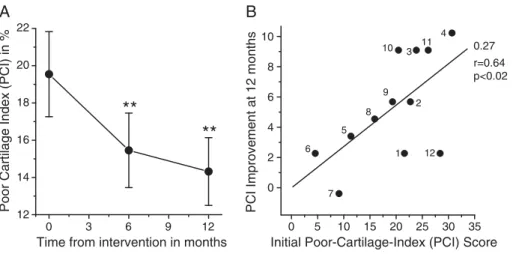
(see Figure S4A,SDC, http://links.lww.com/TP/A811). Be-cause 95% of values should be smaller than (mean+2SD), 50 was chosen as the threshold above which T2 values were considered inordinately high. To quantify T2 mapping, a Poor Cartilage Index (PCI) was estimated as the percentage of T2 values more than 50 ms. A PCI of 100 is the worst possi-ble value, and a value near 5 is considered healthy. A posi-tive correlation was identified between the baseline PCI and VAS scores (r=0.42; PG0.001) (see Figure S4B, SDC, http://links.lww.com/TP/A811). Additionally, the mean PCI significantly decreased from 19.5 to 15.4 during the first 6 months after treatment and further decreased to 14.3 at 12 months after injection (Fig. 2A). Figure S4C details individual patient F2 evolution (seeSDC, http://links.lww.com/TP/A811). The PCI decreased in 11 of 12 patients. Additionally, when PCI im-provement was plotted against the initial PCI, a positive correlation (r=0.64; PG0.020) was noted. The slope of the best-fitting line was 0.27 (Fig. 2B).
DISCUSSION
Both animal experimentation and human case studies suggest that intra-articular MSC injection could be a useful therapeutic alternative for treating knee osteoarthritis. Our preliminary studies in horses and sheep (seeFigure S1,SDC, http://links.lww.com/TP/A811) demonstrated procedural fea-sibility and safety. Here, we present a phase I to II study of
TABLE 1. Total score sum of VAS, WOMAC, and Lequesne severity indices
Test Time n Mean SE Min P25%a P50%a P75%a Max
Knee pain VAS-DA (0Y100) 0 12 46.9 7.5 0.0 35.8 52.5 66.5 80.0
3 months 12 25.1 6.8 0.0 3.8 22.5 36.0 74.0
6 months 12 24.8 6.0 0.0 8.8 16.5 44.0 58.0
12 months 12 15.4 3.8 0.0 3.0 19.0 24.3 38.0
Knee pain VAS-SP (0Y100) 0 8 79.8 6.4 49.0 74.0 88.0 94.5 99.0
3 months 8 16.4 5.8 1.0 6.0 12.0 21.0 48.0
6 months 8 11.1 5.1 0 0.0 12.0 26.0 75.0
12 months 8 15.5 6.4 0 2.3 12.5 31.3 53.0
WOMAC (0Y100)
Pain Subscale 0 12 24.2 4.1 10.0 15.0 17.5 30.0 60.0
12 months 12 5.8 1.6 0.0 0.0 5.0 10.0 15.0
Rigidity Subscale 0 12 10.4 3.7 0.0 0.0 6.3 15.6 37.5
12 months 12 5.2 3.2 0.0 0.0 0.0 3.1 37.5
Function loss Subscale 0 12 19.1 3.8 4.4 8.5 14.0 29.0 41.2
12 months 12 9.4 3.2 0.0 2.2 6.6 12.1 39.7
Total WOMAC Scale 0 12 19.4 3.6 6.3 9.1 14.6 28.1 42.7
12 months 12 8.3 2.7 0.0 1.8 6.3 12.8 32.3
Lequesne (0Y100) 0 12 45.1 5.6 16.7 29.2 43.8 60.4 75.0
12 months 12 14.9 4.1 0.0 7.3 10.4 21.9 50.0
a
P25%, P50%, and P75% represent 25th, 50th (median), and 75th percentiles, respectively.
In all cases, the scale was from 0 to 100%. Measurements were performed before cell transplantation (0) and 3, 6, and 12 months afterwards.
Max, maximum value; Min, minimum value; VAS, Visual Analogue Scale; VAS-DA, Visual Analogue Scale for pain associated to daily activities; VAS-SP, Visual Analogue Scale for pain associated to sports activities; WOMAC, Western Ontario and McMaster Universities Osteoarthritis Index.
Copyright @ 2013 by Lippincott Williams & Wilkins. Unauthorized reproduction of this article is prohibited.
FIGURE 2. Cartilage quality improvement resulting from MSC treatment. Cartilage quality was assessed by MRI T2
mapping and is quantified as the PCI (computed as the percentage of sample points with a T2 relaxation value950 ms). The worse possible value for PCI is 100, and healthy cartilage should approach 5. A, temporal evolution of PCI. MeanTSE values of 12 patients treated with MSCs. **PG0.01 (ANOVA; Bonferroni test for paired values). B, correlation between PCI improve-ment and initial PCI score for the 12 patients included in this study. Codes for each patient are given beside the data points. The best-fitting line is shown with values for the slope and linear regression coefficient (r) at the right. ANOVA, analysis of variance; PCI, Poor Cartilage Index; MRI, magnetic resonance imaging; MSC, mesenchymal stem cells.
Fig
1
4/C
FIGURE 1. Pain improvement resulting from MSC treatment. A, evolution of knee pain over time, as measured by the VAS.
MeanTSE values of 12 patients treated with MSCs.**PG0.01;***PG0.001 (ANOVA; Bonferroni test for paired values). BYD, correlation between improvement of knee pain 1 year after treatment with MSCs and initial pain score, as measured with different tests. The ‘‘perfect’’ treatment (dotted line with slope of 1) is shown for comparison. The best-fitting lines are shown with values for the slope and linear regression coefficient (r) at the right. In case D, the pain and physical function loss subscales of the WOMAC test are shown with different signs (codes at top right). ANOVA, analysis of variance; MSC, mesenchymal stem cells; WOMAC, Western Ontario and McMaster Universities Osteoarthritis Index.
4
www.transplantjournal.com Transplantation&
Volume 95, Number 12, June 27, 2013Copyright @ 2013 by Lippincott Williams & Wilkins. Unauthorized reproduction of this article is prohibited. 12 patients with clinical and objective follow-up coverage for
1 year after intra-articular MSC injection. Our results show that autologous MSC transplantation is both feasible and safe, with no major adverse events recorded. The postimplantation pain observed in 50% of patients responded well to ibuprofen and vanished within 1 to 6 days. Quality control and reproduc-ibility of cell production is essential for meaningful evaluation of cell therapy trials. The GMP-compliant cell preparation (32) was very reproducible with respect to the number of cells (SD=3%) and the expansion time (SD=5%). Immunopheno-typic characteristics were also adequate and stable over time. Cell viability was more than 90% and not affected by trans-port to the administration site.
The analgesic effect of MSC treatment is remarkable, resulting in 65% to 78% improvement in pain (Fig. 1BYD; seeFigure S3,SDC, http://links.lww.com/TP/A811). Improve-ments in function (Fig. 1D) and quality of life are smaller. Our results supersede those of previous case reports, where re-sults were described as ‘‘satisfactory’’ (31) or ‘‘encouraging’’ (30). In these case studies, the number of cells used was smaller (8Y20106), follow-up was for only 6 months, and the MRI study, when performed, was not quantitative.
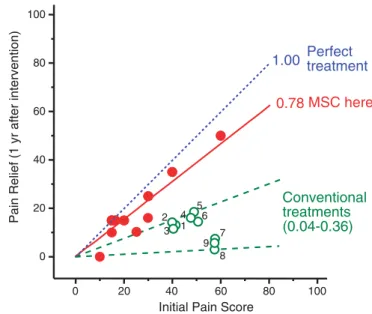
F3 Figure 3 presents a meta-analysis of four recent high-quality clinical trials (8, 11, 12, 14). Data on pain evolution were recalculated and expressed on a 0 to 100% scale. Quan-tification and comparison of several osteoarthritis treatments
were performed using the initial pain score versus pain relief plot (35). The slope of the line defines the treatment efficacy, with complete pain relief reflected in a slope of 1. Each point represents a given condition (for codes, see last column in Table S3 [see SDC, http://links.lww.com/TP/A811], which also provides additional trial details). Overall, the slopes oscillated between 0.04 and 0.36 (meanTSE, 0.21T0.04) for seven conventional treatments (see Table S3, SDC, http://links.lww.com/TP/A811). Our results, labeled ‘‘MSCs here’’ in Figure 3, compare very favorably with previous trials that explored conventional treatments (8, 11, 12, 14).
The analgesic effect of MSC treatment was quite rapid, with more than 50% of the total improvement at-tained by 3 months (Fig. 1A). For sports activities-associated pain, the improvement was even faster (seeFigure S3,SDC, http://links.lww.com/TP/A811). Early action has also been reported for the effects of MSCs on degenerative disc disease (32). After this rapid effect, improvement progressed more slowly and the maximum effect was observed at the 1-year follow-up. Pain improvement associated with sports activi-ties was even larger than the pain improvement associated with daily activities (Figure S3).
Our novel approach for analyzing T2 mapping images filters out most spurious variations and enhances sensitiv-ity by focusing on the evolution of the poor cartilage areas. We demonstrate a significant correlation between the PCI and the VAS (seeFigure S4B,SDC, http://links.lww.com/TP/A811). Additionally, the PCI was improved significantly by MSC treatment (Fig. 2A), although the magnitude of this effect varied among cases (see Figure S4C,SDC, http://links.lww.com/TP/A811). Finally, the slope of the relationship between PCI improve-ment and initial the PCI was 0.27 (Fig. 2B), suggesting that cartilage healing, although significant (PG0.01), was less than the analgesic effect. Further investigation of cartilage healing progression over longer evolution times, and the effect or re-peated MSC application, will be informative.
We can only speculate regarding the mechanisms gov-erning the beneficial effects of MSC treatment. Chondrocytes induce differentiation of cocultured MSCs toward a chondro-cyte phenotype (20). Proliferation and differentiation of MSCs to chondrocytes also happen with MSCs injected into knee joints (28). Importantly, MSCs stimulate cocultured cells to pro-liferate and synthesize extracellular matrix (21, 23, 46). This action may be more important in vivo because few MSCs are required to trigger this effect (22). It was recently shown that transplanted MSCs engraft into the joint, are activated, and express Indian hedgehog and other genes. These genes in turn promote expression of collagen II and other chondro-genic genes by host cells (26). Additionally, MSCs have a well-known immunomodulatory effect (47, 48) and can induce anti-inflammatory cytokine production (22). These data indi-cate that MSCs may help analgesia by reducing inflammation. Because the analgesic effect is more evident than anatomic re-storation, we conclude that the trophic and anti-inflammatory effects of MSCs on the damaged tissue may occur more quickly than the regenerative effects.
In summary, we propose that cell therapy with expanded bone marrowYderived MSCs should be considered as a pu-tative treatment for chronic osteoarthritis. Cell handling and expansion is reproducible, and quality-control tests were satis-factory. The clinical procedure is feasible and safe and requires
Fig
3
4/C
FIGURE 3. Comparison of the efficacy of several
osteo-arthritis treatments for pain relief. Data from four presti-gious clinical trials (8, 11, 12, 14), quantified using the algofunctional WOMAC index, are represented as pain re-lief versus initial pain score (35). The slope of the lines (values at right) represents efficacy. Lines were forced to pass through the origin. The data from the present study (‘‘MSC here’’) are included for comparison (each closed circle corresponds to one patient; three values overlap at 15,15 location). Open circles correspond to results obtained in different trials; the definition of the numerical codes is given in the last column of Table S3 (see SDC, http://links.lww.com/TP/A811). For a more detailed de-scription, see Table S3(SDC, http://links.lww.com/TP/A811). MSC, mesenchymal stem cells; WOMAC, Western Ontario and McMaster Universities Osteoarthritis Index.
Copyright @ 2013 by Lippincott Williams & Wilkins. Unauthorized reproduction of this article is prohibited. only minimally invasive intervention without surgery or
hos-pitalization. The results are better than those obtained with established treatments. Pain relief occurs by 3 months and in-creases for at least 1 year. The recovery of functional losses is less but also significant, and there is quantitative evidence of partial articular cartilage healing. Future studies will involve larger trials focused on efficacy, with greater patient numbers and longer follow-up periods. These studies will track long-term joint evolution and investigate the specific anatomic and functional changes that occur in the knee.
MATERIALS AND METHODS
Patients and Procedures
This pilot phase I to II trial was approved by the Teknon Medical Centre Ethics Committee and the Spanish Drug and Medicines Agency (EudraCT 2009-017405-11) and registered in ClinicalTrials.gov (NCT01183728). Twelve patients with chronic knee osteoarthritis unresponsive to conventional treat-ments (for details, see Table S1,SDC, http://links.lww.com/TP/A811) were included. Detailed inclusion and exclusion criteria are reported in
T2 Table 2.
After clinical, analytical, and imaging evaluations to ensure compliance with these criteria, patients were informed about the protocol characteristics and provided written informed consent.
The protocol included seven visits (V0YV6). V0 involved the final check of compliance with inclusion criteria, performance of necessary complementary evaluations and tests, and scheduling of dates for V1 and V2. V1 involved bone marrow harvesting from the iliac crest (80Y90 mL) for MSC isolation. This intervention was performed under local anesthesia and slight sedation, and patients were discharged after 2 hr of observation. V2 (21Y24 days after V1) involved the injection of MSCs (40106cells per knee from a 5106cells/mL
suspension by medial parapatellar injection). V3 to V6 (8 days and 3, 6, and 12 months after implantation) included clinical evaluation and routine
analysis (V3YV6), VAS for daily activity and for sports (35), WOMAC and Lequesne algofunctional indices (49), SF-36 questionnaire (50), and quanti-tative MRI exploration (V0, V5, and V6). Outcomes were expressed on a 0 to 100% scale in all cases.
Cell Isolation and Expansion
Cell isolation and expansion were performed in the Instituto de Biologı´a y Gene´tica Molecular Cell Production Unit under GMP conditions and with approval of the Spanish Drug and Medicines Agency (PEI No. 10-134), as described previously (32). Bone marrow samples were transported to the Cell Production Unit at 4-C to 12-C within 12 hr of harvesting. The mononu-clear cell fraction was isolated by density-gradient centrifugation, resuspended, and cultured in MSC expansion culture medium (51) in 175-cm2tissue cul-ture flasks, with periodic washing to remove nonadherent cells. When cells reached 80% confluence, they were trypsinized and replated, and the process was repeated for two more passages. At the end of this period (21Y24 days), cells were harvested, resuspended in Ringer’s lactate solution containing 0.5% human albumin (CSL Behring GmbH, Marburg, Germany) and 5 mM glucose, and transported at 4-C to 20-C by air courier (6 hr) to Teknon Medical Centre for application. In addition to quality-control tests, viability and flow cytometric immunophenotypic profiles (34, 51) were determined at this stage.
MRI Assessments
MRI was used to assess cartilage state by T2 mapping using the GE CartiGram sequence (37Y39). Mean T2 relaxation values (ms) were sampled in 88 well-defined regions of interest (ROIs), including patellar cartilage (24 ROIs), femoral condyles (32 ROIs), and tibial condyles (32 ROIs). In-strumental variation, computed as the mean of differences between two consecutive measurements, was approximately 4%. Interobserver variation was 3%. To analyze assay results, values were averaged in each area and those above 50 ms, which represent poor quality, remodeling, inflammatory tissue (40Y42), were counted to compute the PCI (expressed as percentage of all values obtained in the 88 ROIs) as described in Results. Values above 90 were not used for computations. For the PCI, 100% represents the worst possible PCI value and values at or below 5% are considered healthy.
Statistical Analysis
Data are reported as meanTSD (or meanTSE), as indicated. The signifi-cance of differences was assessed either by Student’sttest or by one-way analysis of variance (ANOVA) and the corresponding nonparametric tests. GraphPad Instat3 package software version 3.06 (GraphPad Software, La Jolla, CA) was used for calculations
ACKNOWLEDGMENTS
The authors thank Mr. Jesu´s Ferna´ndez (Instituto de Biologı´a y Gene´tica Molecular, Valladolid, Spain) and Ms. Carmen Barbero (Institut de Tera`pia Regenerativa Tissular, Barcelona, Spain) for technical support, Dr. Juan Carlos Vilanova (Centro Diagno´stico por la Imagen, Girona, Spain) and Dr. Sigfried Trattnig (Medical University of Vienna, Vienna, Austria) for help with T2 mapping, Dr. Xavier Peirau (traumatology consultant at In-stitut de Tera`pia Regenerativa Tissular), and the Banc de Sang i Teixits (Barcelona, Spain) for promoting the study of chondral defect repair in the sheep model.
REFERENCES
1. Arden N, Nevitt MC. Osteoarthritis: epidemiology.Best Pract Res Clin Rheumatol2006; 20: 3.
2. Le Pen C, Reygrobellet C, Gerentes I. Financial cost of osteoarthritis in France. The ‘‘COART’’ France study.Joint Bone Spine2005; 72: 567. 3. Hermans J, Koopmanschap MA, Bierma-Zeinstra SM, et al.
Produc-tivity costs and medical costs among working patients with knee os-teoarthritis.Arthritis Care Res (Hoboken)2012; 64: 853.
4. Hawker GA, Mian S, Bednis K, et al. Osteoarthritis year 2010 in review: non-pharmacologic therapy.Osteoarthritis Cartilage2011; 19: 366.
TABLE 2. Inclusion and exclusion criteria
Inclusion criteria
1. Grade II to IVosteoarthritis according to the KellgrenYLawrence grading scale (33) and concurred by two different observers. 2. Chronic knee pain of mechanical origin.
3. Absence of local or general infection.
4. Hematologic and biochemical analyses with no significant alterations that contraindicate intervention.
5. Patient is able to understand the nature of the study. 6. Informed written consent provided by the patient. Exclusion criteria
1. Age975 orG18 years or legally dependent.
2. Signs of infection or positive serology for HIV, hepatitis, or syphilis. 3. Congenital or acquired diseases leading to significant knee
deformities that may interfere with cell application or inter pretation of results.
4. Obesity, with body mass index930 (calculated as mass in kg/ height in m2).
5. Pregnancy or breast-feeding. 6. Neoplasia.
7. Immunosuppression.
8. Intra-articular injection of any drug during the previous 3 months. 9. Participation in another clinical trial or treatment with an
other investigational product within 30 days before inclusion in the study.
10. Other conditions that may, according to medical criteria, discourage participation in the study.
6
www.transplantjournal.com Transplantation&
Volume 95, Number 12, June 27, 2013Copyright @ 2013 by Lippincott Williams & Wilkins. Unauthorized reproduction of this article is prohibited.
5. American Academy of Orthopaedic Surgery. Treatment of Osteoar-thritis of the Knee (Non-arthroplasty). Full Guideline. Rosemont, IL: American Academy of Orthopaedic Surgeons; 2008.
6. Hochberg MC, Altman RD, April KT, et al. American College of Rheu-matology 2012 recommendations for the use of nonpharmacologic and pharmacologic therapies in osteoarthritis of the hand, hip, and knee.
Arthritis Care Res (Hoboken)2012; 64: 465.
7. Samson DJ, Grant MD, Ratko TA, et al. Treatment of primary and secondary osteoarthritis of the knee.Evid Rep Technol Assess (Full Rep)2007; 1. 8. Pisters MF, Veenhof C, Schellevis FG, et al. Long-term effectiveness of
exercise therapy in patients with osteoarthritis of the hip or knee: a randomized controlled trial comparing two different physical therapy interventions.Osteoarthritis Cartilage2010; 18: 1019.
9. Rutjes AW, Juni P, da Costa BR, et al. Viscosupplementation for oste-oarthritis of the knee: a systematic review and meta-analysis.Ann Intern Med2012; 157: 180.
10. Sawitzke AD, Shi H, Finco MF, et al. Clinical efficacy and safety of glucosamine, chondroitin sulphate, their combination, celecoxib or placebo taken to treat osteoarthritis of the knee: 2-year results from GAIT.Ann Rheum Dis2010; 69: 1459.
11. Moseley JB, O’Malley K, Petersen NJ, et al. A controlled trial of arthro-scopic surgery for osteoarthritis of the knee.N Engl J Med2002; 347: 81. 12. Kirkley A, Birmingham TB, Litchfield RB, et al. A randomized trial of arthroscopic surgery for osteoarthritis of the knee.N Engl J Med
2008; 359: 1097.
13. Manheimer E, Cheng K, Linde K, et al. Acupuncture for peripheral joint osteoarthritis.Cochrane Database Syst Rev2010; CD001977. 14. Witt C, Brinkhaus B, Jena S, et al. Acupuncture in patients with
os-teoarthritis of the knee: a randomised trial.Lancet2005; 366: 136. 15. Rutjes AW, Nuesch E, Sterchi R, et al. Therapeutic ultrasound for
osteoarthritis of the knee or hip.Cochrane Database Syst Rev2010; CD003132.
16. Vasiliadis HS, Wasiak J. Autologous chondrocyte implantation for full thickness articular cartilage defects of the knee.Cochrane Database Syst Rev2010; CD003323.
17. Brittberg M, Lindahl A, Nilsson A, et al. Treatment of deep cartilage defects in the knee with autologous chondrocyte transplantation.
N Engl J Med1994; 331: 889.
18. Gupta PK, Das AK, Chullikana A, et al. Mesenchymal stem cells for cartilage repair in osteoarthritis.Stem Cell Res Ther2012; 3: 25. 19. Yoo JU, Barthel TS, Nishimura K, et al. The chondrogenic potential of
human bone-marrow-derived mesenchymal progenitor cells.J Bone Joint Surg Am1998; 80: 1745.
20. Hwang NS, Im SG, Wu PB, et al. Chondrogenic priming adipose-mesenchymal stem cells for cartilage tissue regeneration.Pharm Res
2011; 28: 1395.
21. Acharya C, Adesida A, Zajac P, et al. Enhanced chondrocyte prolifera-tion and mesenchymal stromal cells chondrogenesis in coculture pellets mediate improved cartilage formation.J Cell Physiol2012; 227: 88. 22. Yang SH, Wu CC, Shih TT, et al. In vitro study on interaction between
human nucleus pulposus cells and mesenchymal stem cells through paracrine stimulation.Spine2008; 33: 1951.
23. Wu L, Prins HJ, Helder MN, et al. Trophic effects of mesenchymal stem cells in chondrocyte co-cultures are independent of culture con-ditions and cell sources.Tissue Eng Part A2012; 18: 1542.
24. Matsumoto T, Okabe T, Ikawa T, et al. Articular cartilage repair with au-tologous bone marrow mesenchymal cells.J Cell Physiol2010; 225: 291. 25. Horie M, Sekiya I, Muneta T, et al. Intra-articular injected synovial stem cells differentiate into meniscal cells directly and promote menis-cal regeneration without mobilization to distant organs in rat massive meniscal defect.Stem Cells2009; 27: 878.
26. Horie M, Choi H, Lee RH, et al. Intra-articular injection of human mesenchymal stem cells (MSCs) promote rat meniscal regeneration by being activated to express Indian hedgehog that enhances expres-sion of type II collagen.Osteoarthritis Cartilage2012; 20: 1197. 27. Lee KB, Hui JH, Song IC, et al. Injectable mesenchymal stem cell therapy
for large cartilage defectsYa porcine model.Stem Cells2007; 25: 2964. 28. Sato M, Uchida K, Nakajima H, et al. Direct transplantation of
mes-enchymal stem cells into the knee joints of Hartley strain guinea pigs with spontaneous osteoarthritis.Arthritis Res Ther2012; 14: R31. 29. Wakitani S, Imoto K, Yamamoto T, et al. Human autologous culture
expanded bone marrow mesenchymal cell transplantation for repair
of cartilage defects in osteoarthritic knees. Osteoarthritis Cartilage
2002; 10: 199.
30. Davatchi F, Abdollahi BS, Mohyeddin M, et al. Mesenchymal stem cell therapy for knee osteoarthritis. Preliminary report of four patients.
Int J Rheum Dis2011; 14: 211.
31. Emadedin M, Aghdami N, Taghiyar L, et al. Intra-articular injection of autologous mesenchymal stem cells in six patients with knee osteo-arthritis.Arch Iran Med2012; 15: 422.
32. Orozco L, Soler R, Morera C, et al. Intervertebral disc repair by au-tologous mesenchymal bone marrow cells: a pilot study. Transplan-tation2011; 92: 822.
33. Kellgren JH, Lawrence JS. Radiological assessment of osteo-arthrosis.
Ann Rheum Dis1957; 16: 494.
34. Dominici M, Le Blanc K, Mueller I, et al. Minimal criteria for defining multipotent mesenchymal stromal cells. The International Society for Cellular Therapy position statement.Cytotherapy2006; 8: 315. 35. Huskisson EC. Measurement of pain.Lancet. 1974; 2: 1127. 36. Hawker G, Melfi C, Paul J, et al. Comparison of a generic (SF-36) and
a disease specific (WOMAC) (Western Ontario and McMaster Univer-sities Osteoarthritis Index) instrument in the measurement of outcomes after knee replacement surgery.J Rheumatol1995; 22: 1193.
37. Trattnig S, Mamisch TC, Welsch GH, et al. Quantitative T2 map-ping of matrix-associated autologous chondrocyte transplantation at 3 Tesla: an in vivo cross-sectional study.Invest Radiol2007; 42: 442. 38. Apprich S, Welsch GH, Mamisch TC, et al. Detection of
degenera-tive cartilage disease: comparison of high-resolution morphological MR and quantitative T2 mapping at 3.0 Tesla.Osteoarthritis Cartilage
2010; 18: 1211.
39. Crema MD, Roemer FW, Marra MD, et al. Articular cartilage in the knee: current MR imaging techniques and applications in clinical practice and research.Radiographics2011; 31: 37.
40. Battaglia M, Vannini F, Buda R, et al. Arthroscopic autologous chon-drocyte implantation in osteochondral lesions of the talus: mid-term T2-mapping MRI evaluation. Knee Surg Sports Traumatol Arthrosc
2011; 19: 1376.
41. Battaglia M, Rimondi E, Monti C, et al. Validity of T2 mapping in characterization of the regeneration tissue by bone marrow derived cell transplantation in osteochondral lesions of the ankle.Eur J Radiol
2011; 80: e132.
42. Giannini S, Battaglia M, Buda R, et al. Surgical treatment of os-teochondral lesions of the talus by open-field autologous chondrocyte implantation: a 10-year follow-up clinical and magnetic resonance imaging T2-mapping evaluation.Am J Sports Med2009; 37: 112S. 43. White LM, Sussman MS, Hurtig M, et al. Cartilage T2 assessment:
dif-ferentiation of normal hyaline cartilage and reparative tissue after ar-throscopic cartilage repair in equine subjects.Radiology2006; 241: 407. 44. Dunn TC, Lu Y, Jin H, et al. T2 relaxation time of cartilage at MR imaging: comparison with severity of knee osteoarthritis.Radiology
2004; 232: 592.
45. Kim HK, Laor T, Graham TB, et al. T2 relaxation time changes in distal femoral articular cartilage in children with juvenile idiopathic arthritis: a 3-year longitudinal study.AJR Am J Roentgenol2010; 195: 1021. 46. Qing C, Wei-ding C, Wei-min F. Co-culture of chondrocytes and bone
marrow mesenchymal stem cells in vitro enhances the expression of cartilaginous extracellular matrix components. Braz J Med Biol Res
2011; 44: 303.
47. Le Blanc K, Ringden O. Immunomodulation by mesenchymal stem cells and clinical experience.J Intern Med2007; 262: 509.
48. Aggarwal S, Pittenger MF. Human mesenchymal stem cells modulate allogeneic immune cell responses.Blood2005; 105: 1815.
49. Faucher M, Poiraudeau S, Lefevre-Colau MM, et al. Assessment of the test-retest reliability and construct validity of a modified WOMAC index in knee osteoarthritis.Joint Bone Spine2004; 71: 121. 50. Kosinski M, Keller SD, Hatoum HT, et al. The SF-36 Health Survey as a
generic outcome measure in clinical trials of patients with osteoarthritis and rheumatoid arthritis: tests of data quality, scaling assumptions and score reliability.Med Care1999; 37: MS10.
51. Blanco JF, Graciani IF, Sanchez-Guijo FM, et al. Isolation and charac-terization of mesenchymal stromal cells from human degenerated nu-cleus pulposus: comparison with bone marrow mesenchymal stromal cells from the same subjects.Spine2010; 35: 2259.
Orozco_Osteoarthritis_MSC_v4.3.doc Mar_1_2013 1
SUPPLEMENTAL DIGITAL CONTENT (SDC)
Contents:
SUPPLEMENTARY TABLES
x Supplementary Table S1. Antecedent history of the patients included in this trial.
x Supplementary Table S2. Minor adverse events
x Supplementary Table S3. Meta-analysis of clinical trials with different osteoarthritis
treatments and comparison of their efficacies
SUPPLEMENTARY FIGURES
x Supplementary Figure S1. Effects of MSCs in horses.
x Supplementary Figure S2. Immunophenotypic characterization of MSCs.
x Supplementary Figure S3. Effects of MSC on sports activity-associated pain
Oro
zc
o
_Osteo
a
rth
ritis_
M
S
C
_
v
4.3
.doc Ma
r_1_2
0
13
Table S1
. Antecedent history
of the patients included in
this trial.
Pat. Num. Sex
Age
S
ide
OA
G
rade
Pr
evious
S
u
rgery
RHB
NSAID
Corti- coids
Hy
aluronic Acid
PRP
Num. (Date
)
Date MSV
1
F
66
L
IV
Yes
Ye
s
3 (2007) 3 (2008)
2010
2
M 41
R
III
ACL
+
MM (1
99
1)
MCL (2004)
Yes
Ye
s
2010
3
M 44
R
II
MM (2009)
ACL (2009)
Yes
Ye
s
2010
4
F 41
L
III
L
M
(2001)
Yes
Ye
s
3 (2002) 3 (2003)
2010
5
F 35
R
II
ACL (1991) MM (2001) LM(2010)
Yes
Ye
s
2010
6
M 33
L
II
ACL (1998) MM (2007) MM(2009)
Yes
Ye
s
4 (2010)
Oro zc o _Osteo a rth ritis_ M S C _ v 4.3 .doc Ma r_1_2 0 13 3 7 M 29 R II ACL (2001) MM + L M (2 007) Yes Ye s 3 (2009) 2010 8 M 43 L IV L M (2001) Yes Ye s
3 (2003) 3 (2006) 3(2007)
2010 9 F 39 R III ACL + MM (1 99 1) Yes Ye s 2 (2010) 2010 10 M 75 R IV Yes Ye s
3 (2005) 3 (2006)
2011 11 M 71 L IV Yes Ye s 3 (2007) 3 (2010) 2011 12 F 72 L IV O S TEOT. (20 00) Yes Ye s 2 (2007)
4 (2009) 4 (2010)
2011 OA , Osteoarthritis; RHB, Rehabilit ati on; NS AI D, non-s teroidal antiinf lammatory d
rug; Cort
ic ., Infiltration with c ort ic o s tero id s; P R P, Plate le t-rich p lasma; MSV, Me senchyma l St
em Cells; ACL,
Ante ri or cru c iate ligament; MM, Media l menisc us; LM, Latera l meniscus ; MCL, media l collatera l lig
ament; OS
TEOT., O
s
teotomy (t
Oro zc o _Osteo a rth ritis_ M S C _ v 4.3 .doc Ma r_1_2 0 13 Table S2
. Minor adverse events
Minor adve
rse event (Comments)
Par ticipants affected ( % ) Pos t-im p lant ati
on pain at
day
s 1-6 (E
, SR) 6/12 (50%) Artic ular inf lammat ion at tributabl e to knee ov erlo adi
ng (E, PSR)
3/12 (25%) Une x p e c
ted knee i
n fl ammati on with sy no vial f lu id effus ion a nd artic u lar swe
lling (UE, PS
R)
3/12 (25%)
Low back
p
a
in (UE, P
S R ) 3/12 (25%) Pai n
in the contral
ateral
knee (UE, P
S R) 1/12 (8%) Isc h iot ibia l tendonitis (U E , PSR) 1/12 (8%) Arthroscopic s urgery in t he contralateral
knee (UE, NSR)
1/12 (8%) Dental im p la n
t (UE, NSR)
1/12 (8%) Influenz a (UE , NS R) 1/12 (8%) Intolerance t o
gluten and and to lacto
s e (UE , NSR) 1/12 (8%) Comments: (E) E x pecte
d; (UE) Une
x
pected; (S
R) Study-Re
lated; (NS
R) Not S
tudy-Related; (P SR) Po ssib ly Study -Re lated. In all cas e s, the adv er s e e v ents resp
onded to medi
ca
l/ph
ysical
thera
Oro zc o _Osteo a rth ritis_ M S C _ v 4.3 .doc Ma r_1_2 0 13 5 Table S3 . Meta-anal y s is of clinical tr ials w ith differ
ent osteoarthritis treatments and comparison of their efficac
ies. Clinical Tr ial Inter vention (a) Duration (b ) n Basal (c) +Treatment (c) Improvement (d) Impr./Basal slope (e) Cod e (f )
Pisters et a
Oro zc o _Osteo a rth ritis_ M S C _ v 4.3 .doc Ma r_1_2 0 13 SHA M 1 y r 75 53± 19 38± 23 14 0.27 6 Mose le y et al., 2002 ( 10 ) PCB 0.5 m 59 60± 19 48± 24 12 0.19 - LAV 0.5 m 61 59± 17 52± 20 7 0.12 - DEBR 0.5 m 58 59± 22 53± 22 6 0.10 - PCB 2 y r 59 60± 19 53± 25 7 0.10 7 LAV 2 y r 61 59± 17 57± 24 3 0.04 8 DEB R 2 y r 58 59± 22 54± 23 5 0.09 9 This
Study, 2012
MSCs 1 y r 12 24± 14 6± 6 18 0·78 x (a) B GA, Beha v ior-Grade d Act ivity (n
o drugs); UC, Us
ua l Ca re (onl y ph ysic a
l; no drugs
); ACCUP , ac upuncture (non-s teroi dal ant i-infl ammatorie
as needed; compare
s with s
h a m -tre ated); PCB, P la c ebo; LA V, Lav age ( m edica
l treatment as ne
eded); DEB
R, Debride
ment (med
ical tre
atment
as needed); CONT,
cont rol; SURG , surgery (la vage plus de bri dement; p h ysic al and medica l ther apy as requ ired); MSCs, Mes en chymal st e m cells e x pand
ed from bone marrow
s
a
mple
s
in the c
u
rrent study sho
wn here. The West
ern O ntari o and McMa s ter Universi ti es A rthriti s (WOMAC) index (pa in comp onent) has been used; sca le 0-1 00. (b)
Duration in years
(yr) or mont
hs
(m);
(c
) Where appropri
ate, mean ± SD is g iv (d) Di fferenc e between “bas
al” and “tr
eatment” v a lues. (e) Impr./ Basa l sl ope, rat io impro vement/basa l = sl ope
in Figure 3;
(f
) Code in Figure
Orozco_Osteoarthritis_MSC_v2.7.doc Dic_28_2012 7
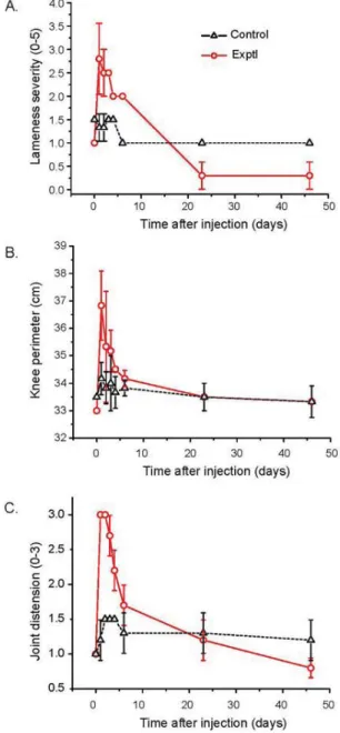
Figure S1. Effects of MSCs in horses.
The intended clinical protocol was first tested in three horses to assess feasibility and safety.
Experiments in horses were approved by the Autonomous University of Barcelona’s Animal
Care and Use Committee. The tibiotarsal joint was chosen for the experiments. Limited
lesions (1.5×1.5 cm laterally, and 4 mm deep, avoiding the subchondral plate) were
approximately 80-ml sternum bone marrow samples, which were expanded for 21 days to
obtain 50×106 MSCs, following the same protocol used in humans (see Methods). Cells were
suspended in 10 ml of autologous plasma and injected intra-articularly 2 weeks after the
lesion was created. A volume of 10 ml of MSC suspension was injected into one joint and 10
ml of vehicle (phosphate-buffered saline) into the contralateral joint. The follow-up period was
46 days. The horses were maintained in stalls during the first 3 days and then in a 10×10 m
fenced space for 43 additional days. Clinical tests were performed at the times shown and
included quantification of (A) lameness severity (0-5 scale), (B) knee diameter, and (C) joint
distension (0-3 scale, appreciated by palpation). The values shown are the mean ± SE of
three independent experiments. The lesion produced lameness and inflammation, estimated
from knee diameter and joint distension. Injection of MSCs produced considerable additional
inflammation and worsened lameness during the first 1-2 days. Then symptoms declined
slowly during the whole observation period with a half-time period of 6-12 days. In controls
injected with saline the inflammatory peak was much smaller. By the end of the observation
period, inflammation was less in the joints injected with MSCs. The necropsy, performed at
the end of the 6 months period, did not reveal local nor general alterations. Overall, results
supported feasibility and safety of the procedure. Our team also performed a preliminary
study in 10 sheep, in which a limited lesion was generated in the femoral condyles and the
internal meniscus. Five sheep were injected with 8 ml of saline as controls and the other five
animals received the same solution containing 50×106 autologous bone marrow MSCs. We
observed clear regeneration of cartilage and the meniscus in the MSC-treated sheep at 12
months post-treatment compared to no improvement in control animals. Necropsy did not
Orozco_Osteoarthritis_MSC_v2.7.doc Dic_28_2012 9
Figure S2. Immunophenotypic characterization of MSCs.
Flow cytometric analysis of MSCs (blue) compared with isotype controls (orange). MSCs
were strongly positive for CD90 and CD166; moderately positive for CD105, CD106, and
Figure S3.Effects of MSC on sports activity-associated pain.
A.Graph showing evolution of knee pain associated to sports activity, as measured by VAS
(VAS-SA), over time. Mean ± standard error (SE) values of 8 patients treated with MSC.
Data from 4 patients were not included because these series were not complete. ***p<0.001
(ANOVA; Bonferroni test for paired values). B. Correlation between improvement of knee
pain 1 year after treatment with MSCs and initial pain score, as measured with VAS-SA. The
“perfect” treatment (dotted line with slope of 1) is shown for comparison. The best-fitting line
is shown with values for the slope and linear regression coefficient (r) at the right. The figures
besides data points are the patient codes. Patients 4 and 8 were not included because data
Orozco_Osteoarthritis_MSC_v2.7.doc Dic_28_2012 11
Figure S4. T2 mapping results.
A. Distribution of the T2 relaxation values (ms) obtained in nine measurements in healthy
individuals; 88 areas were analyzed in each knee articulation: 24 in the patella, 32 in the
femoral condyles, and 32 in the tibial condyles. Mean±SD = 39.0±6.8 (n=792). Percentile
95=50; Gaussian fitting is also shown (r=0.984). B. Correlation between baseline values of
Poor Cartilage Index (PCI) and VAS. PCI was computed as the percentage of T2 relaxation
readings >50 ms. Numbers beside data points correspond to patient codes. The best-fitting
line is also shown. Linear regression analysis: r=0.34; p<0.001. C. Temporal evolution of PCI
Transplantation
®
T H E O F F I C I A L J O U R N A L O F T H E T R A N S P L A N T A T I O N S O C I E T Y