TítuloEffect of hydrogen sulfide sources on inflammation and catabolic markers on interleukin 1β stimulated human articular chondrocytes
13
0
0
Texto completo
(2) In addition to the effects above, OA is also characterized by an increase in PGEs production. PGEs are produced from arachidonic acid in a cascade that involves cyclooxygenases 1 and 2 (COX1 and COX2) which produce the intermediate form PGE H-2. PGE E synthase (PTGES) converts PG (H)-2 to PGE E−2 (PGE-2) downstream of COX1 and COX29. OA cartilage produces high quantities of PGE-2 and PTGES is also upregulated, together with COX2, in IL1β and TNF-α stimulated CHs5 and 9. All these factors, probably synergistically10, contribute to the increase in catabolic processes that lead to cartilage destruction in OA. Hydrogen sulfide (H2S) is emerging as a potential regulator of inflammation and it might offer therapeutic value in the treatment of OA. H2S has been traditionally classified as a toxic gas11 but in the past 10–15 years, scientific opinion has changed as more reports on its biological activity were published and it is now considered as a biologically relevant molecule12 and 13. H2S is synthesized endogenously in mammals from l-cysteine by at least two pyridoxal-5′-phosphate dependent enzymes, cystathionine βsynthase (CBS) and cystathionine γ-lyase (CTH) and to a lower extent by mercaptopyruvate sulfurtransferase (MPST). Together with NO and CO, H2S has been identified as a gasotransmitter14 and 15 and as such it can permeate cellular membranes without a specific transporter. Low (micromolar) levels of H2S have been found to be cytoprotective in several disease models, whereas several inflammation animal models have found increased endogenous levels of H2S (reviewed in12 and 13). With respect to inflammatory rheumatic diseases, Whiteman et al. 16 were able to detect H2S in the synovial fluid from patients with rheumatoid arthritis (RA) and OA as well as in plasma from normal controls (37.6 μM). Levels from RA patients (62.41 μM) were significantly increased over those of the OA group (25.1 μM). This group has also recently shown that human articular CHs and mesenchymal progenitor cells synthesize CBS and CTH, and that their expression was induced by various cytokines, including IL1β 17. Therefore, even though its role has not been clearly elucidated yet, evidence suggests that H2S may have an important function in inflammation and it might be of interest as a therapeutic agent in diseases that have an inflammatory component, such as OA. In this study, two exogenous sources of H2S, NaSH (a fast dissolving salt) and GYY4137 (as a slow releasing agent), were added to cultures of IL1β-stimulated human articular CHs isolated from osteoarthritic tissue. Concentrations of the H2S sources ranged from 0.05 to 1 mM and their effects on several markers of inflammation and cartilage degradation were investigated. Also, since activation of nuclear factor-κB (NFκB) is directly responsible for the upregulation of several proinflammatory cytokines and enzymes, including NOS2, COX2 and IL618 and 19, this signaling route was investigated as a possible mechanism of action for H2S. Methods Materials Dulbecco's Modified Eagle Medium (DMEM), fetal bovine serum (FBS) and Penicillin/Streptomycin (P/S) were from Gibco (Gibco, Madrid, Spain). Culture flasks and other disposable plastic were purchased from BD (BD Bioscience, Madrid, Spain). Sodium hydrosulfide (NaSH), sodium nitrite, human recombinant interleukin-1β (IL1β), trypsin and collagenase were purchased from Sigma–Aldrich (Sigma–Aldrich Química S.A, Madrid Spain). GYY4137 (Morpholin-4-ium 4 methoxyphenyl(morpholino phosphinodithioate)) was purchased from Santa Cruz Biotechnology, Heidelberg, Germany. AlamarBlue® was purchased from Invitrogen S.A., Barcelona, Spain. The Griess reagent was from Enzo Life Science, Madrid, Spain. The PGE-2 EIA was purchased from GE Healthcare, Barcelona, Spain and the ELISAs for matrix metalloproteinase-13 (MMP13), and interleukin-6 (IL6) were purchased from R&D systems (Madrid, Spain). Antibodies for NOS2, MMP13 and NFκB p65 were from Abcam (Abcam plc, Cambridge, UK), Thermo Scientific (Cienytech, A Coruña, Spain) and Santa Cruz Biotechnology, respectively. Reagents for immunocytochemistry (ICC) were from Dako (Dako, Barcelona, Spain) and reagents for molecular biology were from Invitrogen (Invitrogen S.A. Barcelona, Spain), unless otherwise stated. Tissue selection and cell culture Osteoarthritic cartilage donors were patients of the Orthopedic Service of the University Hospital A Coruña (CHUAC) that had knee or hip replacement surgery (seven men and six women, age 77.5 ± 10.0 years old). The local ethics committee approved the study and written informed consent was obtained from all the patients. CHs were isolated by sequential enzymatic digestion from human articular cartilage.
(3) as described previously20. Briefly, cartilage sections were washed with saline and minced into small pieces. This was then incubated with trypsin at 37 °C for 10 min, followed by 12–16 h incubation with type IV clostridial collagenase (2 mg/ml) in DMEM with 5% FBS and 1% P/S. Isolated cells were centrifuged and re-suspended in expansion medium (DMEM containing 10% FBS and 1% P/S) and allowed to grow until pre-confluent at 37 °C in a humidified incubator with 5% CO2. The cells used for the experiments were routinely checked for production of collagen type II and aggrecan to ensure they maintained a chondrocytic phenotype. Cell stimulation and treatment experiments Only cells from passage two were used. For all experiments cells were incubated in expansion medium during 24 h for adherence, then the medium was changed to depleted medium (DMEM containing 0.5% FBS and 1% P/S) with the different concentrations, 0 (Basal), 100, 200, 500 or 1,000 μM of one of the sulfide sources (NaSH or GYY4137) and with or without IL-1β (5 ng/ml), for an additional incubation time of either 24 h (cell viability) or 48 h (rest of the experiments). Cell viability assay The effect of the sulfide-releasing compounds on CH viability was determined with AlamarBlue® cell viability reagent following manufacturer instructions. Cells were seeded (5·104/well, 96 well plates) and treated as described above. AlamarBlue® was added to the cells in a volume equivalent to 10% of the total volume. Reduction of AlamarBlue® was measured with a spectrophotometer (Nanoquant Infinite M200, Tecan Ibérica Instrumentación S.L Barcelona, Spain) at 570 nm and with 600 nm as a reference, after 4 h of incubation. NO production assay The Griess reaction was used to determine the effects of NaSH and GYY4137 on NO production in IL1βstimulated CHs21. CHs were plated (5·104/well, 96 well plates) and treated as described above. Culture supernatants (50 μl) were collected for nitrite measurements and mixed with 50 μl of Griess reagent. NO formation was detected by nitrite accumulation using sodium nitrite as standard. Optical density was measured at 570 nm in a Nanoquant Infinite M200 spectrophotometer (Tecan). NOS2 and MMP13 ICC The effect of NaSH and GYY4137 on the accumulation of NOS2 and MMP13 proteins in IL1βstimulated CHs was measured through ICC. Cells were seeded (3·104/well, 8 well chamber slides), treated as described above and then fixed in acetone at 4 °C for 10 min. The endogenous peroxidase activity was blocked with Dako Real™ peroxidase blocking solution during 10 min at room temperature. Cells were washed with phosphate buffer solution (PBS) and incubated with either rabbit-antihuman polyclonal NOS2 antibody or mouse-antihuman MMP13 monoclonal antibody. The rabbit/mouse peroxidase/DAB DAKO REAL™ EnVision™ detection kit was used to determine antigen–antibody interactions. Slides were dehydrated in graded alcohol, cleared in xylene and mounted in DePeX (Gurr®, VWR International Eurolab S.L., Barcelona, Spain). A negative control was included by omitting the primary antibody in one of the wells. Slides were then visualized in an Olympus Dx61 optical microscope (Olympus España S.A.U., Barcelona, Spain). NFκB immunofluorescence For the NFκB immunofluorescence assay cells were seeded (3·104/well, 8 well chamber slides) and treated as described above, but for only 45 min, to be able to detect NFκB translocation to the cells nuclei and then fixed in paraformaldehyde at 4 °C for 10 min. Cells were then treated with 3% Triton-X for 5 min, washed subsequently with water and PBS. Cell were incubated with the antibody for 3 h in the dark, counterstained with DAPI for 30 min and mounted with Glycergel mounting medium..
(4) PGE-2, IL6 and MMP13 ELISA assays The effect of the addition of the H2S sources on PGE-2, MMP13 and IL6 production in IL1β–stimulated CHs was quantified on cell supernatants using specific enzyme immunoassays, following their manufacturer instructions. Cells were seeded (5·104/well, 96 well plates) and treated as described above. The calibration curves for PGE-2 EIA, the MMP13 ELISA and the IL6 ELISA spanned from 50 to 6,400 ng/ml, from 33 to 4,200 pg/ml and from 9.4 to 1,200 pg/ml, respectively. Samples were diluted accordingly. qRT-PCR analyses The effect of NaSH and GYY4137 on the expression of the genes included in Table I in IL1β-stimulated CHs was quantified using qRT-PCR. Cells were seeded (3·105/well, 12 well plates) and treated as described above. Total RNA was isolated using Trizol® Reagent, following the manufacturer protocol. RNA concentration was quantified at 260 nm using a NanoDrop™ spectrophotometer (Thermo Scientific, Madrid, Spain). The A260/A280 ratio was calculated to verify quality and purity. Integrity of RNA was verified with an Agilent 2100 Bioanalyzer (Agilent Technologies Spain S.L., Madrid, Spain). Total RNA was treated with DNase enzyme (Fermentas, Fisher Scientific, Madrid, Spain) to degrade residual genomic DNA. Complementary DNA was synthesized from 0.5 μg of total RNA using SuperScript® VILO™ Master Mix in a total volume of 10 μl in a Thermocycler (Gene Amp PCR System 9700, Applied Biosystems, Madrid, Spain). qRT-PCR experiments were performed on a LightCycler1 480 Instrument (Roche, Mannheim, Germany) using Taqman probes (Universal Probe Library set, Roche). Primers and probe assays were designed using Roche Assay Design Center available at www.universalprobelibrary.com. Designed probe assays are included in Table I. PCR reactions consisted of a pre-incubation (95 °C, 10 min) (up to) 45 cycles of amplification including incubation (95 °C 10 s), extension (60 °C, 30 s) and cooling (72 °C, 1 s) and final cooling ramp (40 °C, 20 s). HPRT1 (Hypoxanthine-guanine phosphoribosyltransferase) and TBP (TATA box binding protein) genes were used as the reference genes. These genes were previously selected from a reference gene panel using GeNorm software. Relative levels of expression were calculated using qBase + software (www.BioGazelle.com). Data were normalized against the mean value obtained in unstimulated cells (Basal), which was normalized to 1, and were expressed as relative expression levels.. Table I. Primers and probes used for the qRT-PCR experiments Gene. RefSeq (mRNA). HPRT1. NM_000194.2. TBP. NM_003194.4. INOS. NM_000625.4. PTGES. NM_004878.4. COX2. NM_000963.2. IL-6. NM_000600.3. MMP13. NM_002427.2. Primers sequences. Position. 5′-tgatagatccattcctatgactgtaga-3′ 5′-caagacattctttccagttaaagttg-3′ 5′-gcccatagtgatctttgcagt-3′ 5′-cgctggaactcgtctcacta-3′ 5′-gctgccaagctgaaattga-3′ 5′-gatagcgcttctggctcttg-3′ 5′-ctgggatgacaggcatgaat-3′ 5′-gactcacatgggagcctttt-3′ 5′-cttcacgcatcagtttttcaag-3′ 5′-tcaccgtaaatatgatttaagtccac-3′ 5′-gctgagtacaaaagtcctgatcca-3′ 5′-ctgcagccactggttctgt-3′ 5′-ccagtctccgaggagaaaca-3′ 5′-aaaaacagctccgcatcaac-3′. 434–460 535–560 104–124 223–242 3,562–3,580 3,615–3,634 1,237–1,256 1,286–1,305 707–728 777–802 548–571 659–677 909–928 974–993. Probe n#. Amplicon length (bp). 22. 127. 67. 139. 68. 73. 83. 69. 23. 96. 40. 130. 73. 85. Statistical analyses All qRT-PCR experiments were performed with cells from n ≥ 3 biological replicates and duplicate technical repeats. Relative expression values were calculated with qBase + software and are expressed as the geometric mean and 95% lower and upper limit confidence intervals. Statistical analysis (one-way ANOVA followed by a post-hoc test) of these results was done with qBase + software too. For the rest of.
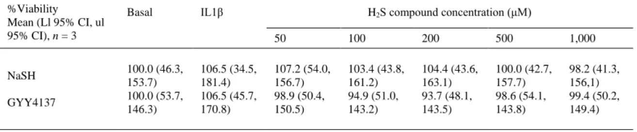
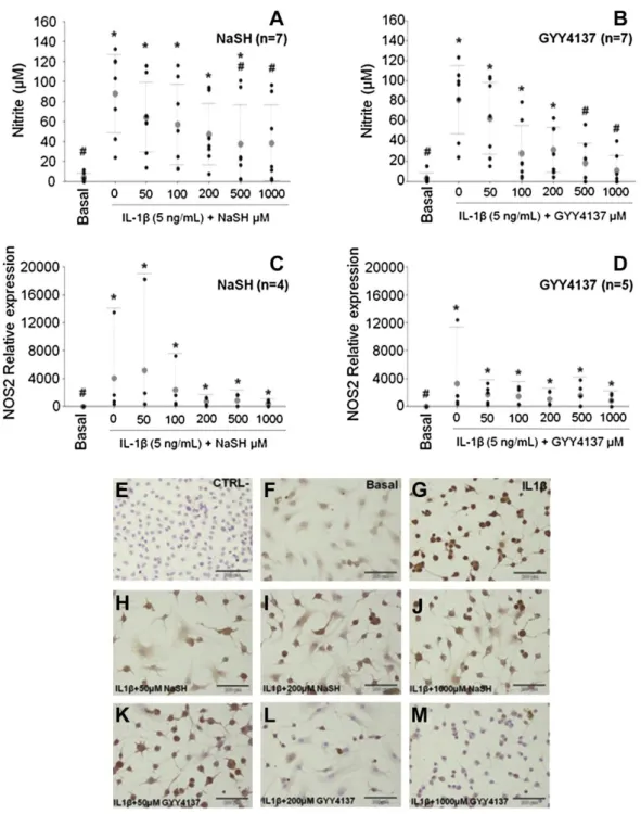
(5) the experiments, results are expressed as mean and 95% lower and upper limit confidence intervals of at least three biological replicates and duplicate technical repeats. Statistical analyses were performed with R software (version 2.15.2) 22. Data were assayed with one-way ANOVA and a Tukey multiple comparisons test. In all cases, a P value lower than 0.05 was considered significant. Results Cell viability Reduction of AlamarBlue® values were normalized with respect to the basal condition which was given an arbitrary value of 100%. None of the concentrations of either NaSH or GYY4137 used reduced the viability of the OA CHs (Table II).. Table II. CH viability, as measured with AlamarBlue reagent. Results are mean and 95% confidence intervals (Lower limit, ll, upper limit ul) of n = 3 independent experiments performed in duplicate. A Kruskal Wallis rank sum test was performed; for NaSH, P = 0.80 and for GYY4137, P = 0.78 %Viability Mean (Ll 95% CI, ul 95% CI), n = 3. NaSH GYY4137. Basal. 100.0 (46.3, 153.7) 100.0 (53.7, 146.3). IL1β. 106.5 (34.5, 181.4) 106.5 (45.7, 170.8). H2S compound concentration (μM) 50. 100. 200. 500. 1,000. 107.2 (54.0, 156.7) 98.9 (50.4, 150.5). 103.4 (43.8, 161.2) 94.9 (51.0, 143.2). 104.4 (43.6, 163.1) 93.7 (48.1, 143.5). 100.0 (42.7, 157.7) 98.6 (54.1, 143.8). 98.2 (41.3, 156,1) 99.4 (50.2, 149.4). Sulfide compounds reduce NO production by downregulating NOS2 gene expression Basal production of NO (measured as nitrite) ranged from around 0.06 μM–12 μM (3.92 (1.64, 6.20), n = 7, performed in duplicate). Stimulation with IL1β (5 ng/ml) increased in production of NO to values that ranged between 23 μM and 133 μM (79.85 (63.70, 96.00), n = 7, performed in duplicate) [ Fig. 1(A) and (B)]. Addition of the different concentrations of H2S compounds did not induce the formation of NO by itself (results not shown). Both NaSH and GYY4137 induced a dose-dependent reduction on NO production in IL1β stimulated cells, with values for 500 μM and 1,000 μM NaSH and GYY4237 being significantly lower [ Fig. 1(A) and (B)]. NOS2 mRNA expression was quantified with qRT-PCR. Stimulation with IL1β induced NOS2 mRNA expression and the addition of H2S-releasing agents dose-dependently attenuated this increase, however, none of the values were significantly different from the IL1β value. For both NaSH and GYY4137, 1,000 μM was the most effective concentration in reducing NOS2 mRNA expression [Fig. 1(C) and (D)]. Results obtained through the Griess reaction and those of qRT-PCR were corroborated also by ICC [Fig. 1(E)–(M)]. In addition, to ensure that these results were due to the H2S released from NaSH and GYY4137, we performed these same experiments with decomposed NaSH and GYY4137 (stock solutions were left uncapped in a chemical hood for 10 days). In this case, all effects were lost, no reduction was observed in NO production or NOS2 relative expression from the IL1βstimulated values (results not shown)..
(6) Fig. 1. Addition of NaSH (A, C) and GYY4137 (B, D) to IL1β-stimulated CHs reduced nitrite accumulation, as measured through the Griess reaction (A and B), NOS2 gene expression, by qRT-PCR (C and D) and NOS2 protein accumulation, by ICC (E–M). Values are mean and 95% CI (Lower limit, upper limit, n number in parenthesis, performed in duplicate). In A and B, Kruskal– Wallis rank sum test was performed followed by multiple comparisons. In C and D, one way ANOVA was performed followed by a Tukey post-hoc multiple comparisons test. *P < 0.0001 (cf. Basal group); #P < 0.0001 (cf. IL1β stimulated group), except for C where P = 0.0001. ICC images include IL1β-stimulated CHs (G) treated with different concentrations of NaSH (H–J) and GYY4137 (K–M). A negative ICC control (without primary antibody (E)), as well as unstimulated cells (F), were included for comparison.. H2S compounds reduce PGE-2 levels by downregulating COX2 and PTGES enzymes PGE-2 basal level was 0.25 (0.13, 0.36) ng/ml (n = 3) in the cells. Stimulation with IL1β led to an increase up to 64.7 (41.78, 87.66) ng/ml PGE-2. Different concentrations of NaSH and GYY4137 were added to the IL1β-stimulated cells to investigate their effect on PGE-2 production [ Fig. 2(A) and (B)]. All concentrations tested, except for 1,000 μM NaSH and 50 μM GYY4137, resulted in a significant decrease from the IL1β-stimulated cells. When looking at the mRNA expression of the main enzymes involved in PGE-2 synthesis, we found a notable dose-dependent reduction in COX2 mRNA expression [.
(7) Fig. 2(C) and (D)] from the IL1β-induced increase. In particular, the IL1β-induced value was 97.5 (19.5, 487,9) fold vs Basal and 1,000 μM NaSH and GYY4137 reduced this value to 12.0 (1.6, 89.13) and 7.6 (0.4, 142,1), respectively, n = 4. Interleukin-1β also induced an elevation of PTGES mRNA expression (68.5 (19.8, 236.7) vs Basal) and 1,000 μM NaSH and GYY4137 considerably reduced this value to 16.3 (3.8, 69.2) and 31.7 (8.3, 120.7), respectively, n = 4 [ Fig. 2(E) and (F)].. Fig. 2. PGE E−2 production in IL1β-stimulated CHs declined after treatment with different concentrations of NaSH (A) and GYY4137 (B). Concurrently, COX2 (C, D) and PTGES (E–F) mRNA expression in the cells was also reduced due to the addition of NaSH (C, E) and GYY4137 (D, F). PGE-2 values, measured with a specific EIA are mean and 95% CI (Lower limit, upper limit, n = 3). Relative mRNA expression values were calculated, with respect to the Basal condition with qBase + software, n indicated in parenthesis and performed in duplicate. One way ANOVA and post-hoc multiple comparisons tests were performed to identify significant differences. In A–C, *P < 0.001 (cf. Basal group) and #P < 0.001 (cf. IL1β stimulated group); in D, *P = 0.003 (cf. Basal group) and #P < 0.001 (cf. IL1β stimulated group) and in E–F, *P < 0.0001 (cf. Basal group) and #P < 0.0001 (cf. IL1β stimulated group)..
(8) H2S compounds also reduce IL6 levels and mRNA expression IL6 levels were increased due to stimulation with IL1β (184.3 (146.6, 222.1) ng/ml vs 106 (36, 176) pg/ml in the unstimulated cells). Measured levels decreased in cells co-stimulated with IL1β and NaSH or GYY4137. The best results were obtained for concentrations 1,000 μM NaSH (86.2 (65.2, 151.5) ng/ml) and 1,000 μM GYY4137 (89.1 (57.13, 121.1) ng/ml) [Fig. 3(A) and (B)]. Similar results were obtained for the mRNA expression levels; IL6 expression was also induced by IL1β (7,743 (3,250, 18,447)) and co-stimulation with the H2S sources reduced it in a dose-dependent manner. In this case, both 1,000 μM NaSH and GYY4137 were the best concentrations and their values were significant reductions from the IL1β value (616.0 (33.2, 11,430) and 543.3 (34.6, 8,540) vs Basal, respectively) [Fig. 3(C) and (D)].. Fig. 3. Interleukin-6 levels (measured with a specific ELISA, A-B) and mRNA expression (by qRT-PCR, C, D) were both reduced in IL1β-stimulated CHs after treatment with different concentrations of NaSH (A, C) and GYY4137 (B, D). For the ELISA experiments, values are mean and 95% CI (Lower limit, upper limit, n = 3, with duplicate measurement). For the qRT-PCR, values were calculated with qBase + software, n = 4 with duplicate technical repeats. One way ANOVA was performed followed by a posthoc multiple comparisons tests, except in B where a Kruskal–Wallis rank sum test followed by multiple comparisons was performed. For A, C and D *P < 0.0001 (cf. Basal group) and #P < 0.0001 (cf. IL1β stimulated group); for B *P < 0.001 (cf. Basal group) and #P < 0.001 (cf. IL1β stimulated group).. H2S compounds show anti-catabolic properties We also looked at the effects of H2S compounds on MMP-13 production. Interleukin-1β increased MMP13 levels to 44.1 (26.7, 61.6) ng/ml (Basal level was 556 (438, 674) pg/ml, n = 3, performed in duplicate) that were subsequently reduced to 33.5 (22.1, 44.9) ng/ml for 1,000 μM NaSH and 17.3 (9.9, 24.6) ng/ml for 1,000 μM for GYY3147 [ Fig. 4]. Also due to the IL1β addition, MMP13 mRNA expression increased up to 48.4 (17.7, 132.2) (fold vs Basal) [ Fig. 4(C) and (D)]. For both NaSH and GYY4137, 1,000 μM was the most efficacious concentration in reducing MMP13 expression (down to 12.2 (3.6, 42.0)) for NaSH, and 8.2 (1.68, 40.1) for GYY4137 [ Fig. 4(C) and (D)]. Also at the protein level in ICC, MMP13 levels were clearly decreased by co-stimulation with both H2S-sources [ Fig. 4(E)– (M)]..
(9) Fig. 4. Addition of NaSH (A, C) and GYY4137 (B, D) to IL1β stimulated CHs resulted in reduced MMP13 production, measured with a specific ELISA (A and B), reduced MMP13 gene expression by qRT-PCR (C and D), and in less MMP13 protein accumulation, by ICC (E–M). Values are mean and 95% CI (Lower limit, upper limit, n = 3, with duplicates). One way ANOVA and a post-hoc multiple comparisons test were performed to identify significant differences, except in A where a Kruskal–Wallis rank sum test followed by multiple comparisons was done. ICC images include IL1β-stimulated CHs (G) treated with different concentrations of NaSH (H–J) and GYY4137 (K–M). A negative ICC control (without primary antibody (E)), as well as unstimulated cells (F), was included for comparison. For B–D *P < 0.0001 (cf. Basal group) and #P < 0.0001 (cf. IL1β stimulated group), and for A *P < 0.00467 (cf. Basal group) and #P < 0.00467 (cf. IL1β stimulated group)..
(10) H2S compounds reduce NFκB p65 translocation to the nucleus In an attempt to elucidate the possible mechanism by which H2S might be exerting the observed effects, an immunofluorescence assay was performed to visualize NFκB p65 nuclear translocation in the cells. Results are included in Fig. 5. In the basal condition, p65 was located mostly in the cytoplasms of the cells [Fig. 5(A)]. Stimulation with IL1β resulted in the translocation of p65 to the nuclei of the cells, as can be clearly seen in Fig. 5(B). Co-stimulation with the different concentrations of H2S compounds resulted in a reduction in the fluorescence intensity in the nuclei [Fig. 5(C)–(G), for GYY4137 and Fig. 5(H)–(L) for NaSH], although p65 could still be seen in the nuclei and the basal condition was not recovered.. Fig. 5. Immunofluorescence images of NFκB p65 protein in IL1β stimulated CHs (45 min). In the basal condition (no stimulation) p65 can be seen mostly in the cells cytoplasms. IL1β stimulation lead to p65 translocation to the nuclei and the different concentrations of GYY4137 (C–G) and NaSH (H–L) resulted in a slight reduction of the nuclear fluorescence intensity, although p65 can still be seen in the nuclei and the basal situation was not fully recovered.. Discussion This paper evaluates the ability of two exogenous H2S sources as anti–inflammatory and anti–catabolic agents in human articular CHs. Interleukin-1β was used as prototype pro-inflammatory cytokine to reproduce the “OA-like effect” in the cells. As expected5 and 6, the stimulation of OA CHs with IL1β induced the production of markers of inflammation and catabolic activity such as NO, PGE-2, IL6 and MMP13. NO is a multifunctional molecule that mediates various biological processes and is a well-known mediator in OA. It has been reported that OA CHs die by NO-induced apoptosis21, that OA CHs produce NO spontaneously23 and that human articular CHs express NOS2 when stimulated in culture24. Later studies reported that the most direct effect of NO in CHs seemed to be the suppression of energy metabolism25, but it has also been suggested that NO can inhibit matrix synthesis by CHs26. Our results show a dose-dependent reduction of NO levels and the inducible synthesis enzyme, NOS2, with both NaSH and GYY4137. Our GYY4137 results are in line with those recently published for lipopolysaccharide (LPS)-activated hCHs18 treated with this compound, but those of NaSH contradict a recent publication27 in which its use led to an increase in NO and NOS2 in rat vascular smooth muscle cells. Also, another study described that NaSH had a biphasic effect and at high concentrations (>200 μm) increased the synthesis of NO in a macrophage cell line28. Some authors reported that H2S can react directly with ROS and reactive nitrogen species (RNS)12, 29 and 30 which might account for part of the reduction they observed in NO, but in our case the NO decline is accompanied by a downregulation of the.
(11) NOS2 gene and a reduction in the NOS2 protein. Therefore the reduction in NO cannot only be the result of a redox reaction and our results in the present in vitro model suggest that the H2S compounds can directly regulate the NOS2 gene. The role of H2S in inflammatory signaling has not been elucidated yet. Many reports attribute H2S both pro- and anti-inflammatory properties depending on the pathology studied, the experimental model used or the source of H2S13 and 31. The effects of NaSH and GYY4137 on the release of pro- and anti-inflammatory mediators in LPStreated murine RAW264.7 macrophages were recently investigated28. GYY4137 inhibited LPS-induced release of pro-inflammatory mediators IL1β, IL6, TNF-α, and PGE-2 by increasing the anti-inflammatory chemokine IL-10. In contrast, NaSH had a biphasic effect and at high concentrations (>200 μm) increased the synthesis of IL1β, IL6, TNF-α, and PGE-2. On the other hand, reductions in PGE-2 and COX2 levels were also found in LPS–activated normal human fibroblast-like synoviocytes (hFLS) and hCHs18. Our results show a dose-dependent reduction in COX2 for both H2S sources. COX2 is considered the “inducible” COX isoform, activated by many factors, and current anti-inflammatory therapy for OA makes use of COX2 inhibitors. These unfortunately, have other disadvantages, including an accompanying inhibition of physiologic COX132. In the present results, COX2 inhibition was achieved at the highest H2S-compound concentration used, without significantly affecting COX1 (results not shown). In addition, our results also show a reduction of PTGES gene expression due to the H2S co-stimulation. PTGES was demonstrated as a key enzyme in the regulation of PGE-2 production in cytokine stimulated CHs from patients with OA9, suggesting that the terminal PG synthase downstream of COX is important for deciding which PG is synthesized, and that the induction of PTGES might be necessary for substantial PGE-2 production by CHs in response to pro-inflammatory stimuli. Therefore, the H2S-sources can regulate the two most important enzymes implicated in PGE-2 production in activated hCHs. Furthermore, in our model, IL6 production was stimulated by IL1β and the co-stimulation with H2Sreleasing compounds both downregulated IL6 gene mRNA expression and reduced IL6 levels produced by the cells. Two different studies have recently showed that NaSH could transiently block IL6 expression, first in RA fibroblast-like synoviocytes33 and later in a CH cell line C-28/I234. IL6 can show both pro-inflammatory and anti-inflammatory effects (since, by itself, it can induce the production of tissue inhibitor of metalloproteinase, TIMP, but not MMPs) but IL1-induced IL6 is required for the inhibition of proteoglycan synthesis by IL1 in human articular cartilage35; also IL6 is synergistically responsible for IL1–induced MMPs increases6 and IL6 or its receptor are therapeutic targets for OA36. Therefore, the H2S-sources as used here can further reduce inflammation by regulating IL6 gene expression, resulting in lower free IL6 cytokine released by the cells. Collagenase 3 (MMP13) is the major type II collagen-degrading collagenase. It is regulated by stress-, inflammation- and differentiation-induced signals. Work by Goldring and colleagues recently reviewed10 has singled out MMP13 as the key gene upon which many of the signaling pathways (i.e., NFκB, JNK or p38 MAPK) and transcription factors (such as NFκB, HIF2α, Ihh, or Runx2) involved at the different stages of OA converge. Most of these regulatory factors directly or indirectly, impact on MMP13 transcription and activity. In our model, 1,000 μM of the two H2S-compounds used, reduced MMP13 below the IL1β-induced levels at the mRNA and at the protein level also. There are, to the best of our knowledge, no other reports available on the effects of NaSH or GYY4137 on MMP13 activity on human osteoarthritic CHs. However, it has been reported that NaSH (20 min, 1 mM) upregulated the expression of the MMP3 gene in RA-FLS, while MMP3 was not detected at the mRNA level in the OA-FLS37. On the other hand, DAS, an oil-soluble organosulfur compound extracted from garlic, reduced MMP13 synthesis in IL1β-activated rabbit CHs38. Additionally, the H2S compounds used in the present model seem to reduce the translocation of NFκB to the CHs nuclei, although not to prevent it completely. Activation of NFκB is directly responsible for the upregulation of several proinflammatory cytokines and enzymes, including NOS2, COX2 and IL618 and 19, so it is a plausible mechanism of action for NaSH and GYY4137. Nevertheless further studies are needed to confirm H2S implication in the NFκB signaling pathway and to investigate what other signaling routes might be implicated. Collectively, our results show that supplementation with exogenous H2S taps on important markers in OA and indicate that OA cartilage might benefit from the exogenous supplementation of H 2S. However, the current results obtained in vitro might not translate to an in vivo situation, and this should be tested directly. Nevertheless, current data should grant further study into the implication of H 2S, both exogenously added and/or endogenously controlled, in OA as a potential therapeutic target..
(12) Author contributions All authors were involved in drafting or critically reading the manuscript for important intellectual content, and all authors approved the final version. Conception and design: EF Burguera, R MeijideFailde, FJ Blanco. Data acquisition: Á Vela-Anero, EF Burguera. Analysis and interpretation of data: Á Vela-Anero, J Magalhaes, FJ Blanco. Obtaining of funding: R Meijide-Failde, FJ Blanco. Competing interest statement FJB has received Grants (Clinical Trials, conferences, advisor and publications) from: Abbvie, Amgen, Bioiberica, Bristol Mayer, Celgene, Celltrion, Cellerix, Grunenthal, Gebro Pharma, Lilly, MSD, Merck Serono, Pfizer, Pierre-Fabra, Roche, Sanofi, Servier, UCB. EFB, AVA, JM and RMF declare they do not have any conflict of interest. No competing financial interests exist. Funding sources This work was supported in part through funding from the Fondo Investigación Sanitaria, Madrid, Spain (CIBER-CB06/01/0040; PI12/00329; RETIC-RIER-RD12/0009/0018; and Proteo-Red/ISCIII); Ministerio Ciencia e InnovaciónPLE2009-0144, FEDER (European Community) and Xunta de Galicia: grant 10 PXIB 310153 PR and Red Gallega REDICENT. The funders did not contribute to data collection, analysis or interpretation of the data, manuscript preparation or submission. Acknowledgments Authors want to express their gratitude to the patients that made possible this study and to the Rheumatology and Orthopedic Services of the University Hospital A Coruña (CHUAC) for their help in obtaining the cartilage samples. EFB was supported by Axencia Galega de Innovación (IPP program) and Ciber-BBN/ISCIII (grant no. CB06/01/0040).. References 1. Y. Zhang, J.M. Jordan. Epidemiology of osteoarthritis. Clin Geriatr Med, 26 (2010), pp. 355–360. 2. L. Sharma, D. Kapoor, S. Issa. Epidemiology of osteoarthritis: an update. Curr Opin Rheumatol, 18 (2006), pp. 147–156. 3. D.T. Felson. The Epidemiology of knee osteoarthritis – results from the Framingham osteoarthritis study. Semin Arthritis Rheum, 20 (1990), pp. 42–50. 4. M.B. Goldring, M. Otero. Inflammation in osteoarthritis. Curr Opin Rheumatol, 23 (2011), pp. 471–478. 5. M. Kobayashi, G.R. Squires, A. Mousa, M. Tanzer, D.J. Zukor, J. Antoniou, et al.. Role of interleukin-1 and tumor necrosis factor a in matrix degradation of human osteoarthritic cartilage. Arthritis Rheum, 52 (2005), pp. 128– 135. 6. J.C. Fernandes, J. Martel-Pelletier, J.P. Pelletier. The role of cytokines in osteoarthritis pathophysiology. Biorheology, 39 (2002), pp. 237–246. 7. W. Droge. Free radicals in the physiological control of cell function. Physiol Rev, 82 (2002), pp. 47–95. 8. A.R. Butler, I.L. Megson, P.G. Wright. Diffusion of nitric oxide and scavenging by blood in the vasculature. Biochim Biophys Acta, 1425 (1998), pp. 168–176. 9. F. Kojima, H. Naraba, S. Miyamoto, M. Beppu, H. Aoki, S. Kawai. Membrane-associated prostaglandin E synthase-1 is upregulated by proinflammatory cytokines in chondrocytes from patients with osteoarthritis. Arthritis Res Ther, 6 (2004), pp. R355–R365. 10. M.B. Goldring, M. Otero, D.A. Plumb, C. Dragomir, M. Favero, K. El Hachem, et al.. Roles of inflammatory and anabolic cytokines in cartilage metabolism: signals and multiple effectors converge upon Mmp-13 regulation in osteoarthritis. Eur Cell Mater, 21 (2011), pp. 202–220. 11. R.O. Beauchamp, J.S. Bus, J.A. Popp, C.J. Boreiko, D.A. Andjelkovich. A critical-review of the literature on hydrogen-sulfide toxicity. Crit Rev Toxicol, 13 (1984), pp. 25–97. 12. C. Szabo. Hydrogen sulphide and its therapeutic potential. Nat Rev Drug Discov, 6 (2007), pp. 917–935. 13. M. Whiteman, P.G. Winyard. Hydrogen sulfide and inflammation: the good, the bad, the ugly and the promising. Expert Rev Clin Pharmacol, 4 (2011), pp. 13–32. 14. K.R. Olson. Is hydrogen sulfide a circulating “gasotransmitter” in vertebrate blood?. Biochim Biophys Acta, 1787 (2009), pp. 856–863. 15. R. Wang. Two's company, three's a crowd: can H2S be the third endogenous gaseous transmitter?. FASEB J, 16 (2002), pp. 1792–1798..
(13) 16. M. Whiteman, R. Haigh, J.M. Tarr, K.M. Gooding, A.C. Shore, P.G. Winyard. Detection of hydrogen sulfide in plasma and knee-joint synovial fluid from rheumatoid arthritis patients: relation to clinical and laboratory measures of inflammation. Ann N Y Acad Sci, 1203 (2010), pp. 146–150. 17. B. Fox, J.T. Schantz, R. Haigh, M.E. Wood, P.K. Moore, N. Viner, et al.. Inducible hydrogen sulfide synthesis in chondrocytes and mesenchymal progenitor cells: is H2S a novel cytoprotective mediator in the inflamed joint?. J Cell Mol Med, 16 (2012), pp. 896–910. 18. L. Li, B. Fox, J. Keeble, M. Salto-Tellez, P.G. Winyard, M.E. Wood, et al.. The complex effects of the slowreleasing hydrogen sulfide donor GYY4137 in a model of acute joint inflammation and in human cartilage cells. J Cell Mol Med, 17 (2013), pp. 365–376. 19. R. Simmonds, B. Foxwell. Signalling, inflammation and arthritis – NF-kappa B and its relevance to arthritis and inflammation. Rheumatology, 47 (2008), pp. 584–590. 20. C. Ruiz-Romero, M.J. Lopez-Armada, F.J. Blanco. Proteomic characterization of human normal articular chondrocytes: a novel tool for the study of osteoarthritis and other rheumatic diseases. Proteomics, 5 (2005), pp. 3048–3059. 21. F.J. Blanco, R.L. Ochs, H. Schwarz, M. Lotz. Chondrocyte apoptosis induced by nitric-oxide. Am J Pathol, 146 (1995), pp. 75–85. 22. R Core Team. R: A Language and Environment for Statistical Computing. Vienna, Austria: R Foundation for Statistical Computing. URL http://www R-project org/2012. 23. A.R. Amin, M. Attur, R.N. Patel, G.D. Thakker, P.J. Marshall, J. Rediske, et al.. Superinduction of cyclooxygenase-2 activity in human osteoarthritis-affected cartilage – influence of nitric oxide. J Clin Invest, 99 (1997), pp. 1231–1237. 24. I.G. Charles, R.M.J. Palmer, M.S. Hickery, M.T. Bayliss, A.P. Chubb, V.S. Hall, et al.. Cloning, characterization, and expression of a CDNA-encoding an inducible nitric-oxide synthase from the human chondrocyte. Proc Natl Acad Sci U S A, 90 (1993), pp. 11419–11423. 25. E. Maneiro, M.A. Martin, M.C. de Andres, M.J. Lopez-Armada, J.L. Fernandez-Sueiro, P. del Hoyo, et al.. Mitochondrial respiratory activity is altered in osteoarthritic human articular chondrocytes. Arthritis Rheum, 48 (2003), pp. 700–708. 26. R. Studer, D. Jaffurs, M. Stefanovic-Racic, P.D. Robbins, C.H. Evans. Nitric oxide in osteoarthritis. Osteoarthritis Cartilage, 7 (1999), pp. 377–379. 27. S.O. Jeong, H.O. Pae, Gs Oh, G.S. Jeong, B.S. Lee, S. Lee, et al.. Hydrogen sulfide potentiates interleukin-1 betainduced nitric oxide production via enhancement of extracellular signal-regulated kinase activation in rat vascular smooth muscle cells. Biochem Biophys Res Commun, 345 (2006), pp. 938–944. 28. M. Whiteman, L. Li, P. Rose, C.H. Tan, D.B. Parkinson, P.K. Moore. The effect of hydrogen sulfide donors on lipopolysaccharide-induced formation of inflammatory mediators in macrophages. Antioxid Redox Signal, 12 (2010), pp. 1147–1154. 29. M. Whiteman, L. Li, I. Kostetski, S.H. Chu, J.L. Siau, M. Bhatia, et al.. Evidence for the formation of a novel nitrosothiol from the gaseous mediators nitric oxide and hydrogen sulphide. Biochem Biophys Res Commun, 343 (2006), pp. 303–310. 30. M. Whiteman, J.S. Armstrong, S.H. Chu, S. Jia-Ling, B.S. Wong, N.S. Cheung, et al.. The novel neuromodulator hydrogen sulfide: an endogenous peroxynitrite ‘scavenger’?. J Neurochem, 90 (2004), pp. 765–768. 31. J.R. Rivers, A. Badiei, M. Bhatia. Hydrogen sulfide as a therapeutic target for inflammation. Expert Opin Ther Targets, 16 (2012), pp. 439–449. 32. F.J. Blanco, R. Guitian, J. Moreno, F.J. De Toro, F. Galdo. Effect of antiinflammatory drugs on COX-1 and COX-2 activity in human articular chondrocytes. J Rheumatol, 26 (1999), pp. 1366–1373. 33. B. Kloesch, M. Liszt, J. Broell. H2S transiently blocks IL-6 expression in rheumatoid arthritic fibroblast-like synoviocytes and deactivates p44/42 mitogen-activated protein kinase. Cell Biol Int, 34 (2010), pp. 477–484. 34. B. Kloesch, M. Liszt, G. Steiner, J. Broell. Inhibitors of p38 and ERK1/2 MAPkinase and hydrogen sulphide block constitutive and IL-1 beta-induced IL-6 and IL-8 expression in the human chondrocyte cell line C-28/I2. Rheumatol Int, 32 (2012), pp. 729–736. 35. J.J. Nietfeld, B. Wilbrink, M. Helle, J.L.A.M. Vanroy, W. Denotter, A.J.G. Swaak, et al.. Interleukin-1-Induced Interleukin-6 is required for the inhibition of proteoglycan synthesis by Interleukin-1 in human articular-cartilage. Arthritis Rheum, 33 (1990), pp. 1695–1701. 36. H. Nakahara, N. Nishimoto. Anti-interleukin-6 receptor antibody therapy in rheumatic diseases. Endocr Metab Immune Disord Drug Targets, 6 (2006), pp. 373–381. 37. B. Kloesch, M. Liszt, D. Krehan, J. Broell, H. Kiener, G. Steiner. High concentrations of hydrogen sulphide elevate the expression of a series of pro-inflammatory genes in fibroblast-like synoviocytes derived from rheumatoid and osteoarthritis patients. Immunol Lett, 141 (2012), pp. 197–203. 38. W.P. Chen, J.L. Tang, J.P. Bao, P.F. Hu, C. Yu, Z.L. Shi, et al.. Effects of Diallyl sulphide in chondrocyte and cartilage in experimental osteoarthritis in rabbit. Phytother Res, 25 (2011), pp. 351–356..
(14)
Figure


+4


Documento similar
The potential displacement of reduction process at less negative values in the voltammograms and the changes in cathodic charge when iridium is present in the solution at
Levels of transcripts corresponding to components of the complement system C3 and Complement Factor D (CFD) showed reduced expression in adipose tissue from the patient with respect
14.- Considera que es necesario que Ud.. 12.-Es sabido que la red también ofrece desventajas y peligros. ¿Ha participado en algún taller formativo acerca del uso de Internet
7.- En el manejo o dominio de las utilidades del correo-web (Enviar y redactar correos, crear listas, adjuntar archivos, etc.) considero que mi nivel es…. A) Bajo (0) B)
Effect of cactus seed oil, olive oil, or colza oil treatment on gene expression of the proin- flammatory markers Il-1β (A,D), iNos (B,E), and Il-6 (C,F), in the brain and
In addition, a signif- icant improvement of tubulointerstitial damage was indicated by reduced gene expression of kidney injury molecule-1 28 in diabetic gapoE compared with apoE
Cytocompatibility of biomimetic magnetic nanoparticles mediated by magnetosome proteins (BMNPs) on 4T1 (A,C) and MCF-7 (B,D) cells in the absence/presence of a gradient magnetic
We used morphological, histological and immunological analysis together with a RT-PCR detection of collagen I and collagen II gene expression to show that chondrocytes isolated
