Current topics in tropical medicine
576
0
0
Texto completo
(2) Current Topics in Tropical Medicine Edited by Alfonso J. Rodriguez-Morales. Published by InTech Janeza Trdine 9, 51000 Rijeka, Croatia Copyright © 2012 InTech All chapters are Open Access distributed under the Creative Commons Attribution 3.0 license, which allows users to download, copy and build upon published articles even for commercial purposes, as long as the author and publisher are properly credited, which ensures maximum dissemination and a wider impact of our publications. After this work has been published by InTech, authors have the right to republish it, in whole or part, in any publication of which they are the author, and to make other personal use of the work. Any republication, referencing or personal use of the work must explicitly identify the original source. As for readers, this license allows users to download, copy and build upon published chapters even for commercial purposes, as long as the author and publisher are properly credited, which ensures maximum dissemination and a wider impact of our publications. Notice Statements and opinions expressed in the chapters are these of the individual contributors and not necessarily those of the editors or publisher. No responsibility is accepted for the accuracy of information contained in the published chapters. The publisher assumes no responsibility for any damage or injury to persons or property arising out of the use of any materials, instructions, methods or ideas contained in the book. Publishing Process Manager Vedran Greblo Technical Editor Teodora Smiljanic Cover Designer InTech Design Team First published March, 2012 Printed in Croatia A free online edition of this book is available at www.intechopen.com Additional hard copies can be obtained from orders@intechweb.org. Current Topics in Tropical Medicine, Edited by Alfonso J. Rodriguez-Morales p. cm. ISBN 978-953-51-0274-8.
(3)
(4)
(5) Contents Preface IX Part 1. Tropical Diseases Due to Bacteria and Viruses. 1. Chapter 1. Rickettsiosis as Threat for the Traveller Aránzazu Portillo and José A. Oteo. 3. Chapter 2. Human Ehrlichioses and Rickettsioses in Cameroon Lucy Ndip, Roland Ndip, David Walker and Jere McBride. Chapter 3. Leptospirosis: Epidemiologic Factors, Pathophysiological and Immunopathogenic Marcia Marinho. 25. 43. Chapter 4. Bartonella Infections in Rodents and Bats in Tropics Ying Bai and Michael Kosoy. Chapter 5. Social Networking in Tuberculosis: Experience in Colombia 67 Diana M. Castañeda-Hernández and Alfonso J. Rodriguez-Morales. Chapter 6. Molecular Characterization of Dengue Virus Circulating in Manaus, the Capital City of the State of Amazonas, Brazil 81 Regina Maria Pinto de Figueiredo. Chapter 7. Genetic Diversity of Dengue Virus and Associated Clinical Severity During Periodic Epidemics in South East Asia 91 E. Khan, R. Hasan, J. Mehraj and S. Mahmood. Chapter 8. Lassa Fever in the Tropics Ute Inegbenebor. Chapter 9. The Re-Emergence of an Old Disease: Chikungunya Fever 117 Bordi Licia, Meschi Silvia, Selleri Marina, Lalle Eleonora, Castilletti Concetta, Carletti Fabrizio, Di Caro Antonino and Capobianchi Maria Rosaria. 109. 51.
(6) VI. Contents. Part 2. Tropical Diseases Due to Protozoa and Helminths. 137. Chapter 10. Malaria Chemoprophylaxis for the International Traveler, Current Options and Future Possibilities 139 Ross Parker and Kevin Leary. Chapter 11. Effects of Irrigated Rice Fields and Seasonality on Plasmodium Transmission in West Africa, Particularly in Central Côte d’Ivoire 155 Benjamin G. Koudou, Marcel Tanner and Juerg Utzinger. Chapter 12. Toxoplasmosis: Advances and Vaccine Perspectives Oscar Bruna-Romero, Dulcilene Mayrink de Oliveira and Valter Ferreira de Andrade-Neto. Chapter 13. Screening of the Prevalence of Antibodies to the Tick Hyalomma lusitanicum in a Province of Northern Spain 185 Consuelo Giménez Pardo and Lourdes Lledó García. Chapter 14. Amoebiasis in the Tropics: Epidemiology and Pathogenesis 201 A. Samie, A. ElBakri and Ra’ed AbuOdeh. Chapter 15. Retrospective Analysis of Leishmaniasis in Central Tunisia: An Update on Emerging Epidemiological Trends 227 Akila Fathallah Mili, Fatma Saghrouni, Zeineb BenSaid, Yusr Saadi- BenAoun, Ikram Guizani and Moncef BenSaid. Chapter 16. Current Advances in Computational Strategies for Drug Discovery in Leishmaniasis 253 Andrés F. Flórez, Stanley Watowich and Carlos Muskus. Chapter 17. Advances in Serological Diagnosis of Chagas’ Disease by Using Recombinant Proteins Iván S. Marcipar and Claudia M. Lagier. Chapter 18. Echinococcosis/Hydatidosis 299 Antoni Soriano Arandes and Frederic Gómez Bertomeu. Chapter 19. A Programme to Control Taeniosis-Cysticercolsis (Taenia solium) in Mexico 323 Aline S. de Aluja, Julio Morales Soto and Edda Sciutto. Chapter 20. Antischistosomal Natural Compounds: Present Challenges for New Drug Screens Josué de Moraes. Chapter 21. 333. Control of Schistosomiasis and Soil-Transmitted Helminthiasis in Sub-Saharan Africa: Challenges and Prospects 359 Louis-Albert Tchuem Tchuenté. 169. 273.
(7) Contents. Chapter 22. Hyperinfection Syndrome in Strongyloidiasis 377 Cristiane Tefé-Silva, Eleuza R. Machado, Lúcia H. Faccioli and Simone G. Ramos. Chapter 23. Molecular Diagnosis and Monitoring of Benzimidazole Susceptibility of Human Filariids 397 Adisak Bhumiratana, Apiradee Intarapuk, Danai Sangthong, Surachart Koyadun, Prapassorn Pechgit and Jinrapa Pothikasikorn. Chapter 24. Lymphatic Filariasis Transmission and Control: A Mathematical Modelling Approach 425 Asep K. Supriatna and N. Anggriani. Part 3. Other Tropical Infectious and Non-Infectious Conditions 443. Chapter 25. Novel Molecular Diagnostic Platform for Tropical Infectious Diseases 445 Yasuyoshi Mori, Norihiro Tomita, Hidetoshi Kanda and Tsugunori Notomi. Chapter 26. Sexually Transmitted Infections in the Tropics John C. Meade and Denise C. Cornelius. Chapter 27. Re-Emergence of Malaria and Dengue in Europe Rubén Bueno Marí and Ricardo Jiménez Peydró. 483. Chapter 28. Neonatal Thermoneutrality in a Tropical Climate Hippolite O. Amadi. 513. Chapter 29. Associations Between Nutritional Indicators Using Geoadditive Latent Variable Models with Application to Child Malnutrition in Nigeria 545 Khaled Khatab. 457. VII.
(8)
(9) Preface Tropical medicine research holds a special place as an important activity that as a consequence of multiple factors, such as globalization and migration has extended and reaffirms its importance not only in tropical developing countries but also in nonendemic areas in the developed world. The update on different aspects related to the practice of tropical medicine and their multiple components needs to be frequently visited. Three of the most important infectious terminal diseases in the world that belong or significantly affect tropical areas are AIDS, Tuberculosis and Malaria. These pathologies, together with other important ones, represent relevant public health problems, particularly in Africa, Asia and Latin America (Franco-Paredes et al. 2007a, Franco-Paredes et al. 2007b, Rodríguez Morales AJ et al. 2006, Rodríguez Morales AJ et al. 2008), secondarily affecting, due to travel, Europe, North America and other areas of the world (Franco-Paredes et al. 2007c). Diseases and conditions as object of the study of tropical medicine are diverse in organ compromise as well as in etiology, including infectious and non-infectious agents. With these concepts in mind, this book includes different topics of tropical medicine of current international interest, trying to update the most significant research in many of them as well as offer a multinational perspective on different relevant conditions. This book has been organized in three major sections: I. Tropical Diseases due to Bacteria and Viruses; II. Tropical Diseases due to Protozoa and Helminths; and III. Other Tropical Infectious and Non-Infectious Conditions. Section I includes topics covering bacterial diseases such as rickettsiosis, ehrlichiosis, leptospirosis, bartonellosis and tuberculosis; as well on viral diseases such as dengue, Lassa fever and Chikungunya. Section II includes topics covering protozoan diseases such as malaria, toxoplasmosis, amebiasis, leishmaniasis and Chagas disease; as well on helminthic diseases such as echinococcosis/hidatidosis, taeniosis/cysticercosis, schistosomiasis, filariasis, strongyloidiasis and soil-transmitted helminths. Section III includes topics on multiple-etiology conditions such as sexually transmitted diseases, new diagnostic tools for tropical diseases and vector-borne diseases; also includes non-infectious conditions particularly related to childhood health in the tropics. This books does not intend to be an exhaustive compilation and this first edition has included not just multiple different topics but also a wide geographical participation from many countries where tropical medicine is of interest. Its online availability allows it to reach a worldwide audience..
(10) X. Preface. I would like to give my thanks to InTech, and particularly to Mr. Vedran Greblo, for the opportunity to edit this interesting and important book. I want to dedicate this book to my family and particularly to my lovely wife, Diana, who actually represents my engine for every activity I made in my professional career up until now, also to my friends and my students around Latin America. We hope our readers enjoy this publication as much as I did reading the chapters of Current Topics in Tropical Medicine. References Franco-Paredes C, Jones D, Rodriguez-Morales AJ, Santos-Preciado JI. 2007a. Commentary: improving the health of neglected populations in Latin America. BMC Public Health 7:11. Franco-Paredes C, Von A, Hidron A, Rodriguez-Morales AJ, Tellez I et al. 2007b. Chagas disease: an impediment in achieving the Millennium Development Goals in Latin America. BMC Int. Health Hum. Rights 7:7. Franco-Paredes C, Dismukes R, Nicolls D, Workowski K, Rodriguez-Morales A, Wilson M, Jones D, Manyang P, Kozarsky P. 2007c. Persistent and Untreated Tropical Infectious Diseases among Sudanese Refugees in the U.S. Am J Trop Med & Hyg 77: 633-635. Rodríguez-Morales AJ, Barbella RA, Case C, Arria M, Ravelo M, Perez H, Urdaneta O, Gervasio G, Rubio N, Maldonado A, Aguilera Y, Viloria A, Blanco JJ, Colina M, Hernández E, Araujo E, Cabaniel G, Benitez J, Rifakis P. 2006. Intestinal Parasitic Infections among Pregnant Women in Venezuela. Infect Dis Obstet Gynecol 14:23125. Rodríguez Morales AJ, Lorizio W, Vargas J, Fernández L, Durán B, Husband G, Rondón A, Vargas K, Barbella RA, Dickson SM. 2008. Malaria, Tuberculosis, VIH/SIDA e Influenza Aviar: ¿Asesinos de la Humanidad? Rev Soc Med Quir Hosp Emerg Perez de Leon 39:52-76.. Prof. Alfonso J. Rodriguez-Morales MD, MSc, DTM&H, FRSTMH(Lon), FFTM RCPS(Glasg), PhD(c). Editor, Current Topics in Tropical Medicine Infection and Immunity Research Group, Universidad Tecnológica de Pereira, Pereira, Colombia Immunoparasitology Section, Tropical Medicine Institute, Universidad Central de Venezuela, Caracas, Venezuela Instituto José Witremundo Torrealba, Universidad de Los Andes, Trujillo, Venezuela.
(11)
(12)
(13) Part 1 Tropical Diseases Due to Bacteria and Viruses.
(14)
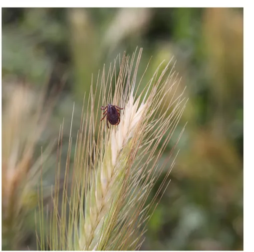
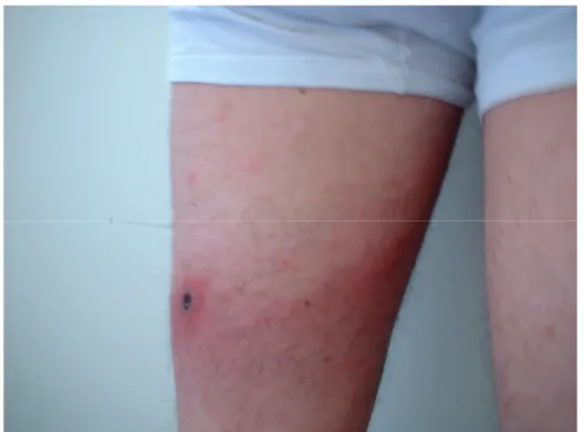
(15) 1 Rickettsiosis as Threat for the Traveller Aránzazu Portillo and José A. Oteo. Hospital San Pedro-Centre of Biomedical Research (CIBIR) Spain 1. Introduction. Over the past six decades, tourism has experienced continued expansion and diversification becoming one of the largest and fastest growing economic sectors in the world. Many new destinations have emerged alongside the traditional ones of Europe and North America. In the next years an increase of travelling is expected, and the number of related infections will also be higher (http://www.unwto.org/facts/menu.html). Rickettsioses are an important chapter in the field of travel medicine. Rickettsiae are small gram-negative intracellular bacteria (belonging to the alpha-1 proteobacteria) mainly transmitted by arthropods (lice, fleas, ticks and other acari) with two genera: Orientia with a unique specie (Orientia tsutsugamushi) and Rickettsia with several species. The clinical pictures that they cause are named rickettsioses (Raoult, 2010a). Rickettsioses have been a threat all along the History and nowadays they are an important cause of morbi-mortality in some areas of the world. To know the distribution of the different diseases caused by these bacteria and how the clinical pictures are recognized may be essential for a quick diagnoses and starting the correct treatment. Some of these infections can be also easily prevented with basic rules. Main rickettsioses with their distribution area are showed in the table 1. In the 21st Century in most parts of the world hygienic conditions have improved and epidemic typhus is absent. To acquire this condition it is necessary to be in contact with body lice. Furthermore, if people have personal hygiene and change their clothing, body lice are removed. Nevertheless it is possible that if we travel for cooperation to catastrophic areas or other places with poverty, we may take body lice (refugees’ camps) and may develop exanthematic typhus. There are a lot of references of rickettsioses acquired by travellers and considered imported diseases (McDonald et al., 1988; Bottieau et al. 2006; Freedman et al., 2006; Askling et al. 2009; Chen & Wilson, 2009; Jensenius et al., 2009; Stokes & Walters, 2009). Nowadays ticks cause most travel-associated rickettsioses. Ticks are considered to be one of the most important vectors of infectious diseases in the world, preceded only by mosquitoes. Therefore, tick-borne rickettsioses are endemic all over the world (Hechemy et al., 2006). The majority of travel-associated rickettsioses refer to Sub-Saharan Africa tourists who develop African tick-bite fever (ATBF), mainly transmitted by Amblyomma hebraeum (Figure 1). In addition to malaria, ATBF is an important cause of fever in people returning from the tropic (Field et al., 2010). Other reports describe Mediterranean spotted fever (MSF) acquired by tourists bitten by Rhipicephalus spp. ticks (Figure 2) when visiting Europe, being.
(16) 4. Current Topics in Tropical Medicine. more scarce references about other rickettsioses. Flea-borne rickettsioses and chiggertransmitted rickettsioses are less frequent in travellers and tourists, and some of them as murine typhus are associated with poor hygienic conditions. Most travel-acquired rickettsioses are related to outdoors leisure activities, like camping, trekking, hunting, safaris, etc. It will be impossible to describe all rickettsioses in few pages. Since rickettsioses have very similar clinical pictures and they can be grouped in different syndromes, we will describe these syndromes emphasizing the typical features (i.e.: Presence of eschar or type of rash). Afterwards, distribution can be observed in the table 1. We will also write a specific paragraph for some infections (i.e.: Diseases caused by Rickettsia akari and Orientia tsutsugamushi).. Fig. 1. Amblyomma hebraeum, the principal vector of African-tick bite fever (ATBF) in southern Africa..
(17) 5. Rickettsiosis as Threat for the Traveller. Fig. 2. Rhipicephalus spp., the main vector of Mediterranean spotted fever (MSF). DISEASE Epidemic typhus. CAUSATIVE AGENT R. prowazekii. Murine typhus. R. typhi. Flea-borne spotted fever. R. felis. VECTOR. DISTRIBUTION. Body lice (Pediculus humanus corporis). Peru, northern Africa, Senegal, Burundi, Rwanda, Russia, sporadic cases in USA associated with flying squirrels. Potentially, all over the world associated to poverty and dirt. All over the world (more prevalent in tropical and subtropical areas) All over the world. Fleas (Xenopsylla cheopis and Ctenocephalides felis) Cat fleas (Ctenocephalides felis).
(18) 6. Current Topics in Tropical Medicine. DISEASE Scrub typhus. Rickettsialpox RMSF1 RMSF-like MSF2. MSF-like MSF-like MSF-like DEBONEL / TIBOLA2 LAR4 ATBF5. CAUSATIVE AGENT Orientia tsutsugamushi. VECTOR. DISTRIBUTION. Thailand, Laos, India, Pakistan, Kashmir, SriLanka, Afghanistan, Nepal, China, Japan, Korea, Indonesia. Philippines, Papua-New Guinea, Australia R. akari Mouse mites Eastern Europe, Korea, (Liponyssoides sanguineus) South Africa, USA R. rickettsii Dermacentor variabilis USA, Mexico, Colombia, and other American Brazil, Argentina, ticks Panama, Costa Rica R. parkeri Amblyomma spp. ticks USA, Uruguay, Brazil, Argentina R. conorii conorii, Rhipicephalus spp. ticks Mediterranean area, R. conorii israelensis, Central Europe, Russia, R. conorii caspia, India and Africa R. conorii indica R. monacensis Ixodes ricinus ticks Europe R. massiliae Rhipicephalus Mediterranean area, sanguineus ticks Argentina, USA? R. aeschlimannii Hyalomma marginatum Africa, Europe? ticks R. slovaca Dermacentor marginatus Europe R. rioja ticks R. raoultii R. sibirica Hyalomma spp. and Europe, Africa. mongolitimonae Rhipicephalus pusillus ticks R. africae Amblyomma spp. ticks Sub-Saharan Africa and West Indies R. sibirica sibirica Dermacentor spp. ticks Russia, Pakistan, China. Siberian tick typhus R. helvetica R. helvetica infection R. japonica Japanese spotted fever R. australis Queensland tick typhus R. honei Flinder’s Island spotted fever Far Eastern spotted R. heilonjgiangensis fever. Trombiculid mite larvae (chiggers). Ixodes ricinus ticks Ticks. Central and northern Europe, Asia Japan. Ixodes spp. ticks. Eastern Australia. Ticks. Australia, Southeast Asia. Dermacentor spp. ticks. Eastern Asia. 1RMSF: Rocky Mountain spotted fever; 2MSF: Mediterranean spotted fever; 3DEBONEL/TIBOLA: Dermacentor-borne, necrosis, erythema, lymphadenopathy/Tick-borne lymphadenopathy; 4LAR: Lymphangitis-associated rickettsiosis; 5ATBF: African tick-bite fever.. Table 1. Rickettsia spp. causing medical diseases, vectors and distribution.
(19) Rickettsiosis as Threat for the Traveller. 7. 2. Typhus syndrome Typhus syndrome refers to a febrile syndrome with mental status impairment and rash. It is caused by Rickettsia prowazekii (epidemic typhus) and Rickettsia typhi (formerly, R. mooseri). Rickettsia felis may also produce a typhus syndrome named flea-borne spotted fever, which is similar to R. typhi infection (perhaps less severe) (Walker & Raoult, 2010; Dumler & Walker 2010; Oteo et al., 2006). Nowadays epidemic typhus is only present in some regions of Africa, Russia and in Peru. It is associated with bad hygienic conditions that are necessary for body lice parasitization. Sporadic cases associated with contact with flying squirrels and their parasite arthropods, which have been involved as new reservoirs of the infection, have also been reported in USA. A possible source of R. prowazekii infection may be a recrudescent case (Brill-Zinsser disease) of R. prowazekii infection. If hygienic conditions are altered and an epidemic of body lice appears may be an epidemic of typhus, as occurred in Burundi with hundreds of affected people (Raoult et al., 1998). Some cases of louse-borne typhus in travellers have been published (Zanetti et al., 1998; Kelly et al., 2002). Endemic typhus or murine typhus is associated with the presence of fleas. The main vector is the rat flea (Xenopsylla cheopis) associated with dirt and poor hygienic conditions. Fleaborne spotted fever is associated with the cat flea, and in this case bad hygienic conditions are not necessary. Murine typhus and flea-borne spotted fever are distributed all over the world. Although they are more frequent in tropical and subtropical areas, cases have also been reported in the Mediterranean area (Greece, Italy, Spain, France and Portugal) (Bernabeu-Wittel et al., 1999; Angel-Moreno et al., 2006; Gikas et al,. 2009; Pérez-Arellano et al., 2005; Oteo et al., 2006). The clinical pictures of murine typhus and flea-borne spotted fever are less severe than the one of epidemic typhus. Thus, 1-2 weeks after the flea exposure, patients begin with fever, headache, myalgia, nausea and vomiting. Rash can be difficult to see in some cases, but is present until 80%. For R. typhi infection, rash is macular or maculo-papular and typically affects trunk and less frequently extremities. In epidemic typhus, petechial rash is more frequent than in endemic typhus, and cough, nausea and vomiting are frequent features. On the contrary of tick-borne rickettsioses or scrub typhus, an inoculation eschar (tache noire) is not observed. In most cases, fever and rash disappear in a few weeks but complications can be developed (central nervous, kidney involvement with renal insufficiency, respiratory failure, etc.). These complications are more frequent for epidemic typhus and in older people or patients suffering chronic diseases (Walker & Raoult, 2010; Dumler & Walker 2010). In all these conditions a raise in hepatic enzymes, C reactive protein as well as in leucocytes and platelets counts can be observed. We can also observe hepatosplenomegaly. In severe cases mainly associated with epidemic typhus, evolution to a multiple-organ dysfunction syndrome and coagulation disorders may appear. Some references related to travellers are: Zanetti et al., 1998; Niang et al., 1999; Kelly et al., 2002; Jensenius et al., 2004; Azuma et al., 2006; Angelakis et al., 2010; Walter et al., 2011.. 3. Tick-borne spotted fever Tick-borne spotted fever are worldwide distributed and the clinical picture is very similar, although the severity is different related with the Rickettsia species involved..
(20) 8. Current Topics in Tropical Medicine. ATBF and MSF are the most frequent tick-borne spotted fever rickettsioses in travellers (Smoak et al., 1996; Fournier et al., 1998; Oteo et al., 2004a; Raoult et al., 2001; Caruso et al., 2002; Jensenius et al., 2003; Roch et al., 2008; Tsai et al., 2008; Consigny et al., 2009; Stephany et al., 2009; Althaus et al., 2010; Jensenius et al., 2004; Boillat et al., 2008; Laurent et al., 2009). For this reason, we will refer to these conditions taking into account that few differences in the incubation period and severity may exist. For Rocky Mountain spotted fever (RMSF) and MSF caused by R. conorii israelensis, higher mortality than with the rest of spotted fever rickettsioses has been communicated (de Sousa et al., 2003). Some features of the main spotted fever rickettsioses are shown in table 2. From 4 to 21 days after the tick bite, fever suddenly starts in 100% cases (less severe in ATBF). A characteristic inoculation lesion (eschar) (figure 3) is typically found until 72% of MSF cases and until 95% for ATBF. Multiple eschars are observed in some cases. This is more frequent in ATBF. Fever is accompanied of chills, headache, etc. (table 2). From 3 to 5 days after the onset of fever, the rash appears. This is a maculo-papular rash with purpuric elements in some cases (figure 4). It is more frequent in extremities and typically affects palms and soles. In ATBF the rash can be vesicular (figure 5), as occurs in R. akari and R. australis infections. For R. sibirica mongolitimonae infection, known as lymphangitisassociated rickettsiosis (LAR), lymphangitis from the eschar may appear in approximately. Fig. 3. Eschar (tache noire) and maculo-papular rash in a patient with Mediterranean spotted fever..
(21) 9. Rickettsiosis as Threat for the Traveller. 50% cases. It also can be observed in ATBF (Figure 6). There are few reported cases of tickborne spotted fever caused by other of Rickettsia species (R. monacensis, R. aeschlimannii, R. massiliae, R. helvetica, R. sibirica mongolitimonae, R. parkeri, R. japonica and R. honei, among others) but it seems that the clinical pictures are very similar to the one of MSF cases. Data about the incidence of these infections among travellers to endemic areas are very scarce (Jensenius et al., 2004; Socolovschi et al., 2010). For R. helvetica infections rash can be absent and fever is often the unique clinical manifestation. All these diseases are more frequent in spring and summer, when the vectors are more active. In all these conditions a raise in hepatic enzymes, C reactive protein and in leucocytes and platelets counts can be observed. We can also observe hepatosplenomegaly. In severe cases mainly associated to RMSF or MSF, evolution to a multiple-organ dysfunction syndrome and coagulation disorders may appear. Distribution of human cases of tick-borne rickettsioses in Europe, Africa and Americas are showed in figures 7-10. Human cases of tick-borne rickettsioses and scrub typhus in Asia and Oceania are showed in figure 11. DISEASE. ESCHAR 72%. Multiple in 32% (children) RMSF2 90% 45% purpuric rash <1% ATBF3 30% Vesicular rash 100%. Frequently multiple DEBONEL/TIBOLA4 Possible Lymph nodes 100%. Larger than in other rickettsioses LAR5 >90% 50% lymphangitis Frequent R. aeschlimannii Possible Possible infection R. helvetica infection Possible Absent MSF1. RASH SPECIFITIES >95% 10% purpuric rash. 100% 100% 30% 100% 100%. Possible. Possible. Rash can be purpuric -. Not always 100%. ?. 100%. 100%. Vesicular rash. 65%. 100%. 85%. 8% purpuric rash. 28%. 100%. 100% 100%. -. 77% 90%. 100% 100%. Possible. -. Possible. 100%. R. massilliae infection Possible R. monacensis infection Queensland tick typhus Flinder’s Island spotted fever Siberian tick typhus Japanese spotted fever Far eastern spotted fever. FEVER 100%. 1MSF: Mediterranean spotted fever; 2RMSF: Rocky Mountain spotted fever; 3ATBF: African tick-bite fever; 4DEBONEL/TIBOLA: Dermacentor-borne, necrosis, erythema, lymphadenopathy/Tick-borne lymphadenopathy; 5LAR: Lymphangitis-associated rickettsiosis.. Table 2. Main clinical characteristics of tick-borne rickettsioses.
(22) 10. Current Topics in Tropical Medicine. Fig. 4. Vasculitic rash affecting soles in a patient with Mediterranean spotted fever.. Fig. 5. Vesicular rash in a patient with African-tick bite fever..
(23) Rickettsiosis as Threat for the Traveller. Fig. 6. Eschar and lymphangitis in a patient with African tick-bite fever.. Fig. 7. Map showing distribution of human cases of tick-borne rickettsioses in Europe.. 11.
(24) 12. Current Topics in Tropical Medicine. Fig. 8. Map showing distribution of human cases of tick-borne rickettsioses in Africa.. Fig. 9. Map showing distribution of human cases of tick-borne rickettsioses in Latin America..
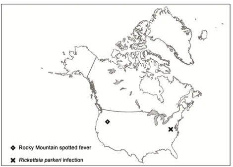
(25) Rickettsiosis as Threat for the Traveller. Fig. 10. Map showing distribution of human cases of tick-borne rickettsioses in North America.. 13.
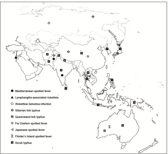
(26) 14. Current Topics in Tropical Medicine. Fig. 11. Map showing distribution of human cases of tick-borne rickettsioses and srub typhus in Asia and Oceania.. 4. Eschar and lymphadenopathy This clinical syndrome has been recently reported in Europe, where it is named TIBOLA (TIck-BOrne LimphAdenopaty) or DEBONEL (DErmacentor-BOrne Necrosis-ErythemaLymphadenopathy). R. slovaca, R. rioja and R. raoultii are the etiological agents, and Dermacentor marginatus is the main vector. This tick species is distributed all over Europe as well as in the North of Africa. Since this rickettsiosis appears in the coldest months of the year, the risk of acquisition for the travellers is lower than for the rickettsioses that are prevalent in spring and summer. In most cases (>90%) the tick-bite is located on the scalp (head) and always in the upper site of the body. After 1-15 days (mean: 4.8 days) of incubation period, the characteristic skin lesion starts as a crusted lesion at the site of the tick-bite (frequently on the scalp). A honey-like discharge from the lesion is observed in some cases. Few days later, a necrotic eschar appears (figure 12). This eschar is usually bigger than the one observed in MSF cases, and it is surrounded by an erythema. When the.
(27) Rickettsiosis as Threat for the Traveller. 15. tick-bite is out of the head, the skin lesion resembles the erythema migrans of Lyme disease. Other typical manifestation, which is always present when the bite is on the head, is the presence of regional and very painful lymphadenopathies. On the contrary of other rickettsioses, in DEBONEL/TIBOLA there are not systemic clinical signs (or they are rare), such as fever or maculo-papular rash (Oteo et al., 2004b). The clinical course is sub-acute and no severe complications have been described.. Fig. 12. DEBONEL/TIBOLA patient with the typical crusted lesion on the scalp.. 5. Scrub typhus The etiological agent of scrub typhus is Orientia tsutsugamushi, which is transmitted by chigger bites (trombiculid mite larvae). It is mainly distributed in Afghanistan, India, Pakistan, Sri-Lanka, Kashmir, China, Nepal, Japan, Korea, Vietnam, Indonesia, Laos, Philippines, Papua New Guinea and Australia (Figure 11). Cases are mainly observed in autumn and spring, in temperate zones where the bite of this arthropod, which is on vegetation, is frequent. The incubation period is about 10 or more days and the clinical signs and symptoms are similar to typhus syndrome, including the rash which is transient and easily missed. A difference with typhus syndrome is the presence of eschar that is frequently multiple. The presence of regional lymphadenopathy is also more frequent. The mortality can be high despite the correct antimicrobial treatment. Outbreaks related to military operations have been reported (Pages et al., 2010). Most travel acquired cases of scrub typhus occur in patients returning from Southeast Asia (Jensenius et al., 2004, 2006)..
(28) 16. Current Topics in Tropical Medicine. 6. Rickettsialpox Rickettsialpox is a worldwide (North America, Eastern Europe, Korea and southern Africa) rickettsiosis caused by Rickettsia akari and transmitted by the bite of the mouse mite Lyponyssoides sanguineus. We can consider it a remerging infection since several cases have been detected in New York City after September 11 attacks (Paddock et al., 2003). Patients have fever, a prominent eschar -which is the best sign of the disease- and rash that, as occurs in ATBF and Queensland tick typhus, may be vesicular. Palms and soles are not involved. The presence of regional lymphadenopathy is common. Patients recover without treatment in most cases (Raoult, 2010b).. 7. Laboratory diagnostic tools As occurs for all infectious diseases, the most definitive diagnostic method is the rickettsial isolation in culture. The main problem is that Rickettsia spp. are strictly intracellular bacteria, conventional growth media cannot be used, and a laboratory with P3 safety level (not generally available in clinical microbiology labs) is necessary. Furthermore, culture is not very sensitive and the yield decreases when clinical samples are taken after antibiotic treatment or when samples are not processed within 24 hours. It is a slow technique that is used for research purposes but not for the routine clinical practice. Centrifugation shell-vial technique is a commercially available adaptation of cell cultures that is easier to handle, faster and less hazardous. Isolation attempts on cell cultures may be performed using buffy coat or tissue samples (eschar biopsies when possible). If not processed within 24 h, samples must be frozen at -70ºC or in liquid nitrogen. Detection of rickettsiae by Giménez or Giemsa staining from blood and tissue samples would allow the confirmation of the diagnosis, but these techniques are non-specific and their sensitivity is very low. In some laboratories molecular biology tools, such as polymerase chain reaction (PCR) and sequencing, are also available. PCR-based assays from anticoagulated blood, biopsies and arthropod tissue samples targeting Rickettsia spp. genes are quite sensitive and useful for a quick diagnosis of these infections. The evaluation of several primer sets for the molecular diagnosis of rickettsioses demonstrated that the performance of three sequential PCRs (nested or semi-nested ones) allowed the detection and identification of Rickettsia species in a high percentage of the samples with previous clinical diagnosis or microbiological confirmation (serological analysis) of rickettsiosis (Santibáñez et al., 2011). Blood and tissue samples should be stored at -20ºC or lower if PCR-based diagnosis is delayed for more than 24 hours. The European guidelines for the diagnosis of tick-borne bacterial diseases contain useful information for clinicians and microbiologists (Brouqui et al., 2004). Indirect diagnostic tests and specifically, immunofluorescence assays (IFA) are considered the standard tests. Besides, since most traveller patients are investigated after returning, IFA are the most available tools for diagnosis. Acute and convalescent sera (collected 4-6 weeks after the onset of the illness) should be taken. In many cases we cannot observe seroconvertion but a high titre of antibodies. Cross-reactions among Rickettsia spp. make very difficult to definitively identify the causative agent by means of IFA. This can only be achieved in reference centres in which different antigens and other serological assays, such as western-blot, are available. Serum samples can be preserved at -20ºC or lower for several months without significant degradation of antibodies..
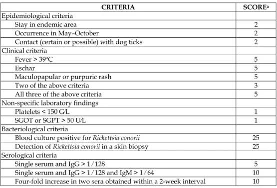
(29) Rickettsiosis as Threat for the Traveller. 17. Ticks removed from patients can be used as tools for the diagnosis of tick-borne rickettsioses. The strategy includes the identification of the tick to the species level, and the detection or isolation of rickettsias (Table 3). 1.. Identification of the ticks to the species level. 2.. Detection of bacteria in ticks with the use of staining tests (haemolymph for viable ticks; salivary glands if ticks were frozen), or PCR-based methods (using one-half of the tick, the other half being kept frozen). PCR may also be done using only ticks that stain positive.. 3.. Sequencing of the amplified PCR fragment and comparison with available sequences in sequence databases.. 4.. If there is 100% similarity between the tested sequence and the corresponding sequence of a known organism, the presumptive identification is confirmed. 5.. If the tested sequence appears to be different from all corresponding sequences available, the organism is probably a new strain and should be isolated and characterized from the stored frozen part of the tick. Table 3. Strategy for detecting and/or isolating rickettsias from ticks Diagnostic scores with epidemiological, clinical and laboratory tests for some tick-borne rickettsioses (ATBF and MSF) have been proposed (Tables 4 and 5). a.. Direct evidence of R. africae infection by culture and ⁄ or PCR or. b.. Clinical and epidemiological features highly suggestive of ATBF, such as multiple inoculation eschars and ⁄ or regional lymphadenitis and ⁄ or a vesicular rash and ⁄ or similar symptoms among other members of the same group of travellers coming back from an endemic area (sub-Saharan Africa or French West Indies) and Positive serology against spotted fever group rickettsiae or. c.. Clinical and epidemiological features consistent with a spotted fever group rickettsiosis such as fever and ⁄ or any cutaneous rash and ⁄ or single inoculation eschar after travel to sub-Saharan Africa or French West Indies and Serology specific for a recent R. africae infection (seroconversion or presence of IgM ‡ 1:32), with antibodies to R. africae greater than those to R. conorii by at least two dilutions, and ⁄ or a Western blot or cross-absorption showing antibodies specific for R. africae. Table 4. Diagnostic criteria for African-tick bite fever (ATBF). A patient is considered to have ATBF when criteria A, B or C are met.
(30) 18. Current Topics in Tropical Medicine. CRITERIA Epidemiological criteria Stay in endemic area Occurrence in May–October Contact (certain or possible) with dog ticks Clinical criteria Fever > 39ºC Eschar Maculopapular or purpuric rash Two of the above criteria All three of the above criteria Non-specific laboratory findings Platelets < 150 G⁄L SGOT or SGPT > 50 U⁄L Bacteriological criteria Blood culture positive for Rickettsia conorii Detection of Rickettsia conorii in a skin biopsy Serological criteria Single serum and IgG > 1 ⁄ 128 Single serum and IgG > 1 ⁄ 128 and IgM > 1 ⁄ 64 Four-fold increase in two sera obtained within a 2-week interval. SCOREa 2 2 2 5 5 5 3 5 1 1 25 25 5 10 10. SGOT, serum glutamate–oxaloacetate transaminase; SGPT, serum glutamate–pyruvate transaminase. aA positive diagnosis is made when the overall score is ≥ 25.. Table 5. Diagnostic criteria for Mediterranean spotted fever caused by Rickettsia conorii. 8. Prophylaxis An important chapter in the field of rickettsioses is related to prophylaxis. Since the majority of rickettsioses associated to travels are transmitted by ticks, the main preventive measure is to avoid tick-bites. Measures to avoid chiggers’ attacks are the same as the ones used against ticks. Only fleas can be more difficult to avoid when cats and other pets are abundant. If there is risk of getting lice, hygiene measures such as changing clothing (they live in the seams of clothing) may be sufficient. How can we avoid tick-bites? There are some rules that can be useful to avoid arthropodbites: 1. You must not wear dark clothes to see the ticks and remove them before attaching. Curiously, dark clothes attract less arthropods than clear ones. But, in our opinion, to look for the arthropods and remove them as soon as possible is more effective. 2. For outdoor activities (grass areas or mountains) you do not have to exposure your body to ticks. Thus, it is very useful to wear clothing that covers the majority of your body. The trousers must be tucked in your shocks with boots. Long sleeves shirt must be tucked into trousers. You must also wear a cap (especially children). 3. Permethrin-based repellents can be used on clothing, although their effect is short in time and the application should be repeated every few hours. 4. A careful inspection of clothing and body looking for ticks after returning from outdoors activities in endemic areas as well as removing them correctly has been.
(31) Rickettsiosis as Threat for the Traveller. 19. effective for the prevention of Lyme disease. The tick needs at least 24-48 hours for the transmission of Borrelia burgdorferi. This measure can be less efficient for Rickettsia spp. because these microorganisms can be transmitted since the first hours. But, anyway, the removal of the tick has to be done. 5. The contact with parasitized pets and wild animals must be avoided. There are two questions that physicians have linked up with tick-bites: How must I remove the tick? Must I take prophylactic drugs after a tick-bite? The first question is easy to answer. The most useful method to remove an attached tick is using forceps. Smooth forceps (without teeth) must be introduced between the tick’s head and the skin in a 90º angle and then pull (Oteo et al., 1996). Other traditional methods as using oil, burning or freezing must be forgotten. The other question is the use of prophylactic drugs after arthropod bites. There are no studies to answer this question. The transmission of rickettsias may be very quick, so we cannot extrapolate the recommendations for Lyme disease. Anyway, when people have been bitten by several ticks in an endemic area for a determinate disease (i.e.: Kruger National Park in South Africa and ATBF) and if the patient is anxious, we can offer doxycycline. It has been demonstrated that 3 doses of 100 mg. every 12 hours is safety and sufficient as treatment for the majority of rickettsioses. We must be cautious with the sun to avoid photo-sensibility. Children can take doxycycline for a short period of time. It is only contraindicated for pregnant women and in this case we can use macrolides (i.e. azythromycin). Vaccine approaches for prevention of rickettsial diseases have been developed since the past century, but currently no vaccine is available. Major surface protein antigens (OmpA and OmpB) of R. rickettsii and R. conorii are candidate vaccine antigens. Molecular biology techniques such as selection, cloning and expression of genes encoding R. prowazekii virulence-associated proteins, offer the opportunity to develop new rickettsial vaccines against typhus group rickettsiae. Further research is needed to develop effective vaccines without undesirable toxic reactions (Azad & Radulovic, 2003; Walker, 2009).. 9. Treatment The treatment of rickettsiosis should be initiated as soon as possible. Antibiotics are very effective and may avoid severe complications and death. In all cases if rickettsiosis is suspected, samples should be sent for laboratory confirmation. In DEBONEL/TIBOLA, in which the clinical signs and symptoms are less severe, recovery without antimicrobials occurs but the use of antibiotics shortens the clinical course and improves the clinical picture (Ibarra et al., 2005). Doxycycline is the most useful drug in children and adults. Doxycycline can be administered in short course (100 mg. every 12 hours for one day) for the treatment of typhus and scrub typhus. In the case of MSF, 2 doses of 200 mg./12 hous are also very effective (in children, 5 mg./kg./12hours); although most physicians use 100 mg. every 12 hours for 3-7 days after fever disappears. The same can be recommended for ATBF. This antibiotic regimen could probably be followed in other tick-borne rickettsioses but there are not good evidences (clinical assays) to support a recommendation. In RMSF the administration of doxycycline for 7 days is recommended. Other drugs that can be prescribed when not using doxycycline (allergy or pregnancy) are chloramphenicol (50-75 mg./kg./day given in 4 doses for 7-10 days) and azythromycin (500 mg./day for 5 days). Doxycycline for 7 days is the treatment of choice for rickettsialpox. Although there is in vitro.
(32) 20. Current Topics in Tropical Medicine. susceptibility to quinolones, the use of these drugs has been associated with worse clinical course (Botelho-Nevers et al., 2011).. 10. Conclusion In conclusion, rickettsioses are a worldwide threat that must be suspected in travellers returning from endemic areas. Most cases are caused by tick-bites, although in some areas of the world old diseases as typhus are present, and the risk exists. Rickettsiosis must be suspected in all patients with fever, exanthema with or without rash. Starting treatment with doxycycline when possible may be essential to rapidly recover and avoid complications. ATBF along with malaria is the leading cause of fever after returning from Sub-Saharan Africa.. 11. Acknowledgment We are grateful to all members from the Centre of Rickettsiosis and Arthropod-Borne Diseases, Hospital San Pedro-Centre of Biomedical Research (CIBIR), Logroño (La Rioja), Spain. Financial support was provided in part by a grant from ‘Instituto de Salud Carlos III’ (EMER 07 ⁄ 033), Ministerio de Ciencia e Innovación (Spain).. 12. References Althaus, F., Greub, G., Raoult, D. & Genton, B. (2010). African tick-bite fever: a new entity in the differential diagnosis of multiple eschars in travelers. Description of five cases imported from South Africa to Switzerland. International Journal of Infectious Diseases, Vol.14, Suppl.3 (September 2010), pp. e274-276, ISSN 1201-9712 Angelakis, E., Botelho, E., Socolovschi, C., Sobas, C.R., Piketty, C., Parola, P. & Raoult, D. (2010). Murine typhus as a cause of fever in travelers from Tunisia and Mediterranean areas. Journal of Travel Medicine, Vol.17, No.5, (September 2010), pp. 310-315, ISSN 1195-1982 Angel-Moreno, A., Bolaños, M., Santana, E., Pérez-Arellano, J.L. (2006). Murine typhus imported from Senegal in a travelling immigrant. Enfermedades Infecciosas y Microbiología Clínica, Vol.24, No.6, (June-July 2006), pp. 406-407, ISSN 0213-005X Askling, H.H., Lesko, B., Vene, S., Berndtson, A., Björkman, P., Bläckberg, J., Bronner, U., Follin, P., Hellgren, U., Palmerus, M., Ekdahl, K., Tegnell, A. & Struwe J. (2009). Serologic analysis of returned travelers with fever, Sweden. Emerging Infectious Diseases, Vol.15, No.11, (November 2009), pp. 1805-1808, ISSN 1080-6059 Azad, A.F. & Radulovic, S. (2003). Pathogenic rickettsiae as bioterrorism agents. Annals of the New York Academy of Sciences, Vol.990 (June 2003), pp. 734-8, ISSN 0077-8923 Azuma, M., Nishioka, Y., Ogawa, M., Takasaki, T., Sone, S. & Uchiyama, T. (2006). Murine typhus from Vietnam, imported into Japan. Emerging Infectious Diseases, Vol.12, No.9, (September 2006), pp. 1466-1468, ISSN 1080-6059 Bernabeu-Wittel, M., Pachón, J., Alarcón, A., López-Cortés, L.F., Viciana, P., Jiménez-Mejías, M.E., Villanueva, J.L., Torronteras, R. & Caballero-Granado, F.J. (1999). Murine typhus as a common cause of fever of intermediate duration: a 17-year study in the south of Spain. Archives of Internal Medicine, Vol.159, No.8, (April 1999), pp. 872-876, ISSN 0003-9926.
(33) Rickettsiosis as Threat for the Traveller. 21. Boillat, N., Genton, B., D'Acremont, V., Raoult, D. & Greub, G. (2008). Fatal case of Israeli spotted fever after Mediterranean cruise. Emerging Infectious Diseases, Vol.14, No.12, (December 2008), pp. 1944-1946, ISSN 1080-6059 Botelho-Nevers, E., Rovery, C., Richet, H. & Raoult, D. (2011). Analysis of risk factors for malignant Mediterranean spotted fever indicates that fluoroquinolone treatment has a deleterious effect. Journal of Antimicrobial Chemotherapy, Vol.66, (June 2011), pp. 1821-1830, ISSN 0305-7453 Bottieau, E., Clerinx, J., Schrooten, W., Van den Enden, E., Wouters, R., Van Esbroeck, M., Vervoort, T., Demey, H., Colebunders, R., Van Gompel, A. & Van Den Ende, J. (2006). Etiology and outcome of fever after a stay in the tropics. Archives of Internal Medicine, Vol.166, No.15, (August 2006), pp. 1642-1648, ISSN 0003-9926 Brouqui, P., Bacellar, F., Baranton, G., Birtles, R.J., Bjoërsdorff, A., Blanco, J.R., Caruso, G., Cinco, M., Fournier, P.E., Francavilla, E., Jensenius, M., Kazar, J., Laferl, H., Lakos, A., Lotric-Furlan, S., Maurin, M., Oteo, J.A., Parola, P., Perez-Eid, C., Peter, O., Postic, D., Raoult, D., Tellez, A., Tselentis, Y. & Wilske, B.; ESCMID Study Group on Coxiella, Anaplasma, Rickettsia and Bartonella; European Network for Surveillance of Tick-Borne Diseases. (2004). Guidelines for the diagnosis of tick-borne bacterial diseases in Europe. Clinical Microbiology and Infection, Vol.10, (December 2004), pp. 1108-1132, ISSN 1198-743X Caruso, G., Zasio, C., Guzzo, F., Granata, C., Mondardini, V., Guerra, E., Macrì, E. & Benedetti, P. (2002). Outbreak of African tick-bite fever in six Italian tourists returning from South Africa. European Journal of Clinical Microbiology & Infectious Diseases, Vol.21, No.2, (February 2002), pp. 133-136, ISSN 0934-9723 Chen LH, Wilson ME. (2009). Tick-borne rickettsiosis in traveler returning from Honduras. Emerging Infectious Diseases, Vol.15, No.8, (August 2009), pp. 1321-3, ISSN 1080-6059 Consigny, P.H., Schuett, I., Fraitag, S., Rolain, J.M., Buffet, P. (2009). Unusual location of an inoculation lesion in a traveler with African tick-bite fever returning from South Africa. Journal of Travel Medicine, Vol.16, No.6, (November-December 2009), pp. 439440, ISSN 1195-1982 de Sousa, R., Nóbrega, S.D., Bacellar, F., Torgal, J. (2003). Mediterranean spotted fever in Portugal: risk factors for fatal outcome in 105 hospitalized patients. Annals of the New York Academy of Sciences, Vol.990 (June 2003), pp. 285-294, ISSN 1749-6632 Dumler J.S. & Walker, D.H. (2010). Rickettsia tyhi (Murine typhus), In: Mandell, Douglas, and Bennett´s Principles and Practice of Infectious Diseases, Mandell G.L., Bennett J.E., Dolin R. (Eds.), pp. 2525-2528, Churchill Livingstone Elsevier, ISBN 978-0-44306839-3, Philadelphia-USA. Field, V., Gautret, P., Schlagenhauf, P., Burchard, G.D., Caumes, E., Jensenius, M., Castelli, F., Gkrania-Klotsas, E., Weld, L., Lopez-Velez, R., de Vries, P., von Sonnenburg, F., Loutan, L. & Parola, P.; EuroTravNet network. (2010). Travel and migration associated infectious diseases morbidity in Europe, 2008. BMC Infectious Diseases, Vol.10, (November 2010), 330, ISSN 1471-2334 Fournier, P.E., Roux, V., Caumes, E., Donzel, M. & Raoult, D. (1998). Outbreak of Rickettsia africae infections in participants of an adventure race in South Africa. Clinical Infectious Diseases, Vol.27, No.2, (August 1998), pp. 316-323, ISSN 1058-4838 Freedman, D.O., Weld, L.H., Kozarsky, P.E., Fisk, T., Robins, R., von Sonnenburg, F., Keystone, J.S., Pandey, P. & Cetron, M.S.; GeoSentinel Surveillance Network..
(34) 22. Current Topics in Tropical Medicine. (2006). Spectrum of disease and relation to place of exposure among ill returned travelers. The New England Journal of Medicine, Vol.354, No.2, (January 2006), pp. 119-130, ISSN 0028-4793 Gikas, A., Kokkini, S., Tsioutis, C., Athenessopoulos, D., Balomenaki, E., Blasak, S., Matheou, C., Tselentis, Y. & Psaroulaki, A. (2009). Murine typhus in children: clinical and laboratory features from 41 cases in Crete, Greece. Clinical Microbiology and Infection, Vol.15, Suppl. 2 (December 2009), pp. 211-212, ISSN 1198-743X Hechemy, K.E., Oteo, J.A., Raoult, D., Silverman, D.J. & Blanco, J.R. (2006). A century of rickettsiology: emerging, reemerging rickettsioses, clinical, epidemiologic, and molecular diagnostic aspects and emerging veterinary rickettsioses: an overview. Annals of the New York Academy of Sciences, Vol.1078 (October 2006), pp. 1-14, ISSN 0077-8923 Ibarra, V., Blanco, J.R., Portillo, A., Santibáñez, S., Metola, L., Oteo, J.A. (2005). Effect of antibiotic treatment in patients with DEBONEL/TIBOLA. Annals of the New York Academy of Sciences, Vol.1063 (December 2005), pp. 257-258, ISSN 0077-8923 Jensenius, M., Davis, X., von Sonnenburg, F., Schwartz, E., Keystone, J.S., Leder, K., LopézVéléz, R., Caumes, E., Cramer, J.P., Chen, L. & Parola, P.; GeoSentinel Surveillance Network. (2009). Multicenter GeoSentinel analysis of rickettsial diseases in international travelers, 1996-2008. Emerging Infectious Diseases, Vol.15, No.11, (November 2009), pp. 1791-1798, ISSN 1080-6059 Jensenius, M., Fournier, P.E. & Raoult, D. (2004). Rickettsioses and the international traveler. Clinical Infectious Diseases, Vol.39, No.10, (November 2004), pp. 1493-1499, ISSN 1058-4838 Jensenius, M., Fournier, P.E., Vene, S., Hoel, T., Hasle, G., Henriksen, A.Z., Hellum, K.B., Raoult, D. & Myrvang, B.; Norwegian African Tick Bite Fever Study Group. (2003). African tick bite fever in travelers to rural sub-Equatorial Africa. Clinical Infectious Diseases, Vol.36, No.11, (June 2003), pp. 1411-1417, ISSN 1058-4838 Jensenius, M., Montelius, R., Berild, D. & Vene, S. (2006). Scrub typhus imported to Scandinavia. Scandinavian Journal of Infectious Diseases, Vol.38, No.3, pp. 200-202, ISSN 0036-5548 Kelly, D.J., Richards, A.L., Temenak, J., Strickman, D., Dasch, G.A. (2002). The past and present threat of rickettsial diseases to military medicine and international public health. Clinical Infectious Diseases, Vol.34, Suppl.4, (June 2002), pp. S145-169, ISSN 1058-4838 Laurent, M., Voet, A., Libeer, C., Lambrechts, M., Van Wijngaerden, E. (2009). Mediterranean spotted fever, a diagnostic challenge in travellers. Acta Clinica Belgica, Vol.64, No.6, (November-December 2009), pp. 513-516, ISSN 0001-5512 McDonald, J.C., MacLean, J.D. & McDade, J.E. (1988). Imported rickettsial disease: clinical and epidemiologic features. American Journal of Medicine, Vol.85, No.6, (December 1988), pp. 799-805, ISSN 0002-9343 Niang, M., Brouqui, P., Raoult, D. (1999). Epidemic typhus imported from Algeria. Emerging Infectious Diseases, Vol.5, No.5, (September-October 1999), pp. 716-718, ISSN 10806059 Oteo, J.A., Ibarra, V., Blanco, J.R., Martínez de Artola, V., Márquez, F.J., Portillo, A., Raoult, D., Anda, P (2004b). Dermacentor-borne Necrosis Erythema and Lymphadenopathy:.
(35) Rickettsiosis as Threat for the Traveller. 23. Clinical and epidemiological features of a new tick-borne disease. Clinical Microbiology and Infection, Vol.10, pp. 327-331, ISSN 1198-743X Oteo, J.A., Martínez de Artola, V., Gómez-Cadiñanos, R., Casas, J.M., Blanco, J.R. & Rosel, L. (1996). Evaluation of methods of tick removal in human ixodidiasis. Revista Clínica Española Vol.196, No.9, (September 1996), pp. 584-587, ISSN 0014-2565 Oteo, J.A., Portillo, A., Blanco, J.R., Ibarra, V. & Santibáñez, S. (2004a). Medicina Clínica, Vol.122, No.20, (May 2004), pp. 786-788, ISSN 0025-7753 Oteo, J.A., Portillo, A., Santibáñez, S., Blanco, J.R., Pérez-Martínez, L. & Ibarra, V. (2006). Cluster of cases of human Rickettsia felis infection from southern Europe (Spain) diagnosed by PCR. Journal of Clinical Microbiology, Vol.44, No.7, (July 2006), pp. 2669-2671, ISSN 0095- 1137 Paddock, C.D., Zaki, S.R., Koss, T., Singleton, J. Jr., Sumner, J.W., Comer, J.A., Eremeeva, M.E., Dasch, G.A., Cherry, B. & Childs, J.E. Rickettsialpox in New York City: a persistent urban zoonosis. Annals of the New York Academy of Sciences, Vol.990, (June 2003), pp. 36-44, ISSN 1749-6632 Pages F., Faulde M., Orlandi-Pradines E. & Parola P. (2010). The past and present threat of vector-borne diseases in deployed troops. Clinical Microbiology and Infection, Vol.16, No.3, (March 2010), pp. 209-224, ISSN 1198-743X Pérez-Arellano, J.L., Fenollar, F., Angel-Moreno, A., Bolaños, M., Hernández, M., Santana, E., Hemmersbach-Miller, M., Martín, A.M. & Raoult, D. (2005). Human Rickettsia felis infection, Canary Islands, Spain. Emerging Infectious Diseases, Vol.11, No.12, (December 2005), pp. 1961-1964, ISSN 1080-6059 Raoult D. (2010b). Rickettsia akari (Rickettsialpox), In: Mandell, Douglas, and Bennett´s Principles and Practice of Infectious Diseases, Mandell G.L., Bennett J.E., Dolin R. (Eds.), pp. 2509-2510, Churchill Livingstone Elsevier, ISBN 978-0-4430-6839-3, Philadelphia-USA. Raoult, D. (2010a). Introduction to Rickettsioses, Ehrlichioses, and Anaplasmosis, In: Mandell, Douglas, and Bennett´s Principles and Practice of Infectious Diseases, Mandell G.L., Bennett J.E., Dolin R. (Eds.), pp. 2495-2498, Churchill Livingstone Elsevier, ISBN 978-0-4430-6839-3, Philadelphia-USA. Raoult, D., Fournier, P.E., Fenollar, F., Jensenius, M., Prioe, T., de Pina, J.J., Caruso, G., Jones, N., Laferl, H., Rosenblatt, J.E. & Marrie, T.J. (2001). Rickettsia africae, a tick-borne pathogen in travelers to sub-Saharan Africa. The New England Journal of Medicine, Vol.344, No.20, (May 2001), pp. 1504-1510, ISSN 0028-4793 Raoult, D., Ndihokubwayo, J.B., Tissot-Dupont, H., Roux, V., Faugere, B., Abegbinni, R. & Birtles, R.J. (1998). Outbreak of epidemic typhus associated with trench fever in Burundi. Lancet, Vol.352, No.9125, (August 1998), pp. 353-358, ISSN 0140-6736 Roch, N., Epaulard, O., Pelloux, I., Pavese, P., Brion, J.P., Raoult, D. & Maurin M. (2008). African tick bite fever in elderly patients: 8 cases in French tourists returning from South Africa. Clinical Infectious Diseases, Vol.47, No.3, (August 2008), pp. e28-35, ISSN 1058-4838 Santibáñez, S., Portillo, A., Santibáñez, P., Ibarra, V., Palomar, A., Oteo, J.A. (2011). Utility of five PCR targets for molecular diagnosis of human rickettsioses, Proceedings of the 6th International Meeting on Rickettsiae and Rickettsial Diseases, Heraklion, Crete, Greece, June 2011..
(36) 24. Current Topics in Tropical Medicine. Smoak, B.L., McClain, J.B., Brundage, J.F., Broadhurst, L., Kelly, D.J., Dasch, G.A. & Miller, R.N. (1996). An outbreak of spotted fever rickettsiosis in U.S. Army troops deployed to Botswana. Emerging Infectious Diseases, Vol.2, No.3, (July-September 1996), pp. 217-221, ISSN 1080-6059 Socolovschi, C., Barbarot, S., Lefebvre, M., Parola, P. & Raoult, D. (2010). Rickettsia sibirica mongolitimonae in traveler from Egypt. Emerging Infectious Diseases, Vol.16, No.9, (September 2010), pp.1495-1496, ISSN 1080-6059 Stephany, D., Buffet, P., Rolain, J.M., Raoult, D. & Consigny, P.H. (2009). Rickettsia africae infection in man after travel to Ethiopia. Emerging Infectious Diseases, Vol.15, No.11, (November 2009), pp. 1867-1870, ISSN 1080-6059 Stokes, P.H. & Walters, B.J. (2009). Spotted fever rickettsiosis infection in a traveler from Sri Lanka. Journal of Travel Medicine, Vol.16, No.6, (November-December 2009), pp. 436438, ISSN 1195-1982 Tsai, Y.S., Wu, Y.H., Kao, P.T., Lin, Y.C. (2008). African tick bite fever. Journal Formosan Medical Association, Vol.107, No.1, (January 2008), pp. 73-76, ISSN 0929-6646 Walker, D.H. & Raoult, D. (2010). Rickettsia prowazekii (Epidemic or louse-borne typhus), In: Mandell, Douglas, and Bennett´s Principles and Practice of Infectious Diseases, Mandell G.L., Bennett J.E., Dolin R. (Eds.), pp. 2521-2524, Churchill Livingstone Elsevier, ISBN 978-0-4430-6839-3, Philadelphia-USA. Walker, D.H. (2009). The realities of biodefense vaccines against Rickettsia. Vaccine, Vol. 27, Suppl. 4 (November 2009), pp. D52-5, ISSN 0264-410X Walter, G., Botelho-Nevers, E., Socolovschi, C., Raoult, D. & Parola, P. (2011). Murine typhus in returned travelers: a report of thirty-two cases, Proceedings of the 6th International Meeting on Rickettsiae and Rickettsial Diseases, Heraklion, Crete, Greece, June 2011. Zanetti, G., Francioli, P., Tagan, D., Paddock, C.D. & Zaki, S.R. (1998). Imported epidemic typhus. Lancet, Vol.352, No.9141, (November 1998), pp. 1709, ISSN 0140-6736.
(37) 2 Human Ehrlichioses and Rickettsioses in Cameroon Lucy Ndip1, Roland Ndip1, David Walker2 and Jere McBride2 2University. 1University. of Buea of Texas Medical Branch, Galveston 1Cameroon 2USA. 1. Introduction Human ehrlichioses and rickettsioses are important arthropod borne infectious diseases which are transmitted by ticks, mites, lice and fleas. Infections result in mild to fatal outcomes, with clinical presentations that resemble other tropical infectious diseases such as malaria making clinical diagnosis difficult. Despite recognition as important causes of lifethreatening diseases in the United States, the geographic distribution of these diseases worldwide remains undefined due to their recent emergence, challenges in diagnosis and lack of comprehensive epidemiological studies needed to determine incidence in developing countries. Recently, the transfer of technological developments to other parts of the world especially developing countries has encouraged basic epidemiological inquiry and generated scientific interest in understanding the epidemiology of these tick borne diseases and their role as causes of undifferentiated febrile illnesses. In this chapter, we review the current knowledge of human monocytotropic ehrlichiosis (HME) and spotted fever rickettsiosis (African tick bite fever) in Cameroon.. 2. Ehrlichiosis 2.1 Etiologic agents Ehrlichioses are diseases caused by small (approximately 0.4–1.5 μm diameter) Gram negative, obligately intracellular bacteria belonging to the genus Ehrlichia of the family Anaplasmataceae, Order Rickettsiales and the alpha sub-division Proteobacteria (Dumler et al., 2001). Although they have a characteristic Gram negative cell wall structure, they lack the necessary enzymes to synthesize cell membrane components such as lipopolysaccharide and peptidoglycan (Lin & Rikihisa, 2003). As intracellular pathogens, Ehrlichia reside in cytoplasmic membrane-bound vacuoles inside host cells (granulocytes or monocytes) forming microcolonies called morulae, derived from the Latin word “morus” for mulberry (Popov et al., 1995; Paddock et al., 1997; Ismail et al., 2010). These morulae (ranging in size from 1.0 to 6.0 µm in diameter) may contain 1 to >40 organisms of uniform or mixed cell types (Popov et al., 1995; Rikihisa, 1999). Organisms in the family Anaplasmataceae were first described in 1910 when Theiler described Anaplasma marginale, the etiologic agent of an economically important and severe disease of.
(38) 26. Current Topics in Tropical Medicine. cattle (Mahan, 1995). This discovery was followed shortly thereafter by the description of E. ruminantium (formerly Cowdria ruminantium) by Cowdry in 1925; E. canis by Donatien and Lestoquard in 1935; and A. phagocytophilum (formerly E. phagocytophila) by Gordon in 1940. Hence, the genus Ehrlichia was established in 1945 in honour of the German microbiologist Paul Ehrlich (Uilenberg, 1983). Ehrlichia species cause significant diseases in their natural hosts (livestock and companion animals) and emerging zoonoses in humans (McBride & Walker, 2010). The first human ehrlichial infection (sennetsu fever) was reported in 1953 ( Rapmund, 1984; Dumler et al., 2007). Sennetsu fever, caused by Neorickettsia sennetsu, was identified in Japan and Malaysia (Dumler et al., 2001; Dumler et al., 2007). However, recent phylogenetic reclassifications based on molecular analysis revealed that E. sennetsu is not a member of the Ehrlichia genus (Dumler et al., 2001). Presently, the genus Ehrlichia consists of five recognized species including E. canis, E. chaffeensis, E. ewingii, E. muris, and E. ruminantium, all of which are at least 97.7% similar in 16S rRNA gene sequence (Perez et al., 1996; Paddock et al., 1997; Dumler et al., 2001; Perez et al., 2006). Ehrlichiae have relatively small genomes (0.8–1.5 Mb) with low G+C content and a high proportion of non-coding sequences but can synthesize all nucleotides, vitamins and cofactors (Dunning et al., 2006). They also have small subsets of genes associated with hostpathogen interactions (Ismail et al., 2010). E. chaffeensis have immunodominant outer membrane proteins (OMP-1/MSP2/P28) (Ohashi et al., 1998; Yu et al., 2000; Huang et al., 2008), and in infected macrophages ehrlichiae express the p28-Omp 19 and 20 genes as dominant protein products (Ganta et al., 2009; Peddireddi et al., 2009). Ehrlichiae also express several targets of the humoral immune response including tandem repeat and ankyrin repeat containing proteins (Yu et al., 1997; Sumner et al., 1999; McBride et al., 2003; McBride et al., 2007). E. chaffeensis, a human pathogen that was first recognised in the United States in 1986 and isolated in 1991 ( Maeda et al., 1987; Dawson et al., 1991) is the cause of human monocytotropic ehrlichiosis (HME) (Anderson et al., 1992), a moderate to severe disease with a case fatality rate of 3% (Fishbein et al., 1994; McBride & Walker, 2010). E. chaffeensis is an obligately intracellular bacterium that primarily infects mononuclear leukocytes and replicates by binary fission. E. chaffeensis morulae can be detected in peripheral blood smears obtained from infected patients when observed with a light microscope (Rikihisa, 1991). When tissues (including clinical samples), mononuclear leucocytes or cell lines of mammalian origin infected with E. chaffeensis are viewed by electron microscopy, two distinct morphologic cell types are identified: a predominantly coccoid form which has a centrally condensed nucleoid DNA and ribosomes (dense-cored cells) measuring between 0.4 and 0.6 µm in diameter and reticulate or the coccobacillary form, which measures about 0.4 to 0.6 µm by 0.7 to 1.9 µm (Paddock et al., 1995; Popov et al., 1997). 2.2 Vectors and reservoirs Investigative studies following the discovery of E. chaffeensis in the late 1980s revealed that the agent is transmitted to humans by the tick Amblyomma americanum, commonly referred to as the lone star tick which has a limited geographic distribution to the United States (Anderson et al., 1993). Molecular analysis (PCR) has demonstrated E. chaffeensis DNA in adult A. americanum ticks collected from different states. The increased recognition of E. chaffeensis as an emerging problem has evoked renewed interest in this and other tick borne diseases, and this has stimulated epidemiologic investigations of this pathogen and its vector in other regions where the tick A. americanum is not found. Results not only indicate.
(39) 27. Human Ehrlichioses and Rickettsioses in Cameroon. that E. chaffeensis has a wider distribution than the United States (Ndip et al., 2010), but also indicates that the pathogen exists outside of the known range of A. americanum and is harbored by other tick species. These tick species include Ixodes pacificus in California (Kramer et al., 1999), Dermacentor variabilis in Missouri (Roland et al., 1998), Ixodes ricinus in Russia (Alekseev et al., 2001), Amblyomma testudinarium in China, (Cao et al., 2000), Haemaphysalis longicornis (Lee et al., 2003), and Ixodes persulcatus (Kim et al., 2003) in Korea.. (a). (b). Fig. 1. a) Rhipicephalus sanguineus (brown dog tick) and b) Male Amblyomma variegatum tick (courtesy Laboratory for Emerging Infectious Diseases, University of Buea) Studies carried out by Ndip and colleagues in Cameroon identified Ehrlichia chaffeensis in Rhipicephalus sanguineus ticks. R. sanguineus, commonly known as the brown dog tick (Figure 1a) is a species that infests canids worldwide. In one study in Limbe, Cameroon, a very high prevalence of E. chaffeensis was detected in R. sanguineus ticks infesting dogs inhabiting one kennel (Ndip et al., 2010). E. chaffeensis DNA was detected in 33 (56%) of 63 R. sanguineus ticks collected from five dogs as opposed to 4 (6%) ticks infected with E. canis. Furthermore, co-infection with more than one pathogen was not uncommon. The E. chaffeensis strain circulating in Cameroon is similar to the North American strain AF403710 based on the analysis of the 378 bp fragment of the disulphide bond formation (Dsb) protein gene (Ndip et al., 2010). Earlier reports revealed E. canis, E. chaffeensis, and E. ewingii in R. sanguineus ticks collected from 51 dogs from different localities in Cameroon (Figure 2), suggesting that dogs could be a reservoir for E. chaffeensis and that R. sanguineus is the probable vector (Ndip et al., 2007). In the United States, the white-tailed deer (Odocoileus virginianus) has been recognised as the primary natural reservoir of E. chaffeensis (Dugan et al., 2000). However, animals such as goats, dogs, and coyotes have also been identified as reservoirs which could play a limited role in the transmission of the pathogen to humans (Breitschwerdt et al., 1998; Dugan et al., 2000; Kocan et al., 2000). Unlike rickettsial species, ehrlichial species are not transmitted trans-ovarially (ie., larvae are uninfected) suggesting that the pathogen is maintained transstadially after the infection is acquired (Ismail et al., 2010). Although the reservoirs for E. chaffeensis in Cameroon have not yet been conclusively identified, preliminary studies detected antibodies reactive to E. chaffeensis in 56% of goats analysed suggesting a probable role of goats in maintaining the pathogen in nature. Moreover, E. chaffeensis DNA was detected in 17% of ticks collected from these animals (Ndip, unpubished data)..
(40) 28. Current Topics in Tropical Medicine. Fig. 2. Known distribution of ehrlichiae and rickettsiae in Cameroon 2.3 Clinical manifestations The comprehensive data available in literature today on symptoms observed in HME infection is based on cases reported to the United States’ Centers for Disease Control and Prevention in addition to a series of patients studied since the disease was described. After exposure to an infecting tick, an incubation period of 1 to 2 weeks (median, 9 days) ensues after which patients develop a febrile illness (often >39°C) characterized by general malaise, low-back pain, or gastrointestinal symptoms (Paddock & Childs, 2003). These signs and symptoms most often resemble manifestations caused by other infectious and non-infectious causes. After 3 to 4 days, symptoms progress and patients may seek medical attention presenting with fever (>95%), headache (60 to 75%), myalgias (40 to 60%), nausea (40 to 50%), arthralgias (30 to 35%), and malaise (30 to 80%) (Fishbein et al., 1994). Some patients (10-40%) may present with cough, pharyngitis, diarrhea, or abdominal pain and may even progress to changes in mental status (Fishbein et al., 1994; Olano et al., 2003). Some populations especially HIV-infected patients (Paddock et al., 2001) and children (Jacobs & Schutze, 1997) may develop a rash on the extremities, trunk and face (Edwards, 1991). Hematological changes include leukopenia in approximately 60 to 70% of patients and thrombocytopenia (Fishbein et al., 1994; Olano & Walker, 2002). Liver enzymes (hepatic transaminases) may become slightly elevated (Nutt & Raufman, 1999). About 60 to 70% of patients require hospitalization and untreated cases last for 2-3 weeks or progress to a fatal outcome during the second week (Fishbein et al., 1994; Standaert et al., 2000). About 20% of patients develop neurologic signs, cough or other respiratory symptoms (Fishbein et al., 1994; Olano et al., 2003). Case-fatality ratio is approximately 3% (McQuiston et al., 2003) with risk factors for severe or fatal disease including older age (Paddock et al., 2001), underlying debilitating diseases such as HIV infection, immunosuppressive therapies (Olano & Walker, 2002) and sickle cell disease (Paddock & Childs, 2003). These reported symptoms are quite similar to those manifested by Cameroonian HME patients. In one series of 206 acutely ill patients studied, 30 (14.6%) demonstrated anti-.
(41) Human Ehrlichioses and Rickettsioses in Cameroon. 29. ehrlichial IgM antibodies, and these probable HME patients presented with headache (83%), fatigue (37%), abdominal pain (47%), joint pain (60%), anorexia (37%) and diarrhoea (13%) in addition to fever (>38oC). Their mean hematocrit, AST and ALT values were 48±21%, 46±23% and 36± 21%, respectively. Five (17%) of the patients were anaemic while 10 (33%) and 5 (17%) had abnormal AST and ALT values, respectively (Ndip, unpublished data). In another series of 118 acutely ill febrile patients studied with HME diagnosed by detection of E. chaffeensis DNA in patient’s blood (n=12), these patients presented with fever (100%), headache (seen in 72% of the patients), arthralgia (58%), myalgia (42%), cough (17%) and a diffuse maculopapular rash (17%). The rash was present on the trunk of one patient and the arms of another. One patient of the 12 with detectable E. chaffeensis DNA required hospitalization (see Table 1) (Ndip et al., 2009). 2.4 Epidemiology The epidemiology and ecology of HME worldwide is not well documented. Since its description in 1986 more than 1000 cases of HME from at least 30 U.S. states have been reported to the Centers for Disease Control and Prevention in Atlanta, Georgia with nearly all occurring in the southeastern and south-central United States where the vector, A. americanum is common (Paddock & Childs, 2003; Dumler et al., 2007). However, the evidence of the disease and/or the pathogen is increasingly being reported in other parts of the world. This includes Africa (Uhaa et al., 1992; Brouqui et al., 1994; Ndip et al., 2009; Ndip et al., 2010), Israel (Dawson et al., 1991; Keysary et al., 1999; Brouqui & Dumler, 2000), Latin America (Gongora-Biachi et al., 1999; Calic et al., 2004;) and Asia (Heppner et al., 1997; Cao et al., 2000; Heo et al., 2002; Kim et al., 2003; Park et al., 2003, Lee & Chae, 2010). In Cameroon, HME has been identified in patients along the coast of Cameroon, in Buea (4o10’0’’N9o14’0’’E), Limbe (4o01’N 9o13’13’’E), Muyuka (4o43’18’’N 9o38’27’’E), Tiko (4o4’0’’N 9o22’60’’E), and Kumba (4o38’38’’ N9o26’19’’E) and the agent, E. chaffeensis, in ticks collected from Limbe (Figure 2). HME was observed in both males and females as well as in children and adults although results suggested that older age was a risk factor for the disease (Ndip et al., 2009). The majority of the patients were adults which suggests that exposure to infected ticks may have occurred during outdoor activities such as farming. Another risk factor is that of owning a companion or domestic animal since most Cameroonian HME patients indicated they had tick-infested pets and domestic animals. 2.5 Microbiological diagnosis The diagnosis of HME requires specialized microscopy equipment and skills which are not readily available in many diagnostic laboratories. Several methods have been proposed for the diagnosis of HME (Paddock & Childs, 2003; Ismail et al., 2010), including serologic tests such as immunofluorescent assay (IFA), western immunoblot employing specific proteins or ehrlichial whole cell antigens or the recently developed Ehrlichia recombinant protein or peptide ELISA for detection of the antibody (Cardenas et al., 2007; Luo et al., 2010; O'Connor et al., 2010). Though these tests can be used to confirm diagnosis retrospectively, some patients may not sero-convert during the early days of the disease and cannot be diagnosed with serologic tests. However, collecting paired sera (at acute and convalescent phases of illness) is confirmatory as a four-fold rise in titer indicates current infection. However, this.
(42) 30. Current Topics in Tropical Medicine. always presents a problem because patients who recover may not return to the hospital for follow up. Moreover, another issue with the interpretation of serological tests such as IFA is cross-reactive antibodies against other organisms, including Anaplasma species. PCR has also been employed to identify ehrlichial DNA in acutely ill patients when antibodies have not reached detectable levels. Several genes have been proposed and used including the VLPT gene (TRP32), TRP36, 16S rRNA, the TRP120, the Dsb, 28-kDa outer membrane protein gene have been used as genus or species specific targets (Yu et al., 1999; Doyle et al., 2005). IFA, western blot and PCR have been used to study the prevalence of ehrlichiae in blood of acutely ill patients, reservoirs, or in suspected tick vectors and anti-ehrlichial antibody in sera (Ndip et al., 2005; Ndip et al., 2007; Ndip et al., 2009; Ndip et al., 2010). Figure 3 shows IFA photomicrographs of whole cell of E. chaffeensis reacting with antibodies in an HME patient serum. A rapid method to detect E. chaffeensis is the observation of morulae in smears of peripheral blood buffy coat using the Diff Quik or Giemsa stain. However, this technique is very insensitive, and morulae are detected in leukocytes in only 10% of HME patients. Patients. 1. 2. 3. 4. 5. 6. 7. 8. 9. 10. 11. 12. Gender F Age (yr) 63 Location A Clinical Manifestations. F 40 B. M 5 C. F 23 D. M 22 C. M 20 D. F 21 A. M 1 C. F 16 A. F 26 A. M 35 B. M 25 A. Fever >38oC Yes. Yes. Yes. Yes. Yes. Yes. Yes. Yes. Yes. Yes. Yes. Yes. 6. 8. 4. 7. 4. 3. 1. 1. 2. 7. 2. 2. Yes Yes Yes No. Yes Yes Yes No. Yes No No No. No No No Yes. No No No No. Yes Yes Yes No. Yes No Yes No. No No No Yes. Yes Yes Yes No. Yes No Yes No. Yes Yes Yes No. No No No No. *Day(s) Headache Myalgia Arthralgia Rash. *Days after onset (i.e., before collection of sample). Locations: A - Buea , B – Limbe, C – Tiko, D - Muyuka. Table 1. Epidemiologic and clinical characteristics of twelve Cameroonian patients with HME 2.6 Treatment The drug of choice for the treatment of E. chaffeensis infection is the tetracyclines (particularly doxycycline) and their derivatives. Generally, between 1 and 3 days after a patient with HME commences treatment with doxycycline, the patient becomes afebrile (Olano & Walker, 2002). However, treatment may continue for up to 10 days or at least 3 days after the patient becomes afebrile (Chapman et al., 2006). Clinical experience and invitro susceptibility testing of E. chaffeensis to some classes of antibiotics have revealed that fluoroquinolones, penicillins, aminoglycosides, macrolides and cotrimoxazole are not effective therapeutics (Dumler et al., 1993; Brouqui et al., 1994; Brouqui & Raoult, 1994; McBride & Walker, 2010)..
Figure


+7


Documento similar
Another notable work in the identification of Academic Rising Stars using machine learning came in Scientometric Indicators and Machine Learning-Based Models for Predict- ing
The current study discusses initiatives to achieve environmental sustainability in manufacturing processes, and focuses in particular on the application of the environmental
The nutritional indexes, PNI and NNI are higher in crops than in halophytes, similarly to the plant nutrient content (Table 2). In contrast, the nutritional indexes
In this paper the design and operation of a HLDS is presented, for the air conditioning of a high latent load application with high ambient humidity levels.. An analysis of
The Dwellers in the Garden of Allah 109... The Dwellers in the Garden of Allah
a) To systematically review the indicators of PPF in breast cancer. i) To study the associations between the main constructs from positive psychology and breast cancer. ii)
Moreover, the tool supported the collaborative design of the final application, resulting in an application with features and components agreed between the members of the
For example, the displacement of a particle in a mechanical problem can be related either to the voltage or to the current variable; in the first case the instantaneous
