Microsolvation of biomolecular models by microwave spectroscopy: structure and cooperative effects
436
0
0
Texto completo
(2)
(3) La investigación llevada a cabo en esta Tesis Doctoral ha sido financiada por: Ministerio de Economía y Competitividad CTQ2013-40717-P CTQ2016-75253-P. Junta de Castilla y León Consejería de Educación VA334U14. Universidad de Valladolid Vicerrectorado de Investigación y Política Científica Ayudas para estancias breves en el desarrollo de tesis doctorales Movilidad Doctorandos UVa 2015 Movilidad Doctorandos UVa 2016.
(4)
(5) “You can't always get what you want, But if you try sometimes you might find You get what you need” The Rolling Stones.
(6)
(7) Aunque solo lo defiende una persona, el trabajo que se hace durante una tesis es un trabajo de equipo, y como en todos los equipos hay personas que lideran y guían. Quiero expresar mi más sincero agradecimiento a mis directores, la Prof. Susana Blanco Rodríguez y el Prof. Juan Carlos López Alonso por haber hecho una excelente labor de guía. Quiero agradecer a Juan Carlos por haberme enseñado tantísimas cosas (y las que me quedan por aprender de él). A Susana, además de por haberme enseñado tanto, quiero agradecerle especialmente por haber estado codo con codo a mi lado y por haber conseguido literalmente, que esta Tesis saliera adelante.. I have had the tremendous luck of doing short research stays in two of the best laboratories. I would like to thank PD Dr. Melanie Schnell for the stay in the Structure and Dynamics of Cold and Controlled Molecules group at the Max Planck Institute for the Structure and Dynamics of Matter in Hamburg. She is a great group leader and all the team mates I met there are awesome, thanks to all! I also would like to thank Prof. Dr. Hab. Zbigniew Kisiel for the stay in the Group of Vibrational and Rotational Spectroscopy group in the Institute of Physics of the Polish Academy of Sciences. He is one of the leading experts in the rotational spectroscopy field and I have learnt so many things from him!. Durante este periodo han pasado varios compañeros de TFG, TFM o doctorado por el departamento, quiero agradecer a todos, con mención propia a Alberto Macario, por tantos buenos momentos.. La lista de amigos a quien querría agradecer es demasiado extensa como para ponerlos a todos. ¡Gracias a todos! En especial a Pablo y Edu.. Por supuesto quiero agradecer a mi familia, especialmente a mis padres Soledad y Jose, que siempre me han apoyado en mis decisiones y ayudando a que continuara formándome. En casa también han estado siempre Balder y Clara, distrayéndome con sus juegos cuando lo necesitaba, y dándome muchísimo cariño. Son los mejores amigos que alguien podría tener.. Durante la última etapa de este doctorado he tenido la suerte de conocer a Teresa, gracias a ella, a su cariño y apoyo el finalizar la Tesis no ha sido tan duro..
(8)
(9) CONTENTS Chapter I – Introduction………………………………………….…… Page 1 Chapter II – Methodology………………………………….................... Page 17. Chapter III – Instrumentation……………………………….................... Page 41. Chapter IV – Hydrogen-bond Cooperativity in Formamide2–water: A Model for Water-Mediated Interactions……………....….. Page 57. Supplementary Material for Chapter IV…………………............... Page 69. Chapter V – Structure and Dynamics in Formamide-(H2O)3: A Water Pentamer Analogue……………………….………………. Page 101. Supplementary Material for Chapter V……………………..…….. Page 117 Chapter VI – Microsolvation of Formanilide Conformers: A Model for Peptide Solvation ……...……………………..................... Page 167 Supplementary Material for Chapter VI…………………….…….. Page 191 Chapter VII – The Effect of Microsolvation Over Structure, Nuclear Quadrupole. and. Internal. Rotation:. The. Methyl. Carbamate-(H2O) and The Methyl Carbamate-(H2O)2 complexes…….................................................................... Page 233.
(10) Supplementary Material for Chapter VII……………….……...….. Page 253 Chapter VIII – Microsolvation of Ethyl Carbamate Conformers: Effect of. Carrier. Gas. on. the. Formation. of. Complexes…………………..…………………….…….. Page 275 Supplementary Material for Chapter VIII..………………..……… Page 297 Chapter IX – Microsolvated Complexes of Ibuprofen as Revealed by High-Resolution Rotational Spectroscopy……………… Page 329 Supplementary Material for Chapter IX…………..…………….… Page 343 Conclusions…………………………………………………………… Page 361 Appendix I – Resumen en Español………………….…………………. Page 369 Appendix II – Flexibility. Unleashed. in. Acyclic. Monoterpenes:. Conformational Space of Citronellal Revealed by Broadband Rotational Spectroscopy……………………. Page 389 Appendix III – Prediction of the Rotational Spectra of Microsolvated Complexes with Low Cost DFT Methods…...………….. Page 401. Appendix IV – Structure Determination, Conformational Flexibility, Internal Dynamics, and Chiral Analysis of Pulegone and Its Complex with Water………………………...……….. Page 413.
(11)
(12)
(13) Chapter I Introduction. 1.
(14)
(15) The activity of biomolecules is function of their shapes, which are in turn determined by interand intramolecular interactions,1,2 corresponding in most of the cases to hydrogen bonding (HB).3-5 The presence of water in the biological environment is of special importance, not only as a solvent but as an active component. For example, the crystals of proteins can be considered as a highly concentrated solution with ≈ 20-50% of the total volume occupied by water.6 The small size of water and its double donor/acceptor capabilities give water a particular flexibility to form hydrogen bonds, which have a central role in the solvation processes. These subtle noncovalent forces may control different kind of phenomena, such as conformational7,8 or tautomeric equilibria,9,10 charge stabilization,11,12 or molecular recognition,13,14 and may influence the structure, dynamics and function of biomolecules.15,16 It has been observed that water inserts between the carbonyl and amino functional groups in α– helices and is involved in the protein folding process.17-20 On it, the protein minimizes the interaction with water of the hydrophobic chains located in the core, where the main interaction is the peptide hydrogen bond N–H···O=C. On the other hand, the polar side chains are located on the surface, exposed to interactions with water. It has been reported that water plays also a determinant function in the nucleation of protein folding as a mediator in the formation of the peptide hydrogen bond. The effect of the interaction with water on different kind of molecules has been extensively studied in condensed phases by 1H-NMR,21-23 X-Ray spectroscopy,24,25 or IR spectroscopy.26-29 However, the understanding at a molecular level of the role of the subtle non-covalent interactions forces responsible for solvation requires isolating the molecules from the condensed phase environment to study them in the gas phase with a controlled hydration degree. This environment, in which a limited number of molecules of water interact with another molecule (the solute) in isolation is called microsolvation. Microsolvation is a consolidated field of research, as shown by the number of works30-33 dedicated to bring light on it. Pioneering studies in the gas phase on molecules with biological interest were performed by electronic spectroscopy coupled with supersonic expansion.34-37 In microwave spectroscopy, detailed works with molecules and clusters of different nature have set the basis for the research on hydrogen bond complexes,38-42 opening the possibility to study many structural aspects of microsolvation. 3.
(16) Rotational spectroscopy coupled with supersonic expansion has proved to be an adequate experimental tool to characterize microsolvated complexes.43 These microsolvated complexes can be easily generated in the supersonic expansion and further studied in an isolated environment34,44-47 to determine their intrinsic properties. Complexes with different degrees of hydration can be formed in the supersonic expansion, furthermore, the number of water molecules in the complexes can be somehow controlled through the expansion conditions. The spacing between the rotational levels is intimately related to the mass distribution within the molecule or complex. Therefore the molecular structures obtained from rotational spectroscopy are characterized by its high accuracy. An important aspect of the microsolvation studies using rotationally resolved spectroscopy is the possibility to determine the rotational constants for a variety of isotopologues, allowing to derive accurate molecular structures by different approaches.48,49 Coordinates, distances and angles for the water molecules can be directly obtained for conformers and clusters, so it is possible to obtain information about the evolution of distances and angles from the bare molecule to the first steps of solvation. Apart from accurate structures, microwave spectroscopy techniques provide other relevant molecular properties. From their high-resolution it is possible to precisely determine the values for the nuclear quadrupole coupling constants determined by the presence in many biomolecules of certain nuclei as 14N, which give rise to a hyperfine structure in the spectrum.50,51 Those constants are related to the electric field gradient at the N nucleus and thus to the electronic environment. As a consequence, they can serve to reveal the subtle polarization changes occurring upon microsolvation. The information obtained from rotational spectra is not limited to static structural properties, in many cases, it is possible to obtain information about dynamical motions associated with tunneling effects. An example of this kind of dynamics that can be measured from the microwave spectra is the internal rotation for systems presenting an internal rotation top,52,53 and to determine the barriers hindering the rotation. Internal rotation shows a high dependence on the structure around the rotating top,54 and therefore, it is possible to correlate the values for the internal rotation barrier with structural changes occurring from isolation to microsolvation. Microsolvation studies on small organic molecules by rotational spectroscopy,8-10,55-69 provide a better knowledge about the solvent interactions in larger biomolecules,70 showing the interplay existing between self-association and solvation. The majority of those studies have been done for monohydrated complexes, giving information about dynamics of water and characterizing the preferred sites of interaction. When increasing the number of water molecules, it is possible to obtain new information, such as the role of HB cooperativity in the solvation process,4 the contribution to the stabilization of weak interactions apart from HB,67,69 or the way in which solvation induces structural changes.68 In most cases, water self-association dominates the interactions since water molecules tend to link to other water molecules forming chains or cycles, as occurs in complexes where there is only one HB acceptor group.66,67,69 However, in molecules with two donor/acceptor positions, as amides,55-59 acids60-63 or esters,64,65 water molecules tend to close donor-acceptor-donor sequential cycles. The observation by microwave spectroscopy of organic molecules microsolvated by more than two molecules of water is complicated, and just a few examples have been reported.61,62,67,69 This is noticeable since the research on pure water clusters71-80 has significantly advanced with the development of microwave spectroscopy techniques in supersonic jets.45,46. 4.
(17) The importance of HB cooperativity contributing to the stabilization of hydrated biomolecules has been widely described in the literature.81-83 σ-bond cooperativity is associated with complexes forming chains or cycles such as the -OH group or any other group capable of acting simultaneously as hydrogen donor and acceptor.3,4 When water molecules establish hydrogen bonds, they become polarized, enhancing their hydrogen donor and hydrogen acceptor capacity and further strengthening the interactions they involve in. σ-cooperativity has been described in detail elsewhere,3,4 its main effects are the shortening of the HB distances and an increase in the HB stabilization energy. These effects are stronger as the number of cooperative molecules increase. In amides as the peptide group, another kind of effect has been reported to occur when those participate in HB, which causes a polarization of the molecule.3,4 This is represented by a major contribution of the partially charged resonant structures (see Scheme 1.1), further enhancing its donor/acceptor capabilities. The main effect of this polarization is the shortening of the C–N bond, due to a major double bond contribution, and the enlargement of the C=O bond, due to a major presence of the single bond resonant form. In addition, if the peptide bond has any substitution group, they can participate in the resonance interaction, extending the polarization effects over a wider range of groups in the molecule, as occurs in carbamates, which are esterderivatives of amides (see Scheme 1.2).. Scheme 1.1. Resonant contributing forms for peptide groups water complexes. Formamide: R1,2 = H. Formanilide: R1 = phenyl, R2 = H. Methyl carbamate: R1 = H, R2 = O–CH3. Ethyl carbamate: R1 = H, R2 = O–C2H5.. Scheme 1.2. Additional resonant contributing forms for carbamates water complexes. Methyl carbamate: R1 = O–CH3. Ethyl carbamate: R1 = O–C2H5. Thus, HB interactions are enhanced by resonance with the π–delocalized conjugated system, the so-called Resonance Assisted Hydrogen Bonding (RAHB), which in some works has been named π-cooperativity.84-86 RAHB is a synergistic mechanism that causes the reinforcement of 5.
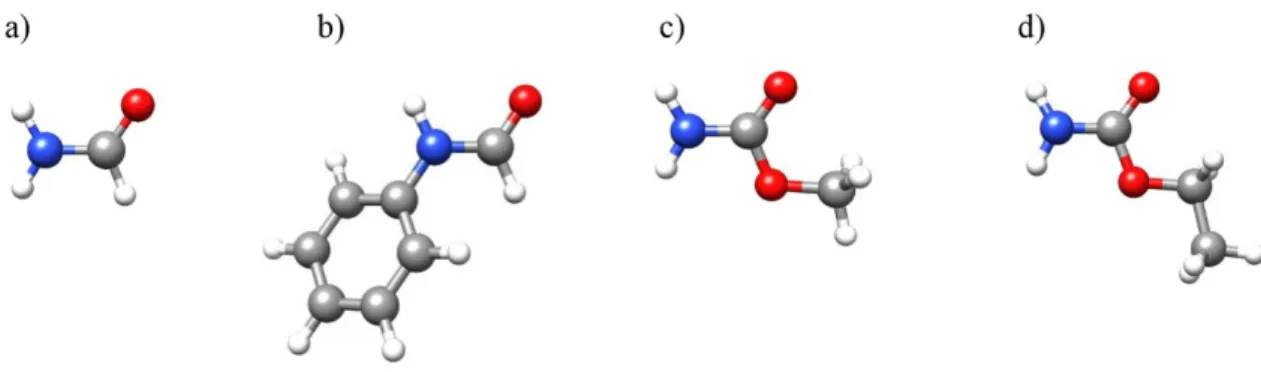
(18) both HB and π–delocalization, being the effects stronger as the number of cooperative HBs increases.84,85 For the peptide linkage, π-cooperative bonding was first pointed out to occur in Xray crystallographic studies of small amides.86,87 RAHB cooperative effects have been also described for chains of amide molecules, stabilizing the secondary structure of proteins.88 The polarization effects due to RAHB have been rarely observed in the gas phase.56 The changes in the C–N and C=O distances associated with π-cooperativity are usually in the limit of detection of usual methods due to vibrational effects, and therefore it is challenging to determine directly whether those changes occur or not. This missing experimental information can be supplied however by other properties directly determinable from the rotational spectra. One of such properties is the nuclear quadrupole coupling of 14N present in amide groups. As has been mentioned above, it is possible to resolve the hyperfine structure and determine the associated nuclear quadrupole coupling constants, which are related to the electric field gradient and the electronic environment at the coupled nucleus.51 These constants are very sensitive to the small changes associated with inductive cooperative effects. The electric field gradient has been in turn attributed to the unequal filling of the valence shell p orbitals in the coupling nucleus,51 thus, it is possible to expect a possible correlation between the nuclear quadrupole coupling constant and the unbalanced 2p electronic charge obtained from Natural Bond Orbital analysis.89 The inductive cooperative effects could be demonstrated by analyzing the tendencies in the values of the nuclear quadrupole coupling constants from isolation to the complexes with increasing hydration degrees, and consequently, quadrupole coupling constants could be used as a probe of the subtle effects due to π-cooperativity. A second probe of the RAHB effects could be the values of the internal rotation parameters of groups as methyl directly bonded to some of the amide group atoms. Investigations by microwave spectroscopy of monohydrated and dihydrated complexes of different amides,55-59 have led to remarkable pieces of information, as the preference of water to interact with the carbonyl group. This Thesis presents the microsolvation study of amides with different substitutes (see Figure 1.1) with the aim to model the local interactions between water and the peptide bond in different situations. Subsequent objectives in this work were i) to extend the previous studies to microsolvated complexes with additional water or solute molecules, ii) to characterize the structural changes due to HB cooperative effects, whereas those associated to σcooperativity are well known, the inductive polarization effects associated to π-cooperativity are less understood, iii) to determine the relation between the nuclear quadrupole coupling constants and the subtle effects caused by polarization, iv) to explore the HB interactions occurring between peptide groups, which has not received much attention in the gas phase.. Figure 1.1. Amides for which microsolvated complexes have been studied in this work. a) Formamide. b) Formanilide. c) Methyl carbamate. d) Ethyl carbamate.. 6.
(19) The experimental studies carried out are presented in the corresponding chapters within this Thesis, following a brief description of them will be given.. Formamide2···(H2O)90 Hydrogen-Bond Cooperativity in Formamide2-Water: A Model for Water Mediated Interactions. Susana Blanco, Pablo Pinacho, Juan Carlos López. Angewandte Chemie International Edition, 2016, 55, 9331–9335.. Formamide is the simplest amide, and thus it can be taken as a reference system. Its microsolvated complexes with one and two molecules of water have been already described in detail.55,58 It presents diverse interaction sites, and its complexes with water can model the interaction with water of the cis and trans peptide linkage. Our aim by studying this molecule was to extend the understanding of microsolvation to complexes increasing the number of molecules of formamide. We focused the search on a complex in which water acts as a bridge between two formamide molecules involving three hydrogen bonds (see Figure 1.2). The interest of searching for this complex lies in that it presents a unique hydrogen bond network that could serve to model the amide-water, amideamide and amide-amide water-mediated interactions. The study of this complex could lead to a better understanding of the interplay between microsolvation and self-association. We took special care in trying to determine very accurately the structure of the complex and the nuclear quadrupole coupling constants to test their possible use as a probe of the subtle inductive effects associated to π-cooperativity combining the results with the previous ones.58. Figure 1.2. The formamide2···(H2O) complex.. Formamide···(H2O)391 Structure and Dynamics in Formamide-(H2O)3: A Water Pentamer Analogue. Susana Blanco, Pablo Pinacho, Juan Carlos López. The Journal of Physical Chemistry Letters, 2017, 8, 6060–6066.. After the detection of formamide2···(H2O), we searched for the most stable complex of formamide with three molecules of water since hitherto no complex of this type has been reported for amides. In this complex, water molecules are supposed to interact with the amide group closing a 10-membered sequential cycle (see Figure 1.3), and could be considered as a continuation of the monohydrated and dihydrated complexes of formamide showing similar 7.
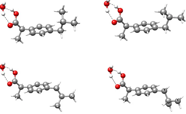
(20) interactions with an extended network. Thus, the microsolvation of formamide···(H2O)3 could serve as a model of the interactions between water and the peptide bond in a cis arrangement. In this context, we wanted to explore its planarity or not, and similarities between its structure and dynamics with those of pure water clusters (H2O)n n = 2–5.71-76 We wanted to continue the investigation of π-cooperative effects and the use of the nuclear quadrupole constants as a probe for the effects associated to polarization, in planar or not planar complexes.. Figure 1.3. The most stable configuration for the formamide···(H2O)3 complex.. Formanilide···(H2O)n n = 1,2 Manuscript in preparation.. The bonding between peptides occurs through amide linkages that can adopt cis or trans dispositions, regarding the relative arrangement of the amino and carbonyl groups, being the trans–peptide link much more represented in nature. Formanilide, or N-phenylformamide (see Figure 1.4) exists in two conformations in the gas phase,92 with the peptide group in cis or trans arrangement that model the peptide group arrangement in large systems. We proposed the investigation of its water complexes in the gas phase with microwave spectroscopy to model the local interactions of water with both forms of the peptide bond, and to find the main differences between them. An interesting point we wanted to study was the possible alteration on the structures of formanilide forms as the number of water molecules increases. The cis arrangement is not planar, and its complexes are expected not to alter significantly the angle between the phenyl and the amide groups. Opposite, the trans arrangement is planar, and the sterical impediment of the phenyl ring blocks the possibility to form sequential cycles as in formamide. We also wanted to continue the investigation of π-cooperativity in complexes of amides and the use of the nuclear quadrupole constants as a probe for the effects associated to polarization, in planar and not planar complexes.. 8.
(21) Figure 1.4. The most stable monohydrated and dihydrated complexes of formanilide for cis (up) and trans (down) conformations.. Methyl carbamate···(H2O)n n = 1,2 Manuscript in preparation.. Methyl carbamate is an ester-derivative of an amide (see Figure 1.5). The aim for investigating its microsolvated complexes was to model the HB interactions with a substituted cis-peptide bond. The starting point for the search were complexes showing similar interactions as those in formamide and formanilide. Methyl carbamate presents a heavy atom planar skeleton, and the water molecules in the complexes are expected to dispose in the plane of methyl carbamate. Therefore, these complexes could continue the investigation of quadrupole coupling as a probe of the πcooperative effects started in previous studies. Another main goal of this study was trying to find experimental evidence showing that the barrier to internal rotation can be a probe of RAHB effects. In this context, we searched for a correlation between nuclear quadrupole constants and internal rotation.. 9.
(22) Figure 1.5. The complexes of methyl carbamate - water.. Ethyl carbamate···(H2O)n n = 1,2,3 Manuscript in preparation.. Ethyl carbamate (see Figure 1.6) presents two conformations, planar and non-planar, that can be interconverted through a periodical potential function with a low barrier. This causes the observation of both conformers in a supersonic expansion to depend on the carrier gas used due to conformational relaxation in the supersonic jet. The aim of investigating its microsolvation complexes was, apart from modelling the interactions of water with the carbamate group, to study the influence or not of water microsolvation in the corresponding conformational equilibrium.. Figure 1.6. Complexes of ethyl carbamate I (up) and II (down) with up to three water molecules.. 10.
(23) Ibuprofen···(H2O) Manuscript in preparation.. Ibuprofen is one of the most popular medicines, it presents a carboxylic acid group. We searched for the microsolvated complex with the aim of model the local interaction between water and acids in larger molecules. The starting point of the investigation were the monohydrated complexes for the four ibuprofen monomer conformations previously studied in the gas phase.93 The aim was to observe the complexes with the four rotamers, characterize them, and to analyze the possible influence of water on the conformational equilibrium. It is expected that water molecule closes a cycle with the carboxylic group, as depicted in Figure 1.7.. Figure 1.7. The predicted most stable complexes of water with the four rotamers observed for ibuprofen.. 11.
(24) References Hofacker G.L. “Intra- and Intermolecular Interactions” in Biophysics, eds. Hoppe W., Lohmann W., Markl H., Ziegler H. Springer, Berlin, 1982. 2 Baker, E. N., Hubbard, R. E. “Hydrogen bonding in globular proteins” Prog. Biophys. Mol. Biol., 1984, 44, 97–179. 3 Saenger, W., Jeffrey, G. A. Hydrogen Bonding in Biological Structures, Springer-Verlag, Berlin, 1991. 4 Jeffrey, G. A. An Introduction to Hydrogen Bonding, Oxford University Press, Oxford, 1997. 5 McNaught, A. D., Wilkinson, A. IUPAC, Compendium of Chemical Terminology (The "Gold Book"), 2nd ed., Blackwell Scientific Publications, Oxford, 1997. 6 Saenger, W. “Structure and Dynamics of Water Surrounding Biomolecules” Ann. Rev. Biophys. Chem., 1987, 16, 93–114. 7 Schmitt, M., Böhm, M., Ratzer, C., Vu, C., Kalkman, I., Meerts, W. L. “Structural Selection by Microsolvation: Conformational Locking of Tryptamine” J. Am. Chem. Soc., 2005, 127, 10356–10364. 8 Caminati, W., López, J. C., Blanco, S., Mata, S., Alonso, J. L. “How Water Links to Cis and Trans Peptidic Groups: The rotational Spectrum of N-Methylformamide-Water” Phys. Chem. Chem. Phys., 2010, 12, 10230–10234. 9 Maris, A., Ottaviani P., Caminati, W. “Pure Rotational Spectrum of 2-Pyridone···Water and Quantum Chemical Calculations on the Tautomeric Equilibrium 2-Pyridone···Water/2-Hydroxypyridine···Water” Chem. Phys. Lett., 2002, 360, 155–160. 10 Mata, S., Cortijo, V., Caminati, W., Alonso, J. L., Sanz, M. E., López, J. L., Blanco, S. “Tautomerism and Microsolvation in 2-Hydroxypyridine/2-Pyridone” J. Phys. Chem. A, 2010, 114, 11393–11398. 11 Wyttenbach, T., Paizs, B., Barran, P., Breci, L., Liu, D., Suhai, S., Wysocki, V. H., Bowers, M. T. “The Effect of the Initial Water of Hydration on the Energetics, Structures, and H/D Exchange Mechanism of a Family of Pentapeptides: An Experimental and Theoretical Study” J. Am. Chem. Soc., 2003, 125, 13768– 13775. 12 Xu, S. J., Nilles, M., Bowen, K. H., “Zwitterion Formation in Hydrated Amino Acid, Dipole Bound Anions: How Many Water Molecules Are Required?” J. Chem. Phys., 2003, 119, 10696–10701. 13 Robinson, C. R., Sligar, S. G. “Molecular Recognition Mediated by Bound Water: A Mechanism for Star Activity of the Restriction Endonuclease EcoRI” J. Mol. Biol., 1993, 234, 302–306. 14 Jayaram, B., Jain, T. A. “The Role of Water in Protein-DNA Recognition” Annu. Rev. Biophys. Biomol. Struct., 2004, 33, 343–361. 15 Levy, Y., Onuchic, J. N. “Water Mediation in Protein Folding and Molecular Recognition” Annu. Rev. Biophys. Biomol. Struct., 2006, 35, 389–415. 16 Biedermannová, L., Schneider, B. “Hydration of Proteins and Nucleic Acids: Advances in Experiment and Theory. A Review” Biochimica et Biophysica Acta, 2016, 1860, 1821–1835. 17 Groot, B. L., Grubmüller, H. “Water Permeation across Biological Membranes: Mechanism and Dynamics of Aquaporin-1 and GlpF” Science, 2001, 294, 2353–2357. 18 Ball, P. “Water as an Active Constituent in Cell Biology” Chem. Rev., 2008, 108, 74–108. 19 Venkatachalam, C. M. “Stereochemical criteria for Polypeptides and Proteins. V. Conformation of a System of Three Linked Peptide Units” Biopolymers, 1968, 6, 1425–1436. 20 Chaplin, M. “Do We Underestimate the Importance of Water in Cell Biology?” Nat. Rev. Mol. Cell Biol., 2006, 7, 861–866. 21 Modig, K., Liepinsh, E., Otting, G., Halle, B. “Dynamics of Protein and Peptide Hydration” J. Am. Chem. Soc., 2004, 126, 102–114. 22 Nucci, N. V., Pometun, M. S., Wand, A. J. “Site-Resolved Measurement of Water-Protein Interactions by Solution NMR” Nat. Struct. Mol. Biol., 2011, 18, 245–249. 23 Hilser, V. J. “Structural Biology: Finding the Wet Spots” Nature, 2011, 469, 166–167. 24 Egli, M., Portmann, S., Usman, N. “RNA Hydration: A Detailed Look” Biochemistry, 1996, 35, 8489– 8494. 25 Schotte, F., Lim, M., Jackson, T. A., Smirnov, A. V., Soman, J., Olson, J. S., Phillips Jr, G. N., Wulff, M., Anfinrud, P. A. “Watching a Protein as it Functions with 150-ps Time-Resolved X-Ray Crystallography” Science, 2003, 300, 1944–1947. 26 Nibbering, E. T. J., Elsaesser, T. “Ultrafast Vibrational Dynamics of Hydrogen Bonds in the Condensed Phase” Chem. Rev., 2004, 104, 1887–1914. 27 Bakker, H. J., Skinner, J. L. “Vibrational Spectroscopy as a Probe of Structure and Dynamics in Liquid Water” Chem. Rev., 2010, 110, 1498–1517. 1. 12.
(25) Tielrooij, K. J., García-Araez, N., Bonn, M., Bakker, H. J. “Cooperativity in Ion Hydration” Science, 2010, 328, 1006–1009. 29 Perera, A. S., Thomas, J., Poopari, M. R., Xu, Y. “The Clusters-in-a-Liquid Approach for Solvation: New Insights from the Conformer Specific Gas Phase Spectroscopy and Vibrational Optical Activity Spectroscopy” Front. Chem., 2016, 4, 1–17. 30 Chem. Rev., 2016, 116, Issue 9 “Noncovalent interactions”, 4911–5688. 31 Chem. Rev., 2016, 116, Issue 13 “Water – The most anomalous liquid”, 7459–7726. 32 Bagchi, B. Water in Biological and Chemical Processes: From structure and Dynamics to Function, Cambridge University Press, Cambridge, 2013. 33 Gerhards, M. “Spectroscopy of Neutral Peptides in the Gas Phase: Structure, reactivity, Microsolvation, Molecular Recognition” in Principles of Mass Spectrometry Applied to Biomolecules, eds. Laskin, J., Lifshitz, C., 2006. 34 Levy, D. H. “Laser Spectroscopy of Cold Gas-Phase Molecules” Annu. Rev. Phys. Chem., 1980, 31, 197–225. 35 Rizzo, T. R., Park, Y. D., Petenau, L., Levy, D. H. “Electronic Spectrum of the Amino Acid Tryptophan Cooled in a Supersonic Molecular Beam” J. Chem. Phys., 1985, 83, 4819–4820. 36 Philips, L. A., Levy, D. H. “The Rotationally Resolved Electronic Spectrum of Indole in the Gas Phase” J. Chem. Phys., 1986, 85, 1327–1332. 37 Philips, L. A., Levy, D. H. “Rotationally Resolved Electronic Spectroscopy of Tryptamine Conformers in a Supersonic Jet” J. Chem. Phys., 1988, 89, 85–90. 38 Legon, A. C., Soper, P. D., Flygare, W. H. “The Rotational Spectrum, H, 19F Nuclear Spin-Nuclear Spin Coupling, D Nuclear Quadrupole Coupling, and Molecular Geometry of a Weakly Bound Dimer of Carbon Monoxide and Hydrogen Fluoride” J. Chem. Phys., 1981, 74, 4944–4950. 39 Legon, A. C., Aldrich, P. D., Flygare, W. H. “The Rotational Spectrum, Chlorine Nuclear Quadrupole Coupling Constants, and Molecular Geometry of a Hydrogen-Bonded Dimer of Cyclopropane and Hydrogen Chloride” J. Am. Chem. Soc., 1982, 104, 1486–1490. 40 Legon, A. C., Millen, D. J. “Gas-Phase Spectroscopy and the Properties of Hydrogen-Bonded Dimers: HCN···HF as the Spectroscopic Prototype” Chem. Rev., 1986, 86, 635–657. 41 Kisiel, Z., Fowler, P. W., Legon, A. C. “Investigation of the Rotational Spectrum of the HydrogenBonded Dimer Formed between Methylenecyclopropane and HCl” J. Chem. Phys., 1994, 101, 4635– 4643. 42 Legon, A. C. “Prereactive Complexes of Dihalogens XY with Lewis Bases B in the Gas Phase: A Systematic Case for the Halogen Analogue B···XY of the Hydrogen Bond B···HX” Angew. Chem. Int. Ed., 1999, 38, 2686–2714. 43 Becucci, M., Melandri, S. “High-Resolution Spectroscopic Studies of Complexes Formed by MediumSize Organic Molecules” Chem. Rev., 2016, 116, 5014–5037. 44 Levy, D. H. “The Spectroscopy of Very Cold Gases” Science, 1981, 214, 263–269. 45 Miller, D. R. “Free Jet Sources” in Atomic and Molecular Beam Methods ed. Scoles, G. Oxford University press, Oxford, 1988, Volume 1, pp. 14–53. 46 Montero, S., Maté, B., Tejeda, G., Fernández, J. M., Ramos, A. “Raman Studies of Free Jet Expansion” in Atomic and Molecular Beams. The state of Art 2000 ed. Campargue, R. Springer-Verlag, Berlin, 2001, pp. 295–306. 47 Maté, B., Graur, I. A., Elizarova, T., Chirokov, I., Tejeda, G., Fernández, J. M., Montero, S. “Experimental and Numerical Investigation of an Axisymmetric Supersonic Jet” J. Fluid Mech., 2001, 426, 177–197. 48 Domenicano, A., Hargittai, I. Accurate Molecular Structures: Their Determination and Importance Oxford University Press, New York, 1992. 49 Demaison, J. “Accurate Structures of Non-Rigid Molecules by Microwave Spectroscopy” in Structures and Conformations of Non-Rigid Molecules eds. Laane, J., Dakkouri, M., van der Veken, B., Oberhammer, H. Springer, Dordrecht, 1993, Volume 410, pp. 239–256. 50 Braag, J. K. “The Interaction of Nuclear Electric Quadrupole Moments with Molecular Rotation in Asymmetric-Top Molecules. I” Phys. Rev., 1948, 74, 533–538. 51 Gordy, W., Cook, R. L. Microwave Molecular Spectra, Wiley-Interscience, New York, 1984. 52 Lin, C. C., Swalen, J. D. “Internal Rotation and Microwave Spectroscopy” Rev. Mod. Phys., 1959, 31, 841–892. 53 Lister, D. G., Macdonald, J. N., Owen, N. L. “Internal Rotation and Inversion. An Introduction to Large Amplitude Motions in Molecules” Academic Press, London, 1978. 28. 13.
(26) Herschbach, D. R. “Calculation of Energy Levels for Internal Torsion and Over-All Rotation. III” J. Chem. Phys., 1959, 31, 91–108. 55 Lovas, F. J., Suenram, R. D., Fraser, G. T., Gillies, C. W., Zozom, J. “The Microwave Spectrum of Formamide-Water and Formamide-Methanol Complexes” J. Chem. Phys., 1988, 88, 722–729. 56 Held, A., Pratt, D. W. “Hydrogen Bonding in Water Complexes. Structures of 2-Pyridone-H2O and 2Pyrididone-(H2O)2 in Their S0 and S1 Electronic States” J. Am. Chem. Soc., 1993, 115, 9708–9717. 57 Lavrich, R. J., Tubergen, M. J. “Conformation and Hydrogen Bonding in the Alaninamide-Water van der Waals Complex” J. Am. Chem. Soc., 2000, 122, 2938–2943. 58 Blanco, S., López, J. C., Lesarri, A., Alonso, J. L. “Microsolvation of Formamide: A Rotational Study” J. Am. Chem. Soc., 2006, 128, 12111–12121. 59 López, J. C., Sanchez, R., Blanco, S., Alonso, J. L. “Microsolvation of 2-Azetidinone: A Model for the Peptide Group-Water Interactions” Phys. Chem. Chem. Phys., 2015, 17, 2054–2066. 60 Canagaratna, M., Phillips, J. A., Ott, M. E. Leopold, K. R. “The Nitric-Acid Complex: Microwave Spectrum, Structure and Tunneling” J. Phys. Chem. A, 1998, 102, 1489–1497. 61 Ouyang, B., Starkey, T. G., Howard, B. J. “High-Resolution Microwave Studies of Ring-Structured Complexes between Trifluoroacetic Acid and Water” J. Phys. Chem. A, 2007, 111, 6165–6175. 62 Ouyang, B., Howard, B. J. “Hydrates of Trans- and Gauche-Difluoroacetic Acids: A High-Resolution Microwave Spectroscopic Study” J. Phys. Chem. A, 2010, 114, 4109–4117. 63 Schnitzler, E. G., Jäger, W. “The Benzoic Acid-Water Complex: A Potential Atmospheric Nucleation Precursor Studied Using Microwave Spectroscopy and Ab Initio Calculations” Phys. Chem. Chem. Phys., 2014, 16, 2305–2314. 64 Thomas, J., Sukhorukov, O., Jäger, W., Xu, Y. “Direct Spectroscopic Detection of the Orientation of Free OH Groups in Methyl Lactate-(Water)1,2 Clusters: Hydration of a Chiral Hydroxy Ester” Angew. Chem. Int. Ed., 2014, 53, 1156–1159. 65 Gall, J. T. A., Thomas, J., Xie, F., Wang, Z., Jäger, W., Xu, Y. “Rotational Spectroscopy of the MethylGlycidate-Water Complex: Conformation and Water and Methyl Rotor Tunneling Motions” Phys. Chem. Chem. Phys., 2017, 19, 29508–29515. 66 Su, Z., Xu, Y. “Hydration of a Chiral Molecule: The Propylene Oxide···(Water) 2 Cluster in the Gas Phase” Angew. Chem. Int. Ed., 2007, 119, 6275–6278. 67 Pérez, C., Neill, J. L., Muckle, M. T., Zaleski, D. P., Peña, I., López, J. C., Alonso, J. L., Pate, B. H. “Water-Water and Water-Solute Interactions in Microsolvated Organic Complexes” Angew. Chem. Int. Ed., 2014, 54, 979–982. 68 Pérez, C., López, J. C., Blanco, S., Schnell, M. “Water-Induced Structural Changes in Crown Ethers from Broadband Rotational Spectroscopy” J. Phys. Chem. Lett., 2016, 7, 4053–4058. 69 Pérez, C., Krin, A., Steber, A. L., López, J. C., Kisiel, Z., Schnell, M. “Wetting Camphor: MultiIsotopic Substitution Identifies the Complementary Roles of Hydrogen Bonding and Dispersive Forces” J. Phys. Chem. Lett., 2016, 7, 154–160. 70 Bellissent-Funel, M.-C.; Hassanali, A.; Havenith, M.; Henchman, R.; Pohl, P.; Sterpone, F.; van der Spoel, D.; Xu, Y.; Garcia, A. E. “Water Determines the Structure and Dynamics of Proteins” Chem. Rev., 2016, 116, 7673–7697. 71 Dyke, T. R.; Mack, K. M.; Muenter, J. S. “The Structure of Water Dimer from Molecular Beam Electric Resonance Spectroscopy” J. Chem. Phys., 1977, 66, 498–510. 72 Keutsch, F. N.; Cruzan, J. D.; Saykally, R. J. “The Water Trimer” Chem. Rev., 2003, 103, 2533–2577. 73 Cruzan, J. D.; Braly, L. B.; Liu, K.; Brown, M. G.; Loeser, J. G.; Saykally, R. J. “Quantifying Hydrogen Bond Cooperativity in Water: VRT Spectroscopy of the Water Tetramer” Science, 1996, 271, 59–62. 74 Liu, K.; Brown, M. G.; Cruzan, J. D.; Saykally, R. J. “Vibration-Rotation Tunneling Spectra of the Water Pentamer: Structure and Dynamics” Science, 1996, 271, 62–64. 75 Ramírez, F.; Hadad, C. Z.; Guerra, D.; David, J.; Restrepo, A. “Structural Studies of the Water Pentamer” Chem. Phys. Lett., 2011, 507, 229–233. 76 Cole, W. T. S.; Fellers, R. S.; Viant, M. R.; Saykally, R. J. “Hydrogen Bond Breaking Dynamics in the Water Pentamer: Terahertz VRT Spectroscopy of a 20 µm Libration” J. Chem. Phys., 2017, 146, 14306. 77 Pérez, C., Muckle, M. T., Zaleski, D. P., Seifert, N. A., Temelso, B., Shields, G. C., Kisiel, Z., Pate, B. H. “Structures of Cage, Prism, and Book Isomers of Water Hexamer from Broadband Rotational Spectroscopy” Science, 2012, 336, 897–901. 78 Richardson, J. O., Pérez, C., Lobsiger, S., Reid, A. A., Temelso, B., Shields, G. C., Kisiel, Z., Wales, D. J., Pate, B. H., Althorpe, S. C. “Concerted Hydrogen-Bond Breaking by Quantum Tunneling in the Water Hexamer Prism” Science, 2016, 351, 1310–1313. 54. 14.
(27) 79. Pérez, C., Lobsiger, S., Seifert, N. A., Zaleski, D. P., Temelso, B., Shields, G. C., Kisiel, Z., Pate, B. H. “Broadband Fourier Transform Rotational Spectroscopy for Structure Determination: The Water Heptamer” Chem. Phys. Lett., 2013, 571, 1–15. 80 Pérez, C., Zaleski, D. P., Seifert, N. A., Temelso, B., Shields, G. C., Kisiel, Z., Pate, B. H. “Hydrogen Bond Cooperativity and the Three-Dimensional Structures of Water Nonamers and Decamers” Angew. Chem. Int. Ed., 2014, 53, 14368–14372. 81 Del Bene, J. E. “Molecular Orbital Theory of the Hydrogen Bond. 18. Methyl Substituent Effects on Amide Hydrogen Bonding” J. Am. Chem. Soc., 1978, 100, 1387–1394 82 Liang, W., Li, H., Hu, X., Han, S. “Proton Transfer of Formamide + nH2O (n = 0-3): Protective and Assistant Effect of the Water Molecule” J. Phys. Chem. A, 2004, 108, 10219–10224. 83 Venkataramanan, N. S. “Cooperativity of Intermolecular Hydrogen Bonds in Microsolvated DMSO and DMF Clusters: A DFT, AIM, and NCI Analysis” J. Mol. Model, 2016, 22, 151–162. 84 Gilli, G., Bellucci, F., Ferretti, V., Bertolasi, V. “Evidence for Resonance-Assisted Hydrogen Bonding from Crystal-Structure Correlations on the Enol Form of the β–Diketone Fragment” J. Am. Chem. Soc., 1989, 111, 1023–1028. 85 Bertolasi, V., Gilli, P., Ferretti, V., Gilli, G. “Evidence for Resonance-Assisted Hydrogen Bonding. 2. Intercorrelation between Crystal Structure and Spectroscopic Parameters in Eight Intramolecularly Hydrogen Bonded 1,3.Diaryl-1,3-propanedione Enols” J. Am. Chem. Soc., 1991, 113, 4917–4925. 86 Gilli, P., Bertolasi, V., Ferretti, V., Gilli, G. “Evidence for Intramolecular N-H···O Resonance Assisted Hydrogen Bonding in β-Enaminones and Related Heterodienes. A Combined Crystal-Structural, IR, and NMR Spectroscopic, and Quantum-Mechanical Investigation” J. Am. Chem. Soc., 2000, 122, 10405– 10417. 87 Ottersen, T. “On the structure of the Peptide Linkage. The Structures of Formamide and Acetamide at 165 Degrees C and an Ab Initio Study of Formamide, Acetamide and N-methylformamide” Acta Chemica Scandinavica., 1975, 29a, 939–944. 88 Kobko, N., Dannenberg, J. J. “Cooperativity in Amide Hydrogen Bonding Chains. Relation between Energy, Position, and H-Bond Chain in Peptide and Protein Folding Models” J. Phys. Chem. A, 2003, 107, 10389–10395. 89 Reed, A. E., Weinstock, R. B., Weinhold, F. “Natural Population Analysis” J. Chem. Phys., 1985, 83, 735–746. 90 Blanco, S., Pinacho, P., López, J. C. “Hydrogen-Bond Cooperativity in Formamide2-Water: A Model for Water Mediated Interactions” Angew. Chem. Int. Ed., 2016, 55, 9331–9335. 91 Blanco, S., Pinacho, P., López, J. C. “Structure and Dynamics in Formamide-(H2O)3: A Water Pentamer Analogue” J Phys. Chem. Lett., 2017, 8, 6060–6066. 92 Blanco, S., López, J. C., Lesarri, A., Caminati, W., Alonso, J. L. “Conformational Equilibrium of Formanilide: Detection of the Pure Rotational Spectrum of the Tunnelling Cis Conformer” Mol. Phys., 2005, 103, 1473-1479. 93 Betz, T., Zinn S., Schnell, M. “The Shape of Ibuprofen in the Gas Phase” Phys. Chem. Chem. Phys., 2015, 17, 4538–4541.. 15.
(28)
(29) Chapter II Methodology. 17.
(30) 18.
(31) 2.1 Introduction Spectroscopy is defined by IUPAC as “the study of physical systems by the electromagnetic radiation with which they interact or that they produce”.1 It is an experimental discipline that studies the resonant or non-resonant interaction between the electromagnetic spectrum and the quantized states that matter presents at the microscopic scale. The application of Quantum Mechanics to analyze the spectrum obtained by spectroscopic techniques, allows us to obtain the energy for those quantized states, which are closely related to the molecular structures and the dynamics of the system.2,3 This work is focused on rotational spectroscopy which uses microwave radiation lying in the centimeter-wave (1-30 GHz), in the millimeter-wave (30-300 GHz) and in the submillimeter-wave (>300 GHz) regions. The quantum-mechanical description of the mechanisms of molecular rotation can be found in the bibliography4-8 and only a concise description focused on molecular rotation will be given here.. 2.2 Rotational Spectroscopy 2.2.1 Rigid-Rotor Hamiltonian The analysis of the microwave spectrum begins by obtaining the energies for the rotational levels. For a system at the microscopic scale, the quantized energy for the states can be calculated as the eigenvalues of the Hamiltonian operator in the Schrödinger equation: ̂ 𝜓 = 𝐸𝜓 𝐻 Considering the Born-Oppenheimer approximation, the nuclear Hamiltonian takes into account expressions for the kinetic and potential energies for the nuclei of the system and is a sum of the translation, vibrational and rotational motions. In a particular axis system with origin fixed at the center of mass, the rotational Hamiltonian can be approximately separated from the vibrational term. For a rotating system formed by i particles, each one with mass mi and spatial coordinates xi, yi, zi in a Cartesian axis system with origin fixed at the center of mass, the rotational kinetic energy is: 1 𝑡 1 2 2 2 𝐸rot = 𝜔 ⃗ 𝐼𝜔 ⃗ = [𝐼xx 𝜔xx + 𝐼yy 𝜔yy + 𝐼zz 𝜔zz + 2𝐼xy 𝜔x 𝜔y + 2𝐼xz 𝜔x 𝜔z + 2𝐼yz 𝜔y 𝜔z ] 2 2 where ωαβ (α,β = x, y or z) are the components of the angular velocity vector for the system in rotation, and Iαβ (α,β = x, y or z) the components of the inertia tensor. The tensor of inertia can be written in matrix representation: 𝐼xx (𝐼yx 𝐼zx. 𝐼xy 𝐼yy 𝐼zy. 𝐼xz 𝐼yz ) 𝐼zz. The diagonal components are the moments of inertia and the non-diagonal elements are the products of inertia. The tensor of inertia depends on the masses of the particles and their spatial disposition:. 19.
(32) 𝐼xx = ∑𝑖 𝑚𝑖 (𝑦𝑖2 + 𝑧𝑖2 ) 𝐼yx = 𝐼xy = ∑𝑖 𝑚𝑖 (𝑥𝑖 𝑦𝑖 ). 𝐼yy = ∑𝑖 𝑚𝑖 (𝑥𝑖2 + 𝑧𝑖2 ) 𝐼zx = 𝐼xz = ∑𝑖 𝑚𝑖 (𝑥𝑖 𝑧𝑖 ). 𝐼zz = ∑𝑖 𝑚𝑖 (𝑥𝑖2 + 𝑦𝑖2 ) 𝐼zy = 𝐼yz = ∑𝑖 𝑚𝑖 (𝑦𝑖 𝑧𝑖 ). Any rotating system (molecule or cluster) has a characteristic inertia ellipsoid (Figure 2.1) defined by three semiaxes, each one proportional to the reciprocal of the square root of the correspondent moment of inertia.. Figure 2.1. The inertial ellipsoid (transparent gray) for ethyl carbamate I – water-a complex (see Chapter VIII), an almost prolate top (κ ≈ -0.97), with the a, b and c inertial axes. To simplify the solution we can choose the axes in such a manner that the products of inertia become zero and the matrix results in a diagonal tensor. For each molecular system, there is a unique definition of the axes that fulfill this criterion, they are called the principal axes of inertia, labeled a, b and c, so that Ia ≤ Ib ≤ Ic. The values of the principal moments of inertia allow classifying the molecules as: - Linear tops (Ia = 0, Ib = Ic). - Spherical tops (Ia = Ib = Ic). - Oblate symmetric tops (Ia = Ib < Ic). - Prolate symmetric tops (Ia < Ib = Ic). - Asymmetric tops (Ia < Ib < Ic). In the principal axis system the rotational energy takes the form: 1 𝐸rot = [𝐼𝑎 𝜔𝑎2 + 𝐼𝑏 𝜔𝑏2 + 𝐼𝑐 𝜔𝑐2 ] 2 This expression can be written in function of the angular momentum components (𝐽 = 𝐼𝜔 ⃗ ): 𝐸rot. 1 𝐽𝑎2 𝐽𝑏2 𝐽𝑐2 = [ + + ] 2 𝐼𝑎 𝐼𝑏 𝐼𝑐. 20.
(33) The quantum mechanical equivalent to 𝐽 is the angular momentum operator 𝐽̂. The operators of the angular momentum components in the molecule-fixed system are related to the overall angular momentum operator by 𝐽̂2 = 𝐽̂ 2𝑥 + 𝐽̂ 2𝑦 + 𝐽̂ 2𝑧 . Therefore, the rigid-rotor Hamiltonian results: ̂rig 𝜓 = 𝐸rot 𝜓 𝐻 ̂rig = 𝐻. ℏ2 𝐽̂𝑎2 ℏ2 𝐽̂𝑏2 ℏ2 𝐽̂𝑐2 + + = 𝐴𝐽̂𝑎2 + 𝐵𝐽̂𝑏2 + 𝐶𝐽̂𝑐2 2𝐼𝑎 2𝐼𝑏 2𝐼𝑐. where 𝐴 = ℏ2 /2𝐼𝑎 , 𝐵 = ℏ2 /2𝐼𝑏 and 𝐶 = ℏ2 /2𝐼𝑐 are the rotational constants. The Hamiltonian commutes with 𝐽̂2 . For symmetric tops, 𝐽̂𝑧 , the angular momentum operator component along the z axis in the principal axis system and 𝐽̂2 commute, that is, they have a common set of eigenfunctions because they are defined simultaneously. Furthermore, 𝐽̂𝑍 , the angular momentum operator component along the Z axis in the laboratory system and 𝐽̂2 also commute because 𝐽̂2 is independent of the coordinate system chosen. This implies that there exist a common set of four eigenfunctions 𝜓𝐽,𝐾,𝑀 for the operators. Each operator has a correspondent eigenvalue (kJ, kK and kM) which are the solutions for the eigenvalue equation. Using bracket notation (𝜓𝐽,𝐾,𝑀 ≡ ⟨𝐽, 𝐾, 𝑀|): ⟨𝐽, 𝐾, 𝑀|𝐽̂2 |𝐽, 𝐾, 𝑀⟩ = 𝑘𝐽 = ℏ2 𝐽(𝐽 + 1) ⟨𝐽, 𝐾, 𝑀|𝐽̂𝑧 |𝐽, 𝐾, 𝑀⟩ = 𝑘𝐾 = 𝐾ℏ ⟨𝐽, 𝐾, 𝑀|𝐽̂𝑍 |𝐽, 𝐾, 𝑀⟩ = 𝑘𝑀 = 𝑀ℏ The eigenvalues of these equations are the quantized values of the energy for a wave function defined by the three quantum numbers J, K, and M. Quantum number J is related to the total rotational angular momentum of the system and takes 0 or any positive integer value. K and M are related to the projections of the angular momentum over the molecule-fixed axis z or over the laboratory-fixed axis Z respectively. Both K and M can take any integer value from –J to +J. All the levels with K ≠ 0 are doubly degenerated. It is possible to rearrange the Hamiltonian to be only a function of 𝐽̂2 , 𝐽̂𝑧 , and 𝐽̂𝑍 . From the quantum numbers simple expressions for the energy, and therefore for the frequencies can be obtained. Asymmetric tops represent a different situation. Only J and M are good quantum numbers and it is not possible to solve directly the Schrödinger equation. To obtain the energy levels, the wave functions for an asymmetric top are described by a linear combination of the symmetric top functions and the problem is solved using a linear variational method. The energy levels for an asymmetric top obtained by this procedure can be correlated to the levels of the symmetric top limits as described in Diagram 2.1. The energy levels are not labeled with the K quantum number as in symmetric tops, but with two pseudo-quantum numbers, K-1 and K+1 which describe the values of K in the corresponding limits, prolate (K-1) and oblate (K+1).6-9. 21.
(34) Diagram 2.1. Correlation diagram for the asymmetric top energy levels with those of the symmetric rotor in the prolate and oblate limits. Only the three lowest J levels are depicted. Asymmetric tops present 2J+1 rotational levels for each value of J. Each rotational level is degenerated in 2J+1 levels for each value of M, but the quantization on M is only relevant in the presence of an external electric field. The degree of asymmetry of a molecular system can be evaluated by the value of the asymmetry parameter of Ray,6,10 κ = (2B-A-C)/(A-C) which can take values from -1 to +1. The limit situations correspond to the prolate (B = C, κ = -1) and the oblate (A = B, κ = +1) symmetric tops.. 2.2.2 Distortable-Rotor Hamiltonian The rigid-rotor model is an approximation in which molecules are treated as point nuclei held by rigid-bonds. However, the frequencies observed experimentally present shifts from those predicted by the rigid-rotor, being the differences larger with increasing J. This effect can be explained by the centrifugal distortion forces that elongate the bonds, changing the values of the moments of inertia. Therefore, chemical bonds are better represented as springs, with a finite restoring force, that can be distorted according to the rotation of the molecule. In order to obtain more accurate frequencies, it is needed to introduce correcting terms to the rotational Hamiltonian taking into account for the centrifugal distortion forces.11-13 The Hamiltonian including up to sixth-order correction in the angular momentum operator terms centrifugal distortion constants is of the form: ̂rot = 𝐻 ̂rig + 𝐻. ℏ4 ∑ 𝜏𝛼𝛽𝛾𝛿 𝐽̂𝛼 𝐽̂𝛽 𝐽̂𝛾 𝐽̂𝛿 + ℏ6 ∑ 𝜏𝛼𝛽𝛾𝛿𝜀𝜂 𝐽̂𝛼 𝐽̂𝛽 𝐽̂𝛾 𝐽̂𝛿 𝐽̂𝜀 𝐽̂𝜂 4 𝛼𝛽𝛾𝛿. 𝛼𝛽𝛾𝛿𝜀𝜂. where α, β, γ, δ, ε and η in the summations can take any value of a, b or c coordinates in the principal axis system. It has been demonstrated thanks to symmetry and commutation properties that the number of those terms can be reduced to six terms for the fourth-order and 10 for the sixth-order corrections, being not possible to determine experimentally all those terms.6,13 Watson demonstrated that only five linear combinations of the fourth-order and seven combinations of the sixth-order can be determined. Watson also proposed two possible combinations of terms giving rise to the reduced Hamiltonians usually used to analyze the centrifugal distortion, the S (symmetric) and the A (asymmetric) reductions.5,14 Watson´s A reduced Hamiltonian in the Ir representation up to the sixth-order correction is of the form: 22.
(35) ̂rot = 𝐻 ̂rig + 𝐻 ̂ (4) + 𝐻 ̂ (6) 𝐻 dis dis (A) ̂rig 𝐻 = 𝐴(A) 𝐽̂𝑎2 + 𝐵(A) 𝐽̂𝑏2 + 𝐶 (A) 𝐽̂𝑐2. ̂ (4) = −𝛥𝐽 𝐽̂4 − 𝛥𝐽𝐾 𝐽̂2 𝐽̂𝑎2 − 𝛥𝐾 𝐽̂𝑎4 − 2𝛿𝐽 𝐽̂2 (𝐽̂𝑏2 − 𝐽̂𝑐2 ) − 𝛿𝐾 [𝐽̂𝑎2 (𝐽̂𝑏2 − 𝐽̂𝑐2 ) + (𝐽̂𝑏2 − 𝐽̂𝑐2 )𝐽̂𝑎2 ] 𝐻 dis ̂ (6) = 𝛷𝐽 𝐽̂6 + 𝛷𝐽𝐾 𝐽̂4 𝐽̂𝑎2 + 𝛷𝐾𝐽 𝐽̂2 𝐽̂𝑎4 + 𝛷𝐾 𝐽̂𝑎6 + 2𝜙𝐽 𝐽̂4 (𝐽̂𝑏2 − 𝐽̂𝑐2 ) + 𝜙𝐽𝐾 𝐽̂2 [𝐽̂𝑎2 (𝐽̂𝑏2 − 𝐽̂𝑐2 ) + (𝐽̂𝑏2 𝐻 dis − 𝐽̂𝑐2 )𝐽̂𝑎2 ] + 𝜙𝐾 [𝐽̂𝑎4 (𝐽̂𝑏2 − 𝐽̂𝑐2 ) + (𝐽̂𝑏2 − 𝐽̂𝑐2 )𝐽̂𝑎4 ] where 𝛥𝐽 , 𝛥𝐽𝐾 , 𝛥𝐾 , 𝛿𝐽 and 𝛿𝐾 are the quartic centrifugal distortion constants and 𝛷𝐽 , 𝛷𝐽𝐾 , 𝛷𝐾𝐽 , 𝛷𝐾 , 𝜙𝐽 , 𝜙𝐽𝐾 and 𝜙𝐾 are the sextic centrifugal distortion constants in the A reduction. However, if the system is very close to the symmetric top limits the A reduction may not be appropriate since 𝛿𝐾 , which depends on (B-C) in the denominator gets very large values.6 In this situation the S reduction may result in a better description of the centrifugal forces. Watson´s S reduced Hamiltonian in the Ir representation up to the sixth-order energy terms is of the form: (S) ̂rig 𝐻 = 𝐴(S) 𝐽̂𝑎2 + 𝐵(S) 𝐽̂𝑏2 + 𝐶 (S) 𝐽̂𝑐2. ̂ (4) = −𝐷𝐽 𝐽̂4 − 𝐷𝐽𝐾 𝐽̂2 𝐽̂𝑎2 − 𝐷𝐾 𝐽̂𝑎4 + 𝑑1 𝐽̂2 (𝐽̂+2 + 𝐽̂−2 ) + 𝑑2 (𝐽̂+4 + 𝐽̂−4 ) 𝐻 dis ̂ (6) = +𝐻𝐽 𝐽̂6 + 𝐻𝐽𝐾 𝐽̂4 𝐽̂𝑎2 + 𝐻𝐾𝐽 𝐽̂2 𝐽̂𝑎4 + 𝐻𝐾 𝐽̂𝑎6 + ℎ1 𝐽̂4 (𝐽̂+2 + 𝐽̂−2 ) + ℎ2 𝐽̂2 (𝐽̂+4 + 𝐽̂−4 ) + ℎ3 (𝐽̂+6 + 𝐽̂−6 ) 𝐻 dis being 𝐽̂± = (𝐽̂𝑏 ± 𝑖𝐽̂𝑐 ) and where 𝐷𝐽 , 𝐷𝐽𝐾 , 𝐷𝐾 , 𝑑1 and 𝑑2 are now the quartic and 𝐻𝐽 , 𝐻𝐽𝐾 , 𝐻𝐾𝐽 , 𝐻𝐾 , ℎ1 , ℎ2 and ℎ3 the sextic centrifugal distortion constants. From the analysis of the frequencies observed experimentally, it is possible to determine the values of the centrifugal distortion constants. Transitions between rotational levels with high values of J usually help to better determine the values of the fourth-order or sixth-order centrifugal distortion constants since the centrifugal distortion effects are larger with increasing J.. 2.2.3 Nuclear quadrupole The majority of the molecules studied in this work present a nitrogen atom. The most abundant isotope of nitrogen (≈ 99.6%), 14N, has a nuclear spin angular momentum I = 1, that is, it presents a non-spherical distribution of the nuclear charge and a non-vanishing electric quadrupole moment, eQ. The nuclear quadrupole moment interacts with the electric field gradient created by the molecular charges at the nucleus, when the molecule or the cluster rotates in an external-field-free environment, and gives rise to the coupling between the nuclear spin angular momentum (𝐼 ) and the molecule rotational angular momentum (𝐽) to give a total angular momentum, 𝐹 , (𝐹 = 𝐽 + 𝐼 ). This angular momentum is quantized by the quantum number F that can take values F = J + I, J + I - 1,…, |J - I| (see Figure 2.2). The spectrum of molecular systems presenting quadrupole coupling shows a characteristic hyperfine structure pattern as a result of the splitting of each rotational transition into the several F´ F´´ allowed components.. 23.
(36) Figure 2.2. Schematic coupling of the nuclear spin angular momentum (I) with the molecular rotational angular momentum (J) in formamide. The Hamiltonian for these systems needs a term to account for the nuclear quadrupole coupling from the Hamiltonian previously described: ̂=𝐻 ̂rot + 𝐻 ̂q 𝐻 ̂q = 𝐻. 𝑒𝑄𝑞𝑗 3 [ 𝐶(𝐶 + 1) − 𝐽(𝐽 + 1)𝐼(𝐼 + 1)] 2𝐽(2𝐽 − 1)𝐼(2𝐼 − 1) 4 𝐶 = 𝐹(𝐹 + 1) − 𝐽(𝐽 + 1) − 𝐼(𝐼 + 1). being eQ the nuclear quadrupole moment, and qj represent the average field gradient in the direction of the axis on which 𝐽 has maximum projection in the Cartesian axis system. The tensor qj can be expressed in the principal axis system by a transformation that depends on the asymmetry of the molecule.15 In the principal axis system its components are: 𝑞αβ =. 𝜕2𝑉 𝜕𝛼𝜕𝛽. The qαβ (α,β = a, b or c) coefficients, are directly related to the nuclear quadrupole coupling constants χij = eQqij, which are the parameters that can be actually fitted from the analysis of the hyperfine structure observed in high-resolution rotational spectroscopy. The nuclear quadrupole coupling tensor in the principal axis system takes the form: 𝜒aa 𝜒 = (𝜒ba 𝜒ca. 𝜒ab 𝜒bb 𝜒cb. 𝜒ac 𝜒bc ) 𝜒cc. For 14N, usually only the diagonal elements of the quadrupole coupling tensor can be obtained from the experimental transitions available. The diagonal elements satisfy the rule: χaa + χbb + χcc = 0, implying that only two of them are linearly independent, and only two values can be resolved from the hyperfine structure of the spectrum. The value of those constants allows discriminating between conformers with similar rotational constants but with different electronic distribution around the quadrupolar nucleus.16. Molecules with two nuclei with quadrupole moment. If there are two nuclei with quadrupole moment, the coupling scheme is more complex. Two coupling schemes are possible depending on the amplitude of the coupling of the nuclei.15,17,18 The Bardeen-Townes scheme,17 considers that the coupling of one of the nucleus is much larger 24.
(37) than the other, then, the nuclear spin angular momentum of the first nucleus, 𝐼1 couples with 𝐽 resulting 𝐹1 . The nuclear spin angular momentum of the second nucleus, 𝐼2 couples then with 𝐹1 to give 𝐹 (see Figure 2.3a). The possible values F1 and F can take are: F1 = J + I1, J + I1 -1,…, |J - I1|, and F = F1 + I2, F1 + I2-1,…, |F1 - I2|.17. Figure 2.3. Coupling schemes for a system with two nuclei having quadrupole moment. a) Bardeen – Townes scheme,17 considering the coupling of I1 to be larger than that of I2. b) Foley scheme,18 considering both couplings of similar magnitude. The Foley scheme18 is adopted when the two nuclei have similar quadrupole moment coupling, as happens for systems with two 14N atoms, the nuclear spin angular momentum of both nuclei, 𝐼1 and 𝐼2 , couples first giving a total spin angular momentum, 𝐼 (see Figure 2.3b). This couples then with 𝐽 giving the total 𝐹 . The possible values for I and F are: I = I1 + I2, I1 + I2 - 1,…, |I1 I2|, and F = J + I, J + I -1,…, |J - I|.18. 2.2.4 Selection rules The Hamiltonian described so far implements distortable-rotor and nuclear quadrupole coupling corrections for an asymmetric top. It gives the energies for the rotational levels but it does not provide information whether a transition involving two levels is allowed or forbidden. A molecular system will be active in rotational spectroscopy if it has at least one non-zero component of the electric dipole moment. Asymmetric tops may have non-zero values for the three components of the dipole moment on each of the principal axes, thus, rotational transitions may be of a-, b- or c-type. A rotational transition (𝐽𝐾´ ´. ´ −1 ,𝐾+1. ← 𝐽𝐾´´ ´´. ´´ −1 ,𝐾+1. ) is permitted when the. 6. transition moment integral not vanishes. ⟨𝐽𝐾´ ´. ´ −1 ,𝐾+1. |𝜇𝛼 |𝐽𝐾´´ ´´. ´´ −1 ,𝐾+1. ⟩ ≠ 0. This integral do not vanish when J´ = J´´ or J´ = J´´ ± 1. Therefore, rotational transitions are only allowed for ΔJ = 0, ± 1, giving rise to the P (ΔJ = -1), Q (ΔJ = 0) or R (ΔJ = 1) branches. In the transition moment integral, µα (α = a, b or c) is the projection of the electric dipole moment on the principal axes. It is possible to evaluate by symmetry whether this integral vanishes or not. When the direct product of the irreducible symmetric representation of the integrands contains the totally symmetric representation for the point group of the inertia ellipsoid (D2), the integral will not vanish.. 25.
(38) Γ𝐽´. 𝐾´−1 ,𝐾´+1. ⨂Γ𝜇𝛼 ⨂Γ𝐽´´. ´´ 𝐾´´ −1 ,𝐾+1. ⊆𝐴. The electric dipole moment components, µα (α = a, b or c), will belong to the corresponding Bα (α = a, b or c) reducible representations for the D2 point group (see Table 2.1). Therefore, we need only to evaluate the symmetry of the initial and the final levels to know if the direct product contains the totally symmetric representation. The symmetry classification of any level can be given in terms of the parity (even or odd) of the pseudo-quantum numbers, K-1 and K+1. Table 2.1 gives the symmetry classification for an asymmetric top: Table 2.1. Symmetry classification of the rotational levels for an asymmetric top for the D2 point group indicated by the parity (e = even, o = odd) of K-1 and K+1. Γ µα Rotation Parity K-1K+1. A ee Ba µa Ra eo Bb µb Rb oo Bc µc Rc oe The selection rules in terms of the parity, or the allowed changes in K-1 and K+1 are collected in Table 2.2 and an example is depicted in Diagram 2.2. Table 2.2. Selection rules for an asymmetric top in terms of the parity and the allowed changes in K-1 and K+1. Transition Selection rule P-branch ΔJ = -1 Q-branch ΔJ = 0 R-branch ΔJ = +1. a-type b-type c-type. Allowed changes ΔK-1 = 0, ±2,… ΔK+1 = ±1, ±3,… ΔK-1 = ±1, ±3,… ΔK+1 = ±1, ±3,… ΔK-1 = ±1, ±3,… ΔK+1 = 0, ±2,…. Allowed transitions ee ↔ eo; oe ↔ oo ee ↔ oo; oe ↔ eo ee ↔ oe; eo ↔ oo. Diagram 2.2. a-, b- and c-type rotational transitions for an asymmetric rotor involving the 000 level. Besides this, there is an additional selection rule for the hyperfine transitions due to nuclear quadrupole coupling, which is ΔF = 0, ±1.. 26.
(39) 2.2.5 Large amplitude motions It is possible, and a quite common situation for molecular systems, that vibrational motions lead the molecules in several equivalent configurations. All the equivalent configurations, having the same energy, correspond to minima in the potential energy surface (PES) along the vibrational coordinate (see Figure 2.4). The interconversion between them passes through at least one maximum that represents the value of the hindering barrier. The motions connecting the minima in the potential functions correspond to large amplitude motions.4,6 Large amplitude motions are vibrational motions with a low mechanical frequency. Furthermore, some vibrational transitions may lie in the radiofrequency, in the microwave or in the far-infrared regions. The vibrational spacing is not constant, in molecules with n equivalent minima in the potential energy function, the levels below the barrier or close to it, appear in groups of n levels. In the limit of an infinite barrier, the levels are degenerated. For barriers with finite values, this degeneration is broken and the levels appear to be split in n components according to the symmetry of the motion. The magnitude of the separation of the doublets is sensitive to the hindering barrier height and width, as those in Figure 2.4 for a double minimum potential function. Experimentally, this may be observed as a fine structure of the rotational spectrum. In a supersonic expansion (see Chapter III - Instrumentation), the rotational and vibrational temperatures are cooled down, and the molecules populate mainly the lowest energy states. Therefore, it is possible to observe the corresponding doublets in the spectrum.6. Figure 2.4. Schematic potential energy functions (cross sections of PES) showing the spacing between vibrational levels for a) a harmonic function with one minimum, b) a low hindering barrier, and c) a moderately high hindering barrier for the interconversion of two equivalent forms arising from a large amplitude motion. There are large amplitude motions of different nature. Inversion, internal rotation, ringpuckering or pseudorotation among others can be identified as large amplitude motions. In this work, we have studied molecular systems presenting hindered internal rotation of a methyl group in the methylcarbamate···(H2O)n (n =1, 2 and 3) clusters, an almost-free internal rotation of a molecule of water in the trans-formanilide···(H2O) Ic cluster and a complex motion resulting in the inversion of the structure for the formamide···(H 2O)3 complex. Hence a further description on these motions will be presented.. 27.
(40) 2.2.5.1 Internal rotation Internal rotation is a vibration in which the motion connecting the n equivalent forms is the rotation of one part of the molecule (internal top) with respect to the rest of the molecule19,20 (molecular frame). The coordinate that describes the motion is the internal rotation angle (α). This motion is hindered by a barrier. The potential function is symmetric and periodic in the angle α. If the barrier is low enough, the system may move from one minimum to another through tunneling effect. The levels below the barrier or close to it, are split into n components as discussed above. We have studied the hindered internal rotation of a –CH3 methyl group, which lead to a threefold potential energy function with three equivalent minima and three equivalent maxima (see Figure 2.5). This potential function can be expressed in a cosine series:6,19 1 1 𝑉(𝛼) = 𝑉3 (1 − 𝑐𝑜𝑠3𝛼) + 𝑉6 (1 − 𝑐𝑜𝑠6𝛼) + … 2 2 where V3 and V6 are the three-fold and six-fold barriers. Usually only the first term (V3) is considered and the other terms in the series are neglected in a first approximation to the problem.. Figure 2.5. Schematic potential function energy for the internal rotation of the methyl group by an angle α along the internal axis (purple) for the methyl carbamate water-a complex (see Chapter VII). The potential function is depicted showing the corresponding minimum and maximum forms and the A-E splitting of each vibrational state.. 28.
(41) If the three-fold potential barrier is low-enough, the levels are split into a non-degenerated A component and a doubly-degenerated E component (matching with the symmetry of the irreducible representations of the C3 point group). As a result, each rotational transition appears split into two components of equal intensity (see Figure 2.6). The observed splitting in the spectrum depends on the barrier height principally, so their analysis allows determining the value of the barrier height and derive structural information about the orientation of the internal rotation axis.6. Figure 2.6. Excerpt of the chirped-pulse spectrum (see Chapter III - Instrumentation) for the 10,100,0 rotational transition of the methyl carbamate water-a complex (see Chapter VII) showing the observed A-E doublets due to internal rotation and the quadrupole coupling components. The Hamiltonian of a system with internal rotation yielding n equivalent forms is a combination of the reduced Hamiltonian for an asymmetric top and a term describing the internal rotation: ̂=𝐻 ̂rot + 𝐻 ̂int 𝐻 The internal rotation Hamiltonian term includes the F constant, which can be calculated from the moment of inertia of the internal top (Iα) and the direction cosines giving the orientation for the axes of the internal top with respect to the principal axis system (λx, x = a, b, c), and the potential function, V(α):21-23 ̂int = 𝐹(𝑗̂int − 𝐽̂)2 + 𝑉(𝛼) 𝐻 𝐹=. ℏ2 2𝑟𝐼𝛼. 𝑟 =1−∑ 𝑥. 𝜆2𝑥 𝐼𝛼 𝐼𝑥. where 𝑗̂int is the operator for the angular momentum of the internal rotation top and 𝐽̂ is the total angular momentum operator. The (𝑗̂int − 𝐽̂) term, represents then the relative angular momentum of the rotating top in the molecular frame.. 29.
(42) In a molecule with a methyl group internal rotation motion, an additional selection rule arises, transitions are only allowed between states of the same symmetry, A A or E E. We have also studied the almost-free internal rotation of a hydrogen-bonded water molecule along the N–H···OH2 axis. The rotation leads to n = 2 equivalent minima in the periodic potential function, which has a two-fold barrier (see Figure 2.4b). In this case, the members of the observed doublets have been labeled as 0+ and 0-. The potential function is represented by a cosine series:6,19 1 𝑉(𝛼) = 𝑉2 [1 − cos(2𝛼)] 2 where V2 is the two-fold barrier. In general, allowed transitions for this situation are those occurring from 0+ 0+ or 0- 0-. 2.2.5.2 Coriolis interaction In some cases, when there are two vibrational states very close in energy a shift in the frequencies of certain rotational transitions from those predicted using the semirigid rotor model may be observed. The shift appears as a result of an interaction between rotational levels from both vibrational states. Coriolis interaction occurs when the vibration generates an angular momentum that couples the rotational functions of the vibrational states close in energy. Coriolis coupling effects are observed when the direct product for the irreducible representation of the vibro-rotational levels involved and the irreducible representation of the vibrational angular momentum components Pα (α = a, b or c) connecting both states, contains the totally symmetric representation: Γ𝑣0 ⨂Γ𝑣1 ⨂Γ𝐽𝛼 ⊆ 𝐴.6 The energy of the rotational levels in the two vibrational states can be described by a 2x2 block matrix Hamiltonian diagonal in J. The diagonal elements are the semirigid rotational Hamiltonians for the 0 and 1 vibrational states, and ΔE01 is the difference in energy between 0,1 ̂Cor them. The non-diagonal elements (𝐻 ) take account of the Coriolis interaction:24 ̂=( 𝐻. 0,1 ̂rot|0⟩ ̂Cor ⟨0|𝐻 ⟨0|𝐻 |1⟩ ) 0,1 ̂Cor|0⟩ ⟨1|𝐻 ̂rot |1⟩ + ∆𝐸01 ⟨1|𝐻. 0,1 ̂Cor In the principal inertial axis system: 𝐻 = 𝑖𝐺α 𝐽̂α. where 𝐺α (α = a, b or c) is the Coriolis coupling constant for a Coriolis interaction between two rotational levels connected by 𝐽̂a .. 2.2.6 Nuclear spin statistical weights An alternation of the line intensities of the rotational transitions belonging to different vibrational substates is observed experimentally when the system presents equivalent nuclei that can be exchanged (see Figure 2.7). Any symmetry operation over the molecular system may leave the total wave function unchanged or changed only in sign, that is, the wave function is either symmetric or antisymmetric with respect to that symmetry operation. For the exchange of two equivalent nuclei, the total wave function is symmetric if the nuclei obey the Bose-Einstein. 30.
(43) statistic (spin equal to zero or an integer), while for nuclei that obey the Fermi-Dirac statistic (spin equal to half odd integers) the total wave function is antisymmetric.6 We have studied the effect of the nuclear spin statistical weight in the formanilide···(H 2O) Ic complex (see Chapter VI), showing an almost-free internal rotation of the water molecule (see section 2.2.5.1 internal rotation), that exchanges the two hydrogen nuclei. Hydrogen atoms have a nuclear spin of ½ and obey the Fermi-Dirac statistic, therefore the total wave function 𝜓𝑡𝑜𝑡 = 𝜓𝑒 𝜓𝑣 𝜓𝑟 𝜓𝑛𝑠 of the system, must be antisymmetric for the exchange of those hydrogen atoms. 𝜓𝑒 , 𝜓𝑣 , 𝜓𝑟 , 𝜓𝑛𝑠 represent the corresponding electronic, vibrational, rotational and nuclear spin functions. Table 2.3 resumes the symmetry properties of the torsional, rotational and nuclear spin wave functions. The other wave functions are symmetric in this case.6,25,26. Figure 2.7. The 70,760,6 rotational transition for the trans-formanilide···(H2O) Ic complex (see Chapter VI) showing the intensity ratio between the v = 0 (blue solid lines) and v = 1 (red dashed lines) vibrational states. Each transition appears as a doublet due to the Doppler effect. 𝑠𝑦𝑚. 𝑎𝑠𝑦𝑚. The number for the symmetric (𝑔𝐼 ) and antisymmetric (𝑔𝐼 depends on the nuclear spin (I) of the atom exchanged:6 𝑠𝑦𝑚. 𝑔𝐼. =. (2𝐼+1)(𝐼+1). 𝑎𝑠𝑦𝑚. 𝑔𝐼. (2𝐼+1)2. ) nuclear spin wave functions. (2𝐼+1)𝐼. = (2𝐼+1)2. Experimentally is observed an intensity ratio between the two states, which can be calculated by the relation: 𝑠𝑦𝑚. 𝑔𝐼. 𝑎𝑠𝑦𝑚 𝑔𝐼. =. (𝐼 + 1) 𝐼. For two hydrogen atoms, with nuclear spin I = ½, the ratio between symmetric/antisymmetric nuclear spin wave functions is 3.. 31.
Figure


+7


Documento similar
Method: This article aims to bring some order to the polysemy and synonymy of the terms that are often used in the production of graphic representations and to
No obstante, como esta enfermedad afecta a cada persona de manera diferente, no todas las opciones de cuidado y tratamiento pueden ser apropiadas para cada individuo.. La forma
Regarding the first aspect, it is enough to think in the multiplication of the inter-laboratories studies, multi-centered clinical tests, as well as the emergence of cooperative
In the previous sections we have shown how astronomical alignments and solar hierophanies – with a common interest in the solstices − were substantiated in the
Díaz Soto has raised the point about banning religious garb in the ―public space.‖ He states, ―for example, in most Spanish public Universities, there is a Catholic chapel
Using a combination of optical photometry and spectroscopy we esti- mate stellar masses, ages, and foreground extinction values of the brightest part of the population
(Color online) (a) We show tunneling current vs bias voltage (upper panels) and normalized tunneling conductance (lower panels) curves obtained at 0.15 K and zero field, with the Al
Vibrationally resolved photoelectron spectroscopy al- lows detecting intramolecular electron diffraction in an ele- gant and consistent way, because (i) the problem of the
