Preclinical evidence for the therapeutic potential of Physalis Peruviana l to treat inflammatory bowel disease
149
0
0
Texto completo
(2) 2.
(3) PRECLINICAL EVIDENCE FOR THE THERAPEUTIC POTENTIAL OF Physalis peruviana L TO TREAT INFLAMMATORY BOWEL DISEASE.. Doctoral Thesis by Yanet Cecilia Ocampo Buendía. In partial fulfillment of the requirements for the degree of Doctor of Philosophy in Biomedical Sciences. Supervisor Luis Alberto Franco Ospina, Ph.D.. University of Cartagena Institute for Immunological Research Ph.D. Program in Biomedical Sciences Cartagena, Colombia 2017. 3.
(4) 4.
(5) To my Lord and Savior: Jesus Christ “And whatever you do in word or deed, do all in the name of the Lord Jesus, giving thanks to God the Father through Him”. Colossians 3:17-21. 5.
(6) 6.
(7) TABLE OF CONTENTS ABSTRACT ...................................................................................................................................... 17 1. CHAPTER ONE. Thesis Overview ........................................................................................ 19 1.1 Study Conception and Pertinence .................................................................................. 19 1.2 Hypothesis ......................................................................................................................... 21 1.3 Objectives.......................................................................................................................... 21 1.4 References ......................................................................................................................... 22. 2. CHAPTER TWO. General Introduction ............................................................................... 23 2.1 Inflammatory Bowel Disease (IBD) ................................................................................ 23 2.1.1 Definition and Epidemiology ..................................................................................... 23 2.1.2 Immunobiology of IBD.............................................................................................. 23 2.1.3 IBD Treatment ........................................................................................................... 28 2.2 Physalis peruviana L. ....................................................................................................... 30 2.2.1 Botanical description ................................................................................................. 30 2.2.2 Ethnopharmacology ................................................................................................... 31 2.2.3 Anti-inflammatory activity ........................................................................................ 32 2.3 References ......................................................................................................................... 33. 3 CHAPTER THREE. Sucrose esters from Physalis peruviana calyces with antiinflammatory activity. ..................................................................................................................... 37 3.1 Abstract ............................................................................................................................. 37 3.2 Introduction ...................................................................................................................... 37 3.3 Material and Methods ..................................................................................................... 39 3.3.1 Experimental Instrumentation and Chemicals ........................................................... 39 3.3.2 Plant Material ............................................................................................................. 39 3.3.3 Extraction and Isolation ............................................................................................. 39 3.3.4 Chemical modifications ............................................................................................. 40 3.3.5 Experimental animals................................................................................................. 40 3.3.6 Acute toxicity ............................................................................................................. 41 3.3.7 l-Carrageenan paw edema ......................................................................................... 41 3.3.8 MTT assay ................................................................................................................. 41 3.3.9 NO production ........................................................................................................... 41 3.3.10 TNF-α and PGE2 release ........................................................................................... 42 3.3.11 Statistical Analysis ..................................................................................................... 42 3.4 Results and Discussion ..................................................................................................... 42 3.5 References ......................................................................................................................... 51 4 CHAPTER FOUR. Cape gooseberry (Physalis peruviana L.) calyces ameliorate TNBSinduced colitis in rats ....................................................................................................................... 55 4.1 Abstract ............................................................................................................................. 55 4.2 Introduction ...................................................................................................................... 55 4.3 Materials and Methods .................................................................................................... 56 4.3.1 Reagents ..................................................................................................................... 56 4.3.2 Animals ...................................................................................................................... 57. 7.
(8) 4.3.3 Extract Preparation..................................................................................................... 57 4.3.4 Acute toxicity ............................................................................................................. 57 4.3.5 Induction of colitis and treatments ............................................................................. 58 4.3.6 MPO activity on colonic tissue .................................................................................. 59 4.3.7 Cytokine levels on colonic tissue ............................................................................... 59 4.3.8 Gene expression on colonic tissue by RT-PCR ......................................................... 59 4.3.9 NO production by RAW 264.7 macrophages ............................................................ 59 4.3.10 Antioxidant activity ................................................................................................... 60 4.3.11 Statistical Analysis ..................................................................................................... 60 4.4 Results ............................................................................................................................... 60 4.4.1 Physalis peruviana Extract ........................................................................................ 60 4.4.2 Physalis peruviana anti-inflammatory effect on rats with colitis. ............................. 60 4.4.3 MPO activity .............................................................................................................. 63 4.4.4 Cytokine levels........................................................................................................... 63 4.4.5 RT-PCR ..................................................................................................................... 63 4.4.6 Physalis peruviana effect on NO production............................................................. 63 4.4.7 Antioxidant activity ................................................................................................... 65 4.5 Discussion ......................................................................................................................... 65 4.6 References ......................................................................................................................... 71 5 CHAPTER FIVE. Protective effect of sucrose esters from Cape gooseberry (Physalis peruviana L.) in TNBS-induced colitis ........................................................................................... 75 5.1 Abstract ............................................................................................................................. 75 5.2 Introduction ...................................................................................................................... 75 5.3 Materials and Methods. ................................................................................................... 77 5.3.1 Plant Material ............................................................................................................. 77 5.3.2 Sucrose Esters isolation ............................................................................................. 77 5.3.3 Animals ...................................................................................................................... 77 5.3.4 Sucrose Esters Treatment ........................................................................................... 78 5.3.5 Induction of TNBS colitis .......................................................................................... 78 5.3.6 Histology Analysis ..................................................................................................... 79 5.3.7 MPO Activity Assay .................................................................................................. 79 5.3.8 Measurement of cytokine levels ................................................................................ 79 5.3.9 Quantitative Real-time PCR (RT-PCR) ..................................................................... 79 5.3.10 Western Blot .............................................................................................................. 80 5.3.11 Cell culture ................................................................................................................. 80 5.3.12 MTT reduction assay ................................................................................................. 80 5.3.13 Macrophages stimulation assay. ................................................................................ 81 5.3.14 Statistical Analysis ..................................................................................................... 81 5.4 Results ............................................................................................................................... 81 5.4.1 Intestinal anti-inflammatory effect of peruvioses in TNBS-induced colitis. ............. 81 5.4.2 Peruviose A and B mixture showed a safe toxicological profile. .............................. 86 5.4.3 Peruviose A and B reduce inflammatory gene and protein expression induced by TNBS instillation. ...................................................................................................................... 86. 8.
(9) 5.4.4 Cytokine production is modulated by peruvioses A and B in macrophages activated with LPS. ................................................................................................................................... 92 5.5 Discussion ......................................................................................................................... 92 5.6 References ......................................................................................................................... 96 6 CHAPTER SIX. Safety of sucrose esters from Physalis peruviana L. in a 28-day repeateddose study using CD-1 (ICR) mice. .............................................................................................. 101 6.1 Abstract ........................................................................................................................... 101 6.2 Introduction .................................................................................................................... 101 6.3 Materials and Methods. ................................................................................................. 103 6.3.1 Plant Material, Sucrose Esters isolation and preparation......................................... 103 6.3.2 Animals .................................................................................................................... 103 6.3.3 Animal Treatment and Dosing ................................................................................. 104 6.3.4 Urinalysis ................................................................................................................. 104 6.3.5 Gross Necropsy ........................................................................................................ 104 6.3.6 Histology Analysis ................................................................................................... 105 6.3.7 Blood biochemical profile........................................................................................ 105 6.3.8 Hematology analysis and peripheral blood micronucleus (MN) assay ................... 105 6.3.9 Bone marrow MN assay........................................................................................... 106 6.3.10 Neutral comet assay ................................................................................................. 106 6.3.11 Quantitative Real-time PCR (RT-PCR) ................................................................... 106 6.3.12 Ames Mutagenicity Test .......................................................................................... 107 6.3.13 Statistical Analysis ................................................................................................... 107 6.4 Results ............................................................................................................................. 107 6.4.1 Body weight and clinical score are not modified by treatment with sucrose esters from Physalis peruviana. ......................................................................................................... 107 6.4.2 Treatment with sucrose esters from Physalis peruviana does not alter vital organs at macroscopic or histological levels. .......................................................................................... 108 6.4.3 Biochemical and urine parameters of mice were not affected by repeated administration of Sucrose esters from Physalis peruviana. ..................................................... 111 6.4.4 Sucrose esters did not alter leukocyte subsets proportions or increase the number of MN-peripheral erythrocytes ..................................................................................................... 112 6.4.5 Treatment with sucrose esters from Physalis peruviana did not increase the frequency of MN-PCE in bone marrow smears. ...................................................................... 113 6.4.6 Administration of sucrose esters from Physalis peruviana did not promoted genotoxic effects ...................................................................................................................... 114 6.4.7 Hepatic gene expression of steatosis markers was not modified by treatment with Physalis peruviana. .................................................................................................................. 114 6.5 Discussion ....................................................................................................................... 116 6.6 References ....................................................................................................................... 120 7. CHAPTER SEVEN. General Discussion ............................................................................. 125 7.1 Concluding Remarks ..................................................................................................... 134 7.2 Challenges and Future Perspectives............................................................................. 135. 8. ACKNOWLEDGEMENTS .................................................................................................. 139. 9.
(10) 9. APPENDIXES ........................................................................................................................ 140 Appendix 1. List of publications ............................................................................................... 140 a) Publications related to the doctoral thesis ........................................................................ 140 b) Publications not related to the doctoral thesis.................................................................. 142 Appendix 2. IR, FAB-MS, 1H-NMR, and 13C-NMR (Dep90-Dep135) spectra obtained for peruvioses A and B mixture. ..................................................................................................... 143 Appendix 3. Proof of Approval by the Ethics Committee of the University of Cartagena . 145 Appendix 4. Histological Analysis ............................................................................................ 146 Appendix 5. Real Time PCR Analysis ..................................................................................... 148. 10.
(11) FIGURE INDEX Figure 1.1. Main pathophysiological features of IBD. .................................................................. 20 Figure 2.1. Intestinal Immune Homeostasis and Inflammation. ................................................. 24 Figure 2.2. Pathogenesis of Inflammatory Bowel Disease. ........................................................... 28 Figure 2.3. Images of Physalis peruviana L.................................................................................... 30 Figure 3.1. Structure of Peruviose A (1) and B (2) isolated from calyces of Physalis peruviana. ........................................................................................................................................................ 43 Figure 3.2. Peruviose A and B isolated from Physalis peruviana calyces did not produce an effect on the histological structure of liver (A) and kidney (B)................................................ 47 Figure 3.3. Anti-inflammatory effect of the mixture of Peruviose A and B on l-carrageenan induced paw edema. ..................................................................................................................... 48 Figure 3.4. Effect of the mixture of Peruviose A and B on LPS-induced mouse peritoneal macrophages viability and pro-inflammatory mediators production..................................... 50 Figure 4.1. Experimental design of TNBS- acid induced colitis. ................................................. 58 Figure 4.2. Body weight changes following the TNBS colitis induction. ..................................... 61 Figure 4.3. Colonic segments of the groups included in the colitis experiment. ........................ 62 Figure 4.4. Representative histological colonic tissue sections from groups included in the colitis experiment; stained with hematoxylin and eosin. .......................................................... 64 Figure 4.5. Microscopic images of PAS stained colon sections from the groups included in the Therapeutic approach. ................................................................................................................ 65 Figure 4.6. Myeloperoxidase (MPO) enzyme activity in colonic segments obtained from the groups included in the TNBS-induced colitis experiment. ....................................................... 66 Figure 4.7. Physalis peruviana (P. p) extract effect on TNF-α, INF-γ, IL-1β, IL-4, IL-6 and IL10 levels in colonic tissue. ............................................................................................................ 68 Figure 4.8. Effect of Physalis peruviana (P. p) extract on the expression of COX-2, iNOS, MUC2, NLRP3, IL-1β, IL-6, IL-10 and IL-17 in colonic tissue from the therapeutic approach experiment. .................................................................................................................. 69 Figure 4.9. Effect of Physalis peruviana (P.p) extract on the NO release in LPS-stimulated RAW264.7 macrophages. ............................................................................................................ 70 Figure 5.1. Pre-treatment with peruvioses A and B from Physalis peruviana ameliorates acute TNBS-colitis. ................................................................................................................................. 84 Figure 5.2. Peruvioses A and B from Physalis peruviana diminished the inflammation induced by TNBS. ....................................................................................................................................... 85 Figure 5.3. Peruvioses A and B pre-treatment reduce the expression of pro-inflammatory enzymes (iNOS, COX-2) and cytokines (TNF-α, IL-10) in acute TNBS-colitis. .................... 88 Figure 5.4. Treatment with peruvioses A and B modulates the expression of pro-inflammatory enzymes, cytokines, and NF-κB expression and restores markers of mucus integrity hampered in rats with established TNBS-induced colitis. ....................................................... 89 Figure 5.5 Peruvioses A and B reduce NO and cytokines production probably by inhibition of NF-κB pathway. ........................................................................................................................... 90 Figure 5.6. Peruvioses A and B inhibited pro-inflammatory mediators production by LPSstimulated RAW 264.7 cells. ....................................................................................................... 91 Figure 6.1. Body weight of mice is not affected by sucrose esters from Physalis peruviana. .. 108. 11.
(12) Figure 6.2. Sucrose esters from Physalis peruviana did not alter the histological architecture of liver, kidney, spleen, heart or gonads. ...................................................................................... 110 Figure 6.3. Treatment with sucrose esters from P. peruviana did not increase the frequency of micronucleated peripheral erythrocytes. ................................................................................. 113 Figure 6.4. Sucrose esters from Physalis peruviana did not increase the frequency of micronucleated bone marrow erythrocytes. ............................................................................ 115 Figure 6.5. Gene expression of hepatic steatosis markers was not modified by administration of sucrose esters from Physalis peruviana. ............................................................................... 116 Figure 7.1. Molecular targets involved in the protective effect of the total extract of Physalis peruviana L. in TNBS-induced colitis. ..................................................................................... 128 Figure 7.2. Regulation of transcriptional response by NF-κB. .................................................. 130 Figure 7.3 Model illustrating the molecular targets involved in the protective effect of sucrose esters from Physalis peruviana L. ............................................................................................. 132 Figure 7.4 Schematic overview of the toxicological endpoints assessed to demonstrate the safety of sucrose esters from P. peruviana. .............................................................................. 133. 12.
(13) TABLE INDEX Table 2.1 Traditional Medicinal uses of Physalis peruviana L. ................................................... 25 Table 3.1. NMR spectroscopic data for Peruviose A (1) in CDCl3 (500 MHz for 1H and 125 MHz for 13C) ................................................................................................................................. 38 Table 3.2. NMR spectroscopic data for Peruviose B (2) in CDCl3 (500 MHz for 1H and 125 MHz for 13C) ................................................................................................................................. 39 Table 3.3. Acute toxicity in mice after 24 h of administration of a mixture of Peruviose A and B isolated from Physalis peruviana calyces. ............................................................................... 40 Table 4.1. Effect of Physalis peruviana (P.p) extract on the damage score, damaged area and the weight/length ratio, in the TNBS-induced colitis. ............................................................... 55 Table 6.1. Effect of sucrose esters from Physalis peruviana on absolute and relative organs weight of CD-1 (ICR) mice. ....................................................................................................... 101 Table 6.2. Effect of sucrose esters from Physalis peruviana on biochemistry serum parameters in CD-1 (ICR) mice. ................................................................................................................... 103 Table 6.3. Effect of sucrose esters from Physalis peruviana on hematological parameters in CD-1 (ICR) mice. ....................................................................................................................... 104 Table 6.4. DNA damage measured by neutral comet assay in peripheral blood cells from CD-1 (ICR) mice treated with sucrose esters from Physalis peruviana. ......................................... 106. 13.
(14) ABBREVIATIONS • • • • • • • • • • • • • • • • • • • • • • • • • • • • • • • • • • • •. IBD: Inflammatory bowel disease TNBS: Trinitrobenzenesulfonic acid MPO: Myeloperoxidase IL: Interleukin TNF: Tumor necrosis factor NO: Nitric Oxide iNOS: Inducible nitric oxide synthase COX: cyclooxygenase NF-κB: Nuclear factor κB Muc: Mucin CD: Crohn's disease UC: Ulcerative colitis Th: T helper IFN: Interferon DSS: Dextran sodium sulfate TLR: Toll like receptors NOD: Nucleotide oligomerization domain STAT: signal transducer and activator of transcription MAPKs: mitogen-activated protein kinases PPARγ : Peroxisome proliferator-activated receptor gamma LRR: Leucine-rich repeat MDP: Muramyl dipeptide ATG16L1: Autophagy-related protein 16-1 UPR: Unfolded protein response ER: Endoplasmic reticulum GWAS: Genome wide associated studies XBP1: X-box-binding protein 1 Treg: Regulatory T cells TGF: Transforming growth factor JAK2: Janus kinase 2 FDA: Food and drugs administration LPS: Lipopolysaccharide PGE2: Prostaglandin E2 TPA: 12-O-tetradecanoyl-phorbol-13-acetate PDA: Photodiode array MTT: 3-(4,5-dimethylthiazol-2-yl)-2,5-diphenyltetrazolium bromide. 14.
(15) • • • • • • • • • • • • • • • • • • • • • • • •. LD50: Lethal Dose 50 IC50: Inhibitory concentration 50 HPLC: High performance liquid chromatography NMR: Nuclear magnetic resonance MS: Mass spectrometry DMEM: Dubelco’s modified eagle medium DPPH: 2,2-diphenyl-1-picrylhydrazyl ABTS: 2,2’-azinobis-(3 ethylbenzothiazoline-6-sulfonic acid Trolox: 6-hydroxy 2,5,7,8-tetramethylchroman-2-carboxylic acid PVP: Polyvinylpyrrolidone FBS: Fetal bovine serum PAS: Periodic Acid-Schiff GAPDH: Glyceraldehyde-3-phosphate dehydrogenase PBS: Phosphate buffer saline H&E: Hematoxylin and eosin MMC: Mitomycin C SG: Specific gravity ALT: Alanine transaminase AST: Aspartate transaminase ALP: Alkaline phosphatase PCE: Polychromatic erythrocytes NCE: Normochromatic erythrocytes MN: Micronuclei OTM: Olive tail moment. 15.
(16) 16.
(17) ABSTRACT Phytotherapy constitutes an emerging alternative strategy for the treatment of inflammatory bowel disease (IBD) that combine efficacy and an adequate safety profile. Although medicinal plants are integral part of Colombian culture, only few of them have been studied deeply. One example is Physalis peruviana (Cape gooseberry), which has previously demonstrated anti-inflammatory activity. This doctoral thesis aimed to investigate the therapeutic potential of P. peruviana calyces to treat IBD, using trinitrobenzenesulfonic acid (TNBS)-induced rat colitis, a well characterized experimental model with some resemblance to human IBD. In order to do so, the study started with the isolation and identification of the main anti-inflammatory compounds of the total ethereal extract using column chromatography and spectroscopy, and continued with the evaluation of the effect of total extract and pure compounds on TNBS-induced colitis after 3 days (preventive setup, acute model) and 15 days (therapeutic set-up, chronic model) of colitis induction. At the end of each experiment, colonic inflammation was evaluated measuring macroscopic/histologic damage, MPO activity, changes in cytokines levels, and gene or protein expression. In addition, the safety of long-term administration of isolated compounds was evaluated using a toxicity study in mice, including necropsy, histology, hematology, serum biochemistry, gene expression, micronucleated erythrocytes, and DNA damage, as toxicological endpoints. The chemical study of the total extract of P. peruviana led to the purification of a mixture of two new sucrose esters: Peruviose A and B, which only differ in the substituent at the C3 position of fructose: isobutyryl and 3-methylbutanoyl, respectively. Pharmacological studies demonstrated the intestinal antiinflammatory activity of P. peruviana total extract with a significant improvement in the colonic tissue at both macroscopic and histological levels, along with a significant reduction of IL-1β and TNF-α. Similarly, peruviose A and B, when administered for two weeks, remarkable ameliorated TNBS-induced colitis, promoting the inhibition of inflammatory mediators (MPO, NO, TNF-α, IL-1β, IL-6, IL-10, IL-17), gene (iNOS, COX2, NF-κB), and protein (nuclear NF-κB and iNOS) expression, while increasing MUC-2 mRNA expression. Although the 3 days treatment with total extract or sucrose esters from P. peruviana did not produced a potent anti-inflammatory effect, it was sufficient to significantly reduce the extent and severity of tissue damage as well as microscopic disturbances. Taken together, these results provide the first evidence of intestinal antiinflammatory activity of P. peruviana calyces positioning sucrose esters as the main bioactive compounds to effectively treat IBD. With regard to toxicological evaluation, the results showed no significant differences between treated animals and control group in any evaluated parameter. Thus, peruvioses A and B can be safely employed at therapeutic dosage levels. Overall, this doctoral thesis provides encouraging and sufficient evidence to start a clinical study to demonstrate the efficacy of P. peruviana as individual or adjuvant treatment for IBD.. 17.
(18) 18.
(19) 1 CHAPTER ONE. Thesis Overview Inflammatory Bowel Disease (IBD) represents a group of chronic, relapsing, and remitting inflammatory disorders of the gastrointestinal tract, that affect millions of people worldwide.[1] Because of the lack of efficacy and poor tolerability of conventional drugs, the employment of complementary and alternative medicines, especially herbal drugs for the management of IBD is increasing.[2] In this chapter, the conception of the thesis project, including the hypothesis and objectives are presented. Since this research is based on the rationale for P. peruviana to have a positive impact on intestinal inflammation, chapter 2 will discuss the pathogenesis and treatments available for IBD, together with a brief description of botanical aspects and ethnopharmacological employment of P. peruviana. The chemical, pharmacological, and toxicological study of P. peruviana was developed in several stages and results are described in separate chapters (3-6). Such chapters are the center of attention in this document and were prepared using the general format acceptable for most scientific publications (abstract, introduction, methods, results, discussion, and conclusion). In chapter 3, the isolation, purification, and structural elucidation of the major bioactive compounds of P. peruviana calices is described, as well as the evaluation of their acute toxicity and biological effect in a model of general inflammation (λ-carrageenan paw edema). Subsequently, the beneficial effect of total extract (chapter 4) and sucrose esters (chapter 5) from P. peruviana in the rat model of colitis induced by (TNBS), and the molecular targets involved in their bioactivity are discussed. Results in chapter 6 demonstrate the safety of sucrose esters from P. peruviana in a 28-day repeated-dose study in mice. Finally, a general discussion about the main contributions of this thesis is presented in chapter 7, in addition to a section for concluding remarks and future perspectives. The Appendix includes a list of publications related to this doctoral thesis; a copy of the IR, FAB-MS, and NMR spectra used for structural elucidation; proof of the approval by the Ethics Committee; as well as 2 tables describing the histological analysis and primers used to evaluate gene expression. 1.1. Study Conception and Pertinence. IBD is classified in two major forms, Crohn's Disease (CD) and Ulcerative Colitis (UC), which are distinguished by their clinical, histological, immunological and genetic features.[3, 4] CD develops mostly in the terminal ileum and colon but can affect different portions of the gastro-intestinal tract; the lesions are patchy and segmental, and inflammation typically transmural with formation of deep fissuring ulcers, non-caseating granulomas, fistulae or intestinal strictures.[5, 6] Inflammation is oriented to a T helper (Th)1 and Th17 immune response, characterized by an enhanced expression of interleukin (IL)-12/IL-23 and interferon (IFN)γ/IL-17 [7, 8]. In contrast, key features of UC include a purely mucosal and continuous inflammation restricted to colon and rectum. Histologically UC is characterized by depletion of goblet cells and the presence of a significant number of. 19.
(20) neutrophils within the lamina propria and the crypts, where they form crypt abscesses.[6] Unlike CD, inflammation is oriented to a Th2 response with excess of IL-5 and IL-13 production.[8, 9] However, regardless of these differences both UC and CD patients have increased levels of tumor necrosis factor (TNF)-α and several pro-inflammatory cytokines involved in innate and adaptive immune responses such as IFN-γ, IL-1β, IL-6, IL-12 and IL-17.[6, 7] Cytokines have been directly implicated in the pathogenesis of IBD in recent genetic and immunological studies, and they seem to have a crucial role in controlling intestinal inflammation and the associated clinical symptoms of IBD.[10]. Figure 1.1. Main pathophysiological features of IBD. In the upper part, general features of IBD are presented; cytokine/chemokine unbalance, leaky gut, and oxidative stress are important targets for pharmacological intervention. The lower part summarizes specific features of ulcerative colitis (UC) and Crohn’s Disease (CD). Although UC and CD are defined clinicopathological subtypes, specific immunological differences are still debatable. Abbreviations; ARPC2, actin related protein 2/3 complex, subunit 2; ATG16L1, autophagy related 16-like 1; CEBP4, Cytoplasmic polyadenylation element binding protein 4; IL-12B, IL-12 beta; IL-23R, IL-23 receptor; JAK2, Janus kinase 2; NLRP3, NLR family pyrin domain containing 3; NOD2, Nucleotide-binding oligomerization domain-containing protein 2; RORC, RAR related orphan receptor C; STAT3, signal transducer and activator of transcription 3; TLR4, toll like receptor 4; XBP1, X-box binding protein 1. Adapted from [8]. Both disorders are associated with marked morbidity and no medical cure.[11] Current pharmacological strategies to reduce the chronic inflammation in the intestinal mucosa. 20.
(21) include aminosalicylates, corticosteroids, thiopurines, methotrexate, cyclosporine, and biologics such as anti-TNFα.[12] A huge drawback is that long-term employment of these agents produce systemic immunosuppression with serious clinical complications, [13] which highlights the need for optimized anti-inflammatory therapy. In this context, the employment of alternative therapies, including herbal medicines, is increasing and gaining popularity worldwide [14], since their effectiveness and safety are presumed. In parallel, the raised interest in the scientific community to evaluate alternative IBD drugs has led to studies showing natural products with the ability to neutralize proinflammatory cytokines/pathways to prevent and treat intestinal inflammation at both preclinical and clinical levels.[15-17] As a consequence, the investigation of herbal extracts and secondary metabolites from plants used in folk medicine constitutes an invaluable strategy to develop new therapies for IBD. Nevertheless, the success in this field has been significantly limited by questionable research with poor chemical characterization of active compounds, insufficient pre-clinical studies using animal models, unclear mechanism of action, small size of tested patients and short time of evaluation.[12] The laboratory of Biological Evaluation of Promising Substances recently started a program to study anti-inflammatory extracts and compounds from Colombian medicinal plants. As a result, we identified Physalis peruviana L. calyces as an important source of immunomodulatory compounds.[18] Indeed, this species is widely used in South American folk medicine to treat malaria, asthma, hepatitis, dermatitis, and rheumatoid arthritis.[19] Considering this background, this doctoral thesis was conceived to investigate the effect of P. peruviana calyces during intestinal inflammation trough the employment of animal models and in vitro studies. This knowledge will add value to P. peruviana crops and will give a function to calyces which are currently treated as a waste product. 1.2. Hypothesis. Extract and compounds from the calyces of P. peruviana L. have immunomodulatory effects at intestinal level, and might constitute an effective and safe new strategy for the treatment of IBD. 1.3. Objectives. The main objective of this doctoral thesis was to study P. peruviana calyces to provide sufficient evidence of their efficacy to treat experimental IBD. Thus, our study was designed to accomplish the following specific objectives: •. To isolate, purify, and identify the major anti-inflammatory compounds from P. peruviana calyces using chromatography and spectrometry.. 21.
(22) •. •. 1.4 1. 2. 3. 4. 5. 6. 7. 8. 9. 10. 11. 12. 13. 14. 15. 16. 17. 18. 19.. To investigate the effect and protective mechanisms of the total extract and sucrose esters from P. peruviana calyces on TNBS-induced colitis in rats, at macroscopic, histologic, and biochemical level. To assess the toxicological effect of sucrose esters from P. peruviana following protocols for single and repeated dose administration. References Heylen, M., et al., Of worms, mice and man: An overview of experimental and clinical helminth-based therapy for inflammatory bowel disease. Pharmacology & Therapeutics, 2014. 143(2): p. 153-167. Rahimi, R., S. Nikfar, and M. Abdollahi, Induction of clinical response and remission of inflammatory bowel disease by use of herbal medicines: a meta-analysis. World J Gastroenterol, 2013. 19(34): p. 5738-49. Xavier, R.J. and D.K. Podolsky, Unravelling the pathogenesis of inflammatory bowel disease. Nature, 2007. 448(7152): p. 427-434. Louis, E., C. Van Kemseke, and C. Reenaers, Necessity of phenotypic classification of inflammatory bowel disease. Best Pract Res Clin Gastroenterol, 2011. 25 Suppl 1: p. S2-7. Lichtenstein, G.R., S.B. Hanauer, and W.J. Sandborn, Management of Crohn's disease in adults. Am J Gastroenterol, 2009. 104(2): p. 465-83; quiz 464, 484. Monteleone, G., R. Caruso, and F. Pallone, Targets for new immunomodulation strategies in inflammatory bowel disease. Autoimmunity Reviews, 2014. 13(1): p. 11-14. Hisamatsu, T., et al., Immune aspects of the pathogenesis of inflammatory bowel disease. Pharmacology & Therapeutics, 2013. 137(3): p. 283-297. Dothel, G., et al., Animal models of chemically induced intestinal inflammation: predictivity and ethical issues. Pharmacol Ther, 2013. 139(1): p. 71-86. Cho, J.H., The genetics and immunopathogenesis of inflammatory bowel disease. Nat Rev Immunol, 2008. 8(6): p. 458-66. Neurath, M.F., Cytokines in inflammatory bowel disease. Nat Rev Immunol, 2014. 14(5): p. 329-342. Cosnes, J., et al., Epidemiology and natural history of inflammatory bowel diseases. Gastroenterology, 2011. 140(6): p. 1785-94. Li, R., et al., An old herbal medicine with a potentially new therapeutic application in inflammatory bowel disease. Int J Clin Exp Med, 2011. 4(4): p. 309-19. Jackson, L.N., et al., Alternative medicine products as a novel treatment strategy for inflammatory bowel disease. Am J Chin Med, 2008. 36(5): p. 953-65. Singh, U.P., et al., Alternative medicines as emerging therapies for inflammatory bowel diseases. Int Rev Immunol, 2012. 31(1): p. 66-84. Rahimi, R., S. Mozaffari, and M. Abdollahi, On the use of herbal medicines in management of inflammatory bowel diseases: a systematic review of animal and human studies. Dig Dis Sci, 2009. 54(3): p. 471-80. Hur, S.J., et al., Review of natural products actions on cytokines in inflammatory bowel disease. Nutr Res, 2012. 32(11): p. 801-16. Debnath, T., D.H. Kim, and B.O. Lim, Natural products as a source of anti-inflammatory agents associated with inflammatory bowel disease. Molecules, 2013. 18(6): p. 7253-70. Franco, L.A., et al., Antiinflammatory activity of extracts and fractions obtained from Physalis peruviana L. calyces. Biomedica, 2007. 27(1): p. 110-5. Lim, T.K., Physalis peruviana, in Edible Medicinal And Non-Medicinal Plants2013, Springer Netherlands. p. 300-309.. 22.
(23) 2 CHAPTER TWO. General Introduction 2.1. Inflammatory Bowel Disease (IBD). 2.1.1 Definition and Epidemiology IBD is a heterogeneous group of illnesses, including ulcerative colitis (UC) and Crohn’s Disease (CD), characterized by chronic and relapsing inflammation of the gastro-intestinal tract, which is mediated by a dysfunctional interaction of the tightly regulated crosstalk between lumen microbiota and mucosal immune system.[1, 2] The complex nature of IBD pathogenesis involves the interaction of genetic, environmental, microbial and immunologic factors that could intersect in several ways, altering intestinal homeostasis.[3, 4] Despite of this currently well-established view, our understanding of the disease is still limited, which is reflected in the uncertainty to what extent each factor contributes to the disease progression, and whether some are more important than others.[5, 6] Taking into account the objectives of this doctoral thesis, only a brief discussion of immunobiology of IBD will be presented in the next section of this chapter. The incidence of IBD is dramatically increasing worldwide both in adults and pediatric patients.[7] The burden of this disease varies in different countries and populations, showing traditionally high-incidence in northern Europe, United Kingdom and North America.[8, 9] However, recent trends indicate a change in the traditional patterns of IBD epidemiology, with previously low-risk areas now reporting a continuous rise in incidence, including regions such as Asia, South America and southern and eastern Europe [10-12]; suggesting that changes in lifestyle, diet, urbanization and other environmental factors play an important etiological role in IBD. Indeed, disease incidence appears to increase in parallel to per capita income.[13] Despite data from developing countries are generally scarce and lack methodological quality, most suggest low incidence rates. In the case of Colombia, there is not consistent data showing the current epidemiological situation of the country. Nonetheless, some studies suggest a similar trend with increasing incidence of IBD.[14, 15] 2.1.2 Immunobiology of IBD In health, the intestinal immune system is tightly controlled, while in patients suffering from IBD, immune regulation is disturbed resulting in elevated expression of proinflammatory cytokines.[16] Available evidence suggests that dysregulated innate and adaptive immune pathways play a pivotal role in disease onset and maintenance.[17] In this section we attempted to describe the major perturbations that produce IBD, at both levels.. 23.
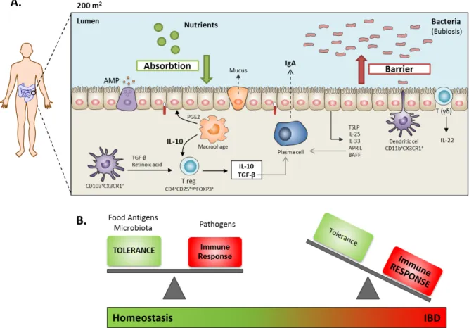
(24) Figure 2.1. Intestinal Immune Homeostasis and Inflammation. From the immunological perspective, the intestinal mucosa has two remarkable features. First it has an extensive surface area (approx. 200 m2) that allows maximum absorption of nutrients, water and electrolytes. Second the gut lumen is challenged with perpetual presence of commensal microbes. (A) In the presence of a balanced microbiota (eubiosis), the intestinal immune system induce and maintains tolerance to food microbes and food antigens through a complex set of innate and adaptive recognition strategies and effector mechanisms. Intestinal epithelial cells are essential for immune response with secretion of mucin (produced by goblet cells), antimicrobial peptides (AMPs – produced by Paneth Cells), epithelial cytokines (TSLP, IL-33, IL-25, BAFF, and APRIL) that promote development of tolerogenic dendritic cells (DCs; CD103+CX3CR1-) and macrophages (Mφ). Tolerogenic DCs and Mφ, in turn, induce the development of induced regulatory T cells (iTreg) through processes dependent on TGF-β and retinoic acid, or IL-10. Additionally, TGF-β from Tregs, as well as epithelial-derived BAFF and APRIL, promote generation of IgA plasma cells. Immune system also participates in the maintenance of epithelium integrity with secretion of PGE2 (Mφ and IEC) and IL-22 (γδ intraepithelial lymphocyte) (B) The equilibrium between tolerance Vs. Immune response is complex and fragile. In the case of IBD, alterations of the microbiota (dysbiosis), injury or xenobiotics, stimulate the production of pro-inflammatory cytokines that promotes intestinal inflammation and lack of resolution in genetically predisposed individuals. Abbreviations; APRIL, a proliferation-inducing ligand; BAFF, B-cell-activating factor of the TNF family; IBD, Inflammatory Bowel Disease; IgA, immunoglobulin A, PGE2, Prostaglandin E2; TGF- β, transforming growth factor; TSLP, thymic stromal lymphopoietin. Adapted from [18].. 24.
(25) 2.1.2.1 Innate immunity in IBD The first physical barrier that intestinal bacteria and food antigens encounter on the mucosal surface is represented by the intestinal epithelium covered by a mucous layer.[19] An important characteristic shared by IBD patients is a dysfunctional intestinal epithelial barrier, which in turn facilitates translocation of harmful substances and pathogens to the lamina propria, promoting inflammation.[20] Alterations in the intestinal epithelium also promotes aberrant secretion of mucin compromising the thickness and activity of mucus layer.[21] In fact, experiments using MUC2-/- mice demonstrated an increased sensitivity to dextran sodium sulfate (DSS) when compared to wild type animals.[22] Similarly, the expression of Toll Like Receptors (TLR), especially TLR4, is significantly increased in epithelial cells and lamina propria mononuclear cells (LPMNCs) during active IBD.[23, 24] Indeed, mutations of TLR4 are strongly associated to IBD development, particularly if they coexist with mutations of the nucleotide oligomerization domain 2 (NOD2) gene. Activation of TLR4 promotes the expression of nuclear transcription factorkappa B (NF-κB), signal transducer and activator of transcription 1 (STAT1), mitogenactivated protein kinases (MAPKs), or Peroxisome proliferator-activated receptor gamma (PPARγ), promoting pro- as well as anti- inflammatory effects. Studies in mice support the hypothesis that mutations over-activating the TLR4 signaling promote intestinal inflammation via excessive production of cytokines (TNF, IL-1β, and IL-6).[25] On the other hand, mutations leading to functional loss of TLR4, or its downstream signaling, worsen DSS-induced colitis by disturbing intestinal homeostasis.[26, 27] Thus, dysfunction of TLR4 in both directions aggravates intestinal inflammation.[28] NOD2 was the first susceptibility gene identified for CD and represents the best investigated and most well-established susceptibility gene for this disease.[29] Mutations within the NOD2 gene have only been associated with CD, but not UC, and are present in about 40% of CD patients.[28] These mutations are located in the C-terminal leucine-rich repeat domain (LRR), which is responsible for muramyl dipeptide (MDP) recognition.[30] In addition to the common NOD2 variants associated with CD [SNP8 (R702W), SNP12 (G908R) and SNP13 (1007fs)], other infrequent variables that are also localized to LRR have been discovered.[31] Although the specific mechanisms by which disease-associated NOD2 polymorphisms contribute to the increased susceptibility to develop CD remains incompletely understood; some models have been proposed. First, it has been suggested that NOD2 negatively regulates TIR signaling, and hence NOD2 deficiency would promote excessive response to TLR ligands. Alternatively, the frame-shift variant (SNP 13) has been suggested to actively suppress IL10 transcription via inhibiting the nuclear ribonucleoprotein hnRNP-A1.[31] Finally, a third model proposes that mutated isoforms of NOD2 impair the production of αdefensins by Paneth cells, promoting alterations in gut microbiota composition.[30] This is 25.
(26) supported by Wehkamp et al., which reported a remarkable reduced expression of αdefensins in patients with ileal CD featuring NOD2 mutations (SNP13), when compared to control patients.[32] Interestingly, recent evidence indicates that NOD2 is directly involved with autophagy trough physical interaction with autophagy-related protein 16-1 (ATG16L1), another CD susceptibility gene associated with Paneth cell dysfunction.[33] Indeed, gene variants of NOD2 and ATG16L1 are defecting in implementing proper autophagy.[34] Particularly, CD patients with T300A polymorphism of ATG16L1 display a large proportion of Paneth cells with abnormal granules; these abnormalities resemble those in mice with deficient expression of this protein.[35] Another homeostatic cellular pathway essential for secretory cells, including Paneth cells and goblet cells, is the Unfolded Protein Response (UPR) that is triggered by Endoplasmic Reticulum (ER) stress to control proper folding of synthetized proteins.[29] UPR response might be mediated through IRE1/XBP1, ATF6p90/ATF6p50 and PERK/ATF4 pathways. In any case, this response reduces the rate of protein synthesis, decreases the load of proteins entering the ER, and increases the capacity of ER to handle unfolded proteins.[36] Recent genome wide associated studies (GWAS) showed a significant association of IBD with X-box-binding protein 1 (XBP1) polymorphisms. Kaser et al demonstrated that specific deletion of XBP1 in intestinal epithelial cells caused severe depletion of Paneth cells and goblet cells, whereas enterocytes remained completely intact.[37] XBP1 malfunction not only promotes dysbiosis by inhibiting the production of antimicrobial peptides, it also restrain the immune response to pathogens. As shown by Martinton et al, XBP1 is fundamental for the optimal response of TLR4 to its ligands, since UPR response allows the survival of inflammatory cells to the stress promoted by immune activation and cytokine production. Thus, XBP1 deficient mice developed spontaneous intestinal inflammation with ulceration, neutrophils infiltration, and crypt abscesses- main features of human IBD.[38] 2.1.2.2 Adaptive immunity in IBD Although innate immune responses seem to be a prerequisite for the excessive activation of adaptive immunity, the latter undoubtedly drives the major tissue damage that is displayed in IBD patients.[39] Based mainly on the levels of T cell-derived cytokines detected in IBD mucosa, several studies have associated active disease with low proportion of regulatory Tcells (Treg) and abnormal development of activated T cells. It is classically considered that naïve CD4+ T cells are differentiated to T helper cell type (Th)1 in CD whereas UC is mediated by Th2 immune responses.[40] However, the notion that UC is a Th2-mediated disease remains controversial. For instance, UC patients produce low levels of IFN-γ instead of high expression of IL-4. Similarly, lower levels of IL-13 were found in the colonic mucosa of UC patients compared to CD patients and control subjects.[41] 26.
(27) CD Th1 cells, triggered by increased mucosal level of IL-12, produce high amounts of IL2, IL-18, IFN-γ, and TNF-α, which in turn enhances the activation of intestinal macrophages.[17] Although CD has historically been considered a Th1 mediated disease, recent discovery of Th17 pathway has prompted to reconsider this paradigm, as key molecules associated with the development, function, and maintenance of Th17 cells are up-regulated in CD patients compared to healthy subjects.[40] The pivotal role of Th17 cells in intestinal inflammation is not exclusive to CD. Indeed, recent data from GWAS have revealed several Th17 risk genes for IBD. The meta-analysis published by Jostins et al. demonstrated that variants of CCR6, STAT3, JAK2, IL-23R, and IL12B are associated to both UC and CD phenotypes and hence are included in the IBD genome.[42, 43] Th17 cells are characterized by the production of large amounts of IL17A, IL-17F, IL-21 and IL-22, induced by IL-6 and transforming growth factor (TGF)-β, with expansion promoted by IL-23.[43] Following engagement of IL 23R by IL 23, Janus kinase 2 (JAK2) is activated, resulting in JAK2 autophosphorylation and phosphorylation of IL 23R. This in turn results in the recruitment, phosphorylation, homodimerization and nuclear translocation of STAT3.[44] The variant R381Q of IL-23R has shown a high level of association to IBD susceptibility, probably by affecting JAK2 activity. Moreover, JAK2-STAT3 signaling is also related to IL-10 signaling, that is also involved in IBD pathogenesis.[45] An additional important gene is CCR6, expressed by various immune cells, mainly Th17 cells, promoting gut homing.[46] Overall, these evidence strongly implicate the IL-23 pathway and Th17 cells as central players in intestinal inflammation. Intestinal homeostasis relays in Treg (CD4+CD25highFOXP3+) and their ability to suppress abnormal immune response in the gut environment filled of microbiota and dietary antigens.[47] Interestingly, Treg cells are depleted in peripheral blood of patients with active IBD when compared to quiescent IBD and control subjects. Additionally, Treg exert a potent anti-inflammatory action in experimental colitis.[48] Although, several mechanisms are involved in Treg immune suppression, the production of anti-inflammatory cytokines (IL-10 and TGF-b) is probably the most notorious.[47] Studies in mice highlight the importance of Il-10 and TGF-b expression during intestinal inflammation. However, L-10 has particular significance in IBD because IL-10 and IL10R2 deficient mice do not develop lethal systemic autoimmunity, as seen in TGF-β1 deficient mice, but instead develop colitis. Moreover, established intestinal inflammation in mice can be ameliorated via treatment with recombinant IL-10 protein, IL-10 expressing transgenic T cells, or intestinal bacteria engineered to produce IL-10.[49]. 27.
(28) Nevertheless, Treg implication in human IBD remains controversial since Treg are increased in the intestinal mucosa of IBD patients, and their function is normal, as demonstrated by the ability to suppress the proliferation of effector T cells. Recent studies suggest that the intestinal milieu in IBD patients alters Treg functional properties rendering them to a non-suppressive or even pathogenic phenotype. These hypothesis is supported from the observation that elevated production of IL-1, IL-6 and IL-23 in IBD mucosa, together with TGF-β, promotes the differentiation T cells into effector cells that sustain chronic inflammation.[47]. Figure 2.2. Pathogenesis of Inflammatory Bowel Disease. Although the exact etiology of IBD remains elusive, it is well recognized that genetic and environmental factors induce impaired barrier function, along with inadequate immune response. Initiating triggers that cause structural changes to the intestinal epithelium subsequently induces the translocation of bacteria and microbial products from the lumen to the lamina propria, which leads to innate immune cells activation in particular dendritic cells and macrophages, amplification of adaptive response by Th1, Th2, or Th17, and elevated pro-inflammatory molecules (NO and PGE2), cytokines/chemokines (IL-1β, IL-5, IL-6, IL-12, IL-13, IL-15, IL-17A/F, IL-18, IL-22, IFN-γ), and growth factors (TGF-β). If acute inflammation cannot be resolved by anti-inflammatory mechanisms, then chronic inflammation is developed with promotion of tissue destruction and causing complications such as fibrosis, stenosis, fistulas, cancer, and/or extraintestinal manifestations, which are common to all types of IBD. Abbreviations; AMP, antimicrobial peptides; IBD, inflammatory bowel disease; NSAIDs, non-steroidal anti-inflammatory drugs; NO, nitric oxide; PGE2, prostaglandin E2; mucosal effector T cells (T helper 1 (Th1), Th2 and Th17); UPR, unfolded protein response. Adapted from [16]. 2.1.3 IBD Treatment The primary goal of medical therapy for IBD patients is to suppress intestinal inflammation to provide relief of symptoms and achieve mucosal healing.[50] Routinely, treatment is 28.
(29) dependent on disease severity, localization, and associated complications. Regardless of this, medical management relays in pharmacological therapy and/or surgery to induce and maintain remission, while preventing complications (megacolon, abscesses, and fistula).[51] Standard pharmacological therapy includes the use of aminosalicylates (5-aminosalicylic acid), steroids (prednisone and prednisolone), and immunosuppressants (cyclosporine, tacrolimus, azathioprine, and methotrexate).[51] Notwithstanding the cost of the treatment, these drugs do not induce complete clinical remission while producing serious secondary effects.[52] Under these circumstances, biological drugs appeared as a hopeful strategy for IBD management. Infliximab, a chimeric anti-TNF antibody, was the first biologic agent approved by the Food and Drugs Administration (FDA) in August of 1998. Subsequently, Adalimumab and Certolizumab pegol were approved. Since then, biological therapy has been increasingly employed in the treatment of IBD, showing a significant improvement for patient’s clinical condition.[53] However, the evidence indicates that treatment with these molecules has not reduced the need of emergency surgery for patients with severe disease; instead it involves several risks and side effects. Renal complications, delayed hypersensitivity-like reaction, new onset of autoimmunity, and opportunistic infections are some examples of complications resulting from the immunosuppression induced by biological therapy.[52, 53] All these evidence, combined with the significant cost of biologic therapy compared to traditional drugs (236.370 Vs. 147.763 USD/patient), indicates the need for new therapies.[54] Given the low efficacy and safety of standard therapies, the use of herbal medicines is rising globally. IBD patients favor the use of phytotherapy for two fundamental reasons: the desire of ending steroid consumption and the lack of confidence in their physician due to the unsatisfactory treatment.[55] In spite of the great potential of traditional herbal medicines, few high-quality investigations have been developed.[56, 57] At basic level, the claim that herbal medicines are useful to treat IBD is often exaggerated. Most of the studies are performed using plant extracts without any chemical characterization and conclusions are often based on results of in vitro experiments to evaluate antioxidant or antiinflammatory activity. When animal studies are used, researchers often employ a single acute model of rodent IBD, frequently chemically induced (TNBS/DSS). Similarly, clinical investigations have serious drawbacks regarding number of patients, short time frames of administration without long-term follow- up, lack of placebo group, standardized scores for endoscopic and histologic evaluation, and poor biochemical characterization. In agreement, Meta-analysis by Rahimi et al showed that herbal medicines may induce clinical efficacy in patients with IBD, but the evidence is too limited to make any confident conclusion.[58] As a result, the application of medicinal plants to treat IBD is significantly restricted.[55]. 29.
(30) 2.2. Physalis peruviana L.. 2.2.1 Botanical description Physalis is a genus of herbaceous plants in the Solanaceae family. The genus comprises about 120 species distributed throughout the tropical and subtropical regions of the world, naturally found in America, with a few introduced species in Europe and the countries of the Southwest and Center Asia.[59, 60] Typical Physalis species are herbaceous and possess solitary flowers, a yellow corolla, and an accrescent and inflated fruiting calyx.[59] Physalis peruviana L. [Synonyms: Alkekengi pubescens Moench, Boberella peruviana (L.) E.H.L. Krause, Physalis esculenta Salisbury, Physalis latifolia Lam., Physalis peruviana var. latifolia (Lam.) Dunal, Physalis tomentosa Medik][61], is probably one of the best known species of this botanical genus due to its economic importance.[62] This plant is indigenous to South American Andes, in the high altitude of tropical Colombia, Chile, Ecuador and Peru, where the plant grows wild. Currently it can be found in tropical and subtropical regions including Malaysia, China and the Caribbean, where is grown from sea level (New Zealand) to 3000 m. elevations (Andes).[63]. Figure 2.3. Images of Physalis peruviana L. The upper part describes the complete taxonomical classification of cape gooseberry/uchuva (P. peruviana). Pictures of (A.) Leaves and flowers; (B.) Close-up of flowers; (C.) Fruits enclosed in the parchment-like husk (calyces); and (D.) Fruit with calix removed. Adapted from [63].. P. peruviana is an herbaceous or soft-wooded, semi-shrub, upright, perennial plant, profusely branched, with densely pubescent and ribbed, often purplish branches. In natural, non-managed conditions, plant is known to reach 1.0 to 1.5 m in height.[64] The vegetative stage is characterized by the occurrence of simple, petiolated, alternate, heart-shaped leaves that are densely pubescent, with dimensions ranging from 5 to15 cm long and 4 to 10 cm wide.[64] Flowers (1.2-1.5 × 1.2-2 cm) occur in leaf axils on 1.5 cm pedicel with bellshaped purplish-green pubescent calyx, yellow corolla and purple spotted in throat.[63] The 30.
(31) flower can be easily pollinated by insects, wind and also by auto-pollination. After fertilization, the calyx (or husk), which is small at the beginning of fruit development, grows to a bladder-like, papery organ, which completely encloses the ripening fruit.[64, 65] This unique structure protects the fruit against insects, birds, disease and adverse climatic situations. Moreover calyces represent an essential source of carbohydrates during the first 20 days of growth and development.[65] The fruit is a juicy berry, 1.25-2 cm diameter that turn from green to yellow upon ripening, with numerous flat seeds (2 mm diameter) embedded in the pulp.[63] When fully ripe the fruit is sweet but with a pleasant grape-like tang. The calyx is bitter and inedible. 2.2.2 Ethnopharmacology Most species of the genus Physalis have been used for a long time in the medicinal folk traditions of Asian and American populations to treat different illnesses, such as malaria, asthma, hepatitis, dermatitis, rheumatism, liver disorders, and as an anti-mycobacterial, anticancer, anti-leukemic, antipyretic, and immunomodulatory agents.[60] P. peruviana is a valued medicinal plant widely used in folk medicine of South America and Africa. The traditional uses reported for this species are presented in Table 2.1. Table 2.1 Traditional Medicinal uses of Physalis peruviana L. Part of the Traditional Use Preparation Country plant Diuretic and anti-asthmatic Decoction Colombia Inflammation Poultice South Enema for abdominal ailments Infusion Africa Parasites and bowel complaints Juice Southern Africa Leaves Diarrhea Decoction South Africa Diabetes, Malaria, Pneumonia Decoction Kenya Skin fungal infections Juice Tanzania Labor Inducer Juice Uganda Cough medicine Decoction Colombia Flower Skin infections/problems. Ecuador Decoction Promote wound healing Uganda Inflammation Calyces Cancer Poultice Colombia Diuretic Pterygium, Cataracts Juice Colombia Fruit Albuminuria Peru Diabetes Daily intake. Ref. [66-71]. [72-74] [75] [65, 72, 76, 77]. 31.
(32) 2.2.3 Anti-inflammatory activity The majority of bioassay data describing the pharmacological activity of P. peruviana L. indicates an important antimicrobial, cytotoxic, antioxidant and anti-inflammatory effect. This section focuses on the studies related to inflammation. Wu et al reported that a supercritical carbon dioxide extract from P. peruviana leaves (SCEPP-5; 30 µg/mL) possessed a strong inhibitory activity in lipopolysaccharide (LPS)activated RAW264.7 cells, reducing nitric oxide (NO) and prostaglandin E2 (PGE2) levels through attenuation of nitric oxide synthase (iNOS) and cyclooxygenase-2 (COX-2) protein expression.[78] Recently, Sang-ngern et al described the isolation of 4βhydroxywhitanolide E from aerial parts of P. peruviana, which inhibited TNF-α induced NF-κB activity in stably transfected human embryonic kidney cells NF-κB Luc293. This effect correlated with a strong inhibition of NO production by LPS-induced RAW264.7 macrophages.[79] The bioactivity of P. peruviana fruit juice has also been studied. Pardo et al found a mild anti-inflammatory effect of the juice in a rabbit eye inflammatory model. In addition, authors established a concentration-dependent cytostatic effect on cultured fibroblasts. Overall, the evidence might correlate with the traditional employment of P. peruviana to treat pterygium.[76] In agreement with the traditional employment of P. peruviana calyces, several studies have demonstrated a strong anti-inflammatory activity in vivo and in vitro. The ethereal extract and various fractions from P. peruviana calyces were found to potentially inhibit the ear edema induced by 12-O-tetradecanoyl-phorbol-13-acetate (TPA). Authors highlighted the activity of fraction Pp-D28-LF, which demonstrated a dose-dependent activity as well as easy isolation and good yield.[75] In another study, this fraction was shown to exert a 40% inhibition of the acetic acid induced writhing (acute visceral nociception) and the licks during phase II (acute inflammatory nociception) of the formalin test; which suggests that anti-nociceptive effect of P. peruviana calyces might be related to NSAID-like mechanisms.[80] A complementary work from the same research team, showed the bioguided isolation of rutin and nicotoflorin from the ethanolic extract of P. peruviana calyces, using TPA-induced ear edema.[81] Furthermore, a strong immunomodulatory effect of ethereal extract and fractions from P. peruviana calyces was demonstrated by Martinez et al when studying Leishmania panamensis infection to murine macrophages. In this study, extracts and fractions increased the proportion of infected cells with parasites by down-regulating macrophage activation, and a strong inhibition of pro-inflammatory cytokines (IL6, TNF-α, and MCP-1).[82]. 32.
(33) 2.3 1. 2. 3. 4. 5. 6. 7. 8. 9. 10. 11. 12. 13. 14. 15. 16. 17. 18. 19. 20. 21. 22.. References Hisamatsu, T., et al., Immune aspects of the pathogenesis of inflammatory bowel disease. Pharmacology & Therapeutics, 2013. 137(3): p. 283-297. Louis, E., C. Van Kemseke, and C. Reenaers, Necessity of phenotypic classification of inflammatory bowel disease. Best Pract Res Clin Gastroenterol, 2011. 25 Suppl 1: p. S2-7. Fiocchi, C., IBD: advances in pathogenesis, complications, diagnosis, and therapy. Curr Opin Gastroenterol, 2012. 28(4): p. 297-300. Castiglione, F., et al., Risk factors for inflammatory bowel diseases according to the "hygiene hypothesis": a case-control, multi-centre, prospective study in Southern Italy. J Crohns Colitis, 2012. 6(3): p. 324-9. Sartor, R.B., Mechanisms of Disease: pathogenesis of Crohn's disease and ulcerative colitis. Nat Clin Pract Gastroenterol Hepatol, 2006. 3(7): p. 390-407. Zhang, Y.Z. and Y.Y. Li, Inflammatory bowel disease: pathogenesis. World J Gastroenterol, 2014. 20(1): p. 91-9. Molodecky, N.A., et al., Increasing incidence and prevalence of the inflammatory bowel diseases with time, based on systematic review. Gastroenterology, 2012. 142(1): p. 46-54 Cosnes, J., et al., Epidemiology and natural history of inflammatory bowel diseases. Gastroenterology, 2011. 140(6): p. 1785-94. Ng, S.C., et al., Incidence and phenotype of inflammatory bowel disease based on results from the Asia-pacific Crohn's and colitis epidemiology study. Gastroenterology, 2013. 145(1): p. 158-165. Burisch, J. and P. Munkholm, Inflammatory bowel disease epidemiology. Curr Opin Gastroenterol, 2013. 29(4): p. 357-62. Lovasz, B.D., et al., New trends in inflammatory bowel disease epidemiology and disease course in Eastern Europe. Dig Liver Dis, 2013. 45(4): p. 269-76. Wong, S. and S. Ng, What Can We Learn From Inflammatory Bowel Disease in Developing Countries? Current Gastroenterology Reports, 2013. 15(3): p. 1-9. Gismera, C. and B. Aladren, Inflammatory bowel diseases: a disease (s) of modern times? Is incidence still increasing? World J Gastroenterol, 2008. 14(36): p. 5491-8. Pineda, L., Enfermedad inflamatoria intestinal en Colombia: ¿Está cambiando nuestro perfil epidemiológico? Revista Colombiana de Gastroenterologia, 2010. 25: p. 235-238. Juliao, F., et al., Fenotipo e historia natural de la enfermedad inflamatoria intestinal en un centro de referencia en Medellín-Colombia. Revista Colombiana de Gastroenterologia, 2010. 25: p. 240-251. Neurath, M.F., Cytokines in inflammatory bowel disease. Nat Rev Immunol, 2014. 14(5): p. 329-342. Dave, M., K.A. Papadakis, and W.A. Faubion, Jr., Immunology of inflammatory bowel disease and molecular targets for biologics. Gastroenterol Clin North Am, 2014. 43(3): p. 405-24. de Souza, H.S. and C. Fiocchi, Immunopathogenesis of IBD: current state of the art. Nat Rev Gastroenterol Hepatol, 2016. 13(1): p. 13-27. Peterson, L.W. and D. Artis, Intestinal epithelial cells: regulators of barrier function and immune homeostasis. Nat Rev Immunol, 2014. 14(3): p. 141-153. Konig, J., et al., Human Intestinal Barrier Function in Health and Disease. Clin Trans Gastroenterol, 2016. 7: p. e196. Maloy, K. and F. Powrie, Intestinal homeostasis and its breakdown in inflammatory bowel disease. Nature, 2011. 474(7351): p. 298-306. Petersson, J., et al., Importance and regulation of the colonic mucus barrier in a mouse model of colitis. Am J Physiol Gastrointest Liver Physiol, 2011. 300(2): p. G327-33.. 33.
(34) 23. 24. 25. 26. 27. 28. 29. 30. 31. 32. 33. 34. 35. 36. 37. 38. 39. 40. 41. 42. 43. 44. 45.. Abreu, M.T., Toll-like receptor signalling in the intestinal epithelium: how bacterial recognition shapes intestinal function. Nat Rev Immunol, 2010. 10(2): p. 131-44. Hausmann, M., et al., Toll-like receptors 2 and 4 are up-regulated during intestinal inflammation. Gastroenterology, 2002. 122(7): p. 1987-2000. Fukata, M., et al., Constitutive activation of epithelial TLR4 augments inflammatory responses to mucosal injury and drives colitis-associated tumorigenesis. Inflamm Bowel Dis, 2011. 17(7): p. 1464-73. Rakoff-Nahoum, S., et al., Recognition of Commensal Microflora by Toll-Like Receptors Is Required for Intestinal Homeostasis. Cell, 2004. 118(2): p. 229-241. Gibson, D., et al., MyD88 signalling plays a critical role in host defence by controlling pathogen burden and promoting epithelial cell homeostasis during Citrobacter rodentiuminduced colitis. Cell Microbiol, 2008. 10(3): p. 618-31. Scharl, M. and G. Rogler, Microbial sensing by the intestinal epithelium in the pathogenesis of inflammatory bowel disease. Int J Inflam, 2010. 2010: p. 671258. Khor, B., A. Gardet, and R.J. Xavier, Genetics and pathogenesis of inflammatory bowel disease. Nature, 2011. 474(7351): p. 307-17. Saleh, M. and C.O. Elson, Experimental inflammatory bowel disease: insights into the hostmicrobiota dialog. Immunity, 2011. 34(3): p. 293-302. Fritz, T., et al., Crohn's disease: NOD2, autophagy and ER stress converge. Gut, 2011. 60(11): p. 1580-8. Wehkamp, J., et al., Reduced Paneth cell alpha-defensins in ileal Crohn's disease. Proc Natl Acad Sci U S A, 2005. 102(50): p. 18129-34. Clevers, H.C. and C.L. Bevins, Paneth cells: maestros of the small intestinal crypts. Annu Rev Physiol, 2013. 75: p. 289-311. Negroni, A., et al., NOD2 induces autophagy to control AIEC bacteria infectiveness in intestinal epithelial cells. Inflamm Res, 2016. 65(10): p. 803-13. Cadwell, K., et al., A key role for autophagy and the autophagy gene Atg16l1 in mouse and human intestinal Paneth cells. Nature, 2008. 456(7219): p. 259-63. Kaser, A., L. Niederreiter, and R. Blumberg, Genetically determined epithelial dysfunction and its consequences for microflora-host interactions. Cell Mol Life Sci, 2011. 68(22): p. 3643-9. Kaser, A., et al., XBP1 links ER stress to intestinal inflammation and confers genetic risk for human inflammatory bowel disease. Cell, 2008. 134(5): p. 743-56. Martinon, F., et al., TLR activation of the transcription factor XBP1 regulates innate immune responses in macrophages. Nat Immunol, 2010. 11(5): p. 411-8. Xavier, R.J. and D.K. Podolsky, Unravelling the pathogenesis of inflammatory bowel disease. Nature, 2007. 448(7152): p. 427-434. Maynard, C. and C. Weaver, Intestinal effector T cells in health and disease. Immunity, 2009. 31(3): p. 389-400. Geremia, A., et al., Innate and adaptive immunity in inflammatory bowel disease. Autoimmun Rev, 2014. 13(1): p. 3-10. Jostins, L., et al., Host-microbe interactions have shaped the genetic architecture of inflammatory bowel disease. Nature, 2012. 491(7422): p. 119-24. Bogaert, S., et al., Differential mucosal expression of Th17-related genes between the inflamed colon and ileum of patients with inflammatory bowel disease. BMC Immunology, 2010. 11(1): p. 61. Cho, J.H., The genetics and immunopathogenesis of inflammatory bowel disease. Nat Rev Immunol, 2008. 8(6): p. 458-66. Budarf, M., et al., GWA studies: rewriting the story of IBD. Trends Genet, 2009. 25(3): p. 137-46.. 34.
Figure


+7


Documento similar
In the preparation of this report, the Venice Commission has relied on the comments of its rapporteurs; its recently adopted Report on Respect for Democracy, Human Rights and the Rule
In the in vivo animal experiments, Δ 9 -THCA-A successfully show a potential therapeutic effect in inflammatory diseases as we have observed in a model of
A limiting value of the oxidation potential of a given compound candidate to have antioxidant activity in the DPPH • test was found, this being explained by
145 We will next review the available evidence demonstrating the presence of neuroinflammation and its potential relationship with P2X7Rs, from preclinical in vitro and in
It was not until 2005, when Sawamura reported the copper-catalyzed borylation of allylic carbonates, that the synthetic potential of copper- catalyzed
Circulating interleukin 6 and albumin, and infliximab levels are good predictors of recovering efficacy after dose escalation infliximab therapy in patients with loss of response
(C) Phenotype of human B-cells, monocytes, pDCs, and cDCs from healthy controls (HCs) and inflammatory bowel disease (IBD) patients based on the basal expression of CCR2, CD40,
These results suggest that unconjugated ISG15 is responsible for counteracting the development of an exacerbated inflammatory response after a high dose of VV⌬E3L virus. To analyze
