Improvement in the definition of anti-HLA
antibody profile in highly sensitized patients
Juan Irure1,2, Esther Asensio1,2, Emilio Rodrigo3, I´ñigo Romo´ n2, Javier Go´ mez4, Manuel Arias3, Marcos Lo´ pez-Hoyos1,2☯, David San Segundo1,2☯*
1 Immunology Service, University Hospital Marque´s de Valdecilla-IDIVAL, Santander, Spain, 2 Tissue Typing Laboratory, University Hospital Marque´s de Valdecilla, Santander, Spain, 3 Nephrology Service, University Hospital Marque´s de Valdecilla-IDIVAL, Santander, Spain, 4 Pathology Service, University Hospital Marque´ s de Valdecilla-IDIVAL, Santander, Spain
☯These authors contributed equally to this work.
Abstract
The definition of anti-HLA antibody profile in highly sensitized patients on a waiting list is cru-cial when virtual crossmatch is used in organ allocation systems, but also when used to identify the true deleterious anti-HLA antibodies. Here we propose different levels of risk based on the results of anti-HLA antibody testing in neat serum (N) and after sera dilution (DIL) and C1q test in 18 highly sensitized patients. This group was heterogeneous in terms of anti-HLA antibody titers and their ability to fix complement. After dilution, 15 out of 18 patients (83.3%) showed a reduction of positive bead counts whereas 4 patients showed a prozone effect and complement fixation was demonstrated. The high dilution of sera and ascertaining the complement fixation allow the accurate definition of risk anti-HLA antibody profiles in highly sensitized patients, as demonstrated in 5 of the sensitized patients who were transplanted.
Introduction
The anti-HLA antibody testing was revolutionized by solid-phase assays (SPA) based on Lumi-nex, increasing the sensitivity and the ability to detect low levels of anti-HLA antibodies[1]. With Luminex data, unacceptable HLA antigens from potential donors could be assigned and virtual crossmatch (vXM) was addressed[2]. This is especially interesting in the case of highly-sensitized patients because vXM has allowed the development of specific programs to reduce the waiting list for organ transplantation, and an accurate profile of unacceptable HLA anti-gens should be defined[3].
Despite the improvement of anti-HLA antibody detection by Luminex being well accepted, several questions have risen with the potential pitfalls[4]. When doubtful HLA antigens are considered as unacceptable with mean fluorescence intensities (MFI) near the cut-off value, the knowledge of potential interfering factors should be kept in mind. Two different kinds of errors could appear; a) false positive results: due to a reaction against denatured HLA antigens (neo-epitopes generated during the process of coating the beads with HLA antigens), or due to
a1111111111 a1111111111 a1111111111 a1111111111 a1111111111 OPEN ACCESS
Citation: Irure J, Asensio E, Rodrigo E, Romo´n I´,
Go´mez J, Arias M, et al. (2017) Improvement in the definition of anti-HLA antibody profile in highly sensitized patients. PLoS ONE 12(2): e0171463. doi:10.1371/journal.pone.0171463
Editor: Jose C. Crispin, Instituto Nacional de
Ciencias Medicas y Nutricion Salvador Zubiran, MEXICO
Received: July 13, 2016 Accepted: January 20, 2017 Published: February 3, 2017
Copyright:© 2017 Irure et al. This is an open access article distributed under the terms of the
Creative Commons Attribution License, which permits unrestricted use, distribution, and reproduction in any medium, provided the original author and source are credited.
Data Availability Statement: All relevant data are
within the paper and its Supporting Information files.
Funding: This research did not receive any specific
grant from funding agencies in the public, commercial, or not-for-profit sectors.
Competing Interests: The authors have declared
that no competing interests exist.
Abbreviations: ABMR, antibody-mediated
complement-the lack of consensus about cut-off values to determine positive or negative specificities explained in part to wide intra- and inter-laboratory variability[5]. A significant example is the assessment of unexpected anti-HLA antibodies in non-sensitized, non-transfused males using different cut-off values[6]; on the other hand b) false negative results: due to the potential interaction of complement with IgG anti-HLA antibodies[7] easily overcome after EDTA or heat treatment[8], or due to a prozone effect which could be solved after dilution of the sera [9,10]. Therefore, the overestimation of unacceptable HLA antigens in a patient could lead to an increased time on the waiting list, and an accurate definition of unacceptable and permitted HLA antigens should be addressed.
There is an increasing interest in a clear definition of the anti-HLA antibody profile due to their use in vXM, but also in donor-pair living donors and highly-sensitized programs in order to allocate an organ with negative vXM results. However, in some undesired situations with positive complement-dependent crossmatchs, a negative vXM result is found [11]. This discor-dance could be improved with further study of the IgG anti-HLA antibodies profile.
Sometimes the definition of the anti-HLA antibodies profile is difficult due to low-titer anti-HLA antibodies that produce variable results in sequential serum studies. Another situa-tion that must be completely ruled out in highly-sensitized patients is the prozone effect where high titers of antibodies compete for the antigen and are not detected in SPA. For this reason. it is crucial to assign these dubious specificities as unacceptable or permitted antigens. In order to facilitate this assignment, two recent publications have demonstrated different strength of anti-HLA antibodies[9,10] as an approach to better defining the anti-HLA antibodies profile.
Here we propose an anti-HLA rating based on anti-HLA antibody strength and the ability to fix the complement in highly-sensitized patients. Such a rating is validated in complement-dependent cytotoxicity (CDC) and flow cytometry crossmatch (FCXM) against peripheral blood mononuclear cells (PBMCs) from blood donors with HLA specificities which are recognized by the sensitized patients´ sera and is further probed in 5 patients who could be transplanted.
Materials and methods
The work has been conducted according to the principles expressed by the Declaration of Hel-sinki and approved by the Ethics Comittee of our institution (Comite´ E´tico de Investigacio´n Clı´nica de Cantabria). The patient on the waiting list signed their informed consent and the sera collection is registered in ISCIII with number: C.0003580.
Patients and samples
Serum from 18 highly-sensitized patients, with >98% of calculated panel reactive of antibodies (cPRA) and who did not receive any desensitization treatment with Rituximab or intravenous immunoglobulins, were studied for anti-HLA class-I antibodies by Single Antigen (LABScreen Single Antigen Class-I, One Lambda, Canoga Park, CA) at neat (N) and at a 1/160 serum dilu-tion (DIL) based on a previous report[9] where further dilutions did not find any prozone effect. For identification of fixing-complement anti-HLA antibodies, the C1q test was per-formed (C1qScreen, One Lambda). A total number of 1,746 class-I antigens on beads were evaluated and raw MFI were compared after N, DIL and C1q assays.
The cut-off value was set at 3000 MFI and every single bead with MFI below the cut off value was considered as negative. The number of beads with a positive result was counted in each condition (N, DIL and C1q).
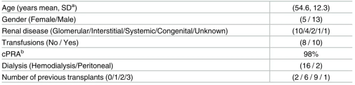
The main demographic, clinical and immunological parameters of the highly-sensitized patients tested are summarized inTable 1.
dependent cytotoxicity; cPRA, calculated-panel reactive of antibodies; DIL, diluted serum; DSA, donor-specific anti-HLA antibodies; FCXM, flow cytometry crossmatches; MFI, mean fluorescence intensity; N, neat serum; PBMCs, peripheral blood mononuclear cells; PBS, phosphate buffer saline;
SPA, Solid phase assay; vXM, virtual
Five out of the 18 kidney transplant recipients (KTR) could be transplanted with negative CDC but positive FCXM and they were biopsied because of clinical suspicion of antibody-mediated rejection (ABMR) or protocol biopsy. Sera at transplantation from these five recipi-ents were retrospectively studied for anti-HLA antibodies, taking into account the three previ-ously-mentioned conditions (N, DIL, C1q) and the risk for rejection was scored/caluclated.
Complement-dependent cytotoxicity crossmatch
Crossmatch was performed by CDC. Donors´ lymphocytes were separated from whole blood by Ficoll gradient. Cells were suspended in phosphate buffer saline (PBS) to achieve a concen-tration of 2x10⁶/mL. 1ul of patients’ serum was dispensed in the wells of an oiled Terasaki plate. ½ and ¼ serum dilutions were also used. All samples and dilutions were in duplicate. Negative and positive control samples were dispensed in the same way. Then, 1ul of lymphocytes and 5ul of rabbit complement (One Lambda) were dispensed in each well. After incubation (90 min-utes, 22˚C), eosin was added to each well and the plate was read in an inverted microscope.
Flow cytometry crossmatch
PBMC isolated by Ficoll gradient were incubated with pronase during 20 minutes in a 37˚C water bath. Neat and 1/160 diluted serum was added to pronase-treated PBMC (Sigma Aldrich, St. Louis, MO) and the mix was incubated during 30 minutes at room temperature. Anti-CD3 Pacific Blue (clone UCHT-1, Immunostep, Salamanca, SPAIN), CD19 APC (clone SJ25C1, BD Biosciences, San Jose, CA) and subsequently Fab´-IgG FITC (Dako, Glostrup, DENMARK) were added and samples were acquired using a FACS-Canto II (BD Biosciences). Flow cytometry crossmatch for class-I antigens was considered positive in the CD3+ gate when the ratio: median fluorescence value Serum / median fluorescence value Negative Con-trol was > 1.5; for class-II antigens, the FCXM was considered positive in CD19+ gate when the ratio was > 2.0.
HLA typing
Donors were typed for HLA locus A, B and DRB1 by low resolution SSP (Life Technologies, Brown Deer, WI).
Donor selection
Once a donor HLA-typing was confirmed, the anti-HLA antibody profile was assessed for all sera. If one serum was eligible for anti-HLA antibody profile testing, subsequent CDC and FCXM was performed.
Table 1. Demographic and clinical parameters of highly-sensitized patients.
Age (years mean, SDa) (54.6, 12.3)
Gender (Female/Male) (5 / 13)
Renal disease (Glomerular/Interstitial/Systemic/Congenital/Unknown) (10/4/2/1/1)
Transfusions (No / Yes) (8 / 10)
cPRAb 98%
Dialysis (Hemodialysis/Peritoneal) (16 / 2)
Number of previous transplants (0/1/2/3) (2 / 6 / 9 / 1)
aSD: Standard deviation
bcPRA: calculated Panel Reactive of Antibodies
Statistical analysis
The correlation of mean fluorescence intensities among N, DIL and C1q was assessed by the Spearman test. The number of positive beads was compared by the Wilcoxon test and p<0.05 was considered significant. All the tests were performed using GraphPad software version 5.0 (San Diego, CA).
Results
Correlation of raw MFI data
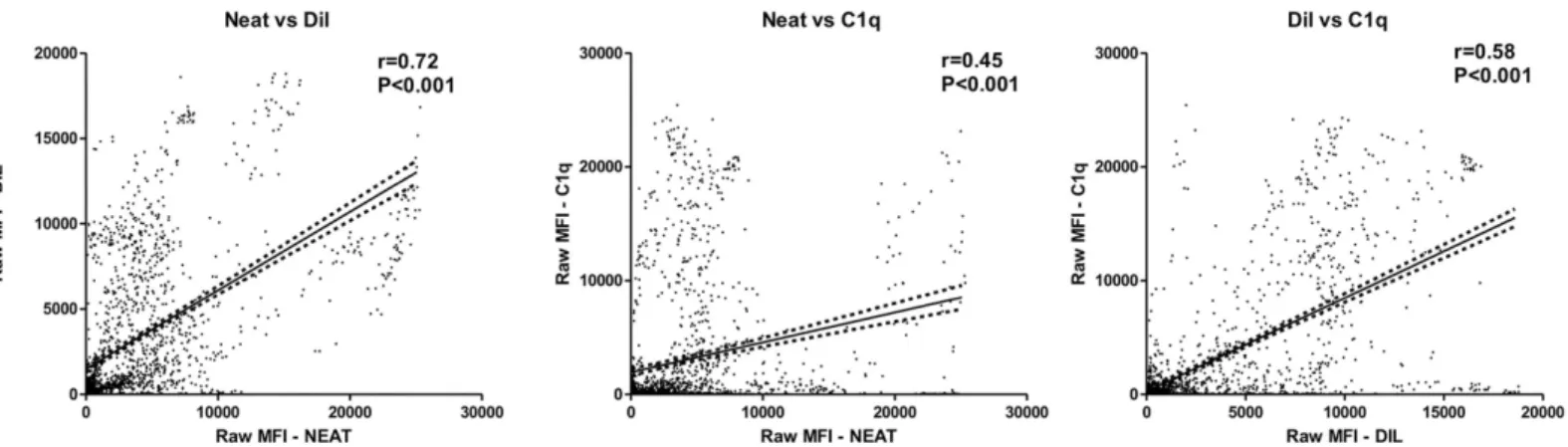
In order to assess the relationship of raw MFI data of neat sera (N), diluted (DIL) and C1q test (C1q), the MFI values of 1,746 beads with HLA-class-I antigens were compared (Fig 1). A posi-tive correlation of raw MFI data between N and DIL serum was observed (r = 0.72; p<0.001), whereas the correlation between N and C1q raw MFI values was poorer (r = 0.45; p<0.001). Nevertheless, a better correlation of raw MFI data C1q and DIL serum was observed (r = 0.58; p <0.001).
Anti-HLA antibody profiles in highly sensitized patients
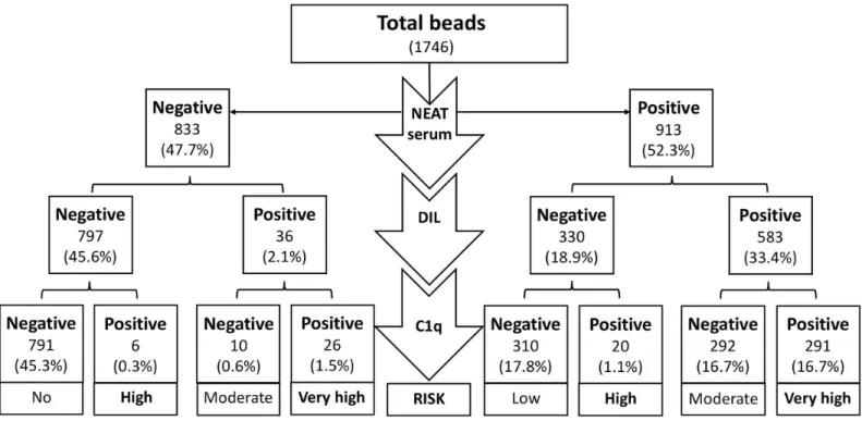
Serum reactivity was stratified based on the combination of N, DIL and C1q positive-beads. The results allowed us to discriminate in high or low titer with DIL serum and fixing and non-fixing antibodies after the C1q test. The persistence of high-titer and fixing-complement antibodies, positive results in N, DIL and C1q (N/DIL/C1q) (+/+/+) profile identified specificities at a very high risk in patients and should be considered unacceptable HLA antigens for a potential donor. The absolute number of beads tested is summarized inFig 2and a wider range of risk can be defined. Theoretically, all C1q positive beads have a high risk of CDC positive result, whereas other combinations can be stratified in low, moderate and high risk. Interestingly, 310 (36.1%) N positive-beads had negative results after DIL and C1q tests and could be considered as low risk specificities. Inversely, a prozone effect was confirmed in 26 (3.1%) N negative-beads, being positive in both after DIL and C1q tests. The HLA antigens associated with this profile (-/+/+) should be considered as very high risk and, consequently, as prohibited specificities.
Description of highly-sensitized group
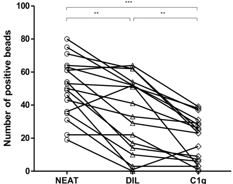
In our cohort of highly sensitized patients, the median of single antigen class-I positive beads was 51.5 beads in neat serum. After 1/160 dilution of the sera, a significant reduction in the
Fig 1. Correlation of raw MFI values after neat, diluted serum and C1q test. The raw MFI value after neat serum (neat), 1/160 diluted serum (DIL) and C1q test (C1q) were compared. The linear regression line and 95% confidence intervals in each plot are depicted (The correlation of MFI values was assessed by Spearman test (***, p<0.001).
median of positive beads was observed, 37.0 beads (Fig 3, p<0.01). In 15 out of 18 highly-sensi-tized patients (83.3%), a number of unacceptable HLA antigens assigned with N serum with the Single Antigen test were negative after DIL, suggesting that these antibodies could be con-sidered as low-titer antibodies. On the contrary, the prozone effect was observed in 7 out of 18 ptients (38.8%) (at least one bead was considered negative in N serum and positive after DIL). Importantly, the ability to fix the complement was confirmed in 4 patients. The values of raw MFI of beads in N, DIL and C1q test are summarized inS1 Table. Furthermore, C1q analysis revealed a significant decrease in the median of the positive beads (24.0) in highly sensitized patients compared with positive beads after N serum test (Fig 3, p<0.001).
From the beads to the patients
After the definition of profiles based on bead-results (Fig 2), in our cohort, we identified patients with different profiles based on the results of Single Antigen beads (N/DIL/C1q). In general, the behavior of most beads individually within a patient lowered the level of sensitiza-tion. Several examples are shown inS1 Fig. Thus, a highly-sensitized patient, defined only by testing neat serum with Single Antigen assay, could have a low risk profile when most of the beads have negative results after dilution and C1q test (+/-/-) (S1 Fig, panel A). The next risk level could be a patient with moderate risk (+/+/-) when most of the beads are negative in C1q test, despite confirmation of high titer with positive results after dilution (S1 Fig, panel B). The third risk level might be when the beads with anti-HLA antibodies are at low titer but are able to fix the complement (+/-/+), (S1 Fig, panel C). And finally, the most problematic case is a false negative in neat serum after Single Antigen, confirmed in the dilution (prozone effect) and which is able to fix the complement by C1q test (-/+/+) (S1 Fig, panel D andS1 Table).
Fig 2. Stratification of the risk regarding positive bead results after neat serum, diluted serum and C1q test. A total number of 1746 beads were distributed according positive (+) and negative (-) results after neat (N), 1/160 diluted (DIL) serum and C1q test. The combination of the results render 8 profiles with potential different risk. The absolute number of beads are in parentheses and the percentage of total beads in every profile are shown.
Assessment of the risk for different anti-HLA antibodies profiles in
complement-dependent cytotoxicity and flow cytometry crossmatches
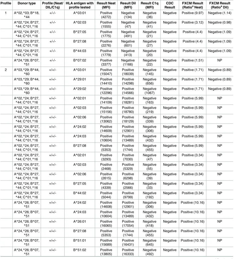
We have tried to assess the risk of different anti-HLA antibody profiles defined in the previous section. In order to test the low-risk profile (+/-/-), donors with compatible HLA class-I anti-gens and sera with at least one HLA class-I antigen with (+/-/-) profile were selected. The MFI of N, DIL and C1q of specific donor-HLA antigens are summarized inTable 2(profile #1). Subsequent CDC and FCXM were performed with negative results (in diluted sera). Further-more, the moderate risk profile (+/+/-) was designed based on the same fashion (profile #2). Twenty reactions were tested and all CDC were negative, whereas the FCXM rendered a
Fig 3. Number of positive beads after study of neat, diluted serum and C1q in highly-sensitized patients. The number of positive beads in 18 highly sensitized patients are depicted after Single Antigen anti-HLA class-I test in neat serum (neat, open circles), in 1/160 diluted serum (DIL, open triangles) and after C1q test (C1q, open diamonds) using the cut off value described in Material and Methods section. Differences of the medians were assessed by Wilcoxon test, (**, p<0.01;***.p<0.001).
Table 2. Assessment of different anti-HLA antibody profiles in highly- sensitized patients. Profile Donor type Profile (Neat/
DIL/C1q)
HLA antigen with profile tested Result Neat (MFI) Result Dil (MFI) Result C1q (MFI) CDC Result FXCM Result (RatioaNeat) FXCM Result (RatioaDil) 1 A*02,*03; B*18, *44 +/-/- A*03:01 Positive (4272) Negative (134) Negative (36)
Negative Positive (9.97) Negative (1.14)
A*02,*24; B*27, *44; C*01,*16 +/-/- A*02:03 Positive (1555) Negative (47) Negative (41)
Negative Positive (3.12) Negative (0.98)
A*02,*24; B*27, *44; C*01,*16 +/-/- B*27:05 Positive (1775) Negative (491) Negative (21)
Negative Positive (4.4) Negative (1.09)
A*02,*24; B*27, *44; C*01,*16 +/-/- B*27:08 Positive (2276) Negative (601) Negative (27)
Negative Positive (4.4) Negative (1.09)
A*02,*24; B*27, *44; C*01,*16 +/-/- B*44:03 Positive (1779) Negative (413) Negative (20)
Negative Positive (4.4) Negative (1.09)
A*24,*26; B*07, *51 +/-/- B*07:02 Positive (3377) Negative (1166) Negative (22) Negative Positive (1.51) NP 2 A*03,*29; B*44, *60 +/+/- A*03:01 Positive (15047) Positive (18039) Negative (145)
Negative Positive (1.71) Negative (0.89)
A*03,*29; B*44, *60 +/+/- A*29:01 Positive (14410) Positive (14298) Negative (656)
Negative Positive (1.71) Negative (0.89)
A*03,*29; B*44, *60 +/+/- A*29:02 Positive (12296) Positive (14566) Negative (1067)
Negative Positive (1.71) Negative (0.89)
A*02,*24; B*27, *44; C*01,*16 +/+/- A*02:01 Positive (14109) Positive (18281) Negative (163) Negative Positive (5.99) NP A*02,*24; B*27, *44; C*01,*16 +/+/- A*02:03 Positive (15156) Positive (18780) Negative (219) Negative Positive (5.99) NP A*02,*24; B*27, *44; C*01,*16 +/+/- A*02:06 Positive (13062) Positive (18129) Negative (339) Negative Positive (5.99) NP A*02,*24; B*27, *44; C*01,*16 +/+/- A*24:02 Positive (14609) Positive (12901) Negative (306) Negative Positive (5.99) NP A*02,*24; B*27, *44; C*01,*16 +/+/- A*24:03 Positive (10604) Positive (13489) Negative (432) Negative Positive (5.99) NP A*02,*24; B*27, *44; C*01,*16 +/+/- B*27:08 Positive (5353) Positive (1744) Negative (455) Negative Positive (5.99) NP A*02,*24; B*27, *44; C*01,*16 +/+/- A*02:01 Positive (3293) Positive (7030) Negative (47) Negative Positive (3.34) NP A*02,*24; B*27, *44; C*01,*16 +/+/- A*02:03 Positive (2468) Positive (5329) Negative (55) Negative Positive (3.34) NP A*02,*24; B*27, *44; C*01,*16 +/+/- A*02:06 Positive (2615) Positive (6298) Negative (39) Negative Positive (3.34) NP A*02,*24; B*27, *44; C*01,*16 +/+/- B*27:05 Positive (4339) Positive (2566) Negative (33) Negative Positive (3.34) NP A*02,*24; B*27, *44; C*01,*16 +/+/- B*44:02 Positive (5044) Positive (9799) Negative (192) Negative Positive (3.34) NP A*24,*26; B*07, *51 +/+/- A*24:02 Positive (14608) Positive (12901) Negative (306) Negative Positive (10.16) NP A*24,*26; B*07, *51 +/+/- A*24:03 Positive (10604) Positive (13489) Negative (432) Negative Positive (10.16) NP A*24,*26; B*07, *51 +/+/- A*26:01 Positive (16065) Positive (17054) Negative (418) Negative Positive (10.16) NP A*24,*26; B*07, *51 +/+/- B*27:08 Positive (5353) Positive (1744) Negative (455) Negative Positive (10.16) NP A*24,*26; B*07, *51 +/+/- B*51:01 Positive (15689) Positive (16431) Negative (645) Negative Positive (10.16) NP A*24,*26; B*07, *51 +/+/- B*51:02 Positive (13805) Positive (16333) Negative (492) Negative Positive (10.16) NP (Continued )
positive result. The high risk (+/-/+) and very high risk profiles (-/+/+) and (+/+/+) were con-firmed with positive CDC and FCXM results (profile #3, #4 and #5 respectively,Table 2).
Clinical risk-assessment of the presence of anti-HLA antibodies before
kidney transplant recipients with positive FCXM
Five out of the 18 KTR from the study cohort with positive FCXM, but negative CDC, were transplanted. Graft biopsy was performed in four patients to discard ABMR and a protocol biopsy was done/carried out in one recipient. All of them presented class-II donor specific anti-HLA antibodies (DSA) in neat serum. Four of these five patients maintained their high titers of DSA in DIL serum, confirmed by C1q test (Table 3).
Table 2. (Continued)
Profile Donor type Profile (Neat/ DIL/C1q)
HLA antigen with profile tested Result Neat (MFI) Result Dil (MFI) Result C1q (MFI) CDC Result FXCM Result (RatioaNeat) FXCM Result (RatioaDil) 3 A*03,*23; B*40, *60 +/-/+ B*40:01 Positive (5119) Positive (1898) Positive (20456)
Positive Positive (2.16) Negative (1.17)
A*03,*23; B*40, *60 +/-/+ B*40:02 Positive (4171) Positive (1686) Positive (20242)
Positive Positive (2.16) Negative (1.17)
A*24,*26; B*07, *51 +/-/+ A*24:02 Positive (5570) Positive (1034) Positive (12202) Positive Positive (1.7) NP A*24,*26; B*07, *51 +/-/+ A*24:03 Positive (5910) Positive (1235) Positive (11710) Positive Positive (1.7) NP A*24,*26; B*07, *51 +/-/+ B*07:02 Positive (4609) Positive (1434) Positive (21129) Positive Positive (2.08) NP 4 A*03,*29; B*44, *60 -/+/+ A*03:01 Negative (519) Positive (9298) Positive (11801)
Positive Positive (3.92) Positive (6.26)
A*03,*29; B*44, *60 -/+/+ A*29:01 Negative (210) Positive (9985) Positive (7132)
Positive Positive (3.92) Positive (6.26)
A*03,*29; B*44, *60 -/+/+ A*29:02 Negative (231) Positive (9917) Positive (7341)
Positive Positive (3.92) Positive (6.26)
5 A*02,*24; B*27, *44; C*01,*16 +/+/+ B*27:05 Positive (3654) Positive (6624) Positive (9842) Positive Positive (4.88) NP A*02,*24; B*27, *44; C*01,*16 +/+/+ B*27:08 Positive (3457) Positive (7032) Positive (9857) Positive Positive (4.88) NP A*02,*24; B*27, *44; C*01,*16 +/+/+ B*27:05 Positive (6007) Positive (2074) Positive (18050) Positive Positive (4.86) NP A*02,*24; B*27, *44; C*01,*16 +/+/+ B*27:08 Positive (5101) Positive (1899) Positive (18113) Positive Positive (4.86) NP A*24,*26; B*07, *51 +/+/+ A*24:02 Positive (21360) Positive (6193) Positive (16041) Positive Positive (6.27) NP A*24,*26; B*07, *51 +/+/+ A*24:03 Positive (22749) Positive (6798) Positive (17821) Positive Positive (6.27) NP A*24,*26; B*07, *51 +/+/+ A*26:01 Positive (24048) Positive (11671) Positive (20357) Positive Positive (6.27) NP A*24,*26; B*07, *51 +/+/+ B*51:01 Positive (24914) Positive (10332) Positive (12486) Positive Positive (6.27) NP A*24,*26; B*07, *51 +/+/+ B*51:02 Positive (25112) Positive (12142) Positive (14275) Positive Positive (6.27) NP
aThe ratio was calculated: median fluorescence value Serum / median fluorescence value Negative Control. Positive when ratio>1.5 N: neat serum
DIL: Diluted serum NP: Not performed
Table 3. Summa ry of anti-HLA antibody profile assessmen t in kidney transpl ant recipients with negative CDC and positive FCXM. Case Donor type
Profile (NEAT/ DIL/C1q)
HLA
antigen with profile
Neat (MFI) Dil (MFI) MFI C1q CDC FXCM (Ratio a Neat) Biopsy indicatio n
Biopsy time after
KT Biopsy result Plasmap heresis Y/ N (number of plasmaph eresis) KTR1 DR * 07,13 DQ * 02,06 + / -/ -DR13 DR13 Positive (3010) DR13 Negative (532) DR13 Negative (390) Negative Positive (2.85) Protocol 13 months t0, i0, g0, v0, ct0, ci0, cg0, cv0, ptc0, ah0, mm1, C4d0 No KTR2 DR * 13,17 DQ * 02,06 + / + / + DQ6 DQ6 Positive (3655) DQ6 Positive (3059) DQ6 Positive (3018) Negative Positive (9.55) Rejecti on suspict ion 2 months t1, i0, g1, v0, cg0, ci1, ct0, cv1, cg0, ah0, ptc1 , mm0, C4d1 Yes (3) KTR3 DR * 04,16 DQ * 05,08 + / + / + DQ8 DQ8 Positive (11517) DQ8 Positive (10259) DQ8 Positive (15002) Negative Positive (3.7) Rejecti on suspict ion 1 month t0, i0, g2, v0, cg0, ci1, ct1, cv0, ptc2 , ah0, mm0, C4d3 Yes (6) KTR4 DR * 07,08 DQ * 02,04 -/ + / + DR7 DQ2 DR7 Negativ e (726) DQ2 Negativ e (1486) DR7 Positive (9457) DQ2 Positive (11583) DR7 Positive (12765) DQ2 Positive (9700) Negative Positive (4.91) Rejecti on suspict ion 3 months t1, i0, g1, v0, cg1, ci1, ct1, cv0, ah0, mm0, ptc3 , C4d1 Yes (6) KTR5 DR * 07,11 DQ * 02,03 + / + / + DQ2 DQA1 * 03 DQA1 * 03 Positive (7709) DQA1 * 03 Positive (3206) DQA1 * 0 3 Positive (6054) Negative Positive Rejecti on suspict ion 18 days t0, i0, g0, v0, ci0, ct0, cg0, cv0, ah2, mm0, ptc1 , Cd42 Yes (7) KT: Kidney transplanta tion KTR: Kidney Transplant Recipient aRatio FCXM: (median fluoresce nce channel of neat serum / median fluorescen ce channel negative control serum); cut off: > 2.0 doi: 10.1 371/journal.pone .0171463.t003
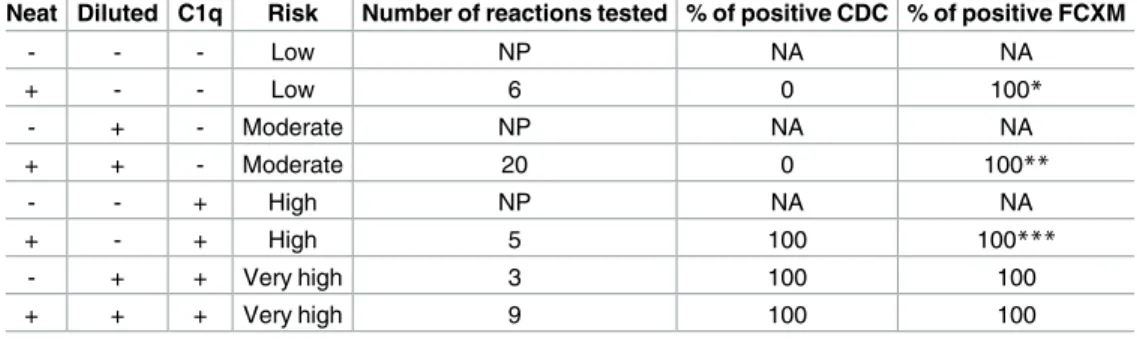
The biopsy confirmed ABMR in the four patients with a high risk profile (1 patient with -/+/+ and 3 patients with +/+/+ profiles) DSA prior transplantation, whereas the patient with low titer and non-fixing complement DSA (+/-/- profile) showed no histological damage in protocol biopsy, without any impairment of graft function. These data confirm the risk-scale for anti-HLA antibodies, proposed and summarized inTable 4.
Conclusions
The accurate assessment of anti-HLA antibody is mandatory for every tissue-typing laboratory which reports anti-HLA antibodies to identify patients who are eligible for highly-sensitized programs.
There are situations where assigning a positive antigen can be difficult due to low fluores-cence signal, and further dilution studies should be performed to rule out a prozone effect[12].
The increased sensitivity of the Luminex test in comparison with earlier ELISA tests has the drawback of using the denatured HLA antigens coated on beads. Besides, the detection of both fixing and non-fixing complement antibodies have led to an increase in false positive results and/or low CDC test, routinely performed in tissue typing laboratories.
Despite the positive correlation between raw MFI data from N and DIL sera shown in this study, a reduction in the number of positive beads was observed after DIL sera in highly-sensi-tized patients. This demonstrates that in a group of highly-sensihighly-sensi-tized patients the number of positive beads could be overestimated, possibly due to low titer of anti-HLA antibodies. How-ever, to figure out the potential risk of the low titer of anti-HLA antibodies (negative specifici-ties after DIL) we suggest performing C1q assay. The complement-binding assay is useful in monitoring kidney transplant recipients at risk for allograft loss[13]. The readout of the possi-ble combinations proposed here opens different scenarios other than just positive or negative specificities. We propose a gradual risk of the specificities derived from the results in N/DIL/ C1q sera, summarized inFig 2andTable 4.
In order to assess the low risk of the specificities of sera with positive N results, but negative after DIL and C1q, we performed CDC and FCXM tests with PBMC with reactive HLA speci-ficities. These sera resulted negative in the CDC, confirming the low risk specificities of the profile (+/-/-) (Table 2).
Table 4. Summary of risk-scale proposed based on neat, diluted serum and C1q test results. Neat Diluted C1q Risk Number of reactions tested % of positive CDC % of positive FCXM
- - - Low NP NA NA + - - Low 6 0 100* - + - Moderate NP NA NA + + - Moderate 20 0 100** - - + High NP NA NA + - + High 5 100 100*** - + + Very high 3 100 100 + + + Very high 9 100 100 NP: not performed NA: not applicable
*5 sera with Negative FCXM after dilution
**3 sera with Negative FCXM after dilution
***2 sera with Negative FCXM after dilution
In contrast, in our cohort we observed a high rate of highly-sensitized patients (22.2%) with at least one HLA class-I specificity with prozone effect and complement-fixing antibodies (S1 Table). The detection of this phenomenon is crucial for accurate definition of an anti-HLA antibody profile. This is especially important to avoid negative vXM results that would render a final positive CDC crossmatch. The limitation of this observation is based on a single serum test, but patients on the renal waiting list are frequently monitored and this situation could be detected in other sera. Recently, the complement interference as a cause of fluctuations of serum anti-HLA antibody strength has been demonstrated[12,14].
As proof of concept, five patients from our cohort could be transplanted despite their hypersensitized status. In four of them, ABMR was demonstrated by biopsy and all of them showed a high risk profile as defined here. On the contrary, the one patient with a low profile who was transplanted from our cohort did not suffer ABMR or graft loss. Due to the scarce number of highly-sensitized patients finally transplanted in our institution, where only 5 patients were tested, a larger number of highly-sensitized patients should be monitored to con-firm these findings.
A limitation of the present study was the use of only class-I coated beads for simplicity. Sim-ilar results were observed in class-II antigens coated beads (S1 File). However the interpreta-tion of the anti-HLA antibody profile is more complex, based on reacinterpreta-tions against the alpha and/or beta chains (DQA, DQB, DPA and/or DPB antigens) and consequently the assessment of the risk profile results are more difficult to demonstrate. Moreover, in our cohort of kidney transplant recipients, all anti-HLA antibody profiles tested were against class-II antigens with similar findings (Table 3).
We consider maintaining HLA-antigens with the (+/+/-) antibody profile (moderate risk) as unacceptable. Non-fixing IgG4 or IgG2 anti-HLA antibodies might sustain this profile, although in the present study we did not perform specific IgG subclass to confirm it. Irrespec-tively of the IgG subclass, the IgG class switch cannot be ruled out and the change to a complement fixing IgG1 or IgG3 would increase the risk of a rejection event. Indeed, the rec-ommendation from recent guidelines is to assign HLA mismatches from previous transplants as unacceptable antigens, despite the lack of complement binding antibodies[8].
The highly-sensitized patients spend prolonged periods of time on the waiting list, and the accurate definition of acceptable HLA antigens with low risk should be a priority in order to minimize the time waiting for an organ to be able to be allocated.
The better definition of anti-HLA antibody profile after dilution and C1q test could open up new opportunities in those highly-sensitized patients with a high number of HLA specifici-ties at low titers and non-complement fixing antibodies but would also allow identification of the prozone effect with undesirable consequences in organ allocation systems based on vXM.
Supporting information
S1 Table. Highly sensitized patients with prozone beads detected.
(DOC)
S1 Fig. Representative patterns of anti-HLA antibody regarding positive beads after neat, dilution and C1q test. Tridimentional scatterplots of MFI values after neat serum (z-axis), 1/
160 dilution (y-axis) and C1q (x-axis) test. The colour scale based on MFI from C1q test repre-sents low level (blue), intermediate levels (red-orange) and high MFI levels (yellow). The risk profile showed after neat serum, 1/160 dilution and C1q tes (neat/Dil/C1q) are A) low risk pro-file (+/-/-), B) moderate risk (+/+/-), C) high risk (+/-/+) and D) very high risk (-/+/+). The Excel macro used (http://www.doka.ch/Excel3Dscatterplot.htm).
S1 File. Raw data of Single Antigen beads for class-I and class-II antigens after assessment of net, diluted serum and C1q test.
(XLSX)
Acknowledgments
The authors would like to thank David Ramos, Celestina Burgueño, Marı´a Luz Valca´rcel and Margarita Gomez for their excellent technical support.
Author contributions
Conceptualization: DSS MLH MA IR. Data curation: JI EA.
Formal analysis: JI EA ER. Investigation: JI EA JG. Methodology: DSS JI EA JG.
Project administration: DSS MLH MA. Resources: MA MLH.
Software: DSS JI EA JG. Supervision: DSS MLH. Validation: DSS JI EA JG MLH. Visualization: JI EA.
Writing – original draft: JI EA IR.
Writing – review & editing: DSS MLH ER MA.
References
1. Minucci PB, Grimaldi V, Casamassimi A, Cacciatore F, Sommese L, Picascia A, et al. Methodologies for anti-HLA antibody screening in patients awaiting kidney transplant: a comparative study. Experimen-tal and clinical transplantation: official journal of the Middle East Society for Organ Transplantation. 2011; 9(6):381–6.
2. Morris GP, Phelan DL, Jendrisak MD, Mohanakumar T. Virtual crossmatch by identification of donor-specific anti-human leukocyte antigen antibodies by solid-phase immunoassay: a 30-month analysis in living donor kidney transplantation. Human immunology. 2010; 71(3):268–73. PMID:20074605
3. Jackson AM. The Virtual Crossmatch: An Essential Tool for Transplanting Sensitized Patients. Clinical transplants. 2014:131–6. PMID:26281137
4. Tait BD, Hudson F, Brewin G, Cantwell L, Holdsworth R. Solid phase HLA antibody detection technol-ogy- -challenges in interpretation. Tissue antigens. 2010; 76(2):87–95. doi:10.1111/j.1399-0039.2010. 01486.xPMID:20403141
5. Bray RA, Gebel HM. Strategies for human leukocyte antigen antibody detection. Current opinion in organ transplantation. 2009; 14(4):392–7. PMID:19610172
6. Endres RO, Kleinman SH, Carrick DM, Steele WR, Wright DJ, Norris PJ, et al. Identification of specifici-ties of antibodies against human leukocyte antigens in blood donors. Transfusion. 2010; 50(8):1749– 60. doi:10.1111/j.1537-2995.2010.02589.xPMID:20158682
7. Visentin J, Vigata M, Daburon S, Contin-Bordes C, Fremeaux-Bacchi V, Dromer C, et al. Deciphering complement interference in anti-human leukocyte antigen antibody detection with flow beads assays. Transplantation. 2014; 98(6):625–31. doi:10.1097/TP.0000000000000315PMID:25058376
8. Tait BD, Susal C, Gebel HM, Nickerson PW, Zachary AA, Claas FH, et al. Consensus guidelines on the testing and clinical management issues associated with HLA and non-HLA antibodies in transplanta-tion. Transplantatransplanta-tion. 2013; 95(1):19–47. doi:10.1097/TP.0b013e31827a19ccPMID:23238534
9. Tambur AR, Herrera ND, Haarberg KM, Cusick MF, Gordon RA, Leventhal JR, et al. Assessing Anti-body Strength: Comparison of MFI, C1q, and Titer Information. American journal of transplantation: offi-cial journal of the American Society of Transplantation and the American Society of Transplant Surgeons. 2015; 15(9):2421–30.
10. Visentin J, Guidicelli G, Taupin JL. Assessing HLA Antibody Strength: Have We Thought About Every-thing? American journal of transplantation: official journal of the American Society of Transplantation and the American Society of Transplant Surgeons. 2015; 15(12):3271.
11. Tambur AR, Ramon DS, Kaufman DB, Friedewald J, Luo X, Ho B, et al. Perception versus reality?: Vir-tual crossmatch- -how to overcome some of the technical and logistic limitations. American journal of transplantation: official journal of the American Society of Transplantation and the American Society of Transplant Surgeons. 2009; 9(8):1886–93.
12. Berth M. Complement interference is not the same as a prozone phenomenon. American journal of transplantation: official journal of the American Society of Transplantation and the American Society of Transplant Surgeons. 2015.
13. Loupy A, Lefaucheur C, Vernerey D, Prugger C, Duong van Huyen JP, Mooney N, et al. Complement-binding anti-HLA antibodies and kidney allograft survival. N Engl J Med. 2013; 369:1215–1226. doi:10. 1056/NEJMoa1302506PMID:24066742
14. Guidicelli G, Anies G, Bachelet T, Dubois V, Moreau JF, Merville P, et al. The complement interference phenomenon as a cause for sharp fluctuations of serum anti-HLA antibody strength in kidney transplant patients. Transplant immunology. 2013; 29(1–4):17–21. doi:10.1016/j.trim.2013.09.005PMID: