TítuloEpidemiology and one‐year outcomes in patients with chronic heart failure and preserved, mid‐range and reduced ejection fraction: an analysis of the ESC Heart Failure Long‐Term Registry
17
0
0
Texto completo
(2) Abstract Aims. The objectives of the present study were to describe epidemiology and outcomes in ambulatory heart failure (HF) patients stratified by left ventricular ejection fraction (LVEF) and to identify predictors for mortality at 1 year in each group. Methods and results. The European Society of Cardiology Heart Failure Long‐Term Registry is a prospective, observational study collecting epidemiological information and 1‐year follow‐up data in 9134 HF patients. Patients were classified according to baseline LVEF into HF with reduced EF [EF <40% (HFrEF)], mid‐range EF [EF 40– 50% (HFmrEF)] and preserved EF [EF >50% (HFpEF)]. In comparison with HFpEF subjects, patients with HFrEF were younger (64 years vs. 69 years), more commonly male (78% vs. 52%), more likely to have an ischaemic aetiology (49% vs. 24%) and left bundle branch block (24% vs. 9%), but less likely to have hypertension (56% vs. 67%) or atrial fibrillation (18% vs. 32%). The HFmrEF group resembled the HFrEF group in some features, including age, gender and ischaemic aetiology, but had less left ventricular and atrial dilation. Mortality at 1 year differed significantly between HFrEF and HFpEF (8.8% vs. 6.3%); HFmrEF patients experienced intermediate rates (7.6%). Age, New York Heart Association (NYHA) class III/IV status and chronic kidney disease predicted mortality in all LVEF groups. Low systolic blood pressure and high heart rate were predictors for mortality in HFrEF and HFmrEF. A lower body mass index was independently associated with mortality in HFrEF and HFpEF patients. Atrial fibrillation predicted mortality in HFpEF patients. Conclusions. Heart failure patients stratified according to different categories of LVEF represent diverse phenotypes of demography, clinical presentation, aetiology and outcomes at 1 year. Differences in predictors for mortality might improve risk stratification and management goals. Keywords Ambulatory; Chronic; Heart failure; Left ventricular ejection fraction; Outcomes. Introduction Although the survival of patients with chronic heart failure (HF) has improved with the introduction of disease‐modifying therapies, these patients still have a substantial risk for death or recurrent decompensation requiring hospitalization.1, 2 Left ventricular ejection fraction (LVEF) has an essential role in phenotyping and guiding the therapy of patients with chronic HF.3 Until recently, two types of HF patient were clinically distinguished based on assessment of LVEF, comprising, respectively, those with HF with reduced ejection fraction (HFrEF) and those with HF with preserved ejection fraction (HFpEF). The specific EF thresholds used to define these HF entities have differed considerably even in recent clinical trials, community‐based studies and HF registries.4, 5 HFrEF has been generally defined according to an EF of <40%, whereas the specific EF thresholds for the definition of HFpEF have varied from >40% to >50%. By defining a cut‐off for HFpEF that is higher than that used to define the HFrEF population, a ‘grey zone’ of EF between 40% and 50% remains to be further characterized.6 This group was formally recognized in the recent 2016 European Society of Cardiology (ESC) guidelines as representing a distinct phenotype, termed HF with mid‐range EF (HFmrEF).3 Heart failure registries7 and community‐based studies8 have systematically excluded HFmrEF, or usually included patients with this condition in the HFpEF group, and therefore specific therapeutic evidence for this group of patients is lacking.3 The ESC HF Long‐Term (ESC‐HF‐LT) Registry is the largest pan‐European cohort providing contemporary generalizable information about ‘real‐world’ chronic HF patients in the full spectrum of EF, from all regions of Europe and from Mediterranean countries. 9, 10.
(3) In the present manuscript we describe and compare for the first time the clinical epidemiology, treatment patterns and long‐term outcomes in ambulatory HF patients stratified by LVEF category according to the 2016 ESC guidelines and identify specific independent predictors for mortality at 1 year in each group.. Methods Study design and clinical setting The ESC‐HF‐LT Registry is an ongoing, prospective, multinational, multicentre, observational study of patients presenting to cardiology centres in European and Mediterranean countries. 9 Site selection in each participating country was performed by national co‐ordinators (supplementary material online, Appendix S1) and targeted a sample of centres of different sizes and levels of complexity from which patients were recruited, focusing on capturing a broad spectrum of cardiology and HF specialty units regularly following outpatients with HF. Inclusion criteria All outpatients with HF seen at the clinics were included during the enrolment period. To facilitate consecutive enrolment, patients were enrolled in the registry on a one‐day‐per‐week basis in Phase 1, and then on five consecutive days per season in Phase 2, and five consecutive working days between 1 October and 31 December in Phase 3. A follow‐up visit at 12 months after the entry visit was mandatory for all patients in order to allow information on morbidity and mortality to be collected. The follow‐up clinical visit could be replaced by a telephone call if the patient was unable to travel to the clinical centre. During the course of the year patients were followed up according to the usual practice of the respective centres. There were no specific exclusion criteria other than patient age, which was required to be higher than 18 years. Echocardiographic assessment of LVEF was performed at enrolment in the registry unless the patient presented with recent echocardiographic data (obtained within the previous week). The ESC‐HF‐LT Registry was approved by the respective local institutional review boards according to the rules of each participating country. No data were collected before detailed information had been provided to the patient and signed informed consent had been obtained. Participating centres Patients were enrolled at centres in the following countries and regions: Lithuania and Sweden (northern countries); Bosnia and Herzegovina, Bulgaria, the Czech Republic, Hungary, Latvia, Poland, Romania and Slovakia (eastern countries); Austria and France (western countries); Greece, Italy, Portugal, Serbia, Slovenia, Spain and Turkey (southern countries); Israel (Middle East), and Egypt (North Africa). Several training meetings were organized for national co‐ordinators and study investigators to assure consistency in data collection among participating centres. Furthermore, in each participating country, data sources were subjected to verification for a random sample of 5% of enrolled patients, by EURObservational Research Programme (EORP) monitors..
(4) Statistical analysis All results were summarized overall and then stratified by LVEF (EF <40%, EF 40–50%, EF >50%). Baseline continuous variables were reported as the mean ± standard deviation (SD) or as the median and interquartile range (IQR), as appropriate. Among‐group comparisons were made using a non‐ parametric test (Kruskal–Wallis test). Categorical variables were reported as percentages and compared using a χ2 test or Fisher's exact test if any expected cell count was less than 5. For categorical variables with more than two possible values, exact P‐values were estimated according to the Monte Carlo method. Univariate analysis was applied to both continuous and categorical variables. In addition, for all‐cause death at 1 year, a pairwise comparison was performed among the three LVEF categories. The outcomes were time to death from any cause, time to admission to hospital for worsening HF, and a composite of time to all‐cause death or HF hospitalization from the date of enrolment to 1 year of follow‐up. Plots of Kaplan–Meier curves for time to all‐cause death, time to admission to hospital for HF and time to all‐cause death or HF hospitalization for the three EF categories were made and survival distributions compared using the log‐rank test. Subsequently, the association between EF group and the three outcomes was assessed in the overall cohort using univariable and multivariable Cox analyses. All variables at entry that were statistically significant in univariable analysis by EF group (P < 0.10), and variables considered to be of relevant clinical interest as fixed covariates were included in the multivariable model (Cox model) to identify the predictors for all‐cause death from study entry to follow‐ up at 1 year in the overall cohort, taking into account EF group. A significance level of 0.05 was required to allow a variable both to be entered into and to stay in the multivariable model. Finally, to identify independent predictors for all‐cause death, separately in the three EF subgroups, three separate multivariable Cox analyses were performed. All variables at entry (supplementary material online, Appendix S2) that were statistically significant in univariable analysis (P < 0.10), and variables considered to be of relevant clinical interest were included in the multivariable model (Cox model) to identify the independent predictors for all‐cause death from study entry to follow‐up at 1 year, separately for subgroups of patients with LVEF of <50%, LVEF of 40–50%, and LVEF of >50%, respectively. A significance level of 0.05 was required to allow a variable both to be entered into and to stay in the multivariable model. As we were assessing associations between a broad range of baseline variables and outcomes, no specific interaction was tested. Missing values were not imputed. A two‐sided P‐value of <0.05 was considered to indicate statistical significance. All analyses were performed using SAS version 9.4 (SAS Institute, Inc., Cary, NC, USA).. Results From April 2011 to January 2015, a total of 16 354 patients were enrolled in the ESC‐HF‐LT Registry. These included 9428 outpatients and 6926 hospitalized patients. Of the 9428 ambulatory patients, 294 patients (3.1%) had no available LVEF data and were not included in the present analysis. The remaining 9134 patients are characterized in the present manuscript. Echocardiography was performed at enrolment in the registry in 91.8% of patients. The remaining 8.2% of patients had undergone a recent echocardiographic examination. The number of patients lost to follow‐up at 1 year was 230 (2.4% of 9428)..
(5) Overall, 59.8% of HF patients were classified as having HFrEF, 24.2% as having HFmrEF and 16.0% as having HFpEF. Classification by geographical region and the distribution of patients by deciles of EF are shown in Figure 1.. Figure 1. (A) Geographic distribution of 9134 heart failure patients stratified by ejection fraction (EF). (B) Distribution of patients by deciles of EF.. At enrolment in the registry, baseline characteristics, in terms of demography, clinical characteristics, aetiologies and co‐morbidities in patients with chronic HF stratified by EF are presented in Table 1..
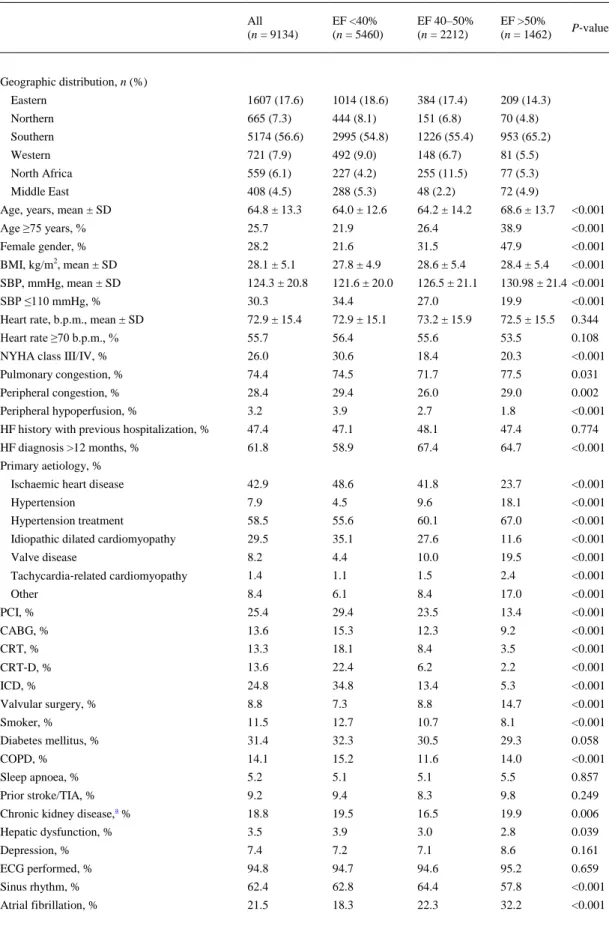
(6) Table 1. Baseline characteristics in chronic heart failure patients stratified by ejection fraction All (n = 9134). EF <40% (n = 5460). EF 40–50% (n = 2212). EF >50% (n = 1462). Eastern. 1607 (17.6). 1014 (18.6). 384 (17.4). 209 (14.3). Northern. 665 (7.3). 444 (8.1). 151 (6.8). 70 (4.8). Southern. 5174 (56.6). 2995 (54.8). 1226 (55.4). 953 (65.2). Western. 721 (7.9). 492 (9.0). 148 (6.7). 81 (5.5). North Africa. 559 (6.1). 227 (4.2). 255 (11.5). 77 (5.3). Middle East. 408 (4.5). 288 (5.3). 48 (2.2). 72 (4.9). Age, years, mean ± SD. 64.8 ± 13.3. 64.0 ± 12.6. 64.2 ± 14.2. 68.6 ± 13.7. <0.001. Age ≥75 years, %. 25.7. 21.9. 26.4. 38.9. <0.001. Female gender, %. 28.2. 21.6. 31.5. 47.9. <0.001. BMI, kg/m2, mean ± SD. 28.1 ± 5.1. 27.8 ± 4.9. 28.6 ± 5.4. 28.4 ± 5.4. <0.001. SBP, mmHg, mean ± SD. 124.3 ± 20.8. 121.6 ± 20.0. 126.5 ± 21.1. 130.98 ± 21.4 <0.001. SBP ≤110 mmHg, %. 30.3. 34.4. 27.0. 19.9. <0.001. Heart rate, b.p.m., mean ± SD. 72.9 ± 15.4. 72.9 ± 15.1. 73.2 ± 15.9. 72.5 ± 15.5. 0.344. Heart rate ≥70 b.p.m., %. 55.7. 56.4. 55.6. 53.5. 0.108. NYHA class III/IV, %. 26.0. 30.6. 18.4. 20.3. <0.001. Pulmonary congestion, %. 74.4. 74.5. 71.7. 77.5. 0.031. Peripheral congestion, %. 28.4. 29.4. 26.0. 29.0. 0.002. Peripheral hypoperfusion, %. 3.2. 3.9. 2.7. 1.8. <0.001. HF history with previous hospitalization, %. 47.4. 47.1. 48.1. 47.4. 0.774. HF diagnosis >12 months, %. 61.8. 58.9. 67.4. 64.7. <0.001. Ischaemic heart disease. 42.9. 48.6. 41.8. 23.7. <0.001. Hypertension. 7.9. 4.5. 9.6. 18.1. <0.001. Hypertension treatment. 58.5. 55.6. 60.1. 67.0. <0.001. Idiopathic dilated cardiomyopathy. 29.5. 35.1. 27.6. 11.6. <0.001. Valve disease. 8.2. 4.4. 10.0. 19.5. <0.001. Tachycardia‐related cardiomyopathy. 1.4. 1.1. 1.5. 2.4. <0.001. Other. 8.4. 6.1. 8.4. 17.0. <0.001. PCI, %. 25.4. 29.4. 23.5. 13.4. <0.001. CABG, %. 13.6. 15.3. 12.3. 9.2. <0.001. CRT, %. 13.3. 18.1. 8.4. 3.5. <0.001. CRT‐D, %. 13.6. 22.4. 6.2. 2.2. <0.001. ICD, %. 24.8. 34.8. 13.4. 5.3. <0.001. Valvular surgery, %. 8.8. 7.3. 8.8. 14.7. <0.001. Smoker, %. 11.5. 12.7. 10.7. 8.1. <0.001. Diabetes mellitus, %. 31.4. 32.3. 30.5. 29.3. 0.058. COPD, %. 14.1. 15.2. 11.6. 14.0. <0.001. Sleep apnoea, %. 5.2. 5.1. 5.1. 5.5. 0.857. Prior stroke/TIA, %. 9.2. 9.4. 8.3. 9.8. 0.249. Chronic kidney disease,a %. 18.8. 19.5. 16.5. 19.9. 0.006. Hepatic dysfunction, %. 3.5. 3.9. 3.0. 2.8. 0.039. Depression, %. 7.4. 7.2. 7.1. 8.6. 0.161. ECG performed, %. 94.8. 94.7. 94.6. 95.2. 0.659. Sinus rhythm, %. 62.4. 62.8. 64.4. 57.8. <0.001. Atrial fibrillation, %. 21.5. 18.3. 22.3. 32.2. <0.001. P‐value. Geographic distribution, n (%). Primary aetiology, %.
(7) Table 1. Baseline characteristics in chronic heart failure patients stratified by ejection fraction All (n = 9134). EF <40% (n = 5460). EF 40–50% (n = 2212). EF >50% (n = 1462). QT duration, ms, mean ± SD. 410.1 ± 56.3. 417.5 ± 55.3. 398.2 ± 59.9. 402.1 ± 50.3 <0.001. LBBB, %. 19.5. 24.2. 15.4. 8.7. <0.001. LVH, %. 31.7. 25.9. 34.6. 48.2. <0.001. EF, %, mean ± SD. 37.6 ± 13.5. 29.1 ± 7.6. 44.0 ± 5.3. 59.7 ± 7.7. <0.001. LVEDD, mm, mean ± SD. 61.0 ± 16.1. 64.9 ± 17.5. 58.0 ± 8.4. 51.1 ± 14.4. <0.001. LA volume, mL, mean ± SD. 73.7 ± 46.0. 79.7 ± 48.8. 60.1 ± 35.7. 78.3 ± 51.2. <0.001. MV inflow pattern‐restrictive (E/A > 2), %. 35.4. 39.6. 28.0. 31.4. <0.001. Mitral regurgitation moderate–severe, %. 31.2. 35.6. 28.4. 19.5. <0.001. Aortic stenosis moderate–severe, %. 3.7. 2.8. 3.4. 7.3. <0.001. Aortic regurgitation moderate–severe, %. 4.7. 4.1. 5.7. 5.0. 0.015. Tricuspid regurgitation moderate–severe, %. 19.2. 19.5. 17.8. 20.3. 0.164. 30.2 ± 11.7. 25.4 ± 10.7. 29.5 ± 14.5. <0.001. Mean pulmonary artery systolic pressure, mmHg, 29.1 ± 12.1 mean ± SD. P‐value. BMI, body mass index; CABG, coronary artery bypass graft; COPD, chronic pulmonary obstructive disease; CRT, cardiac resynchronization therapy; CRT‐D, CRT with defibrillation; ECG, electrocardiogram; EF, ejection fraction; ICD, implantable cardioverter defibrillator; LA, left atrium; LBBB, left bundle branch block; LVEDD, left ventricular end‐diastolic diameter; LVH, left ventricular hypertrophy; MV, mitral valve; NYHA, New York Heart Association; PCI, percutaneous coronary intervention; SBP, systolic blood pressure; SD, standard deviation; TIA, transient ischaemic attack. a Chronic kidney disease: baseline creatinine >1.5 mg/dL.. The subset of patients with EF of >50% included an older population and 48.0% were female. In contrast, patients presenting with EF of <40% and those with EF of 40–50% were on average 4 years younger and more frequently male. The aetiology of HF was ischaemic in a higher percentage of patients with HFrEF and HFmrEF, whereas hypertension was more common in those with HFpEF. At enrolment, systolic blood pressure (SBP) was lower in patients with HFrEF, whereas heart rate (HR) did not differ significantly among EF categories. Pulmonary congestion was seen in similar proportions of HFrEF and HFpEF patients (74.5% and 77.5%, respectively), but New York Heart Association (NYHA) class was higher in HFrEF patients, 30.6% of whom presented with NYHA class III or IV status. A lower body mass index (BMI) was reported in patients with HFrEF compared with HFpEF patients. Rates of most other non‐cardiac associated conditions did not vary significantly among the EF groups, except for chronic obstructive pulmonary disease (COPD), and hepatic and renal dysfunction, which were more frequent in the group with HFrEF. Routine measurements at baseline are presented in the supplementary material online (Table S1). The only clinically significant difference referred to levels of natriuretic peptides, which were higher in patients with HFrEF. Electrocardiography was performed in 94.8% of study patients. Atrial fibrillation (AF) on ECG was most commonly seen in patients with HFpEF (32.2%) (Table 1). The proportion of patients with left bundle branch block (LBBB) was higher in the HFrEF group (24.2%)..
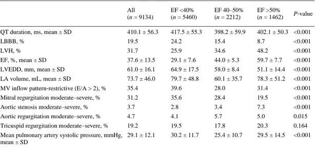
(8) Left ventricular diameter was larger in HFrEF than in HFpEF patients. The proportion of patients with LVH was lower in the HFrEF group than in the other EF subgroups. Left atrial volume was smaller in patients with HFmrEF compared with patients with HFrEF or HFpEF. Mitral regurgitation was commonly seen in the HFrEF group and the proportion of patients with aortic valve disease was higher in the HFpEF group (Table 1). Pharmacological therapy of chronic HF patients at the completion of outpatient visits and at 1 year is presented in Figure 2. In patients with HFrEF, ACE inhibitors/ARBs, beta‐blockers and mineralocorticoid receptor antagonists (MRAs) were used in 91.7%, 92.9% and 67.8% of subjects, respectively. Similar rates of utilization of guideline‐directed medical therapies (GDMTs) were observed in HFmrEF patients, whereas lower rates were noted in patients with HFpEF. The rate of utilization of GDMTs did not change significantly during the follow‐up, regardless of EF category.. Figure 2. Pharmacological treatments administered in 9134 heart failure patients at entry and at 1 year of follow‐up according to ejection fraction (EF) category. ACE, angiotensin‐converting enzyme; ARBs, angiotensin receptor blockers; BB, beta‐blockers; MRAs, mineralocorticoid receptor antagonists.. Outcomes at 1 year Table 2 presents the events (mortality, causes of death and hospitalizations) experienced through the 1‐year follow‐up. Mortality rates at 1 year were 8.8% in patients with HFrEF, 7.6% in patients with HFmrEF and 6.4% in patients with HFpEF. By pairwise comparison, HFrEF patients had a significantly higher mortality rate than HFpEF patients (P = 0.0002), whereas all‐cause mortality in HFmrEF did not differ significantly from mortality in HFrEF (P = 0.07) or in HFpEF (P = 0.17). Non‐cardiovascular mortality was numerically higher in patients with HFmrEF and HFpEF (27.8% and 30.7%, respectively, vs. 20.1% in HFrEF) (Table 2). The percentages of patients hospitalized for HF in the HFrEF, HFmrEF and HFpEF groups were 14.6%, 8.7% and 9.7%, respectively. Combined incidences of death and hospitalization for HF at 1 year were 21.2% in HFrEF patients, 15.0% in HFmrEF patients and 14.6% in HFpEF patients. Figure S1 (supplementary material online) presents rates of these outcomes by deciles of.
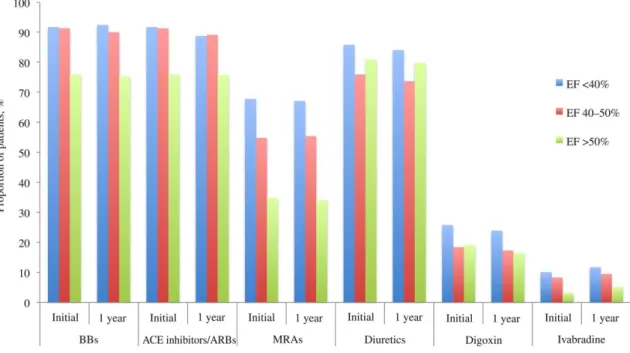
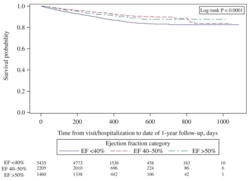
(9) EF. Figures 3 4 and 5 show Kaplan–Meier curves for all‐cause mortality, hospitalization for HF and the composite event of all‐cause mortality and hospitalization for HF stratified by LVEF.. Table 2. Outcomes at 1 year by category of ejection fraction All (n = 9134). EF <40% (n = 5460). EF 40–50% (n = 2212). EF >50% (n = 1462). P‐valuea. 8.1. 8.8. 7.6. 6.3. 0.005. Cardiovascular death, %. 52.1. 53.5. 50.6. 47.2. 0.504. Non‐cardiovascular death, %. 23.2. 20.1. 27.8. 30.7. 0.059. Unknown, %. 24.7. 26.3. 21.6. 21.9. 0.393. All‐cause hospitalization, %. 28.1. 31.9. 22.0. 23.5. <0.001. HF hospitalization, %. 12.4. 14.6. 8.7. 9.7. <0.001. All‐cause death or HF hospitalization, %. 18.6. 21.2. 15.0. 14.6. <0.001. All‐cause death, %. EF, ejection fraction; HF, heart failure. a P‐values for differences among EF <40%, EF 40–50% and EF >50%.. Figure 3. Kaplan–Meier curves for all‐cause mortality in 9134 heart failure patients at 1 year. EF, ejection fraction..
(10) Figure 4. Kaplan–Meier curves for hospitalization for heart failure in 9134 heart failure patients at 1 year. EF, ejection fraction.. Figure 5. Kaplan–Meier curves for all‐cause mortality or hospitalization for heart failure in 9134 heart failure patients at 1 year. EF, ejection fraction..
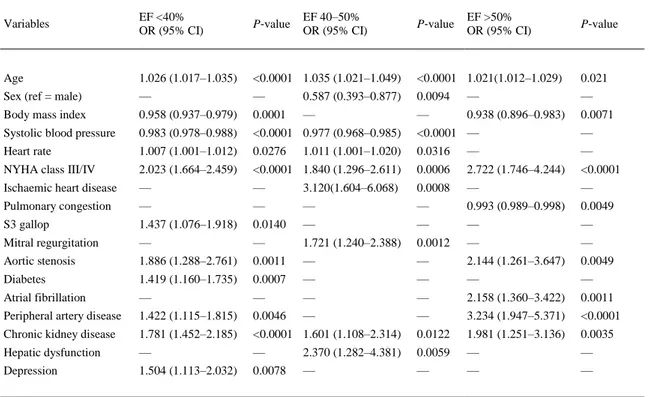
(11) Although not included in the present analysis, high rates of adverse outcomes at 1 year were observed in patients for whom information about EF was not available, including a mortality rate of 11.4% at 1 year, a rate of hospitalization for HF of 9.5% at 1 year, and a mortality/hospitalization for HF rate of 20.1% at 1 year. Variables independently associated with all‐cause mortality at 1 year are shown in Table 3. Older age, NYHA class and chronic kidney disease were independently associated with a worse outcome regardless of EF category. Low SBP and high HR were predictors for mortality in patients with reduced and mid‐ range EF. A higher BMI was independently associated with lower mortality in HFrEF and HFpEF patients. Atrial fibrillation was an independent predictor of mortality in HFpEF patients. Other non‐ cardiovascular co‐morbidities, such as depression, peripheral artery disease and hepatic dysfunction, were predictive of mortality at 1 year in different categories of EF.. Table 3. Predictors for all‐cause mortality within 1 year by ejection fraction category Variables. EF <40% OR (95% CI). P‐value. EF 40–50% OR (95% CI). Age. 1.026 (1.017–1.035). <0.0001 1.035 (1.021–1.049). <0.0001 1.021(1.012–1.029). Sex (ref = male). —. —. 0.587 (0.393–0.877). 0.0094. —. —. Body mass index. 0.958 (0.937–0.979). 0.0001. —. —. 0.938 (0.896–0.983). 0.0071. Systolic blood pressure. 0.983 (0.978–0.988). <0.0001 0.977 (0.968–0.985). <0.0001 —. —. Heart rate. 1.007 (1.001–1.012). 0.0276. 1.011 (1.001–1.020). 0.0316. —. —. NYHA class III/IV. 2.023 (1.664–2.459). <0.0001 1.840 (1.296–2.611). 0.0006. 2.722 (1.746–4.244). <0.0001. Ischaemic heart disease. —. —. 3.120(1.604–6.068). 0.0008. —. —. Pulmonary congestion. —. —. —. —. 0.993 (0.989–0.998). 0.0049. S3 gallop. 1.437 (1.076–1.918). 0.0140. —. —. —. —. Mitral regurgitation. —. —. 1.721 (1.240–2.388). 0.0012. —. —. Aortic stenosis. 1.886 (1.288–2.761). 0.0011. —. —. 2.144 (1.261–3.647). 0.0049. Diabetes. 1.419 (1.160–1.735). 0.0007. —. —. —. —. Atrial fibrillation. —. —. —. —. 2.158 (1.360–3.422). 0.0011. Peripheral artery disease. 1.422 (1.115–1.815). 0.0046. —. —. 3.234 (1.947–5.371). <0.0001. Chronic kidney disease. 1.781 (1.452–2.185). <0.0001 1.601 (1.108–2.314). 0.0122. 1.981 (1.251–3.136). 0.0035. Hepatic dysfunction. —. —. 2.370 (1.282–4.381). 0.0059. —. —. Depression. 1.504 (1.113–2.032). 0.0078. —. —. —. —. P‐value. EF >50% OR (95% CI). P‐value. 0.021. CI, confidence interval; EF, ejection fraction; NYHA, New York Heart Association; OR, odds ratio.. Discussion The ESC‐HF‐LT Registry provides a contemporary dataset based on which chronic HF patients with different categories of EF, as suggested by the 2016 ESC guidelines, 3 can be evaluated. One important finding of the present study is that chronic HF patients stratified by categories of EF (i.e. <40%, 40–50%, >50%) represent different phenotypes in terms of demography, clinical presentation, aetiology, mechanical and electrical remodelling, and pharmacotherapies. Furthermore, the registry provides important information regarding outcomes at 1 year and independent predictors for mortality in patients stratified by EF..
(12) In an ambulatory cardiology setting, the majority of HF patients present with HFrEF. The proportion of patients with HFpEF in our study is lower than those reported in other community‐based studies of HF,8, 11-13 but similar to that reported in a recent meta‐analysis.14 The study population distribution across the deciles of EF shows a unimodal distribution that peaks at EF of 30–40%, indicating a substantial proportion of patients in the ‘middle band’ of EF of 40–50%, and relatively few patients with an EF of >50%. This unimodal pattern of distribution is similar to that found in one HF trial15 and in observational studies enrolling chronic HF patients presenting routinely at ambulatory visits. 2 In contrast, studies including hospitalized patients,7 or a mixed population of hospitalized patients and chronic HF patients included post‐discharge,11, 12 described a bimodal distribution, with substantial proportions of patients with HFrEF and HFpEF, and very few patients with HFmrEF. One possible explanation for this difference refers to the inclusion of a younger and predominantly male population in studies with unimodal distribution in comparison with studies showing a bimodal distribution. Other possible explanations for the lower proportion of HFpEF patients refer to the study methodology. The ESC‐HF‐LT Registry included only patients from cardiology departments or specialized HF units that care mostly for HFrEF patients. Patients with HFpEF commonly present with worsening non‐cardiac co‐morbidities and are more likely to be admitted and followed in internal medicine or geriatric wards. Phenotypes of left ventricular ejection fraction In the present study, patients with HFrEF were typically younger than patients with HFpEF, which confirms previously reported age differences between the two categories.7, 8, 11, 12, 15 Men were more likely to present with HFrEF, which is also consistent with previous studies. 13 An ischaemic aetiology was more commonly reported in HFrEF than in HFpEF, whereas hypertension, diabetes and AF were more prevalent in HFpEF. Although congestion was seen in similar proportions of HFrEF and HFpEF patients, its clinical presentation, including NYHA class and hypotension, was more severe in HFrEF patients. Additionally, natriuretic peptide levels were higher in HFrEF than in HFpEF patients. Chamber dilation seen in HFrEF16 is coupled with pathological electric remodelling, including a longer QT interval and more frequent LBBB than in the other EF subgroups. The rate of use of GDMTs was high at the entry visit and did not change during the follow‐up. The ESC‐HF‐LT Registry demonstrates excellent adherence to guidelines and reduces the gap between the information generated by randomized controlled trials and those provided by observational research that reflects routine clinical practice.9 Compared with those with HFrEF, patients with HFpEF had high prevalences of hypertension, diabetes and AF, but were less likely to have ischaemic heart disease. Because HFpEF is a disease that affects elderly people, it coexists with a high frequency of co‐morbidities. As these may mimic HF symptoms, this may generate concern for the possible misdiagnosis of HF when EF is preserved. However, in the current study, frequencies of co‐morbid conditions were similar in patients with HFpEF and HFrEF. Therefore, it is unlikely that HF‐like symptoms were caused by other conditions, such as COPD or renal failure, more often among patients with preserved compared with those with reduced EF. The pattern of ventricular remodelling in HFpEF is characterized by normal left ventricular size and a high prevalence of LVH. Left atrial dilation and high prevalence of AF suggest predominantly left atrial electrical remodelling. Although no guideline‐specific recommendations exist for this group of patients, substantial proportions of HFpEF patients are treated with beta‐blockers, ACE/ARBs and MRAs, and, as in HFrEF patients, these treatments remain stable over time..
(13) In the ESC‐HF‐LT Registry, the mid‐range group has many features more typical of HFrEF. Patients with HFmrEF were younger, more likely to be male, more frequently had coronary artery disease and less frequently had hypertension compared with those with HFpEF. Left ventricular end‐diastolic diameters were higher in HFmrEF subjects compared with those with HFpEF, but lower than in patients with HFrEF. Similarities between HFrEF and HFmrEF suggest that HFmrEF represents either recovered HFrEF or early‐stage HFrEF,17, 18 but the lack of serial echocardiograms in the present study prohibits more granular insight. The HFmrEF group may have comprised patients with early‐stage HFrEF who recover EF, or well treated patients with disease of ischaemic aetiology in which EF changes were delayed, patients who had recovered after myocarditis and HFpEF patients with a progressive decline in EF. 6 The proportions of patients with previous CRT implants were 8.4% in the HFmrEF group and 3.5% in the HFpEF group, a finding that is highly suggestive of the benefit of the intervention and the dynamic change in LVEF from HFrEF at the time of implant, to HFmrEF or HFpEF at enrolment in the registry. Echocardiography follow‐up is crucial because a substantial proportion of HF patients may show dynamic changes in LVEF over time, especially those with disease of an ischaemic aetiology,19, 20 and patients may transition from one category to another. Unfortunately, the ESC‐HF‐LT Registry does not include echocardiography follow‐up, which is essential to determine the clinical course of chronic HF, especially in the subgroup with HFmrEF. Of note, previous HF clinical trials and epidemiological studies have systematically either excluded HFmrEF or mixed HFmrEF subjects within the HFpEF group. Hence, specific therapeutic evidence for this group of patients is lacking and current guidelines recommend that HFmrEF should be treated similarly to HFrEF.3 Outcomes at 1 year In the ESC‐HF‐LT Registry, the rate of all‐cause mortality at 1 year was 8.1%; 52.1% of these deaths were attributable to cardiovascular causes. The figure is similar to that observed in the ESC‐HF pilot study (7.2%) and in the Italian Registry (5.9%). 1, 2 The rate of HF hospitalization at 1 year was 28.2%, but of all admissions, only 44.1% were attributable to HF, which underscores the importance of non‐cardiac co‐morbidities in the general process of decompensation. Patients with HFpEF had a lower risk for adverse cardiovascular outcomes than those with HFrEF. The results contrast with those of other studies that report similar mortality rates in patients with HFpEF and HFrEF.7, 8, 11 Of note, all of these studies included patients hospitalized for HF and followed up post‐ discharge and thus refer to populations that differ from that of the ESC‐HF‐LT Registry, which enrolled chronic HF patients who presented routinely at outpatient visits. Similarly to the ESC‐HF‐LT Registry, in the CHARM (Candesartan in Heart Failure Reduction in Mortality) programme, patients with an EF of >45% were found to have a much lower risk for mortality at 1 year than those with reduced EF.15 In a recent meta‐analysis of data derived from 31 studies that included both registries and randomized controlled trials, patients with HFpEF were found to have a lower risk for death within 1 year compared with those with HFrEF (12.1% vs. 14.1%), irrespective of gender, age, ischaemic aetiology, hypertension, diabetes and AF.21 In patients with HFmrEF, the rate of mortality at 1 year lies between rates in the HFrEF and HFpEF groups. In terms of all adverse outcomes at 1 year, findings in the HFmrEF group resembled those of the HFpEF group more closely than those of the HFrEF group, an observation similar to those of previous reports.15.
(14) Although not included in the present analysis, patients for whom information about EF was missing showed the highest rate of mortality at 1 year, similar to that reported in a recent meta‐analysis.14 A higher index of non‐cardiovascular co‐morbidities, especially COPD, as well as the lower prescription of GDMTs than in patients with known EF, may explain the increased mortality in this subgroup. 14 Predictors for mortality at 1 year Overall, predictors for mortality in chronic HF outpatients were represented by variables that are easily accessible in clinical practice, including demographic factors, vitals, coexisting severe valvular disease and co‐morbidities. Older age, NYHA class III or IV status and chronic kidney disease were predictive of mortality at 1 year regardless of EF stratification, a finding similar to those observed in previous studies.22, 23 A lower SBP, higher HR and lower BMI were associated with increased mortality at 1 year in patients with HFrEF. Interestingly, although HR extends its predictive value in patients with EF 40–50%, beyond the EF cut‐off used in one clinical trial,24 it has not emerged as a predictor in the subgroup of patients with EF >50%, which suggests that targeting HR in this category may not be beneficial. 25, 26 This is explained by recently reported differences in the prognostic role of HR according to EF. When EF is reduced, HR is a risk factor, and when EF is preserved, HR is simply a marker of the underlying pathological condition. Reducing it may be useful in terms of decreasing symptoms, but not in terms of prognostic improvement.25 In patients with HFrEF or HFpEF, a lower BMI identifies a particularly high risk for death within 1 year. This observation is consistent with that previously reported in chronic HF patients. 27 Ischaemic aetiology was associated with a poor prognosis in the HFmrEF subgroup. Significant mitral regurgitation as a consequence or cause of left ventricular remodelling was associated with higher mortality at 1 year in HFmrEF patients. Mitral regurgitation is a well‐known predictor of mortality in a wide spectrum of EF patients and, additionally, may overestimate EF in patients with borderline EF. 28 Depression, peripheral artery disease and hepatic dysfunction were predictive of mortality in some chronic HF patients, an observation that emphasizes the need to identify non‐cardiac co‐morbidities in chronic HF patients.22, 23 Outpatients with HF should be included in multidisciplinary programmes, in which regular follow‐up in which data about vitals, clinical presentation, aetiologies and co‐morbidities are collected may improve prognosis.3 Limitations The study population included only patients seen at cardiology outpatient clinics and did not include patients with chronic HF seen in other ambulatory facilities or those seen by other professionals such as internists. The diagnosis of HF was not centrally validated and cause of death was not adjudicated by a central committee. Echocardiography was performed in the context of routine clinical practice and, for this reason, was limited to a focused assessment of cardiac structure and function. In the present study, LVEF was obtained only at the moment of enrolment in the registry, and LVEF data from previous hospitalizations or at first diagnosis were not collected. The assessment of LVEF could not be standardized and therefore may have been subject to variations among different operators that may have resulted in the misclassification of some patients. Other potentially important variables, which may have prognostic importance, such as levels of natriuretic peptides, were not selected in the multivariate model as data were not available in many patients..
(15) Conclusions Chronic HF patients stratified by different categories of EF (i.e. <40%, 40–50% and >50%) represent different phenotypes in terms of demography, clinical presentation, aetiology, and mechanical and electrical remodelling. However, classifying chronic HF patients in ambulatory settings is more complex than simply stratifying patients by LVEF cut‐off values because these patients have a high burden of cardiovascular and non‐cardiovascular co‐morbidities, which may interact on different levels of LVEF and may influence prognosis more than LVEF category. Risk stratification and the identification of predictors of worse outcomes may provide opportunities to explore potential interactions between underlying risk and possible treatment effects, especially in EF categories in which guideline‐based recommendations are lacking.. Acknowledgements The Executive Committee of the study had full access to the data and takes complete responsibility for the integrity and accuracy of the data analysis. The authors would like to thank the data monitoring and technical support team: data collection was conducted by Maryna Andarala of the EURObservational Research Programme of the European Society of Cardiology. Statistical analyses were performed by Cecile Laroche. Overall activities were co‐ordinated by Aldo P. Maggioni (Scientific Co‐ordinator, EORP) and Thierry Ferreira (Head of Department, EORP).. Funding The Survey was funded by the ESC. Each participating national cardiology society was given a grant of €10 000 to help with the organizational needs of national network implementation. The following companies support the EURObservational Research Programme: Abbott Vascular Int., Amgen, Bayer Pharma AG, Boston Scientific International, The Bristol Myers Squibb and Pfizer alliance, Boehringer Ingelheim, The Alliance Daiichi Sankyo Europe GmbH and Eli Lilly and Company, Menarini International Operations, Novartis Pharma AG, Merck & Co., Sanofi‐Aventis Group, Servier.. Conflict of interest: O.C. reports the receipt of grants from Servier, Vifor, Novartis and other support from Novartis outside the submitted work. S.D.A. reports the receipt of grants and personal fees from Vifor International, personal fees from Brahms, Novartis and ASTRA, and grants from Abbott Vascular during the conduct of the study. M.G.C.‐L. reports the receipt of grants and personal fees from Novartis outside the submitted work. J.P. received honoraria from Novartis, Servier and Orion Pharma outside the conduct of the present work. A.M. reports the receipt of personal fees from Novartis, Orion, Roche, Servier, Cardiorentis and Zs Pharma, grants and personal fees from Adrenomed, and grants from MyCartis and Critical Diagnostics outside the submitted work. L.H.L. reports the receipt of grants from the Swedish Research Council, Swedish Heart Lung Foundation, AstraZeneca and the Swedish Society of Medicine during the conduct of the study, and the receipt of personal fees from Bayer, Novartis, ViforPharma, St. Jude Medical, HeartWare and Relypsa, grants and personal fees from AstraZeneca and Medtronic, and grants from Boston Scientific outside the submitted work. G.A. reports the receipt of personal fees from Behring, Angelini, Menarini and Merck outside the submitted work. A.J.C. reports the receipt of personal fees from Lone Star Heart, Respicardia, Servier and Resmed outside the submitted work. R.F. reports the receipt of grants from Servier International, Boehringer Ingelheim, Novartis, Merck Serono and Bayer outside the submitted work. F.R. reports the receipt of grants and personal fees from St. Jude Medical, and personal fees from Servier, Zoll, AstraZeneca, HeartWare, Sanofi, Cardiorentis, Novartis and Amgen outside the submitted work. A.P.M. reports the receipt of grants from Novartis, Bayer and Cardiorentis outside the submitted work. G.F. reports having served as a committee member on trials and registries sponsored by Bayer, Novartis, Servier and Vifor outside the submitted work. M.L., P.M.S., V.‐P.H., C.L., M.F.P. and C.F. have nothing to disclose..
(16) Supporting Information Additional Supporting Information may be found in the online version of this article: Appendix S1. Committees and investigators in the European Society of Cardiology Heart Failure LongTerm Registry. Appendix S2. Covariates used in the Cox model and proportions of patients for whom data were available at study entry. Table S1. Laboratory measurements at outpatient visits according to ejection fraction. Figure S1. Rates of adverse outcomes by decile of ejection fraction.. References 1. Maggioni AP, Dahlstrom U, Filippatos G, Chioncel O, Crespo‐Leiro M, Drozdz J, Fruhwald F, Gullestad L, Logeart D, Fabbri G, Urso R, Metra M, Parissis J, Persson H, Ponikowski P, Rauchhaus M, Voors AA, Nielsen OW, Zannad F, Tavazzi L; Heart Failure Association of the European Society of Cardiology (HFA). EURObservational Research Programme: regional differences and 1‐year follow‐up results of the Heart Failure Pilot Survey (ESC‐HF Pilot). Eur J Heart Fail 2013;15:808–817. 2. Tavazzi L, Senni M, Metra M, Gorini M, Cacciatore G, Chinaglia A, Di Lenarda A, Mortara A, Oliva F, Maggioni AP; IN‐HF (Italian Network on Heart Failure) Outcome Investigators. Multicenter prospective observational study on acute and chronic heart failure: one‐year follow‐up results of IN‐HF (Italian Network on Heart Failure) outcome registry. Circ Heart Fail 2013;6:473–481. 3. Ponikowski P, Voors AA, Anker SD, Bueno H, Cleland JG, Coats AJ, Falk V, González‐Juanatey JR, Harjola VP, Jankowska EA, Jessup M, Linde C, Nihoyannopoulos P, Parissis JT, Pieske B, Riley JP, Rosano GM, Ruilope LM, Ruschitzka F, Rutten FH, van der Meer P. 2016 ESC Guidelines for the diagnosis and treatment of acute and chronic heart failure: the Task Force for the diagnosis and treatment of acute and chronic heart failure of the European Society of Cardiology (ESC). Developed with the special contribution of the Heart Failure Association (HFA) of the ESC. Eur J Heart Fail 2016;18:891– 975. 4. Butler J, Fonarow GC, Zile MR, Lam CS, Roessig L, Schelbert EB, Shah SJ, Ahmed A, Bonow RO, Cleland JG, Cody RJ, Chioncel O, Collins SP, Dunnmon P, Filippatos G, Lefkowitz MP, Marti CN, McMurray JJ, Misselwitz F, Nodari S, O'Connor C, Pfeffer MA, Pieske B, Pitt B, Rosano G, Sabbah HN, Senni M, Solomon SD, Stockbridge N, Teerlink JR, Georgiopoulou VV, Gheorghiade M. Developing therapies for heart failure with preserved ejection fraction: current state and future directions. JACC Heart Fail 2014;2:97–112. 5. Vaduganathan M, Michel A, Hall K, Mulligan C, Nodari S, Shah SJ, Senni M, Triggiani M, Butler J, Gheorghiade M. Spectrum of epidemiological and clinical findings in patients with heart failure with preserved ejection fraction stratified by study design: a systematic review. Eur J Heart Fail 2016;18:54– 65. 6. Lam CS, Solomon SD. The middle child in heart failure: heart failure with mid‐range ejection fraction (40–50%). Eur J Heart Fail 2014;16:1049–1055. 7. Fonarow GC, Stough WG, Abraham WT, Albert NM, Gheorghiade M, Greenberg BH, O'Connor CM, Sun JL, Yancy CW, Young JB; OPTIMIZE‐HF Investigators and Hospitals. Characteristics, treatments, and outcomes of patients with preserved systolic function hospitalized for heart failure: a report from the OPTIMIZE‐HF Registry. J Am Coll Cardiol 2007;50:768–777. 8. Bhatia RS, Tu JV, Lee DS, Austin PC, Fang J, Haouzi A, Gong Y, Liu PP. Outcome of heart failure with preserved ejection fraction in a population‐based study. N Engl J Med 2006;355:260–269. 9. Maggioni AP, Anker SD, Dahlstrom U, Filippatos G, Ponikowski P, Zannad F, Amir O, Chioncel O, Leiro MC, Drozdz J, Erglis A, Fazlibegovic E, Fonseca C, Fruhwald F, Gatzov P, Goncalvesova E, Hassanein M, Hradec J, Kavoliuniene A, Lainscak M, Logeart D, Merkely B, Metra M, Persson H, Seferovic P, Temizhan A, Tousoulis D, Tavazzi L. Are hospitalized or ambulatory patients with heart failure treated in accordance with European Society of Cardiology guidelines? Evidence from 12,440 patients of the ESC Heart Failure Long‐Term Registry. Eur J Heart Fail 2013;15:1173–1184. 10. Crespo‐Leiro MG, Anker SD, Maggioni AP, Coats AJ, Filippatos G, Ruschitzka F, Ferrari R, Piepoli MF, Delgado Jimenez JF, Metra M, Fonseca C, Hradec J, Amir O, Logeart D, Dahlström U, Merkely B, Drozdz J, Goncalvesova E, Hassanein M, Chioncel O, Lainscak M, Seferovic PM, Tousoulis D, Kavoliuniene A, Fruhwald F, Fazlibegovic E, Temizhan A, Gatzov P, Erglis A, Laroche C, Mebazaa A. European Society of Cardiology Heart Failure Long‐Term Registry (ESC‐HF‐LT): 1‐year follow‐up outcomes and differences across regions. Eur J Heart Fail 2016;18:613–625. 11. Bursi F, Weston SA, Redfield MM, Jacobsen SJ, Pakhomov S, Nkomo VT, Meverden RA, Roger VL. Systolic and diastolic heart failure in the community. JAMA 2006;296:2209–2216..
(17) 12. Dunlay SM, Roger VL, Weston SA, Jiang R, Redfield MM. Longitudinal changes in ejection fraction in heart failure patients with preserved and reduced ejection fraction. Circ Heart Fail 2012;5:720–726. 13. Owan TE, Hodge DO, Herges RM, Jacobsen SJ, Roger VL, Redfield MM. Trends in prevalence and outcome of heart failure with preserved ejection fraction. N Engl J Med 2006;355:251–259. 14. Poppe K, Squire IB, Whalley, G, Kober L, McAlister FA, McMurray JJV, Pocock S, Earle NJ, Berry C, Doughty RN, Berry C, Doughty R; Meta‐Analysis Global Group in Chronic Heart Failure. Known and missing left ventricular ejection fraction and survival in patients with heart failure: a MAGGIC meta‐ analysis report. Eur J Heart Fail 2013;15:1220–1227. 15. Solomon SD, Anavekar N, Skali H, McMurray JJ, Swedberg K, Yusuf S, Granger CB, Michelson EL, Wang D, Pocock S, Pfeffer MA; Candesartan in Heart Failure Reduction in Mortality (CHARM) Investigators. Influence of ejection fraction on cardiovascular outcomes in a broad spectrum of heart failure patients. Circulation 2005;112:3738–3744. 16. Lam CS, Roger VL, Rodeheffer RJ, Bursi F, Borlaug BA, Ommen SR, Kass DA, Redfield MM. Cardiac structure and ventricular‐vascular function in persons with heart failure and preserved ejection fraction from Olmsted County, Minnesota. Circulation 2007;115:1982–1990. 17. Borlaug BA, Redfield MM. Diastolic and systolic heart failure are distinct phenotypes within the heart failure spectrum. Circulation 2011;123:2006–2014. 18. Kalogeropoulos AP, Fonarow GC, Georgiopoulou V, Burkman G, Siwamogsatham S, Patel A, Li S, Papadimitriou L, Butler J. Characteristics and outcomes of adult outpatients with heart failure and improved or recovered ejection fraction. JAMA Cardiol 2016;1:510–518. 19. Mann DL, Burkhoff D. Is myocardial recovery possible and how do you measure it? Curr Cardiol Rep 2012;14:293–298. 20. Hwang SJ, Melenovsky V, Borlaug BA. Implications of coronary artery disease in heart failure with preserved ejection fraction. J Am Coll Cardiol 2014;63:2817–2827. 21. Meta‐Analysis Global Group in Chronic Heart Failure (MAGGIC). The survival of patients with heart failure with preserved or reduced left ventricular ejection fraction: an individual patient data meta‐ analysis. Eur Heart J 2012;33:1750–1757. 22. Triposkiadis F, Giamouzis G, Parissis J, Starling RC, Boudoulas H, Skoularigis J, Butler J, Filippatos G. Reframing the association and significance of co‐morbidities in heart failure. Eur J Heart Fail 2016;18:744–758. 23. van Deursen VM, Urso R, Laroche C, Damman K, Dahlström U, Tavazzi L, Maggioni AP, Voors AA. Co‐morbidities in patients with heart failure: an analysis of the European Heart Failure Pilot Survey. Eur J Heart Fail 2014;16:103–111. 24. Swedberg K, Komajda M, Böhm M, Borer JS, Ford I, Dubost‐Brama A, Lerebours G, Tavazzi L; SHIFT Investigators. Ivabradine and outcomes in chronic heart failure (SHIFT): a randomised placebo‐controlled study. Lancet 2010;376:875–885. 25. Fox K, Ford I, Steg PG, Tardif JC, Tendera M, Ferrari R. Ivabradine in stable coronary artery disease without clinical heart failure. N Engl J Med 2014;371:1091–1099. 26. Ferrari R, Fox K. Heart rate reduction in coronary artery disease and heart failure. Nat Rev Cardiol 2016;13:493–501. 27. Zafrir B, Salman N, Crespo‐Leiro MG, Anker SD, Coats AJ, Ferrari R, Filippatos G, Maggioni AP, Mebazaa A, Piepoli MF, Ruschitzka F, Paniagua‐Martin MJ, Segovia J, Laroche C, Amir O; Heart Failure Long‐Term Registry Investigators. Body surface area as a prognostic marker in chronic heart failure patients: results from the Heart Failure Registry of the Heart Failure Association of the European Society of Cardiology. Eur J Heart Fail 2016;18:859–868. 28. Bursi F, Barbieri A, Grigioni F, Reggianini L, Zanasi V, Leuzzi C, Ricci C, Piovaccari G, Branzi A, Modena MG. Prognostic implications of functional mitral regurgitation according to the severity of the underlying chronic heart failure: a long‐term outcome study. Eur J Heart Fail 2010;12:382–388..
(18)
Figure
+3
Documento similar
