Low doses of insulin like growth factor I improve insulin resistance, lipid metabolism and oxidative damage in aging rats
10
0
0
Texto completo
(2) 2434. Endocrinology, May 2008, 149(5):2433–2442. Garcı́a-Fernández et al. • Low Doses of IGF-I in Aging. Germany) and divided into aliquots that were stored at ⫺20 C until used. The animals were then killed by decapitation. The liver and brain were dissected. Samples from the cortex and hippocampus were stored separately until assaying at ⫺80 C after immersion in N liquid. In six animals from each group, a part of the fresh liver was used to isolate mitochondria and perform mitochondrial function tests by flow cytometry.. H 2O2 2 H+. 2. FIG. 1. Mechanisms of oxidative cellular damage. Free radicals are reduced into water with the cooperation of the three main antioxidant enzymes: SOD, catalase, and GSHPx. The generation of hydroxyl radicals from hydroperoxide produces the development of oxidative cell injury: DNA damage; carboxylation of proteins; and lipid peroxidation, including lipids of mitochondrial membranes. By these pathways, oxidative damage leads to cellular death.. of brain and liver. In addition, mitochondria function [mitochondrial membrane potential (MMP) and ATP synthesis] was assessed in isolated liver mitochondria (29, 32, 35). Three experimental groups were included in this protocol: young healthy controls (yCO) (17 wk old); untreated old rats (O) (103 wk old); and aging rats (103 wk old) treated with IGF-I (O ⫹ IGF-I) during 1 month [2.25 g IGF-I/100 g body weight (bw)⫺1䡠d⫺1]. Materials and Methods Previous experimental protocol To characterize the experimental model of aging in male Wistar rats and select the age of the controls, healthy rats of increasing age (9, 17, 90, and 103 wk old) were studied. The life span of these rats is estimated at about 2 yr (96 –110 wk). The age of the controls was selected on the basis of the evolution in IGF-I and testosterone serum concentrations, and the plasma TAS (Fig. 2). Thus, 9-wk-old rats were considered to be too young (see Results), whereas 17-wk-olds were judged to be a suitable age for young controls (yCO). Animals of 103 wk old were considered suitable to evaluate the decline in anabolic hormones and the reduction in antioxidant capability (see Fig. 2 and Results), as well as to investigate the impact of IGF-I therapy on these parameters in aging animals. After selecting the age of the controls, the following experimental procedure was performed.. Experimental design Healthy male Wistar rats were divided into two groups according to age: yCO (n ⫽ 10) of 17 wk, and aging control rats of 103 wk. Old animals were randomly assigned to receive either saline (group O, n ⫽ 10) or recombinant human IGF-I (Chiron Co., Emeryville, CA), (2.25 g IGFI/100 g bw⫺1䡠d⫺1 in two divided doses) (group O ⫹ IGF, n ⫽ 10) sc during 30 d. All experimental procedures were performed in conformity with The Guiding Principles for Research Involving Animals (36). Both food (standard semipurified diet for rodents; B.K. Universal, Sant Vicent del Horts, Spain) and water were given ad libitum. Rats were housed in cages placed in a room with a 12-h light, 12-h dark cycle and constant humidity and temperature (20 C). In the morning of the 31st day, blood was obtained from the retroocular plexus with capillary tubes (70 mm; Laboroptik, Marienfeld,. Analytical methods in serum Analytical parameters (total protein, albumin, glucose, cholesterol, triglycerides, free fatty acids, transaminases, aspartate transaminase, and alanine transaminase) were determined in serum by routine laboratory methods using a Hitachi 747 autoanalyzer (Roche Molecular Biochemicals, Mannheim, Germany). Serum levels of hormones (testosterone, IGF-I, and insulin) were assessed by RIA in a GammaChen 9612 Plus (Serono Diagnostics, Roma, Italy) using specific commercial assay systems: free testosterone by a Coat-a-Count, DPC (Diagnostic Products Corp., Los Angeles, CA). The sensitivity (S) of total testosterone assay was 4 ng/dl, and the intraassay coefficient of variation was less than 7%. Assessment of serum IGF-I levels were performed using radioimmunoassay with coated tubes for the determination of IGF-I (IGF binding protein blocked), which included elimination of interference by IGF binding protein through excess IGF-II as well as acid alcohol (ALPCO Diagnostics, Windham, NH). The S of IGF-I assay was 0.1 ng/ml and the interserial coefficient of variation was 7.4. Insulin levels were determined using a specific kit for RIA (LINCO Research, Inc., St. Charles, MO) following protocol instructions. The sensibility was of 0.1 ng/ml. The homeostasis model assessment (HOMA), as an index of insulin resistance, was assessed using the HOMA Calculator Programe version 2.0 based on the HOMA formula (37, 38). The HOMA estimates steadystate -cell function (percent B) and insulin S (percent S) as percentages of a normal reference population. These measures correspond well but are not necessarily equivalent to nonsteady-state estimates of -cell function and insulin S derived from stimulatory models such as the hyperinsulinemic clamp, hyperglycemic clamp, iv glucose tolerance test (acute insulin response, minimal model), and the oral glucose tolerance test (0 –30 ␦ insulin/glucose ratio). Serum TAS (28), as total enzymatic and nonenzymatic antioxidant capability of serum, was evaluated using a colorimetric assay (Randox Laboratories Ltd., Crumlin, UK) using the following principle: Abts [2,2⬘-Azino-di-(‘3-ethylbenzthiazoline sulfonate)] was incubated with a peroxidase (metmyoglobin) and H2O2 to produce the radical cation Abts 䡠 ⫹. This has a relatively stable blue-green color, which is measured at 600 nm. Antioxidants in the added sample cause suppression of this color production to a degree that is proportional to their concentration (27).. Parameters of oxidative damage and antioxidant defenses in liver and brain homogenates Lipid peroxidation marker. MDA was used as an index of lipid peroxidation (30) in liver and brain homogenates, and it was measured after heating samples at 45 C for 60 min in acid medium. It was quantitated by a colorimetric assay using LPO-586 (Bioxytech; OXIS International Inc., Portland, OR), which after reacting with MDA generates a stable chromophore that can be measured at 586 nm (Hitachi U2000 Spectro; Roche Molecular Biochemicals) (28, 30). PCC. PCC was determined by the method of Levine et al. (31). Each sample was divided into two portions containing 1- to 2-mg protein each. An equal volume of 2 m HCl was added to one portion, incubated at room temperature for 1 h, and shaken intermittently. The other portion was treated with an equal volume of 10 mm dinitrophenylhydrazine in 2 m HCl and incubated for 1 h at room temperature. After incubation, the mixture was precipitated with 10% trichloroacetic acid and centrifuged. The precipitate was washed with ethanol-ethyl acetate (1:1), twice dissolved in 1 ml 6 m guanidine HCl, centrifuged at low speed, and the supernatant was extracted. The difference in absorbance between the. Downloaded from endo.endojournals.org at Univ San Pablo Biblioteca De Ciencias on March 10, 2009.
(3) Garcı́a-Fernández et al. • Low Doses of IGF-I in Aging. Endocrinology, May 2008, 149(5):2433–2442 2435. FIG. 2. Evolution with aging of IGF-I and testosterone circulating levels and TAS in serum. According to these parameters, 9-wk-old (wo) rats seemed to be too young because IGF-I and testosterone levels were not high enough in these animals. On the other hand, 90-wk-old rats showed levels of testosterone too high for them to be considered old rats. Thus, 17-wk-old rats were considered to be a suitable age for the yCO, and 103 wk was considered as a suitable age to assess the decline in anabolic hormones (old groups). A close direct correlation was found between TAS and IGF-I levels. CO, Control.. dinitrophenylhydrazine-treated and HCl-treated samples was determined at 366 nm. Activities of antioxidant enzymes. Activities of superoxide dismutase (SOD) [Enzyme Commission of the International Union of Biochemistry (EC) 1. 15. 1. 1.], catalase (EC 1. 11. 1. 6.), and glutathione peroxidase (EC 1. 11. 1. 9.) were measured both in brain and liver tissues. Samples were homogenized in Tris-HCl buffer [20 mmol/liter (pH 7.4); 1 g tissue/10 ml] at 0 C and centrifuged at 25,000 g for 30 min at 4 C. All measurements were performed in the supernatant. SOD activity was determined at 37 C using a commercial kit (Randox Laboratories) and an autoanalyzer (Cobas Mira; Roche Diagnostic System, Basel, Switzerland). Catalase activity was determined at 25 C by measuring the changes of absorbance using final concentrations of 10 mmol/liter H2O2 and 50 mmol/liter phosphate buffer (pH 7.0) at 240 nm during the time interval 15–30 sec after addition of the sample (28, 34, 39). GSHPx was measured at 37 C with the Ransod commercial kit using the Cobas Mira autoanalyzer, and its activity was expressed in units (1 U equals 1 mol substrate turnover/ min) per milligram of protein (39).. Isolation of liver mitochondria Liver mitochondrial fraction was prepared according to the method described by Hogeboom and Schneider (40) with modifications. Liver samples were homogenized (1:10 wt/vol) in an ice-cold isolation buffer (pH 7.4) containing sucrose 0.25 m, KH2PO4 5 mm, 3[N-morholino]propanesulfonic acid 5 mm, and 0.1% BSA. The homogenate was centrifuged at 800 ⫻ g for 10 min. The resulting supernatant was centrifuged at 8500 ⫻ g for 10 min. The supernatant was discarded, and the pellet diluted in cold isolation buffer and centrifuged at 8500 ⫻ g for 10 min three times. The final mitochondria pellet was resuspended in a minimal volume, and aliquots were stored at ⫺80 C until used in enzyme assays. All procedures were conducted at 4 C.. Flow cytometry analysis in isolated mitochondria MMP. MMP was evaluated using rhodamine 123 dye (RH123) obtained from Molecular Probes, Inc. (Eugene, OR). It has an absorbance maximum of 500 nm and an emission maximum of 523 nm. Mitochondrial suspensions (40- to 50-g protein mitochondria/ml) were incubated in. Downloaded from endo.endojournals.org at Univ San Pablo Biblioteca De Ciencias on March 10, 2009.
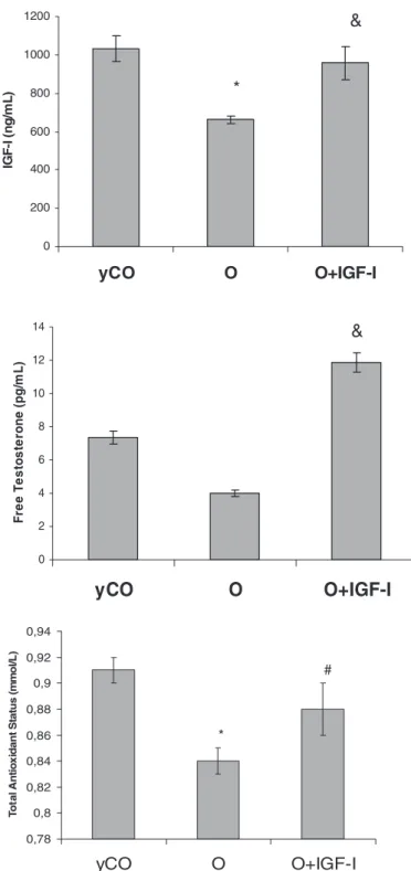
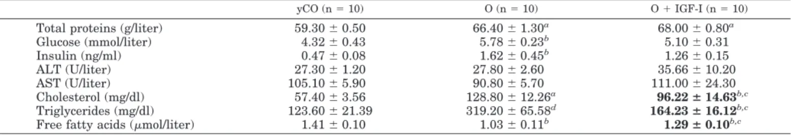
(4) Endocrinology, May 2008, 149(5):2433–2442. isolation buffer for 5 min in the dark with RH123 (0.5 g/ml), after adding various agents. For state 4 conditions, the mitochondrial samples were incubated with rotenone 25 m and energized with sodium succinate 5 mm, and with rotenone 25 m, sodium succinate 5 mm plus ADP 200 m for state 3 conditions. The uncoupler carbonylcyamide-mchlorophenylhydrazone 10 m was added to confirm that the uptake of RH123 was related to MMP. Finally, 2.5 g/ml oligomycin (SigmaAldrich, St. Louis, MO) was added as an inhibitor of ATP synthetase to block all phosphorylation-related respiration (35).. ATP synthesis by mitochondria. Garcı́a-Fernández et al. • Low Doses of IGF-I in Aging. 1200. 600 400 200 0. yCO. Statistical analysis. Figure 2 summarizes the evolution of serum levels of IGF-I, free testosterone, and serum total antioxidant capability in Wistar rats of increasing age (9, 17, 90, and 103 wk old). As mentioned previously, these data allowed us to select the age of the controls (yCO, 17 wk old, and O group, 103 wk old) according to the following criteria: reductions of anabolic hormones (IGF-I and testosterone) and serum TAS with aging. Interestingly, a close direct correlation was found between serum IGF-I levels and total antioxidant capability (r ⫽ 0.7; P ⬍ 0.01; Fig. 2). Thus, in this experimental model of aging, O showed a significant decrease of serum IGF-I (O ⫽ 662.00 ⫾ 17.43 vs. yCO ⫽ 1030.00 ⫾ 67.00 ng/ml; P ⬍ 0.01) and testosterone levels (O ⫽ 4.01 ⫾ 0.19 vs. yCO ⫽ 7.35 ⫾ 0.43 pg/ml; P ⬍ 0.05) as compared with yCO (Fig. 3). O presented a decreased TAS in serum compared with yCO (O ⫽ 0.84 ⫾ 0.01 vs. yCO ⫽ 0.91 ⫾ 0.01 mmol/liter; P ⬍ 0.05). In addition, old animals showed a significant increase in blood glucose (Table 1) with hyperinsulinemia compared with yCO. In addition, the O group presented a significantly higher insulin resistance index (HOMA) compared with yCO (O ⫽ 6.26 ⫾ 1.63 vs. yCO ⫽ 1.23 ⫾ 0.48; P ⬍ 0.01) (Fig. 4). Regarding lipid metabolism, aging rats showed a significant increase of serum cholesterol and triglycerides concentrations, and a significant reduction of free fatty acids compared with yCO (Table 1). Effect of IGF-I on anabolic hormones, serum total capability, and glucose and lipid metabolism. Figure 3 shows that low doses of IGF-I were able to correct IGF-I circulating levels [O ⫹ IGF-I ⫽ 958.00 ⫾ 84.90 ng/ml;. O. O+IGF-I. Free Testosterone (pg/mL). 14. &. 12 10 8 6 4 2 0. yCO. O. O+IGF-I. 0,94 Total Antioxidant Status (mmol/L). Results Characterization of the experimental model of aging. *. 800. ATP synthesis was assessed (41) resuspending a mitochondrial population in buffer and supplemented with 5 mm KH2PO4, 5 mm succinate, and 5 mm ADP, and then incubated for 15 min at 37 C. The reaction was stopped by adding an equal volume of 12% trichloroacetic acid, and the reaction mixture was centrifuged at 1200 g for 15 min at 4 C. The supernatant was neutralized with 6 n KOH and measured for ATP by an enzymatic method using a kit obtained from Sigma Chemical (Tokyo, Japan).. Data are expressed as mean ⫾ sem. To assess the homogeneity among the different groups of rats, a Kruskal-Wallis test was used, followed by multiple post hoc comparisons using Mann-Whitney U tests. A regression model was fitted considering IGF-I levels and serum TAS as the independent and dependent variables, respectively. Any P value less than 0.05 was considered statistically significant. Calculations were performed with SPSSW in v.10.0 program (SPSS, Inc., Chicago, IL).. &. 1000. IGF-I (ng/mL). 2436. 0,92. #. 0,9 0,88. *. 0,86 0,84 0,82 0,8 0,78. yCO *p<0.05 vs yCO;. &p<0.05. O. O+IGF-I. vs O; #p = 0.05 vs O; p = ns yCO vs O+IGF-I. FIG. 3. Effect of low doses of IGF-I on IGF-I and testosterone circulating levels and total antioxidant capability in serum. Low doses of IGF-I increased IGF-I and testosterone circulating levels. IGF-I replacement therapy was also able to restore total antioxidant capability in serum.. P ⫽ not significant (ns) vs. yCO], testosterone levels (O ⫹ IGF-I ⫽ 11.86 ⫾ 0.62 pg/ml; P ⬍ 0.05 vs. O group), and serum total antioxidant capability (O ⫹ IGF-I ⫽ 0.88 ⫾ 0.02; P ⬍ 0.05 vs. O group) in aging rats to values similar to those found in yCO (P ⫽ ns).. Downloaded from endo.endojournals.org at Univ San Pablo Biblioteca De Ciencias on March 10, 2009.
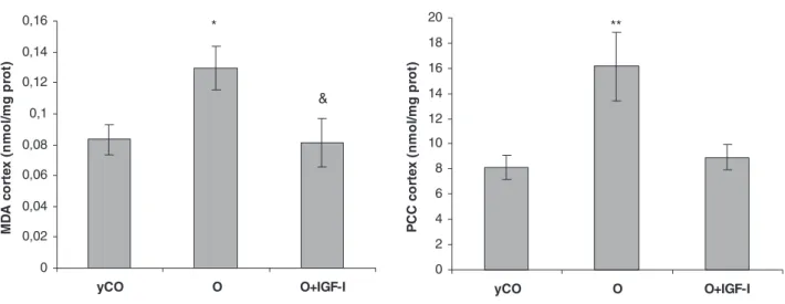
(5) Garcı́a-Fernández et al. • Low Doses of IGF-I in Aging. Endocrinology, May 2008, 149(5):2433–2442 2437. TABLE 1. Analytical parameters in the three experimental groups. Total proteins (g/liter) Glucose (mmol/liter) Insulin (ng/ml) ALT (U/liter) AST (U/liter) Cholesterol (mg/dl) Triglycerides (mg/dl) Free fatty acids (mol/liter). yCO (n ⫽ 10). O (n ⫽ 10). O ⫹ IGF-I (n ⫽ 10). 59.30 ⫾ 0.50 4.32 ⫾ 0.43 0.47 ⫾ 0.08 27.30 ⫾ 1.20 105.10 ⫾ 5.90 57.40 ⫾ 3.56 123.60 ⫾ 21.39 1.41 ⫾ 0.10. 66.40 ⫾ 1.30a 5.78 ⫾ 0.23b 1.62 ⫾ 0.45b 27.80 ⫾ 2.60 90.80 ⫾ 5.70 128.80 ⫾ 12.26a 319.20 ⫾ 65.58d 1.03 ⫾ 0.11b. 68.00 ⫾ 0.80a 5.10 ⫾ 0.31 1.26 ⫾ 0.15 35.66 ⫾ 10.20 111.00 ⫾ 24.30 96.22 ⴞ 14.63b,c 164.23 ⴞ 16.12b,c 1.29 ⴞ 0.10b,c. Values are expressed as ⫾ SEM. ALT, Alanine transaminase; AST, aspartate transaminase. a P ⬍ 0.001 vs. yCO. b P ⬍ 0.05 vs. yCO. c P ⬍ 0.05 O vs. O ⫹ IGF-I. d P ⬍ 0.01 vs. yCO.. In addition, IGF-I replacement therapy reduced glucose and insulin levels (Table 1; P ⫽ ns vs. yCO) correcting the insulin resistance presented in O (HOMA; O ⫹ IGF-I ⫽ 3.72 ⫾ 1.11, vs. yCO; P ⫽ ns) (Fig. 4). IGF-I treatment also induced a significant reduction of serum cholesterol and triglycerides increasing free fatty acids (Table 1).. 7. *. Glucose (mmol/L). 6 5 4 3. Oxidative damage and antioxidant enzyme activities in brain and liver in the three experimental groups. 2 1 0. yCO. O. O+IGF-I. 2,5. *. Insulin (ng/mL). 2. 1,5. 1. 0,5. 0. yCO. 9. O. O+IGF-I. **. 8 7 HOMA. 6. &. 5 4 3 2 1 0. yCO. O. O+IGF-I. *p<0.05, **p<0.01 vs yCO; &p<0.05 O vs O+IGF-I; p = ns yCO vs O+IGF-I. FIG. 4. Circulating glucose (top panel) and insulin levels (middle panel) and index of insulin resistance (HOMA) (lower panel) in the three experimental groups. Compared with yCO, untreated aging rats (O group) presented a significant index of insulin resistance with hyperglycemia and hyperinsulinemia. Low doses of IGF-I were able to improve all these alterations.. Parameters of oxidative damage in brain (cortex and hippocampus) in the three experimental groups are shown in Fig. 5. O presented significant increases of lipid peroxidation products (MDA, expressed as nmol/mg protein) and PCC (expressed as nmol/mg protein) compared with yCO (cortex MDA: O ⫽ 0.13 ⫾ 0.01 vs. yCO ⫽ 0.08 ⫾ 0.01, P ⬍ 0.05; cortex PCC: O ⫽ 16.16 ⫾ 2.72 vs. yCO ⫽ 8.12 ⫾ 0.94, P ⬍ 0.01; hippocampus MDA: O ⫽ 0.80 ⫾ 0.09 vs. yCO ⫽ 0.61 ⫾ 0.04, P ⬍ 0.05; and hippocampus PCC: O ⫽ 19.48 ⫾ 2.76 vs. yCO ⫽ 12.93 ⫾ 1.95, P ⬍ 0.05). Low doses of IGF-I were able to reduce significantly both oxidative damage markers (cortex MDA: O ⫹ IGF-I ⫽ 0.08 ⫾ 0.02; cortex PCC: O ⫹ IGF-I ⫽ 8.93 ⫾ 0.97; hippocampus MDA:O ⫹ IGF-I ⫽ 0.47 ⫾ 0.05; and hippocampus PCC: O ⫹ IGF-I ⫽ 15.46 ⫾ 1.44, nmol/mg protein). These parameters were not significantly different from those found in yCO (P ⫽ ns). Regarding the antioxidant enzyme activities, several significant alterations were found in untreated aging rats (O group) compared with yCO, which are summarized in Table 2. Old rats treated with IGF-I showed similar values to those found in yCO (P ⫽ ns yCO vs. O ⫹ IGF-I) with the only exception of GSHPx in hippocampus (P ⬍ 0.05). In the hepatic tissue, MDA was significantly increased in O (O ⫽ 0.20 ⫾ 0.02 vs. yCO ⫽ 0.11 ⫾ 0.01; P ⬍ 0.001), and IGF-I therapy also reduced this marker of lipid peroxidation (O ⫹ IGF-I ⫽ 0.16 ⫾ 0.01). No significant differences were found in PCC between the three experimental groups (yCO ⫽ 3.76 ⫾ 0.63; O ⫽ 4.71 ⫾ 1.17; and O ⫹ IGF-I ⫽ 4.73 ⫾ 1.31 nmol/mg protein; P ⫽ ns) in liver. Results regarding antioxidant enzymes activities in liver are summarized in Table 2. Only significant differences between groups were found in catalase, which was decreased in O (P ⬍ 0.05), whereas IGF-I-treated old rats presented similar values to those found in yCO.. Downloaded from endo.endojournals.org at Univ San Pablo Biblioteca De Ciencias on March 10, 2009.
(6) 2438. Endocrinology, May 2008, 149(5):2433–2442. A. Garcı́a-Fernández et al. • Low Doses of IGF-I in Aging. Malondialdehyde and Protein Carbonyl Content in cortex. 0,16. 20. *. 0,12. & 0,1 0,08 0,06 0,04 0,02. PCC cortex (nmol/mg prot). MDA cortex (nmol/mg prot). 0,14. 16 14 12 10 8 6 4 2. 0. 0 yCO. O. O+IGF-I. yCO. X ± SEM; *p<0.05 vs yCo; &p<0.05 vs O. B. O. O+IGF-I. X ± SEM; **p<0.01 vs yCO. Malondialdehyde and Protein Carbonyl Content in hippocampus. 25. *. 0,9 0,8 0,7 0,6. &. 0,5 0,4 0,3 0,2 0,1 0. PCC hippocampus (nmol/mg prot). 1 MDA hippocampus (nmol/mg prot). **. 18. * 20. 15. 10. 5. 0. yCO. O. O+IGF-I. X ± SEM; *p<0.05 vs yCO; &p<0.05 vs O. yCO. O+IGF-I. O+IGF-I. X ± SEM; *p<0.05 vs yCO. FIG. 5. Parameters of oxidative damage in brain. Both parameters of oxidative damage (MDA and PCC) (28, 30, 31) in cortex (A) and hippocampus (B) were found to be significantly higher in O compared with yCO. IGF-I replacement therapy reduced brain oxidative damage, reaching similar values to those found in yCO.. MMP and ATP synthesis in liver mitochondria. Figure 6 summarizes the MMP, which is considered a good marker of the mitochondrial functionality, with different substrates. A reduction of MMP was observed in untreated aging rats in all conditions compared with yCO: state 4 (P ⬍ 0.001 vs. yCO), state 3 (P ⬍ 0.01 vs. yCO), and with oligomycin (P ⬍ 0.05 vs. yCO), which deactivating ATPase shows the condition of maximum intramitochondrial negativity. Mitochondria from old rats treated with IGF-I therapy presented similar values to those found in mitochondria from yCO (P ⫽ ns) in all conditions.. According to these data, ATP production was reduced in untreated aging rats (yCO ⫽ 106.40 ⫾ 5.56 vs. O ⫽ 100.32 ⫾ 14.25 mg/mg mitochondrial protein), and IGF-I therapy was able to recover ATP production, reaching similar values to those found in yCO (O ⫹ IGF-I ⫽ 119.41 ⫾ 8.17) (Fig. 7). Discussion. GH and IGF-I concentrations decline with age. There are interesting discrepancies for understanding the physiological relevance of the reduced GH/IGF-I axis in aging. Several studies have suggested that reduced GH/IGF-I activity pro-. Downloaded from endo.endojournals.org at Univ San Pablo Biblioteca De Ciencias on March 10, 2009.
(7) Garcı́a-Fernández et al. • Low Doses of IGF-I in Aging. Endocrinology, May 2008, 149(5):2433–2442 2439. TABLE 2. Antioxidant enzymes activities in brain and liver. Cortex. SOD (U/mg prot) GSHPx (U/mg prot) Catalase (KU/mg prot) SOD (U/mg prot) GSHPx (U/mg prot) Catalase (KU/mg prot) SOD (U/mg prot) GSHPx (U/mg prot) Catalase (KU/mg prot). Hippocampus Liver. Values are expressed as ⫾ a P ⬍ 0.01 vs. yCO. b P ⬍ 0.001 vs. yCO. c P ⬍ 0.01 vs. O. d P ⬍ 0.05 vs. O. e P ⬍ 0.05 vs. yCO.. SEM.. yCO (n ⫽ 10). O (n ⫽ 10). O ⫹ IGF-I (n ⫽ 10). 4.27 ⫾ 0.13 94.46 ⫾ 2.66 24.05 ⫾ 0.86 1.35 ⫾ 0.05 55.92 ⫾ 2.85 9.93 ⫾ 0.47 12.22 ⫾ 1.89 1.13 ⫾ 0.09 2.97 ⫾ 0.30. 5.61 ⫾ 0.38a 82.17 ⫾ 2.91a 19.93 ⫾ 1.10a 2.07 ⫾ 0.17b 88.13 ⫾ 5.05b 15.84 ⫾ 2.23a 12.97 ⫾ 2.14 1.68 ⫾ 0.33 2.03 ⫾ 0.23e. 4.70 ⫾ 0.18 107.18 ⫾ 9.14 20.65 ⫾ 2.83 1.41 ⫾ 0.08c 70.16 ⫾ 5.09d,e 9.83 ⫾ 1.05d 15.74 ⫾ 2.25 2.15 ⫾ 0.22 3.00 ⫾ 0.44d. prot, Protein.. motes longevity (20 –22), and a significant amount of evidence has been accumulated indicating that IGF-I might play a role in several pathological conditions (23–26) commonly seen during aging. These pathologies are associated with oxidative tissular damage. Attending to our experience in experimental liver cirrhosis (9 –19), as a condition of IGF-I deficiency, we hypothesized. that aging could be considered as a novel condition of IGF-I deficiency because circulating levels of this hormone are reduced, anabolism is diminished, and oxidative stress is one of the most important mechanisms of cellular damage in aging (23, 28). In this context the present study analyzed the effect of the exogenous administration of low doses of IGF-I on oxidative. 45 40 yCO. &. O 30. O+IGF-I. 25 20 15 10. MMP in State 4. Fluorescence (AUF). 35. ***. 5 0 Rotenone. Succinate. Suc+ADP. Valinomycin. 60. & &. MMP with oligomycin. MMP in State 3. 50. **. 40. 30. * 20. 10. 0 yCO. O. O+IGF-I. &. X ± SEM; ***p<0.001, **p<0.01, *p<0.01 vs yCO; p<0.05 vs O;. p = ns yCO vs O+IGF-I; n=6 FIG. 6. MMP in isolated liver mitochondria by flow cytometry. MMP is considered a good marker of mitochondrial function (32, 35). Under different substrates MMP was assessed by flow cytometry. Mitochondria from untreated aging rats (O group) showed a significant depletion of MMP in all conditions compared with yCO. IGF-I treatment induced a recovery of MMP. Suc, Succinate; AUF, arbitrary units of fluorescence.. Downloaded from endo.endojournals.org at Univ San Pablo Biblioteca De Ciencias on March 10, 2009.
(8) 2440. Endocrinology, May 2008, 149(5):2433–2442. Garcı́a-Fernández et al. • Low Doses of IGF-I in Aging. 140. &. ATP (mg/mg mitochondrial protein). 130 120. *. 110 100 90 80 70 60 50. yCO. O. O+IGF-I. X ± SEM; *p<0.05 vs yCO; &p<0.05 vs O; p=ns yCO vs O+IGF-I. FIG. 7. ATP production. Liver mitochondria from O showed a significant reduction of ATP synthesis compared with yCO. IGF-I replacement therapy increased ATP production.. damage (in brain and liver), glucose and lipid metabolism, and anabolic hormones in aging rats. Compared with yCO, O showed insulin resistance, hyperlipidemia with reduced free fatty acid levels, increased brain and hepatic oxidative damage, and reduced testosterone concentrations. In addition, these old animals showed a mitochondrial dysfunction with depletion of MMP and a significant reduction of ATP synthesis. These findings provide new data to characterize better this experimental model of aging in rodents. Thus, we can conclude from these results that the reduced circulating levels of IGF-I in aging (specifically in the experimental model) are associated with a reduction in the serum total antioxidant capability, an increase in cerebral oxidative damage, and an increase of hepatic lipid peroxidation associated with mitochondrial dysfunction, with MMP depletion and reduced synthesis of ATP. The major finding in this work is the recognition that the exogenous administration of low doses of IGF-I restore IGF-I circulating levels and are able to exert many beneficial effects on age related-change, improving testosterone levels, insulin resistance, lipid metabolism, and oxidative damage on brain and liver associated with a normalization of antioxidant enzyme activities and mitochondrial function. Oxidative damage is considered one of the predominant mechanisms of cellular and tissular damage in aging. The ensuing oxidative stress leads to lipid peroxidation, mitochondrial dysfunction, and ATP depletion (28, 29, 32, 33). In turn, injured mitochondria and the products of lipid peroxidation are capable of perpetuating cell damage (29, 32, 33). Our results suggest that the observed cytoprotective (neuroprotective and hepatoprotective) effect of IGF-I may be caused by decreased peroxidative cell damage. In an attempt to characterize the protection afforded by IGF-I against free radical damage, we investigated the effect of IGF-I on antioxidant enzymes and mitochondrial function because the former are relevant components of the cell defense against pro-oxidant injury, and damaged mitochondria are an important source of free radicals. In our study the activities of the antioxidant enzymes SOD,. GSHPx, and catalase were altered in untreated aging rats compared with yCO but returned to normal in O ⫹ IGF-I. The effect of IGF-I enhancing GSHPx activity in old rats (cortex) could have special pathophysiological importance because this enzyme is considered to be the main enzymatic defense against the oxidative destruction of biomembranes in mitochondria and other organelles (34). We had previously understood (16) that these changes in SOD, GSHPx, and catalase activities could not reflect a specific effect of IGF-I on antioxidant enzymatic systems but, rather, the general anabolic properties of the hormone, promoting protein synthesis in damaged cells. However, in this model of aging, it is interesting to note that IGF-I-therapy modulated antioxidant enzymes (increasing and reducing the activities) in all of the cases (Table 2) approaching these values to those found in yCO. It is known that high enzyme activities are sometimes associated with an increased oxidative damage (28, 34, 42). These results suggest that another IGF-I mechanism acts against free radicals before the antioxidant enzymes. In fact, in this work IGF-I replacement therapy was associated with a recovery of MMP and ATP synthesis to values similar to those found in isolated mitochondria from yCO. This effect can be an additional mechanism to explain the antioxidant (neuroprotective and hepatoprotective) activity displayed by this hormone in conditions of “IGF-I deficiency” (43). The mechanisms responsible for the effects of IGF-I described in this article are not fully understood. The beneficial effects of IGF-I could be a result of many properties of this hormone that require further investigation. Current studies in our laboratory are designed to characterize mitochondrial dysfunction in aging and mitochondrial protection provided by IGF-I, in the understanding that mitochondria are one of the most important cellular targets of this hormone. In this work IGF-I therapy increased testosterone and reduced brain oxidative damage. IGF-I expression in the brain can be dissociated from plasma IGF-I levels. However, it has been reported that the decrease in some hormones with aging, such as estradiol and IGF-I, may have a negative impact on brain function because the signaling of estradiol and IGF-I interact to promote neuroprotection (44). IGF-I has antiapoptotic and neuroprotective effects, and promotes projection neuron growth, dendritic arborization, and synaptogenesis. All these data are consistent with a causal link between the age-related decline in GH and IGF-I levels and cognitive deficits in older people (23, 26). It has been reported that IGF-I acts in the central nervous system, where it affects many different cell populations (neuronal and nonneuronal cells) (23). Results in this paper are in agreement with a significant amount of evidence, accumulated during the last decade, indicating that IGF-I might play a role in several pathological conditions commonly seen during aging, such as atherosclerosis and cardiovascular diseases (CVDs), the so-called metabolic syndrome (a strong risk factor for type 2 diabetes and CVD in association with hormonal dysregulation), neuronal aging, symptoms of neurodegeneration, cognitive decline, dementia, sarcopenia, and frailty (23, 45–50). Low IGF-I circulating levels have been associated with unfavorable CVD risk factor profiles such as atherosclerosis, abnormal lipopro-. Downloaded from endo.endojournals.org at Univ San Pablo Biblioteca De Ciencias on March 10, 2009.
(9) Garcı́a-Fernández et al. • Low Doses of IGF-I in Aging. Endocrinology, May 2008, 149(5):2433–2442 2441. tein levels, and hypertension, whereas in prospective studies, lower IGF-I levels predict the future development of ischemic heart disease (4). It has been suggested that IGF-I may play a relevant role in the higher brain functions underlying cognition and may serve a homeostatic role during brain aging (23, 26, 49). There is compelling evidence to suggest that inflammation significantly contributes to neurodegenerative changes (51). IGF-I acts to antagonize the interferon␥-induced microglial activation. Our group has previously reported the antiinflammatory proprieties of these doses of IGF-I in cirrhotic rats (16, 18). It has also been reported that IGF-I modulates local cerebral glucose use and ATP levels (48). Type 2 diabetes animal models associated with insulin resistance show reduced insulin brain uptake and content. Recent data point to changes in the insulin receptor cascade in obesity related insulin resistance, suggesting that brain insulin receptors also become less sensitive to insulin, which could reduce synaptic plasticity (52). There is also some indication that reduced S to insulin or IGF-I in the brain, as observed in aging, obesity, and diabetes, decreases the clearance of Abeta amyloid. Such a decrease involves the insulin receptor cascade and can also increase amyloid toxicity (52). Another point that deserves particular mention is that very low doses of IGF-I are able to induce many beneficial activities in aging, promoting similar effects to those found in other conditions of IGF-I deficiency, such as liver cirrhosis (9 –19). In type 2 diabetes mellitus, the use of IGF-I at daily doses of 24 g/100 g body wt induced decreases in fasting and postprandial blood glucose levels. This hypoglycemic effect, together with the reduction in insulin levels, can attenuate the anabolic effect of IGF-I when administrated at these doses (53). In the present study, the neuroprotective, hepatoprotective, and metabolic activities of IGF-I were observed at doses as low as 2.25 g/100 g bw⫺1䡠d⫺1 (12- to 14-fold inferior to those used in the clinical trial mentioned previously). Hypoglycemia was not observed with low doses in cirrhosis (10 –14). In the present work these doses were able to improve the alterations of glucose metabolism observed in O (insulin resistance, hyperglycemia with hyperinsulinemia). A second aspect regarding the employed low doses deserves special mention. After IGF-I replacement therapy, aging rats presented elevated levels of circulating IGF-I, similar to those found in yCO. Considering that blood extraction (d 31) was obtained more than 12 h after the last doses of IGF-I, and the half-life of IGF-I, this result suggests that IGF-I therapy seems to activate the somatotropic axis. In fact, similar doses of IGF-I significantly influenced the GH-IGF-I axis in rats with CCl4 induced cirrhosis. Similar doses of IGF-I restored the reduced somatostatinergic tone controlling GH secretion in cirrhotic rats (54). Further studies should be addressed investigating if some of the beneficial effects of IGF-I in this work may have been due to suppressing endogenous GH release. In conclusion, the effects of low doses of IGF-I described in the present work suggest that a therapeutic approach targeted at lowering oxidative damage and improving glucose and lipid metabolisms could be effective in aging.. Acknowledgments We thank Dr. Bruce Scharschmidt, Chiron Company, Emeryville, California, for providing the recombinant human IGF-I used in this study. We also thank Ms. Yolanda Rico and Mr. Jose Rioja for their generous help. Theses results have been registered as ef. P200502798. Received August 28, 2007. Accepted January 3, 2008. Address all correspondence and requests for reprints to: Inma Castilla de Cortázar Larrea, M.D., Department of Medical Physiology, School of Medicine, University CEU-San Pablo, Boadilla del Monte, 28668 Madrid, Spain. E-mail: [email protected], [email protected]. This work was supported by the Spanish I⫹D Program SAF 200508113. Disclosure Statement: The authors have nothing to disclose.. References 1. Tollet-Egnell P, Flores-Morales A, Odeberg J, Lundeberg J, Norstedt G 2000 Differential cloning of growth hormone-regulated hepatic transcripts in the aged rat. Endocrinology 141:910 –921 2. Sonntag WE, Lynch CD, Cooney PT, Hutchins PM 1997 Decreases in cerebral microvasculature with age are associated with the decline in growth hormone and insulin-like growth factor 1. Endocrinology 138:3515–3520 3. Sun LY, Al-Regaiey K, Masternak MM, Wang J, Bartke A 2005 Local expression of GH and IGF-1 in the hippocampus of GH-deficient long-lived mice. Neurobiol Aging 26:929 –937 4. Ceda GP, Dall’Aglio E, Maggio M, Lauretani F, Bandinelli S, Falzoi C, Grimaldi W, Ceresini G, Corradi F, Ferrucci L, Valenti G, Hoffman AR 2005 Clinical implications of the reduced activity of the GH-IGF-I axis in older men. J Endocrinol Invest 28:96 –100 5. Laron Z, Pertzelan A, Mannheimer S 1966 Genetic pituitary dwarfism with high serum concentration of growth hormone–a new inborn error of metabolism? Isr J Med Sci 2:152–155 6. Laron Z 1996 Short stature due to genetic defects affecting growth hormone activity. N Engl J Med 334:463– 465 7. Klinger B, Laron Z 1995 Three year IGF-I treatment of children with Laron syndrome. J Pediatr Endocrinol Metab 8:149 –158 8. Wu A, Grant DB, Hambley J, Levi AJ 1974 Reduced serum somatomedin activity in patients with chronic liver disease. Clin Sci Mol Med 47:359 –366 9. Picardi A, de Oliveira AC, Muguerza B, Tosar A, Quiroga J, Castilla-Cortázar I, Santidrian S, Prieto J 1997 Low doses of insulin-like growth factor-I improve nitrogen retention and food efficiency in rats with early cirrhosis. J Hepatol 26:191–202 10. Castilla-Cortázar I, Prieto J, Urdaneta E, Pascual M, Nuñez M, Zudaire E, Prieto J 1997 Impaired intestinal sugar transport in cirrhotic rats: correction by low doses of insulin-like growth factor I. Gastroenterology 113:1180 –1187 11. Castilla-Cortázar I, Picardi A, Ainzua J, Urdaneta E, Pascual M, Garcı́a M, Pascual M, Quiroga J, Prieto J 1999 Effect of insulin-like growth factor I on in vivo intestinal absorption of D-galactose in cirrhotic rats. Am J Physiol 276(1 Pt 1):37– 42 12. Castilla-Cortazar I, Pascual M, Urdaneta E, Pardo J, Puche JE, Vivas B, Diaz-Casares A, Garcia M, Diaz-Sanchez M, Varela-Nieto I, Castilla A, Gonzalez-Baron S 2004 Jejunal microvilli atrophy and reduced nutrient transport in rats with advanced liver cirrhosis: improvement by insulin-like growth factor I. BMC Gastroenterol 4:12–20 13. Cemborain A, Castilla-Cortázar I, Garcı́a M, Quiroga J, Muguerza B, Picardi A, Santidrian S, Prieto J 1998 Osteopenia in rats with liver cirrhosis: beneficial effects of IGF-I-treatment. J Hepatol 28:122–131 14. Castilla-Cortázar I, Garcı́a M, Quiroga J, Diez N, Diez-Caballero F, Calvo A, Diaz M, Prieto J 2000 Insulin-like growth factor I reverts testicular atrophy in rats with advanced liver cirrhosis. Hepatology 31:592– 600 15. . Castilla-Cortazar I, Diez N, Garcia-Fernandez M, Puche JE, Diez-Caballero F, Quiroga J, Diaz-Sanchez M, Castilla A, Casares AD, Varela-Nieto I, Prieto J, Gonzalez-Baron S 2004 Hematotesticular barrier is altered from early stages of liver cirrhosis: effect of Insulin-like growth factor 1. World J Gastroenterol 10:2529 –2534 16. Castilla-Cortázar I, Garcı́a M, Muguerza B, Perez R, Quiroga J, Santidrián S, Prieto J 1997 Hepatoprotective effects of insulin-like growth factor I in rats with carbon tetrachloride-induced cirrhosis. Gastroenterology 113:1682–1691 17. Garcia-Fernandez M, Castilla-Cortazar I, Diaz-Sanchez M, Navarro I, Puche JE, Castilla A, Casares AD, Clavijo E, Gonzalez-Baron S 2005 Antioxidant effects of insulin-like growth factor-I (IGF-I) in rats with advanced liver cirrhosis. BMC Gastroenterol 5:7 18. Garcı́a-Fernández M, Castilla-Cortázar I, Dı́az-Sánchez M, Diez-Caballero F, Castilla A, Dı́az Casares A, Varela-Nieto I, González-Barón S 2003 Effect of IGF-I on total serum antioxidant status in cirrhotic rats. J Physiol Biochem 59:145–146 19. Mirpuri E, Garcia-Trevijano ER, Castilla-Cortazar I, Berasain C, Quiroga J, Rodriguez-Ortigosa C, Mato JM, Avila M, Prieto J 2002 Altered liver gene. Downloaded from endo.endojournals.org at Univ San Pablo Biblioteca De Ciencias on March 10, 2009.
(10) 2442. 20. 21. 22.. 23. 24.. 25.. 26. 27. 28. 29. 30. 31. 32. 33. 34. 35. 36. 37. 38.. Endocrinology, May 2008, 149(5):2433–2442. expression in CCl4-cirrhotic rats is partially normalized by insulin-like growth factor-I. Int J Biochem Cell Biol 34:242–252 Holzenberger M 2004 The GH/IGF-I axis and longevity. Eur J Endocrinol 151(Suppl 1):S23–S27 Shimokawa I, Higami Y, Tsuchiya T, Otani H, Komatsu T, Chiba T, Yamaza H 2003 Life span extension by reduction of the growth hormone-insulin-like growth factor-1 axis: relation to caloric restriction. FASEB J 17:1108 –1109 Sonntag WE, Carter CS, Ikeno Y, Ekenstedt K, Carlson CS, Loeser RF, Chakrabarty S, Lee S, Bennett C, Ingram R, Moore T, Ramsey M 2005 Adult-onset growth hormone and insulin-like growth factor I deficiency reduces neoplastic disease, modifies age-related pathology, and increases life span. Endocrinology 146:2920 –2932 Fernandez S, Fernandez AM, Lopez-Lopez C, Torres-Aleman I 2007 Emerging roles of insulin-like growth factor-I in the adult brain. Growth Horm IGF Res 17:89 –95 Thum T, Hoeber S, Froese S, Klink I, Stichtenoth DO, Galuppo P, Jakob M, Tsikas D, Anker SD, Poole-Wilson PA, Borlak J, Ertl G, Bauersachs J 2007 Age-dependent impairment of endothelial progenitor cells is corrected by growth-hormone-mediated increase of insulin-like growth-factor-1. Circ Res 100:434 – 443 Li Q, Wu S, Li SY, Lopez FL, Du M, Kajstura J, Anversa P, Ren J 2007 Cardiac-specific overexpression of insulin-like growth factor 1 attenuates aging-associated cardiac diastolic contractile dysfunction and protein damage. Am J Physiol Heart Circ Physiol 292:1398 – 403 Trejo JL, Carro E, Lopez-Lopez C, Torres-Aleman I 2004 Role of serum insulin-like growth factor I in mammalian brain aging. Growth Horm IGF Res 14(Suppl A):S39 –S43 Miller NJ, Rice-Evans C, Davies MJ 1993 A new method for measuring antioxidant activity. Biochem Soc Trans 21:95S Voss P, Siems W 2006 Clinical oxidation parameters of aging. Free Radic Res 40:1339 –1349 Cardoso SM, Pereira C, Oliveira CR 1999 Mitochondrial function is differentially affected upon oxidative stress. Free Radic Biol Med 26:3–13 Esterbauer H, Schaur RJ, Zollner H 1991 Chemistry and biochemistry of 4-hydroxynonenal, malonaldehyde and related aldehydes. Free Rad Biol Med 11:81–128 Levine RL, Garland D, Oliver CN, Amici A, Climent I, Lenz AG, Ahn BW, Shaltiel S, Stadtman ER 1990 Determination of carbonyl content in oxidatively modified proteins. Methods Enzymol 186:464 – 478 Richter C, Gogvadze V, Laffranchi R, Schlapbach R, Schwelver M, Suter M, Walter P, Yafffee M 1995 Oxidant in mitochondria: from physiology to diseases. Biochim Biophys Acta 1271:67–74 Kowaltowski AJ, Vercesi E 1999 Mitochondrial damage induced by conditions of oxidative stress. Free Radic Biol Med 26:463– 471 Harris ED 1992 Regulation of antioxidant enzymes. FASEB J 6:2675–2683 O’Connor JE, Vargas JL, Kimler BF, Hernandez-Yago J, Grisolia S 1988 Use of rhodamine 123 to investigate alterations in mitochondrial activity in isolated mouse liver mitochondria. Biochem Biophys Res Commun 151:568 –573 National Academy of Sciences 1991 The guiding principles for research involving animals. Bethesda, MD: National Institutes of Health Wallace TM, Levy JC, Matthews DR 2004 Use and abuse of HOMA modeling. Diabetes Care 27:1487–1495 Matthews DR, Hosker JP, Rudenski AS, Naylor BA, Treacher DF, Turner RC 1985 Homeostasis model assessment: insulin resistance and B-cell function from fasting plasma glucose and insulin concentrations in man. Diabetologia 28:412– 419. Garcı́a-Fernández et al. • Low Doses of IGF-I in Aging. 39. Aebi HE 1983 Catalase. In: Bergmeyer HU, Bergmeyer J, Grassl M, eds. Methods of enzymatic analysis. 3rd ed. Weimheim, Germany: Verlag Chemie; 273–286 40. Hogeboom GH, Schneider WC 1950 Sonic disintegration of isolated liver mitochondria. Nature 166:302–303 41. Vega-Nunez E, Alvarez AM, Menendez-Hurtado A, Santos A, Perez-Castillo A 1997 Neuronal mitochondrial morphology and transmembrane potential are severely altered by hypothyroidism during rat brain development. Endocrinology 138:3771–3778 42. Navarro A, Boveris A 2004 Rat brain and liver mitochondria develop oxidative stress and lose enzymatic activities on aging. Am J Physiol Regul Integr Comp Physiol 287:R1244 –R1249 43. Pérez R, Garcı́a-Fernández M, Castilla-Cortázar I, Quiroga J, Delgado G, González-Barón S, Prieto J 2001 Effect of IGF-I on mitochondrial dysfunction in rats with carbon tetrachloride-induced cirrhosis. J Hepatol 34:79 44. Garcia-Segura LM, Sanz A, Mendez P 2006 Cross-talk between IGF-I and estradiol in the brain: focus on neuroprotection. Neuroendocrinology 84:275– 279 45. Groban L, Pailes NA, Bennett CD, Carter CS, Chappell MC, Kitzman DW, Sonntag WE 2006 Growth hormone replacement attenuates diastolic dysfunction and cardiac angiotensin II expression in senescent rats. J Gerontol A Biol Sci Med Sci 61:28 –35 46. Maggio M, Lauretani F, Ceda GP, Bandinelli S, Basaria S, Paolisso G, Ble A, Egan JM, Metter EJ, Abbatecola AM, Zuliani G, Ruggiero C, Valenti G, Guralnik JM, Ferrucci L 2007 Association of hormonal dysregulation with metabolic syndrome in older women: data from the InCHIANTI study. Am J Physiol Endocrinol Metab 292:353–358 47. Svensson J, Diez M, Engel J, Wass C, Tivesten A, Jansson JO, Isaksson O, Archer T, Hökfelt T, Ohlsson C 2006 Endocrine, liver-derived IGF-I is of importance for spatial learning and memory in old mice. J Endocrinol 189: 617– 627 48. Sonntag WE, Bennett C, Ingram R, Donahue A, Ingraham J, Chen H, Moore T, Brunso-Bechtold JK, Riddle D 2006 Growth hormone and IGF-I modulate local cerebral glucose utilization and ATP levels in a model of adult-onset growth hormone deficiency. Am J Physiol Endocrinol Metab 291:604 – 610 49. Aberg ND, Brywe KG, Isgaard J 2006 Aspects of growth hormone and insulinlike growth factor-I related to neuroprotection, regeneration, and functional plasticity in the adult brain. ScientificWorldJournal 6:53– 80 50. Adamo ML, Farrar RP 2006 Resistance training, and IGF involvement in the maintenance of muscle mass during the aging process. Ageing Res Rev 5:310 – 331 51. Maher FO, Clarke RM, Kelly A, Nally RE, Lynch MA 2006 Interaction between interferon gamma and insulin-like growth factor-1 in hippocampus impacts on the ability of rats to sustain long-term potentiation. J Neurochem 96:1560 –1571 52. Messier C, Teutenberg K 2005 The role of insulin, insulin growth factor, and insulin-degrading enzyme in brain aging and Alzheimer’s disease. Neural Plast 12:311–328 53. Zenobi PD, Jaegg-Groisman SE, Riesen WF, Roder ME, Froesch ER 1992 Insulin-like growth factor-I improves glucose and lipid metabolism in type 2 diabetes mellitus. J Clin Invest 90:2234 –2241 54. Castilla-Cortázar I, Aliaga-Montilla MA, Salvador J, Garcı́a M, Quiroga J, Delgado G, González-Barón, Prieto J 2001 Insulin-like growth factor-I restores the reduced somatostatinergic tone controlling growth hormone secretion in cirrhotic rats. Liver 21:405– 409. Endocrinology is published monthly by The Endocrine Society (http://www.endo-society.org), the foremost professional society serving the endocrine community.. Downloaded from endo.endojournals.org at Univ San Pablo Biblioteca De Ciencias on March 10, 2009.
(11)
Figure

Documento similar
Recombinant human insulin‑like growth factor I has significant anabolic effects in adults with growth hormone receptor deficiency: studies on protein, glucose, and lipid
Evolution of insulin resistance measured by glucose tolerance test in CT, MtS and MtS-SYNB rats at the beginning of procedure (a), after 8 weeks of diet intervention (b) and after
(A) Relative GH receptor (GHR) protein levels in control rats (C); rats receiving acute central administration of insulin (CI); pair-fed rats (PF); pair-fed rats treated with
Efecto del entrenamiento con cargas sobre la grasa corporal en personas obesas: revisión sistemática
To investigate the effects of low- intensity resistance training on body fat, muscular strength, cardiovascular fitness and insulin sensitivity on overweight..
In support that this may be a cause for the insulin signalling defects, the replenishment of cholesterol to the hippocampus of old mice reduced PI3K/Akt activity, prevented
Methods Cross-sectional, multicentre, observational study conducted to determine the effectiveness—measured by control of serum insulin-like growth factor 1 (IGF-1) —of
Accordingly, the present study was devised to determine: (i) whether the PG become insulin resistant under obesity conditions in mice, for this may entail important
Treatment of the diabetic rats with RDE ameliorated hyperglycemia, improved glucose tolerance and insulin sensitivity, increased liver glycogen and alleviated the activity of
