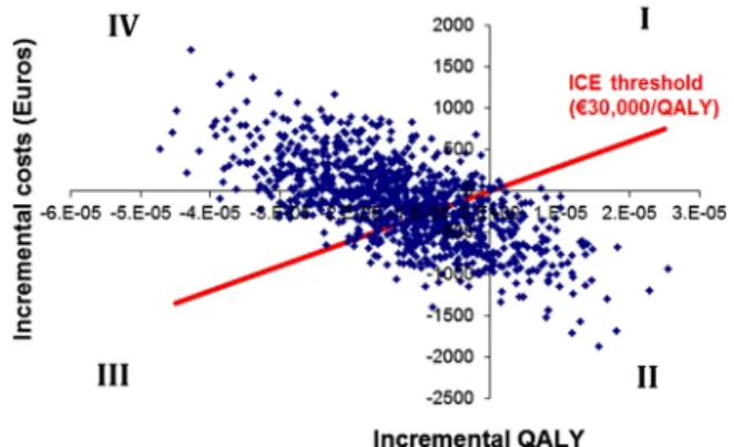
Cost–Utility Analysis of Magnetic Resonance Imaging Management of Patients with Acute Ischemic Stroke in a Spanish Hospital
Texto completo
Figure


Documento similar
Other pieces of anatomy research recently done 4 de- scribe this ligament as present in 100% of the subjects, with a coracoid process origin by two fascicles, a body,
Spectrum of lesions observed by computed tomography and magnetic resonance imaging scans in young athletes that participated in the 2018 Youth Olympic Games in Buenos
2D and 3D biometric parameters were mea- sured from slice-to-volume reconstructed images, including 3D measurements of supratentorial brain tissue, lateral ventricles,
The Dwellers in the Garden of Allah 109... The Dwellers in the Garden of Allah
The intra-reader reliability of bone related and soft-tissue results, the patterns of BML associated with tendon enthesopathy (ICC total = 0.79, range across the locations 0.44 to 1)
We used structural magnetic resonance imaging (MRI) to esti- mate global brain volumes and performed a voxel-based morphometry and lesion symptom mapping analysis in order to
Moreover, a recent magnetic resonance imaging study (Cover- dale et al. 2014) demonstrates a significant increase in middle cerebral artery diameter following acute
Analysis of Left Ventricular Volumes and Function: A Multicenter Comparison of Cardiac Magnetic Resonance Imaging, Cine Ventriculography, and Unenhanced and Contrast-Enhanced
