Rac2 GTPase activation by angiotensin II is modulated
by Ca
2C/calcineurin and mitogen-activated protein kinases
in human neutrophils
Rajaa El Bekay, Gonzalo Alba, M Edith Reyes, Pedro Chaco
´ n, Antonio Vega,
Jose´ Martı´n-Nieto
1, Juan Jime´nez, Eladio Ramos
2, Josefina Oliva´n
2, Elı´zabeth Pintado
and
Francisco Sobrino
Departamento de Bioquı´mica Me´dica y Biologı´a Molecular, Facultad de Medicina, Universidad de Sevilla, Avda. Sa´nchez Pizjua´n 4, E-41009 Sevilla, Spain 1Departamento de Fisiologı´a, Gene´tica y Microbiologı´a, Universidad de Alicante, Alicante, Spain
2Unidad de Hipertensio´n, Hospital Universitario Virgen Macarena, Sevilla, Spain
(Correspondence should be addressed to F Sobrino; Email: fsobrino@us.es)
R El Bekay and G Alba contributed equally to this work
R El Bekay is now at Laboratorio de Investigacio´n, Fundacio´n IMABIS, Hospital Carlos Haya, Ma´laga, CIBER de obesidad y Nutricio´n (CB06/03/0018)
Abstract
Angiotensin II (Ang II) highly stimulates superoxide anion production by neutrophils. The G-protein Rac2 modulates the activity of NADPH oxidase in response to various stimuli. Here, we describe that Ang II induced both Rac2 translocation from the cytosol to the plasma membrane and Rac2 GTP-binding activity. Furthermore,Clostridium difficiletoxin A, an inhibitor of the Rho-GTPases family Rho, Rac and Cdc42, prevented Ang II-elicited OK
2=ROS production, phosphorylation
of the mitogen-activated protein kinases (MAPKs) p38, extracellular signal-regulated kinase 1/2 (ERK1/2) and c-Jun N-terminal kinase 1/2, and Rac2 activation. Rac2 GTPase inhibition byC. difficiletoxin A was accompanied by a robust reduction of the cytosolic Ca2C elevation induced by Ang II in human neutrophils. Furthermore, SB203580 and PD098059 act as inhibitors of p38MAPK and ERK1/2 respectively, wortmannin, an inhibitor of phosphatidylinositol-3-kinase, and cyclosporin A, a calcineurin inhibitor, hindered both translocation of Rac2 from the cytosol to the plasma membrane and enhancement of Rac2 GTP-binding elicited by Ang II. These results provide evidence that the activation of Rac2 by Ang II is exerted through multiple signalling pathways, involving Ca2C/calcineurin and protein kinases, the elucidation of which should be insightful in the design of new therapies aimed at reversing the inflammation of vessel walls found in a number of cardiovascular diseases.
Journal of Molecular Endocrinology(2007)39,351–363
Introduction
Angiotensin II (Ang II), the main peptide hormone of the renin–angiotensin system, induces leukocyte recruitment to the vessel wall, which constitutes a hallmark of early stages of atherosclerosis and several hypertensive diseases (Ross 1999). Ang II acts via high-affinity cell surface receptors (AT1), which are linked to pathways classically associated with G-protein-coupled and tyrosine kinase-mediated responses (Timmermans
et al. 1993). Although most studies on Ang II have been carried out on smooth muscle and endothelial cells, experimental evidence has also been obtained of its effects on circulating cells. Expression of AT1 receptors for Ang II has been evidenced in circulating neutro-phils (Ito et al. 2001) and in human peripheral monocytes (Shimada & Yakazi 1978), and Ang II-induced monocyte activation has been reported
(Hahn et al. 1994). In this context, the adhesion of leukocytes to endothelial cells is a primary event taking place during the pathogenesis of vascular diseases (Mazzoneet al. 1993).
Given that chronic inflammation of vessel walls is a pathological indicator of hypertension (Touyz 2003) and that reactive oxygen species (ROS) such as superoxide anionðOK
2Þand H2O2constitute the main
intermediary molecules responsible for inflammation (Finkel 1998), a link between the development of hypertension and ROS production has been postulated (El Bekay et al. 2003). NADPH oxidase in phagocytic cells is a multi-component enzyme composed of at least two membrane proteins, gp91phoxand p22phox, which together form the flavocytochrome b558, and four cytosolic proteins, namely p47phox, p67phox, p40phox and Rac. In resting cells, this oxidase is inactive and its components are distributed separately between the cell
Journal of Molecular Endocrinology(2007)39,351–363 DOI: 10.1677/JME-07-0074
membrane and the cytosol. When neutrophils are exposed to appropriate stimuli, NADPH oxidase becomes activated through the association of all its components at the plasma membrane, which is followed by OK
2 production (Inanami et al. 1998a,b,
2001,Johnsonet al. 1998,Babior 2000).
The Rac small GTPases, members of the Rho-GTPases family, including Rho, Cdc42 and Rac, constitute a growing subgroup of Ras proteins which act as molecular switches upon their cycling between active GTP- and inactive GDP-bound states (Van Aelst & D’Souza-Schorey 1997). Previous studies have shown that Rho-GTPases are involved in multiple cellular processes, such as actin polymerization and cytoskeleton rearrangement, regulation of gene transcription, cell cycle progression and cell survival (Etienne-Manneville & Hall 2002). The Rac subfamily of Rho-GTPases includes three highly homologous members, namely Rac1, Rac2 and Rac3. Unlike Rac1 and Rac3, which are widely expressed, Rac2 is found only in haematopoietic cells (Didsburyet al. 1989,
Haatajaet al. 1997) In addition to their reported roles in actin remodelling, Rac proteins have been implicated in the generation of OK
2 via the phagocytic NADPH oxidase
complex (Segal & Abo 1993). Rac1 was identified as an oxidase-related factor from peritoneal macrophages (Abo et al. 1991), whereas Rac2 was demonstrated to fulfil such a role in human neutrophils (Knauset al. 1991,
Bokoch 1994). Rac exists in the cytosol as a complex with a GDP dissociation inhibitor (Aboet al. 1991), and upon activation of NADPH oxidase Rac separates from this inhibitor and becomes stably associated with NADPH oxidase components at the plasma membrane (Northup
et al. 1982,Curnutteet al. 1987). The GTP-bound form of Rac binds to p67phox, and also likely to flavocytochrome b558, in the assembled active oxidase complex ( Diek-mannet al. 1994,Heyworthet al. 1994,Kogaet al. 1999).
In a cell-free system, Rac1 and Rac2 recombinant proteins can reconstitute a fully active NADPH oxidase complex able to produce OK
2 (Abo et al. 1991,1992,
Knaus et al. 1991). Moreover, it has recently been reported that neutrophils from Rac2-deficient mice exhibit a diminished NADPH oxidase activity (Roberts
et al. 1999,Ambrusoet al. 2000,Williamset al. 2000,Kim & Dinauer 2001). This finding is consistent with other previous results suggesting that Rac2 is a primary GTPase modulator of NADPH oxidase activation (Heyworthet al. 1994). However, upstream signals and regulatory proteins controlling Rac activity in neutro-phils remain unknown, and only an involvement of phosphatidylinositol-3-kinase (PI3K) in Rac2 activation elicited by the respiratory burst stimulator,N-formyl-L
-methionyl-L-leucyl-L-phenylalanine (fMLP), has been
demonstrated so far (Akasakiet al. 1999,Benardet al. 1999). Therefore, the role of Rac2 in Ang II-stimulated neutrophils remains to be analysed.
Recently, the mitogen-activated protein kinases (MAPKs) family has become a focus of interest in cardiovascular research. In human neutrophils, we have described that Ang II induces a robust phosphorylation of p38MAPK, extracellular signal-regulated kinase 1/2 (ERK1/2) and c-Jun N-terminal kinase 1/2 (JNK1/2), which is hindered by inhibitors of NADPH oxidase and tyrosine kinases, as well as by ROS scavengers (El Bekay
et al. 2003). Also, we have reported that Ang II enhances the synthesisde novoand activity of calcineurin (CaN) and that the immunosuppressant cyclosporin A (CsA) inhibits Ang II-induced CaN activity, but not CaN synthesis (El Bekayet al. 2003). In this study, we present experimental evidence that Ang II promotes Rac2 translocation from the cytosol to the plasma membrane in human neutrophils. Conversely, prevention of Rac2 translocation byClostridium difficiletoxin A, an inhibitor of Rho-GTPases, including Rho, Rac and Cdc42 (Aktories et al. 2000, Voth & Ballard 2005), abolishes ROS production and activation of p38MAPK, ERK1/2 and JNK1/2 elicited by Ang II in human neutrophils. The present data thus indicate that Rac2 translocation to the plasma membrane plays a critical role in Ang II-dependent signalling pathways in human neutrophils.
Materials and methods
Chemicals and reagents
Ang II, SP6000125 and chemicals of general use were purchased from Sigma–Aldrich or Calbiochem and eprosartan was from Solvay Pharma. The rabbit polyclonal antibodies to phosphorylated p38MAPK (Thr180/Tyr182) and total p38MAPK, and mouse monoclonal antibodies to phosphorylated ERK1/2 (Thr202/Tyr204) and total ERK1/2 were obtained from New England Biolabs (Beverly, MA, USA). Mouse antibodies from Santa Cruz Biotechnology (Santa Cruz, CA, USA) to phosphorylated JNK1/2 (Thr183/Tyr185) and total JNK1/2 (sc-6254 and sc-571 respectively), and rabbit antibodies to Rac2 (sc-96) and b-actin (sc-7210) were also used. Goat polyclonal antibodies against p22phoxwere kindly donated by T L Leto (NIH, Bethesda, MD, USA). The p21-activated kinase-1 p21-binding domain (PAK-1 PBD) conjugated to agarose, GTPgS and GDP were products from Upstate Cell Signaling. The inhibitors PD089059, SB203580 and NSC23766 were obtained from Calbiochem. The rabbit antiserum against CaN was kindly provided by Dr C B Klee (NIH).
Human neutrophil extraction and processing
Human peripheral neutrophils were obtained from healthy blood donors and processed as described in
to the treatments indicated in each figure legend, except for the experiments shown inFigs 5 and 7, in which the cells were preincubated at 378C for 7 h in Krebs Ringer (KR)-HEPES before additions. Under these conditions, cell viability ranged between 90 and 97%, as estimated from the lactic dehydrogenase release assay (Gualbertoet al. 1998). The Universidad de Sevilla Ethics Committee approved this study and each subject gave informed consent prior to its undertaking.
Mobilization of Rac2
Human neutrophils (107 cells) were lysed on ice for 30 min in 60ml buffer containing 100 mM HEPES (pH 7
.
3), 100 mM KCl, 3 mM NaCl, 3 mM MgCl2,1
.
25 mM EGTA, and the protease inhibitors phenyl-methylsulphonyl fluoride (1 mM), aprotinin (20mg/ml), leupeptin (20mg/ml) and benzamidine (156mg/ml). Then, the cells were disrupted by sonication (20 W, three bursts of 5 sec each separated by 30-s interval), and unbroken cells and debris were removed by centrifugation at 10 000gfor 5 min at 48C. The supernatant obtained after further ultracentrifuga-tion at 100 000g for 30 min at 48C constituted the cytosolic fraction. The pellet was resuspended in a buffer containing 120 mM NaH2PO4(pH 7.
4), 1 mMMgCl2, 1 mM EGTA, 1 mM dithiothreitol, 20% (v/v)
glycerol, 40 mM octylglucoside and the protease inhibitors as indicated above, and then recentrifuged at 20 000g for 40 min at 48C. The new supernatant obtained, containing solubilized membranes, was used for immunoblotting analysis of Rac2 mobilization (El Bekayet al. 2003). With this purpose, this fraction was subjected to SDS-PAGE on 10% polyacrylamide gels (50mg protein/lane) and electroblotted onto polyviny-lidene difluoride membranes using a semi-dry device (Bio-Rad). Thereafter, antibody probing was carried out overnight without need of prior blocking (Mansfield 1995) with rabbit anti-Rac2 IgG, at a 1:2000 dilution in PBS supplemented with 0
.
02% Tween 20 and 1% BSA, and the 21.
429 kDa Rac2 band (Didsburyet al. 1989) was detected by incubation with horseradish peroxidase (HRP)-conjugated anti-rabbit IgG at a 1:20 000 dilution in PBS with 0.
5% (w/v) BSA, followed by enhanced chemiluminescence as previously indicated (Carballoet al. 1999). Briefly, the membranes were incubated for 1 min in 10 ml fresh luminescent reagent solution, composed 10 mM Tris–HCl (pH 8
.
5), 2.
25 mM lumi-nol, 0.
015% (v/v) H2O2 and 0.
45 mM 4-iodophenol,the latter acting as an enhancer of the chemi-luminescence reaction, and the signals were recorded on X-ray-sensitive films. The membrane levels of the p22phoxsubunit of NADPH oxidase were monitored as a loading control, as previously described (Mankelow & Henderson 2001).
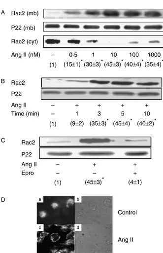
Figure 1Angiotensin II stimulates Rac2 translocation from the cytosol to the plasma membrane in an AT1 receptor-dependent manner in human neutrophils. Neutrophils were suspended in KR-HEPES buffer at 107cells/ml and incubated for 10 min at 378C with Ang II at the indicated concentrations (A), with or without 10 nM Ang II for different times (B), or else previously incubated in the absence or presence of 10 ng/ml eprosartan (Epro) for 30 min and then treated or not with 10 nM Ang II for 10 min (C). The cells were then lysed, and the plasma membrane-enriched fraction was separated from the cytosolic fraction by ultracentrifugation at 100 000g. Membrane (mb) (A–C) and cytosolic (cyt) proteins (A) were subjected to SDS-PAGE (50mg/lane), transferred to PVDF membranes and probed with antibodies against Rac2. Each blot is representative of a set of three experiments yielding similar results, and values given within parentheses correspond to Rac2 membrane levels normalized to those found in the absence of Ang II stimulation (meanGS.E.M. from three separate experiments). Statistically significant *P!0.01. p22phoxmembrane levels are also shown for the sake of
Rac2 GTP-binding activity
Rac2 activity pull-down assays were carried out essentially as described previously (Priceet al. 2003), on the basis of the capacity of PAK proteins to bind to GTP-activated Rac2, but not to Rac2 bound to GDP. After the treatments indicated in each case, neutrophils (107 cells) were washed and then lysed in a magnesium-containing lysis buffer composed 25 mM HEPES (pH 7
.
5), 150 mM NaCl, 10 mM MgCl2, 25 mM NaF, 1 mM EDTA, 1 mM Na3VO4,1% (v/v) Nonidet P-40, 2% (v/v) glycerol, 100mM phenylarsine oxide and the protease inhibitors described above. After centrifugation at 15 000g, the supernatant obtained was incubated with 5–10mg PAK-1 PBD agarose beads, and the reaction mixture was gently rocked at 48C for 1 h. In parallel, lysates from untreated neutrophils were loaded with 100mM GTPgS (positive control) or 1 mM GDP (negative control), for 15 min at 308C before addition of PAK-1 PBD agarose. The beads were then pelleted and washed, and immunoblotting analysis was carried out with anti-Rac2 antibodies as described above.
p38MAPK, ERK1/2 and JNK1/2 phosphorylation
In order to reduce the MAPK basal phosphorylation levels usually found in our preparations of human neutrophils, these cells were preincubated in KR-HEPES at 378C for
7 h. After the treatments indicated in each case, the cells were lysed and immunoblotting analysis with antibodies to phosphorylated and total forms of the three MAPKs was carried out as described (El Bekayet al. 2003). Relative protein levels were determined by scanning densitometry analysis using the Scion Image software (Frederick, MD, USA).
OK
2 and total ROS production
The production of ROS was analysed by two separate methods, each with a different specificity for the location and type(s) of ROS produced. (i) The lucigenin-based luminescence method was specific for OK
2, whereas (ii)
luminol-based luminescence correlated well with total ROS produced by the cells (Liet al. 1998). Since both
Figure 2Cyclosporin A inhibits Rac2 translocation to the plasma membrane in angiotensin II-stimulated neutrophils. Neutrophils (107cells/ml) were preincubated for 1 h at 378C in the absence or presence of 1mg/ml CsA, and then treated with Ang II for 10 min at the indicated doses (A), or else they were preincubated with different concentrations of CsA for 1 h, and then treated or not with 10 nM Ang II for further 10 min at 378C (B). The cells were then lysed, and the plasma membrane-enriched fraction was analysed by immunoblotting with antibodies against Rac2. Each blot is representative of a set of four experiments yielding similar results. Values given within parentheses correspond to Rac2 membrane levels normalized to those found in the absence of CsA and Ang II treatment (meanGS.E.M. from three separate experiments).
Statistically significant *P!0.01. p22phoxmembrane levels are
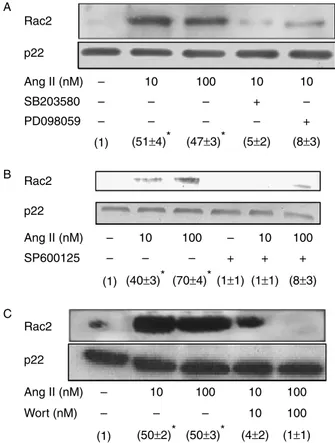
also shown for the sake of loading controls. Figure 3 SB203580, PD098059, SP600125 and wortmannin
inhibit angiotensin II-dependent Rac2 translocation to the plasma membrane in human neutrophils. Neutrophils (107cells/ml) were
preincubated for 30 min at 378C in the absence or presence of 5mM SB203580 or 5mM PD098059 (A), 1mM SP600125 (B), or 10–100 nM wortmannin (Wort) (C), and then treated or not for 10 min with 10–100 nM Ang II. Rac2 membrane levels were analysed by immunoblotting. Each blot is representative of a set of four experiments yielding similar results, and values given within parentheses correspond to Rac2 membrane levels normalized to those found in the absence of treatments (meanGS.E.M. from three separate experiments). Statistically
luminol and lucigenin can permeate freely through the cell membrane, their luminescence was an indi-cation of the sum of intracellular plus extracellular ROS. The assays were carried out as described previously (Monteseirı´net al. 1996), except that HRP (8 mU/ml) was included when luminol was used.
Calcineurin protein and phosphatase activity levels
CaN phosphatase activity was measured as previously described (El Bekayet al. 2003). For immunoblotting
analysis of CaN subunits A and B, neutrophils were lysed and subjected to SDS-PAGE (50mg protein/lane) as described earlier (El Bekay et al. 2003). Detection was carried out by enhanced chemiluminescence as indicated above. To verify even protein loading, the blots were subsequently stripped and reprobed with rabbit polyclonal antibodies againstb-actin at a 1:1000 dilution.
Immunofluorescence microscopy analysis of Rac2 membrane translocation
The membrane translocation of Rac2 in human neutrophils was also assessed by immunofluorescence cell staining, as described previously (Vegaet al. 2004) with minor modifications. After stimulation, neutro-phils (107cells) were harvested, washed with PBS and smeared onto poly-L-lysine-coated glass slides. The cells
were fixed at room temperature with 2% paraformalde-hyde for 30 min. After washing with PBS, unspecific binding was blocked with PBS containing 0
.
2% gelatin. Further, the cells were permeabilized with 0.
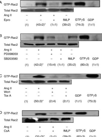
1% Triton X-100 for 4 min, and thereafter incubated with rabbit anti-Rac2 IgG at a 1:100 dilution for 30 min, washedFigure 4Angiotensin II enhances Rac2 GTP-binding activity in an AT1 receptor-dependent manner in human neutrophils. Involvement of p38MAPK, ERK1/2, PI3K and CaN. Neutrophils (107cells/ml) were preincubated at 378C for 30 min in the absence or presence of either 10 ng/ml eprosartan (Epro) (A), 5mM PD098059 or 5mM SB203580 (B), 100 nM wortmannin (Wort) or 10 ng/mlC. difficile toxin A (Tox A) (C), or else they were preincubated for 1 h with or without 1mg/ml CsA (D). Then the cells were stimulated with 10 nM Ang II for 10 min or with 10 nM fMLP for 3 min at 378C, where indicated. Rac2 GTP-binding activity was measured by PAK-1 PBD affinity precipitation followed by Rac2 immunoblotting analysis as described in Materials and methods. Lysates from untreated neutrophils loaded with 100mM GTPgS or 1 mM GDP prior to pull-down assays are shown as positive and negative controls respectively. Each blot is representative of a set of three experiments yielding similar results, and values given within parentheses correspond to GTP-bound Rac2 levels normalized to those found in the absence of treatments (meanGS.E.M. from three
separate experiments). Statistically significant *P!0.01.
Figure 5Cyclosporin A inhibits p38MAPK, ERK1/2 and JNK1/2 activation in angiotensin II-stimulated human neutrophils. (A) Neutrophils (107cells/ml) were preincubated for 7 h at 378C in
KR-HEPES without additions. Then, they were incubated for 1 h in the absence or presence of 1mg/ml CsA, and thereafter for 10 min with 10 nM Ang II, and the levels of phosphorylated ERK1/2 and p38MAPK together with total levels of p38MAPK were analysed by immunoblotting. (B) Idem as in panel A, except that cells were treated or not with different concentrations of Ang II, and the levels of phosphorylated and total JNK1/2 were analysed by immuno-blotting. Each blot is representative of a set of four experiments yielding similar results, and values given within parentheses correspond to levels of phosphorylated MAPKs normalized to those found in the absence of treatments (meanGS.E.M. from
extensively and stained with FITC-conjugated anti-rabbit IgG at a 1:500 dilution for 30 min. After final washing, coverslips were mounted on the slides using 50% glycerol in PBS. Immunostained cells were observed and photographed using a Nikon EFD-3 microscope.
Intracellular Ca2C
levels
Cytosolic [Ca2C
] was measured in cell populations using the fluorescent probe Fura2 as described previously (Sageet al. 1990).
Statistical analysis
Data are expressed as the meanGS.E.M. from a
minimum number of three independent experiments. Protein expression levels were determined by densito-metry of the bands using Scion Image software. This software detects the bands obtained by western blot and gives individual values which are dependent on the light quantification of the corresponding band. Measurements are expressed as arbitrary units. The results were normalized for unstimulated control. The numerical data obtained from Ang II stimulated and the corresponding controls were statistically analysed using Statgraphics plus 5.0 software (Manugistic Inc., Rockville, MD, USA) from ANOVA and the pairedt-test. Asterisks indicatePvalues!0
.
01.Results
Ang II promoted Rac2 translocation from the cytosol to the plasma membrane in human neutrophils
Ang II exogenously added promoted the accumulation of Rac2 at the plasma membrane in human
neutrophils, with maximal levels being reached at a 10 nM concentration of Ang II and with concomitant disappearance of the Rac2 signal from the cytosol, as shown inFig. 1A. The effect of this hormone on OK 2
production by human neutrophils was also examined, and the values found were 0
.
043G0.
009 (in control cellswithout additions), 0
.
165G0.
022 (with 1 nM Ang II),0
.
897G0.
103 (with 10 nM Ang II) and 1.
123G0.
236(with 100 nM Ang II) relative units of chemi-luminescence. It was thus observed that Ang II elicited OK
2 synthesis in a dose-dependent manner, confirming
our previous report (El Bekay et al. 2003). Short-time kinetic experiments (Fig. 1B) revealed that an effective Ang II-dependent translocation of Rac2 from the cytosol to the plasma membrane took place as soon as within 1 min of incubation with the hormone, with maximal membrane levels being reached after 5 min of treatment. Rac2 translocation in Ang II-stimulated neutrophils was prevented by a 10 ng/ml dose of eprosartan (Fig. 1C), an inhibitor of the AT1 Ang II receptor (Brookset al. 1999), indicating a specific role for this receptor in Ang II-induced Rac2 translocation in human neutrophils. In control experiments, the same effects were obtained when the chemoattractant fMLP at 100 nM substituted for Ang II (data not shown). The membrane levels of p22phox at the membrane, in keeping with it being an integral membrane protein (Mankelow & Henderson 2001), were similar in both resting and Ang II-stimulated neutrophils (Fig. 1A–C). We further investigated the effect of Ang II on the subcellular localization of Rac2 using immunofluorescence microscopy. As shown in
Fig. 1D, this protein was primarily cytoplasmic in unstimulated cells (Fig. 1a and b), but was found predominantly located at the plasma membrane upon incubation of neutrophils with 10 nM Ang II for 10 min (Fig. 1c and d). These results corroborated that Ang II promotes the translocation of Rac2 to the cell membrane in human neutrophils.
Involvement of calcineurin, MAP kinases and PI3K in Ang II-dependent Rac2 translocation to the plasma membrane in human neutrophils
We have recently shown that Ang II induces an increase of bothde novosynthesis and activity of CaN in human neutrophils (El Bekayet al. 2003). In order to determine whether inhibition of CaN activity affected Rac2 translocation to the plasma membrane, we pretreated neutrophils with CsA for 1 h.Figure 2A and Bshows that CsA at a 0
.
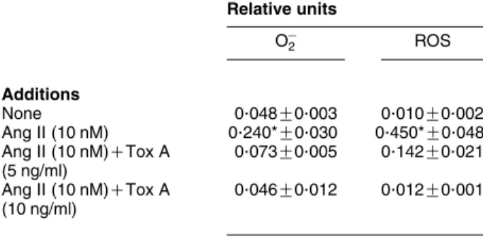
3mg/ml concentration was sufficient to suppress Rac2 translocation induced by Ang II in these cells indirectly suggesting an involve-ment of CaN in Ang II-dependent Rac2 mobilization to the plasma membrane in human neutrophils.Table 1 Clostridium difficiletoxin A prevents OK 2/ROS
production in Ang II-stimulated human neutrophils
Relative units
OK
2 ROS
Additions
None 0.048G0.003 0.010G0.002
Ang II (10 nM) 0.240*G0.030 0.450*G0.048
Ang II (10 nM)CTox A
(5 ng/ml)
0.073G0.005 0.142G0.021
Ang II (10 nM)CTox A
(10 ng/ml)
0.046G0.012 0.012G0.001
Neutrophils (106cells/ml) were incubated for 30 min in the absence or presence of toxin A fromC. difficile(Tox A) at the indicated concentrations. Then 5 pM lucigenin or luminol were added for the measurement of OK
2or ROS
In addition, we have reported that Ang II elicits phosphorylation of both p38MAPK and ERK1/2 in human neutrophils, and that inhibition of these two MAPKs by SB203580 and PD098059 respectively, prevents
Ang II-stimulated ROS=OK
2 production as well as
translo-cation of the p47phox and p67phox NADPH oxidase subunits to the plasma membrane (El Bekayet al. 2003). As shown in Fig. 3A, we now found that these two
Figure 6 Clostridium difficile toxin A inhibits Ca2C
-dependent Rac2 activation in Ang II-stimulated human neutrophils. Neutrophils (107cells/ml) were preincubated at 378C with 10 ng/ml toxin A fromC. difficile(Tox A) for 30 min (A), or with 1
mM thapsigargin (Tg) plus 0.1 mM EGTA for 5 min (B), and then they were treated or not with 10 nM Ang II for further 10 min. Thereafter, Rac2 plasma membrane levels were analysed by immunoblotting, together with p22phoxlevels as a loading control. Each blot is representative of a set of four
experiments yielding similar results, and values given within parentheses correspond to Rac2 membrane levels normalized to those found in the absence of treatments (meanGS.E.M. from three separate experiments). Statistically significant *P!0.01. In separate experiments, neutrophils were preincubated with or without 5–10 ng/ml Tox A for 30 min, and then treated or not with 10 nM Ang II for 5 h at 378C, and CaN phosphatase activity was measured in cell extracts (C), or CaN protein levels were analysed by immunoblotting (D). Arrowheads point to CaN A and B subunits (59 and 19 kDa respectively) in panel D. Finally, Fura2-loaded neutrophils were preincubated with different concentrations of Tox A and intracellular Ca2C
inhibitors were also able to efficiently prevent Rac2 membrane translocation in Ang II-stimulated neutro-phils. In a similar manner, the Ang II-promoted Rac2 translocation was effectively hindered by SP600125, an inhibitor of JNK1/2, in human neutrophils (Fig. 3B).
It has recently been reported that Rac2 becomes activated by fMLP by a PI3K-mediated mechanism in human neutrophils (Akasakiet al. 1999). Since we have previously shown that wortmannin, a pharmacological PI3K inhibitor, inhibits ROS production by these cells (El Bekayet al. 2003), we set to investigate the possible involvement of PI3K in Ang II-induced Rac2 mobili-zation. As shown inFig. 3C, wortmannin blocked partially Rac2 translocation at a 10 nM dose, and totally at 100 nM. These data indicated that PI3K activity is also required for translocation of Rac2 from the cytosol to the plasma membrane in Ang II-stimulated human neutrophils.
Ang II promoted Rac2 GTP-binding activity in human neutrophils
In order to determine whether Ang II-induced Rac2 membrane translocation correlated with an increase in Rac2 GTP-binding activity, a PAK-1 PBD pull-down assay was carried out which was based on the property exhibited by PAK-1/3 Ser/Thr kinases of becoming stimulated upon their binding of active Rac GTPases (Bokoch 2003).Figure 4Ashows that the treatment of human neutrophils with Ang II at 10 nM resulted in an up-regulation of Rac2 GTP binding by up to 40-fold. This enhancement was inhibited by eprosartan, indi-cating that this phenomenon depended on Ang II binding to AT1 receptors. Rac2 GTP-binding activity was also increased upon cell stimulation with 100 nM fMLP (Fig. 4A), as previously described in human neutrophils (Akasaki et al. 1999). As a control, when human neutrophil lysates were incubated with GTPgS, Rac2 was found to massively bind to PAK-1 PBD, whereas no such association was detected when lysates were incubated with GDP (Fig. 4A). We also examined the effect of p38MAPK and ERK1/2 inhibitors on Rac2 GTP binding in Ang II-stimulated neutrophils.Figure 4B
shows that both SB203580 and PD098059 hindered GTP binding by Rac2 suggesting an implication of both MAPKs in the signalling pathways modulating the Ang II-elicited targeting of Rac2 to the plasma membrane.
Besides, Rac2 GTP-binding activity was totally inhibited by the PI3K inhibitor, wortmannin (Fig. 4C). As expected, Rac2 GTP-binding activity was also abolished in the presence of toxin A fromC. difficile, confirming previous data (Aktorieset al. 2000).
Ang II induced MAP kinases activation through calcineurin in human neutrophils
In order to determine whether Rac2 translocation to the plasma membrane elicited by Ang II was mediated by signalling molecules other than MAPKs, the effect of pretreatment with the CaN inhibitor, CsA, prior to Ang II addition was assessed. We found that CsA at 1mg/ml suppressed Rac2 GTP-binding activity induced by Ang II in human neutrophils, as revealed by its hindering of Rac2 GTP-binding activity (Fig. 4D). In addition, given that CsA was also able to inhibit Rac2 translocation to the plasma membrane (Fig. 2), and that crosstalk between CaN and MAPKs has been reported in myocytes (Limet al. 2001) and in neutrophils (El Bekayet al. 2003), the inhibitory effect of CsA could be indirectly exerted through MAPK inhibition. In order to test this possibility, we examined the effect of CsA on Ang II-induced p38MAPK, ERK1/2 and JNK1/2 activation in human neutrophils. As shown in Fig. 5A and B, CsA prevented the phosphorylation, and hence the activation, of these three MAPKs by Ang II. These results allowed us to infer that CaN has an implication in both Rac2 and MAPK activation elicited by Ang II in human neutrophils.
Ang II-dependent Rac2 GTPase activity regulated ROS=OK
2 production, cytosolic Ca
2C
release and calcineurin activity
The treatment of cells with C. difficile toxin A (5–10 ng/ml), an inhibitor of the GTPase activity of GTP-binding proteins such as Rac, Rho and Cdc42 (Aktorieset al. 2000,Voth & Ballard 2005), was found to reduce OK
2 and total ROS release by w81 and 97%
respectively, in Ang II-stimulated human neutrophils when compared with untreated cells (Table 1), this indicating that GTPase activity plays a pivotal role in NADPH oxidase activation by Ang II in human neutrophils. In parallel,C. difficiletoxin A was found to abolish Ang II-dependent Rac2 translocation to the plasma membrane (Fig. 6A). Since it
Figure 7Clostridium difficiletoxin A inhibits angiotensin II-dependent p38MAPK, ERK1/2 and JNK1/2 activation in human neutrophils. Neutrophils (107cells/ml) were preincubated for 7 h at 378C in KR-HEPES without additions. Then they were treated for 30 min with the indicated concentrations of clostridial toxin A (Tox A) and thereafter for 10 min with 10 nM Ang II, where indicated. The levels of phosphorylated p38MAPK (A), ERK1/2 and JNK1/2 (B) were then analysed by immunoblotting. In a separate set of experiments, Tox A was substituted by 100mM NSC23766 and, after incubation with 10–100 nM Ang II or 100 nM PMA for 10 min, the levels of
phosphorylated and total p38MAPK and JNK1/2 (C) or ERK1/2 (D) were analysed by immunoblotting. Each blot is representative of a set of four experiments yielding similar results, and values given within parentheses correspond to levels of phosphorylated MAPKs normalized to those found in the absence of treatments (meanGS.E.M. from three separate experiments). Statistically significant, comparison between unstimulated control and Ang II stimulated *P!0.01 and comparison between Ang II stimulated and Ang II plus inhibitors treated,CP
has previously been shown that Ang II increases cytosolic Ca2C
in human neutrophils (El Bekayet al. 2003), we set to test whether Ca2C
signalling was involved in Ang II-dependent Rac2 activation. With this aim, neutrophils were pretreated simultaneously with thapsigargin, in order to deplete intracellular Ca2C
stores, and with EGTA to chelate extracellular Ca2C
, and then the cells were activated by the addition of Ang II.Figure 6Bshows that under these conditions Ang II-elicited Rac2 mobilization became drastically inhibited, indicating that cytosolic Ca2C
elevation is crucial for Rac2 translocation to the plasma membrane.C. difficiletoxin A treatment also inhibited CaN activity (Fig. 6C), but did not affect thede novosynthesis of CaN induced by Ang II (Fig. 6D). Conversely, we also assessed whether elevation of intracellular Ca2C
required activation of Rac2 GTPase. As shown in Fig. 6E, when increasing doses ofC. difficiletoxin A were added to cells prior to Ang II stimulation, the Ca2C
signal was hindered, suggesting that the Ca2C
mobilization elicited by Ang II in human neutrophils depends, at least in part, on the activation of Rac2. In this context, Rac2 has also been shown to participate in actin polymerization and cytoskele-ton rearrangement. Present observations on the inhibition on Ca2C
mobilization could be thus linked to the disruption of the actin cytoskeleton organization induced by several toxins, as previously observed (Bozemet al. 2000).
Ang II induced p38MAPK, ERK1/2 and JNK1/2 activation through Rac2 GTPase activity
Subsequent experiments were designed to examine whether the inhibition of Rac2 affected MAPK acti-vation in Ang II-stimulated neutrophils.Figure 7A and B
shows that C. difficile toxin A at a 5–10 ng/ml dose efficiently prevented Ang II-dependent p38MAPK, ERK1/2 and JNK1/2 activation. In order to gain insight on the specificity of this inhibitory effect, we also used a chemical compound, NSC23766, identified as a Rac-specific small-molecule inhibitor (Gaoet al. 2004). NSC23766 exhibited an inhibitory effect on the phosphorylation of p38MAPK, JNK1/2 and ERK1/2 similar to that exerted by C. difficile toxin A (Fig. 7C and D). These results thus led support to the idea that the signalling pathways mediated by these three MAPKs are involved in Rac2 targeting to the cell membrane.
Discussion
Recent clinical studies have shown that Ang II is a key player in several pathological processes, including hypertension and atherosclerosis (Ross 1999). We have demonstrated that Ang II activates human neutrophils through the induction of OK
2=ROS
production, consistently with the translocation to the cell membrane of the cytosolic components of NADPH oxidase, p47phox and p67phox (El Bekay et al. 2003). Despite Rac2 having been reported as participating in a wide array of signalling pathways in neutrophils and other cells (Robertset al. 1999,Yanget al. 2000), its role in the stimulation of these cells by Ang II has yet not been addressed. In the present work, we report for the first time the ability of Ang II to induce translocation of Rac2 from the cytosol to the plasma membrane, and to enhance its GTP-binding activity in a dose-dependent manner via AT1 receptors in human neutrophils. Previous and present data also indicate that transloca-tion to the plasma membrane of the p47phox and p67phoxsubunits of NADPH oxidase and of Rac2 occur in parallel in Ang II-activated human neutrophils. In this context, it has been reported that Rac2 mobil-ization to the cell membrane is diminished in p47phox -deficient neutrophils from chronic granulomatous disease (CGD) patients (El Bennaet al. 1994). However, Rac2 translocation is not affected in CGD neutrophils lacking p67phox(Dusiet al. 1995a). In contrast, other authors have shown that Rac2 is able to translocate independently of other cytosolic factors, and to migrate to the cell membrane in the absence of both p67phox and p47phox in human neutrophils (Heyworth et al. 1994,Dusiet al. 1995b).
Also, we have observed that both Ang II-dependent Rac2 translocation and its GTPase activity increase were abrogated by CsA, a CaN inhibitor and immunosup-pressant, and that this compound prevented p38MAPK, ERK1/2 and JNK1/2 activation by Ang II. These data thus underscore a pivotal role of CaN both in Rac2 translocation and the activation of these three MAPKs in Ang II-stimulated human neutrophils. Other authors have also shown that Rac is an upstream mediator of PAK-1 and JNK1/2 activation during Ang II signalling in vascular smooth muscle cells (Schmitz et al. 2001,
showing that in human neutrophils Rac2 becomes activated by fMLP in a PI3K-dependent manner (Akasakiet al. 1999).
C. difficileproduces two toxins, which cause notable pathological disorders. Both, toxins A and B, trans-locate to the cytosol of target cells and inactivate small GTP-binding proteins, which include Rho, Rac and Cdc42. Inactivation of these substrates occurs through monoglycosylation of a single reactive threonine, which lies within their effector-binding loop and coordinates a divalent cation critical for GTP binding. By glycosylat-ing small GTPases, toxins A and B cause actin condensation and cell rounding, which is followed by cell death (Justet al. 1995,Voth & Ballard 2005). Our data illustrate that toxin A dramatically inhibited both OK
2=ROS and ROS production and p38MAPK, ERK1/2
and JNK1/2 phosphorylation, in agreement with the pivotal role assigned to Rac2 in NADPH oxidase activation (Kim & Dinauer 2001,2006) and in MAPK phosphorylation (Yu et al. 2001). These results indicate that Rac2 is necessary both for optimal activity of the assembled oxidase complex towards ROS production and MAPK-mediated signalling elicited by Ang II.
In order to obtain a more specific inhibitory effect on Rac2 activity, we also tested a chemical compound, NSC23766, identified as a Rac-specific small-molecule inhibitor (Gaoet al. 2004). We have observed that this molecule evoked an inhibition of the Ang II-dependent phosphorylation of both p38MAPK, ERK1/2 and JNK1/2 and similar to that provoked by toxin A. These data are in agreement with a previous study showing that T cells from Rac2-deficient mice exhibit a decreased phosphorylation of p38MAPK and ERK1/2, in parallel to a reduced antigen-induced Ca2C
efflux (Yuet al. 2001).
In the search of other intracellular signalling events related to Rac2, increased intracellular Ca2C
seems sufficient to induce Rac2 activation in several epithelial cell lines (Price et al. 2003). However, fMLP-induced Rac2 activation is independent of intracellular Ca2C
in human neutrophils (Geijsenet al. 1999). From our data, a mutual interdependence between Rac2 and Ca2C
mobilization seems operative, since clostridial toxin A inhibited Ang II-stimulated cytosolic Ca2C
elevation and, conversely, Ang II-elicited Rac2 activation was blocked upon the chelation of intra- and extracellular Ca2C
using thapsigargin plus EGTA. These findings thus strongly suggest that cytosolic Ca2C
increase induced by Ang II is necessary for Rac2 activation. Our results are also consistent with recently published data showing that Ang II induces CaN activation in human neutrophils (El Bekayet al. 2003), and suggest the existence of a pathway linking Ca2C
/CaN and MAPK signalling pathways, as it occurs in cardiac myocytes (Lim et al. 2001). This notion is further
supported by our previous observations that Ang II enhances CaN expression and NF-kB DNA-binding activity in human neutrophils, which constitute two crucial intracellular signalling events (El Bekay et al. 2003). It has also been shown that Ang II modulates the cellular immune response through a CaN-dependent pathway (Nataraj et al. 1999). Interestingly, CaN has been shown to synergize with protein kinase C (PKC) to activate Rac in T cells (Werlen et al. 1998), and to engage in crosstalk with PKC and MAPK activation in transducing the Ang II stimulus in cardiomyocytes (Muratet al. 2000). Both proteins, Rac2 and PKC, are constituents of the machinery responsible for the activation of NADPH oxidase (Kwong et al. 1993,
Bokoch & Knaus 1994) and are thus amenable to participate in Ang II-dependent signal transduction pathways. Also, it has been reported that the rapid CsA-induced depression of the respiratory burst in neu-trophils stimulated by fMLP is due to a primary reduction of Ca2C
signalling (Nguyen et al. 1998). In this context, present observations illustrate for the first time the prevention of Ang II-dependent intra-cellular Ca2C
elevation through Rac2 inhibition (by
C. difficiletoxin A), with simultaneous decrease of CaN activity, events which are consistent with the view that Rac2 activation is coupled to the elicitation of intracellular oxidative stress by Ang II in human neutrophils.
Finally, our results also illustrate that MAPK acti-vation and both Rac2 translocation to the plasma membrane and GTP-binding activity in Ang II-stimu-lated neutrophils are mutually reguII-stimu-lated processes, given that specific pharmacological inhibitors acting on either the MAPK or the Rac2 pathway reciprocally abrogated activation of the other. Although the precise hierarchy is difficult to determine, the complex response elicited by Ang II in neutrophils suggests that the presumptive crosstalk between Rac2 and MAPK activation mechanisms is probably dependent on the participation of other putative signalling pathways, such as those mediated by PI3K, Ca2C
elevation or CaN activation.
Acknowledgements
This work was financed by grants from the Ministerio de Educacio´n y Ciencia (BFU2006-13802), and the Con-sejerı´a de Innovacio´n, Ciencia y Empresa (P06-CTS-1936), Junta de Andalucı´a, Spain, awarded to F S. We thank Drs Thomas L Leto and Claude B Klee for kindly providing the anti-p22phoxand anti-CaN antisera respectively. G A is the recipient of a fellowship from the Consejerı´a de Educacio´n y Ciencia, Junta de Andalucı´a. The authors declare that there is no conflict of interest that would prejudice the impartiality of this scientific work.
References
Abo A, Pick E, Hall A, Totty N, Teahan CG & Segal AW 1991 Activation of the NADPH oxidase involves the small GTP-binding protein p21rac1.Nature353668–670.
Abo A, Boyhan A, West I, Thrasher AJ & Segal AW 1992 Reconstitution of neutrophil NADPH oxidase activity in the cell-free system by four components: p67-phox, p47-phox, p21rac1, and cytochrome b-245.
Journal of Biological Chemistry26716767–16770.
Van Aelst L & D’Souza-Schorey C 1997 Rho GTPases and signaling networks.Genes and Development112295–2322.
Akasaki T, Koga H & Sumimoto H 1999 Phosphoinositide 3-kinase-dependent and -in3-kinase-dependent activation of the small GTPase Rac2 in human neutrophils.Journal of Biological Chemistry27418055–18059. Aktories K, Schmidt G & Just I 2000 Rho GTPases as targets of bacterial
protein toxins.Biological Chemistry381421–426.
Ambruso DR, Knall C, Abell AN, Panepinto J, Kurkchubasche A, Thurman G, Gonzalez-Aller C, Hiester A, deBoer M, Harbeck RJ
et al.2000 Human neutrophil immunodeficiency syndrome is
associated with an inhibitory Rac2 mutation.PNAS974654–4659. Babior BM 2000 Phagocytes and oxidative stress.American Journal of
Medicine10933–44.
El Bekay R, A´ lvarez M, Monteseirin J, Alba G, Chaco´n P, Vega A, Martı´n-Nieto J, Jime´nez J, Pintado E, Bedoya FJet al.2003 Oxidative stress is a critical mediator of the angiotensin II signal in human neutrophils: involvement of mitogen-activated protein kinase, calcineurin, and the transcription factor NF-kB.Blood102662–671. Benard V, Bohl BP & Bokoch GM 1999 Characterization of rac and
cdc42 activation in chemoattractant-stimulated human neutrophils using a novel assay for active GTPases.Journal of Biological Chemistry
27413198–13204.
El Benna J, Ruedi JM & Babior BM 1994 Cytosolic guanine nucleotide-binding protein Rac2 operatesin vivoas a component of the neutrophil respiratory burst oxidase. Transfer of Rac2 and the cytosolic oxidase components p47phox and p67phox to the submembranous actin cytoskeleton during oxidase activation.
Journal of Biological Chemistry2696729–6734.
Bokoch GM 1994 Regulation of the human neutrophil NADPH oxidase by the Rac GTP-binding proteins.Current Opinion in Cell
Biology6212–218.
Bokoch GM 2003 Biology of the p21-activated kinases.Annual Review of
Biochemistry72743–781.
Bokoch GM & Knaus UG 1994 Ras-related GTP-binding proteins and leukocyte signal transduction.Current Opinion in Hematology153–60. Bozem M, Kuhlmann S, Blum R, Feick P & Schulz I 2000 Hormone-stimulated calcium release is inhibited by cytoskeleton-disrupting toxins in AR4-2J cells.Cell Calcium2873–82.
Brooks DP, Ohlstein EH & Ruffolo RR, Jr. 1999 Pharmacology of eprosartan, an angiotensin II receptor antagonist: exploring hypotheses from clinical data.American Heart Journal138246–251.
Carballo M, Ma´rquez G, Conde M, Martı´n-Nieto J, Monteseirı´n J, Conde J, Pintado E & Sobrino F 1999 Characterization of calcineurin in human neutrophils. Inhibitory effect of hydrogen peroxide on its enzyme activity and on NF-kB DNA binding. Journal of Biological Chemistry27493–100.
Curnutte JT, Kuver R & Babior BM 1987 Activation of the respiratory burst oxidase in a fully soluble system from human neutrophils.
Journal of Biological Chemistry2626450–6452.
Didsbury J, Weber RF, Bokoch GM, Evans T & Snyderman R 1989 Rac, a novel ras-related family of proteins that are botulinum toxin substrates.Journal of Biological Chemistry26416378–16382. Diekmann D, Abo A, Johnston C, Segal AW & Hall A 1994 Interaction
of Rac with p67phox and regulation of phagocytic NADPH oxidase activity.Science265531–533.
Dusi S, Donini M & Rossi F 1995aMechanisms of NADPH oxidase activation in human neutrophils: p67phox is required for the translocation of rac 1 but not of rac 2 from cytosol to the membranes.Biochemical Journal308991–994.
Dusi S, Donini M & Rossi F 1995bMechanisms of NADPH oxidase activation: translocation of p40phox, Rac1 and Rac2 from the cytosol to the membranes in human neutrophils lacking p47phox or p67phox.Biochemical Journal314409–412.
Etienne-Manneville S & Hall A 2002 Rho GTPases in cell biology.
Nature420629–635.
Finkel T 1998 Oxygen radicals and signaling.Current Opinion in Cell
Biology10248–253.
Gao Y, Dickerson JB, Guo F, Zheng J & Zheng Y 2004 Rational design and characterization of a Rac GTPase-specific small molecule inhibitor.PNAS1017618–7623.
Geijsen N, van Delft S, Raaijmakers JA, Lammers JW, Collard JG, Koenderman L & Coffer PJ 1999 Regulation of p21rac activation in human neutrophils.Blood941121–1130.
Gualberto A, Marquez G, Carballo M, Youngblood GL, Hunt SW, Baldwin AS & Sobrino F 1998 p53 transactivation of the HIV-1 long terminal repeat is blocked by PD 144795, a calcineurin-inhibitor with anti-HIV properties.Journal of Biological Chemistry273
7088–7093.
Haataja L, Groffen J & Heisterkamp N 1997 Characterization of RAC3, a novel member of the Rho family.Journal of Biological Chemistry272
20384–20388.
Hahn AW, Johas U, Buhler FR & Resink TJ 1994 Activation of human peripheral monocytes by angiotensin II.FEBS Letters347
178–180.
Heyworth PG, Bohl BP, Bokoch GM & Curnutte JT 1994 Rac translocates independently of the neutrophil NADPH oxidase components p47phox and p67phox. Evidence for its interaction with flavocyto-chrome b558.Journal of Biological Chemistry26930749–30752. Inanami O, Johnson JL & Babior BM 1998aThe leukocyte NADPH
oxidase subunit p47PHOX: the role of the cysteine residues.
Archives of Biochemistry and Biophysics35036–40.
Inanami O, Johnson JL, McAdara JK, Benna JE, Faust LR, Newburger PE & Babior BM 1998bActivation of the leukocyte NADPH oxidase by phorbol ester requires the phosphorylation of p47PHOX on serine 303 or 304.Journal of Biological Chemistry2739539–9543.
Inanami O, Yamamori T, Takahashi TA, Nagahata H & Kuwabara M 2001 ESR detection of intraphagosomal superoxide in polymor-phonuclear leukocytes using 5-(diethoxyphosphoryl)-5-methyl-L
-pyrroline-N-oxide.Free Radical Research3481–92.
Ito H, Takemory K & Suzuki T 2001 Role of angiotensin II type I receptor in the leukocytes and endothelial cells of brain microvessels in the pathogenesis of hypertensive cerebral injury.
Journal of Hypertension19591–597.
Johnson JL, Park JW, Benna JE, Faust LP, Inanami O & Babior BM 1998 Activation of p47(PHOX), a cytosolic subunit of the leukocyte NADPH oxidase. Phosphorylation of ser-359 or ser-370 precedes phosphorylation at other sites and is required for activity.Journal of
Just I, Selzer J, Eichel-Streiber C & Aktories K 1995 The low molecular mass GTP-binding protein Rho is affected by toxin A from Clostridium difficile.Journal of Clinical Investigation951026–1031. Kim C & Dinauer MC 2001 Rac2 is an essential regulator of neutrophil
nicotinamide adenine dinucleotide phosphate oxidase activation in response to specific signaling pathways.Immunology1661223–1232. Kim C & Dinauer MC 2006 Impaired NADPH oxidase activity in
Rac2-deficient murine neutrophils does not result from defective translocation of p47phox and p67phox and can be rescued by exogenous arachidonic acid.Journal of Leukocyte Biology79223–234. Knaus UG, Heyworth PG, Evans T, Curnutte JT & Bokoch GM 1991
Regulation of phagocyte oxygen radical production by the GTP-binding protein Rac 2.Science2541512–1515.
Koga H, Terasawa H, Nunoi H, Takeshige K, Inagaki F & Sumimoto H 1999 Tetratricopeptide repeat (TPR) motifs of p67(phox) partici-pate in interaction with the small GTPase Rac and activation of the phagocyte NADPH oxidase.Journal of Biological Chemistry274
25051–25060.
Kwong CH, Malech HL, Rotrosen D & Leto TL 1993 Regulation of the human neutrophil NADPH oxidase by rho-related G-proteins.
Biochemistry325711–5717.
Li Y, Zhu H, Kuppusamy P, Roubaud V, Zweier JL & Trush MA 1998 Validation of lucigenin (bis-N-methylacridinium) as a chemilumi-genic probe for detecting superoxide anion radical production by enzymatic and cellular systems.Journal of Biological Chemistry273
2015–2023.
Lim HW, New L, Han J & Molkentin JD 2001 Calcineurin enhances MAPK phosphatase-1 expression and p38 MAPK inactivation in cardiac myocytes.Journal of Biological Chemistry27615913–15919. Mankelow TJ & Henderson LM 2001 Inhibition of the neutrophil
NADPH oxidase and associated HCchannel by diethyl
pyrocar-bonate (DEPC), a histidine-modifying agent: evidence for at least two target sites.Biochemical Journal358315–324.
Mansfield MA 1995 Rapid immunodetection on polyvinylidene fluoride membrane blots without blocking.Analytical Biochemistry
229140–143.
Mazzone A, De Servi S, Ricevuti G, Mazzucchelli I, Fossati G, Pasotti D, Bramucci E, Angoli L, Marsico F & Specchia G 1993 Increased expression of neutrophil and monocyte adhesion molecules in unstable coronary artery disease.Circulation88358–363.
Monteseirı´n J, Camacho MJ, Montan˜o R, Llamas E, Conde M, Carballo M, Guardia P, Conde J & Sobrino F 1996 Enhancement of antigen-specific functional responses by neutrophils from allergic patients.Journal of
Experimental Medicine1832571–2579.
Murat A, Pellieux C, Brunner HR & Pedrazzini T 2000 Calcineurin blockade prevents cardiac mitogen-activated protein kinase acti-vation and hypertrophy in renovascular hypertension.Journal of
Biological Chemistry27540867–40873.
Nataraj C, Oliverio MI & Mannon RB 1999 Angiotensin II regulates cellular immune responses through a calcineurin-dependent pathway.Journal of Clinical Investigation1041693–1701. Nguyen NS, Pulido SM & Ruegg UT 1998 Biphasic effects of
cyclosporin A on formyl-methionyl-leucyl-phenylalanine stimulated responses in HL-60 cells differentiated into neutrophils.British
Journal of Pharmacology1241774–1780.
Northup JK, Smigel MD & Gilman AG 1982 The guanine nucleotide activating site of the regulatory component of adenylate cyclase. Identification by ligand binding.Journal of Biological Chemistry257
11416–11423.
Price LS, Langeslag M, ten Klooster JP, Hordijk PL, Jalink K & Collard JG 2003 Calcium signaling regulates translocation and activation of Rac.
Journal of Biological Chemistry27839413–39421.
Roberts AW, Kim C, Zhen L, Lowe JB, Kapur R, Petryniak B, Spaetti A, Pollock JD, Borneo JB, Bradford GBet al.1999 Deficiency of the hematopoietic cell-specific Rho family GTPase Rac2 is characterized by abnormalities in neutrophil function and host defense.Immunity
10183–196.
Ross R 1999 Atherosclerosis-an inflammatory disease.New England
Journal of Medicine340115–126.
Sage SO, Pintado E, Mahaut-Smith MP & Merritt JE 1990 Rapid kinetics of agonist-evoked changes in cytosolic free Ca2C
concen-tration in fura-2-loaded human neutrophils.Biochemical Journal265
915–918.
Schmitz U, Thommes K, Beier I, Wagner W, Sachinidis A, Dusing R & Vetter H 2001 Angiotensin II-induced stimulation of p21-activated kinase and c-Jun NH2-terminal kinase is mediated by Rac1 and Nck.
Journal of Biological Chemistry27622003–22010.
Segal AW & Abo A 1993 The biochemical basis of the NADPH oxidase of phagocytes.Trends in Biochemical Sciences1843–47.
Shimada K & Yakazi Y 1978 Binding sites for angiotensin II in human mononuclear leucocytes.Journal of Biochemistry841013–1015. Timmermans PB, Wong PC, Chiu AT, Herblin WF, Benfield P, Carini DJ,
Lee RJ, Wexler RR, Saye JA & Smith RD 1993 Angiotensin II receptors and angiotensin II receptor antagonists.Pharmacological Reviews45
205–251.
Touyz RM 2003 Recent advances in intracellular signalling in hypertension.Current Opinion in Nephrology and Hypertension12
165–174.
Vega A, Chaco´n P, Monteseirı´n J, El Bekay R, Alvarez M, Alba G, Conde J, Martı´n-Nieto J, Bedoya FJ, Pintado Eet al.2004 A new role for monoamine oxidases in the modulation of macrophage-inducible nitric oxide synthase gene expression.Journal of Leukocyte Biology75
1093–1101.
Voth DE & Ballard JD 2005Clostridium difficiletoxins: mechanism of action and role in disease.Clinical Microbiology Reviews18247–263. Werlen G, Jacinto E, Xia Y & Karin M 1998 Calcineurin preferentially
synergizes with PKC-theta to activate JNK and IL-2 promoter in T lymphocytes.EMBO Journal173101–3111.
Williams DA, Tao W, Yang F, Kim C, Gu Y, Mansfield P, Levine JE, Petryniak B, Derrow CW, Harris Cet al.2000 Dominant negative mutation of the hematopoietic-specific Rho GTPase, Rac2, is associated with a human phagocyte immunodeficiency.Blood96
1646–1654.
Woolfolk EA, Equchi S, Ohtsu H, Nakashima H, Ueno H, Gerthoffer WT & Motley ED 2005 Angiotensin II-induced activation of p21-activated kinase 1 requires Ca2Cand protein kinase C {delta} in vascular smooth muscle cells.American Journal of Physiology. Cell Physiology289
C1286–C1294.
Yang FC, Kapur R, King AJ, Tao W, Kim C, Borneo J, Breese R, Marshall M, Dinauer MC & Williams DA 2000 Rac2 stimulates Akt activation affecting BAD/Bcl-XL expression while mediating survival and actin function in primary mast cells.Immunity12557–568.
Yu H, Leitenberg D, Li B & Flavell RA 2001 Deficiency of small GTPase Rac2 affects T cell activation.Journal of Experimental Medicine194
915–926.
Received in final form 7 September 2007 Accepted 24 September 2007