Facultad de Odontología Vol. 18, No. 4 October-December 2014
pp 263-270
Revista Odontológica Mexicana
CASE REPORT
www.medigraphic.org.mx
* Professor, National School of Dentistry, National University of Mexico.
§ Head of Maxillofacial and Oral Surgery Service, Morelos Clinic ISSEMYM (Social Security Institute, State of Morelos).
This article can be read in its full version in the following page: http://www.medigraphic.com/facultadodontologiaunam ABSTRACT
The clinical case presented in this article illustrates the combination of multiple surgical techniques geared to the prosthetic rehabilitation of an edentulous patient for the treatment of an atrophic upper jaw with an onlay type iliac crest autologous graft. Treated patient was a 60 year old female presenting a type IV Cawood and Howell atrophic alveolar process. Therefore, treatment conducted was reconstruction of the alveolar process with an iliac crest autologous graft.
Key words: Autologous graft, reconstruction, bone defects, maxillofacial region. Palabras clave: Injerto autólogo, reconstrucción, defectos óseos, región maxilofacial.
RESUMEN
El caso clínico que se muestra ejemplifi ca la conjunción de múl-tiples técnicas quirúrgicas con el objetivo de rehabilitar protésica-mente a un paciente desdentado, para el tratamiento del maxilar superior atrófi co con injerto autólogo de cresta iliaca anterior tipo onlay. Se trata de paciente del sexo femenino de 60 años de edad, la cual presenta un proceso alveolar atrófi co tipo IV de Cawood y Howell, por lo que se realiza la conservación y reconstrucción del proceso alveolar con injerto autólogo de cresta iliaca.
Reconstruction of maxillary alveolar process
with iliac crest autologous graft
Reconstrucción de proceso alveolar maxilar
con injerto autólogo de cresta iliaca
Jorge Pérez Villaseñor,* David Villanueva Jurado§
INTRODUCTION
Patients who have suffered total amputation of teeth are doomed to become invalids for the rest of their lives, unless they receive the benefits of a suitable prosthetic rehabilitation. This implies restitution of maxillary or mandibular alveolar and basal bone in order to achieve long term thickness and height.
Physiopathology of alveolar resorption takes place caused by metabolic components (nutrition, endocrine factors, associated osteopenia, etc.) as well as local elements. These factors exert a signifi cant impact in the fi eld of reconstructive surgery.1
One of the most common local components is the absence of teeth. This elicits resorption in the maxillary bone which is caused by lack of intra-osseous stimulus. Consequently, the proportion of medullar bone compared to cortical bone is modifi ed, nevertheless, cortical bone experiences lesser loss.
It is therefore important to take into consideration the physiological process of mandibular and maxillary bone resorption in order to achieve suitable reconstruction. In maxillary bone, resorption is centripetal, therefore resulting in collapse, nevertheless, in the mandible resorption is centrifuged.
This process initiates in the alveolar ridge, after tooth loss, which elicits gingival collapse and decrease of bone volume. This process takes place from the initial 6 months up to two years after the extraction.1,2
This situation can increase during the rest of the patient´s life, due to the compression produced by the use of mal-adjusted removable prostheses, since the patient tends to adapt to their use and modify thus eating habits as well as functional processes of the stomatognatic system.1,3
Alterations in bony form or function are followed by certain changes in their internal and external architecture. Therefore, if bone is loaded into a new direction, its form and structure can change according to its new function. If a deformed bone is rectifi ed and its function restored, the whole bony structure will return to its original shape.4,5
www.medigraphic.org.mx
or there is an excessive load, a bone regressive
remodeling could occur.4,5 Since there is a certain
equilibrium between the collapse and reparation by means of the osteoma (functional bone unit); this process results in bone shape viability.
This effect of alveolar bone resorption has been described by Cawood and Howell in the following manner:6,7
a) Class I: dentate patients.
b) Class II: post-extraction patients.
c) Class III: convex shaped process with suitable height and width.
d) Class IV: razor’s edge with sufficient height, insuffi cient width of alveolar process.
e) Class V: fl at shape with loss of alveolar process. f) Class VI: loss of basal bone.
Several techniques of pre-prosthetic surgery have been designed in order to rehabilitate different types of alveolar atrophy in both upper and lower jaws. Some of these techniques target the increase of vestibular depth achieved by dissection of soft tissues, such as Wassmund, Kazanjian or Obwgeser vestibuloplasties. Nevertheless, the aforementioned techniques did not meet long term expectations in severe atrophy cases. 7
Currently, procedures are geared towards increasing the remaining bone with the help of autografts, allografts and xenografts. From these derive hybrid techniques such as onlay type grafts, interposition grafts with Lefort I type osteotomy, maxillary sinus grafts, nasal fl oor grafts, morphogenetic protein, microvascular grafts, dental implants, zygomatic implants and osteogenic distraction. All these can be considered successful reconstructive methods of an atrophic maxillary process.2,7,8
In cases of single tooth loss, or loss of three to four teeth, it is possible to achieve grafting techniques with intraoral donor sites such as the chin, mandibular ramus and maxillary tuberosity.7,8
Nevertheless, in cases of full edentulous atrophic maxillary processes, the bone volume that might be provided by these sites is insuffi cient for reconstruction. In this context, the clinical operator has the option of choosing an extra-oral donor site, which might provide the option of full maxillary reconstruction, with enough bone to obtain desired volume.9
Procedures involved in harvesting these grafts might present several complications such as postoperative
bone with high content of cellular bony components. Therefore it is considered the standard for reconstruction treatment of the different degrees of maxillary atrophy, since it allows sufficient corticocancellous bone volume, which is a requirement for success in pre-prosthetic surgery.10-12
International scientific literature concurs in the benefi ts achieved with the reconstruction of atrophic ridges aiming at placing implants. To achieve this aim a treatment plan is required; in that plan it is important to consider the amount and origin of the bone loss. This is essential to obtain successful results in the reconstruction, always bearing in mind the fact that the main objective is long term increase of alveolar process’ width and height.7,10-12
When treating atrophic alveolar processes classifi ed as Cawood and Howell IV-VI, an iliac crest onlay type graft is indicated to re-establish maxillary dimensions. This graft is fi xed with 1.5 to 2 mm screws as sources of retention and stabilization.7,10-12
PATIENT AND METHOD
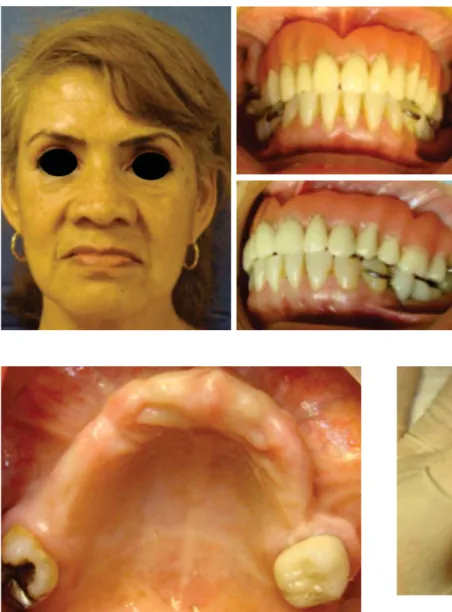
Our patient was a 60 year old female, with no chronic pathological history. The patient´s main complaint was instability of the upper prosthesis, which had been manufactured by many specialists. Intra-oral physical examination revealed incomplete permanent dentition, presence of two teeth with grade III mobility in the upper jaw, Cawood and Howell class IV atrophic alveolar process, which measured 3 mm thickness. These data were confi rmed with a tomographic study
(Figures 1 to 3).
It was decided to extract residual maxillary teeth preserving the alveolus, and to reconstruct the alveolar process by means of onlay-type anterior iliac crest graft, contemplating implant placement in a second surgical event.
SURGICAL TECHNIQUE
Surgical procedure was achieved under balanced general anesthesia. The anterior iliac crest graft was first harvested, following the Kalk technique which consists on taking a corticocancellous block, reciprocating saw, straight and curved chisel, additional cancellous bone was harvested with curettes. Once the graft was harvested, hemostasis
www.medigraphic.org.mx
Figure 1.
Frontal view of the patient and ill-adjusted dentures.
Figure 2. Maxilla alveolar process.
Figure 3. Tomographic tri-dimensional reconstruction.
w
w
w
w
w
w
w
w
w
w
w
w
w
w
w
w
w
w
w
w
w
w
w
w
w
w
w
w
w
w
w
w
w
w
w
w
w
w
w
w
w
w
w
w
w
w
w
w
w
w
w
w
w
w
w
w
w
w
w
w
w
w
w
w
w
w
w
w
w
w
w
w
w
w
w
w
w
w
w
w
w
w
w
w
w
w
w
w
w
w
w
w
w
w
w
ww
w
w
ww
w
w
w
w
w
w
w
w
w
w
w
w
w
w
w
w
w
w
w
w
w
w
w
w
w
w
w
w
w
w
w
w
w
w
w
w
w
w
w
w
w
w
w
w
w
w
w
w
w.
w
w
w
w
w
w
w
w
w
w
w
w
w
w
w
w
w
w
w
w
w
w
w
w
w
w
.
.m
.
.
.
.
m
m
m
m
m
m
m
m
m
m
m
m
m
m
m
m
m
m
m
m
m
m
m
m
m
m
m
m
m
m
m
m
m
m
m
m
m
m
m
m
m
m
m
m
m
m
m
m
m
m
m
m
m
m
m
m
m
m
me
m
m
m
m
m
m
m
m
m
m
m
m
m
m
m
m
m
m
m
m
m
m
m
m
m
m
m
m
m
m
m
m
m
m
m
m
m
m
m
m
m
m
m
m
m
m
m
m
m
m
m
m
m
m
m
m
m
m
m
m
m
m
m
m
m
m
m
m
m
m
m
m
m
m
m
m
m
m
m
m
m
m
m
m
m
m
m
m
m
m
m
m
m
m
m
m
m
m
m
m
m
m
m
m
m
e
e
e
e
e
e
e
e
e
e
e
e
e
e
e
e
e
e
e
e
e
e
e
e
e
e
e
e
e
e
e
e
e
e
e
e
ed
e
e
e
e
e
e
e
e
e
e
e
e
e
e
e
e
e
e
e
e
e
e
e
e
e
e
e
e
e
e
e
e
e
e
e
e
e
e
e
e
e
e
e
e
e
e
e
e
e
e
e
e
e
e
e
e
e
e
e
e
e
e
e
e
e
e
e
e
e
e
e
e
e
e
e
e
e
e
e
e
e
e
e
e
e
e
e
e
e
e
e
e
e
e
e
e
e
e
e
d
d
d
d
d
d
d
d
d
d
d
d
d
d
d
d
d
d
d
d
d
d
d
d
d
d
d
d
d
d
d
d
d
d
d
d
d
d
d
d
d
d
d
d
d
d
d
d
d
d
di
d
d
d
d
d
d
d
d
d
d
d
d
d
d
d
d
d
d
d
d
d
d
d
d
d
d
d
d
d
d
d
d
d
d
d
d
d
d
d
d
d
d
d
d
d
d
d
d
d
d
d
d
d
d
d
d
d
d
d
d
d
d
d
d
d
d
d
d
d
d
d
d
d
d
d
d
d
d
d
d
d
d
d
d
d
d
d
d
d
d
d
d
i
i
i
i
i
i
i
i
i
i
i
i
i
i
i
i
i
i
i
i
i
i
i
i
i
i
i
i
i
ig
i
i
i
i
i
i
i
i
i
i
i
i
i
i
i
i
i
i
i
i
i
i
i
i
i
i
i
i
i
i
i
i
i
i
i
i
i
i
i
i
i
i
i
i
i
i
i
i
i
i
i
i
i
i
i
i
i
i
i
i
i
i
i
i
i
i
i
i
i
i
i
i
i
i
i
i
i
i
g
g
g
g
g
g
g
g
g
g
g
g
g
g
g
g
g
g
g
g
g
g
g
g
g
g
g
g
g
g
g
g
g
g
g
g
g
g
g
g
g
g
g
g
g
g
g
g
g
g
g
g
g
g
g
g
g
g
g
g
g
g
g
g
g
g
g
g
g
g
g
g
g
g
g
g
g
g
g
g
g
g
g
g
g
g
g
g
g
g
g
g
g
g
g
g
g
g
g
g
g
g
g
g
g
g
g
g
g
g
g
g
g
g
g
g
g
g
g
g
g
g
g
g
g
g
g
g
g
g
g
g
g
g
g
g
g
g
g
g
g
g
g
g
g
g
g
g
g
g
g
g
g
g
g
g
g
g
g
g
g
g
g
g
g
g
g
g
g
g
g
g
g
g
g
g
g
g
g
g
g
g
g
g
g
g
g
g
g
g
Figure 4. Incision.Figure 5. Exposition of anterior iliac crest.
p
p
p
p
p
p
p
p
p
p
p
p
p
p
p
p
p
p
p
p
p
p
p
p
p
p
p
p
p
p
p
p
p
p
p
p
p
p
p
p
p
p
p
p
p
p
p
p
p
p
p
p
p
p
p
p
p
p
p
p
p
p
p
p
p
p
p
p
p
p
p
p
p
p
p
p
p
p
p
p
p
p
p
p
p
p
p
p
p
p
p
p
p
p
p
p
p
p
p
p
p
p
p
p
p
p
p
p
p
p
p
p
p
p
p
p
p
p
p
p
p
p
p
p
p
p
p
p
p
p
p
p
p
p
p
p
p
p
p
p
p
p
p
p
p
p
p
p
p
p
p
p
p
p
p
p
p
p
p
p
p
p
p
p
p
p
p
p
p
p
p
p
p
p
p
p
p
p
p
p
p
p
p
p
p
p
p
p
p
p
p
p
p
p
p
p
p
p
p
p
p
p
p
p
p
p
p
p
p
p
p
p
p
p
p
p
p
p
p
p
p
p
p
p
p
p
p
p
p
p
p
p
p
p
p
p
ph
h
h
h
h
h
h
h
h
h
h
h
h
h
h
h
h
h
h
h
h
h
h
h
h
h
h
h
h
h
h
h
h
h
h
h
h
h
h
h
h
h
h
h
h
h
h
h
h
h
h
h
h
h
h
h
h
h
h
h
h
h
h
h
h
h
h
h
h
h
h
h
h
h
hi
h
h
h
h
h
h
h
h
h
h
h
h
h
h
h
h
h
h
h
h
h
h
h
h
h
h
h
h
h
h
h
h
h
h
h
h
h
h
h
h
h
h
h
h
h
h
h
h
h
h
h
h
h
h
h
h
h
h
h
h
h
h
h
h
h
h
h
h
h
h
h
h
h
h
h
h
h
h
h
h
h
h
h
h
h
h
h
h
h
h
h
h
h
h
h
h
h
h
h
h
h
h
h
h
h
h
h
h
h
h
h
h
h
h
h
h
h
h
h
h
h
h
h
h
h
h
h
h
h
h
h
h
h
h
i
i
i
i
i
i
i
i
i
i
i
i
i
i
i
i
i
i
i
i
ic
i
i
i
i
i
i
i
i
i
i
i
i
i
i
i
i
i
i
i
i
i
i
i
i
i
i
i
i
i
i
i
i
i
i
i
i
i
i
i
i
i
i
i
i
i
i
i
i
c
c
c
c
c
c
c
c
c
c
c
c
c
c
c
c
c
c.
c
c
c
c
c
c
c
c
c
c
c
c
c
c
c
c
c
c
c
c
c
c
c
c
.
.o
.
.
.
.
.
o
o
o
o
o
o
o
o
o
o
o
o
o
or
o
o
o
o
o
o
o
o
o
o
o
o
o
o
o
o
o
o
r
r
r
r
r
r
r
r
r
rg
r
r
r
r
r
r
r
r
r
r
r
r
r
r
r
r
r
g
g
g
g
g
g
g
g
g
g
g
g
g
g
g
g
g
g
g
g
g
g
g
g
g
g
g
g
g
g
g
g
g
g
g
g.
g
g
g
g
g
g
g
g
g
g
g
g
g
g
g
g
g
g
g
g
g
g
g
g
g
g
g
g
g
g
g
g
g
g
g
g
g
g
g
g
g
g
g
g
g
g
g
g
g
g
g
g
g
g
g
g
g
g
g
g
g
g
g
g
g
g
g
g
g
g
g
g
g
g
g
g
g
g
g
g
g
g
g
g
g
g
g
g
g
g
g
g
g
g
g
g
g
g
.
.
.
.
.
.
.
.
.m
.
.
.
.
.
.
.
.
.
.
.
.
.
.
.
m
m
m
m
m
m
m
m
m
m
m
m
m
m
m
m
m
m
m
m
m
m
m
m
m
m
m
m
m
m
m
m
m
m
m
mx
m
m
m
m
m
m
m
m
m
m
m
m
m
m
m
m
m
m
m
m
m
m
m
m
m
m
m
m
m
m
m
m
m
m
m
m
m
m
m
m
m
m
m
m
m
m
m
m
m
m
m
m
m
m
m
m
m
m
m
m
m
m
m
m
m
m
m
m
m
m
m
m
m
m
m
m
m
m
m
m
m
m
m
m
m
m
m
m
m
m
m
m
m
m
m
m
m
m
m
m
m
x
x
x
x
x
x
x
x
x
x
x
x
x
x
x
x
x
x
x
x
x
x
x
x
x
x
x
x
x
x
x
x
x
x
x
x
x
x
x
x
x
x
x
x
x
x
x
x
x
x
x
x
x
x
x
x
x
x
x
x
x
x
x
x
x
x
x
x
x
x
x
x
x
x
x
x
x
x
x
x
x
x
x
x
x
x
x
x
x
x
x
x
x
x
x
x
x
x
x
x
x
x
x
x
x
x
x
x
x
x
x
x
x
x
x
x
x
x
x
x
x
x
x
x
x
x
x
x
x
x
x
x
x
x
x
x
x
x
x
x
x
x
x
www.medigraphic.org.mx
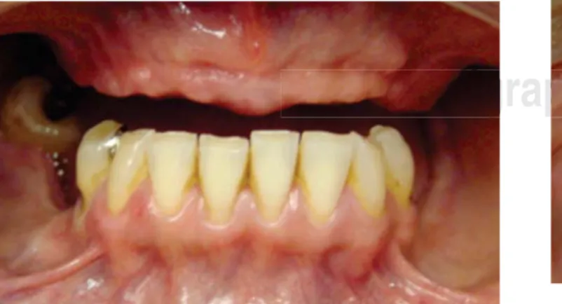
mucosa (Figures 12 and 13).
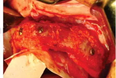
Once the alveolar process was exposed, vestibular cortical decortications were conducted, as well as in one graft wall. Onlay type graft blocks were placed, and graft was fastened with 1.5 mm diameter x 10 mm long screws. Osteosynthesis material was
Figure 6. Corticocancellous block of anterior iliac crest.
Figure 7. Iliac crest corticocancellous graft harvesting.
w
w
w
w
w
w
w
w
w
w
w
w
w
w
w
w
w
w
w
w
w
w
w
w
w
w
w
w
w
w
w
w
w
w
w
w
w
w
w
w
w
w
w
w
w
w
w
w
w
w
w
w
w
w
w
w
w
w
w
w
w
w
w
w
w
w
w
w
w
w
w
w
w
w
w
w
w
w
w
w
w
w
w
w
w
w
w
w
w
w
w
w
w
w
w
w
w
w
w
w
w
w
w
w
w
w
w
w
w
ww
w
w
w
w
w
w
w
w
w
w
w
w
w
w
w
w
w
w
w
w
w
w
w
w
w
w
w
w
ww
w
w
w
w
w
w
w
w
w
w
w
w
w
w
w
w
w
w
w
w
w
w
w
w
w
w
w
w
w
w
w
w
w
w
w
w
w
w
w
w
w
w
w
w
w
w
w
w
w
w
w
w
w
w
w
w
w
w
w
w
w
w
w
w
w
w
w
w
w
w
w
w
w
w
w
w
w
w
w
w
w
w
w
w
w
w
w
w
w
w
w
w
w
w
w
w
w
w
w
w
w
w
w.
w
w
w
w
w
w
w
w
w
w
w
w
w
w
w
w
w
w
w
w
w
w
w
w
w
w
w
w
w
w
w
w
w
w
w
w
w
w
w
w
w
w
w
w
w
w
w
w
w
w
w
w
w
w
w
w
w
w
w
w
w
w
w
w
w
w
w
w
w
w
w
.
.
.
.
.
.
.
.
.
.
.
.
.
.
.
.
.
.
.
.
.
.
.
.
.
.m
.
.
.
.
.
.
.
.
.
.
.
.
.
.
.
.
.
.
..
.
.
.
.
.
.
.
.
.
.
.
.
.
.
m
m
m
m
m
m
m
m
m
m
m
m
m
m
m
m
m
m
m
m
m
m
m
m
m
m
m
m
m
m
m
m
m
m
m
m
m
m
m
m
m
m
m
m
m
m
m
m
m
m
m
m
m
me
m
m
m
m
m
m
m
m
m
m
m
m
m
m
m
m
m
m
m
m
m
m
m
m
m
m
m
m
m
m
m
m
m
m
m
m
m
m
m
m
m
m
m
m
m
m
m
m
m
m
m
m
m
m
m
m
m
m
m
m
m
m
m
m
m
m
m
m
m
m
m
m
m
m
m
m
m
m
m
m
m
m
m
m
m
m
m
m
m
m
m
m
m
m
m
m
m
m
m
e
e
e
e
e
e
e
e
e
e
e
e
e
e
e
e
e
e
e
e
e
e
e
e
e
e
e
e
e
e
e
ed
e
e
e
e
e
e
e
e
e
e
e
e
e
e
e
e
e
e
e
e
e
e
e
e
e
e
e
e
e
e
e
e
e
e
e
e
e
e
e
e
e
e
e
e
e
e
e
e
e
e
e
e
e
e
e
e
e
e
e
e
e
e
e
e
e
e
e
e
e
e
e
e
e
e
e
e
e
e
e
e
e
e
e
d
d
d
d
d
d
d
d
d
d
d
d
d
d
d
d
d
d
d
d
d
d
d
d
d
d
d
d
d
d
di
d
d
d
d
d
d
d
d
d
d
d
d
d
d
d
d
d
d
d
d
d
d
d
d
d
d
d
d
d
d
d
d
d
d
d
d
d
d
d
d
d
d
d
d
d
d
d
d
d
d
d
d
d
d
d
d
d
d
d
d
d
d
d
d
d
d
d
d
d
d
d
d
d
d
d
d
d
d
d
d
i
i
i
i
i
i
i
i
i
i
i
i
i
i
i
i
i
i
i
i
i
i
i
i
i
i
i
i
i
ig
i
i
i
i
i
i
i
i
i
i
i
i
i
i
i
i
i
i
i
i
i
i
i
i
i
i
i
i
i
i
i
i
i
i
i
i
i
i
i
i
i
i
i
i
i
i
i
i
i
i
i
i
i
i
i
i
i
i
i
i
i
i
i
i
i
i
i
i
i
i
i
i
i
i
i
i
i
i
i
i
g
g
g
g
g
g
g
g
g
g
g
g
g
g
g
g
g
g
g
g
g
g
g
g
g
g
g
g
g
g
g
g
g
g
g
g
g
g
g
g
g
g
g
Figure 8. Surgical bed.
Figure 9. Cancellous graft harvesting.
Figure 10. Obtained cancellous graft.