Contents lists available atScienceDirect
Journal of Functional Foods
journal homepage:www.elsevier.com/locate/jff
Wine pomace product ameliorates hypertensive and diabetic aorta vascular
remodeling through antioxidant and anti-in
fl
ammatory actions
Gisela Gerardi, Mónica Cavia-Saiz, Raquel del Pino-García, María D. Rivero-Pérez,
María L. González-SanJosé, Pilar Muñiz
⁎Department of Biotechnology and Food Science, Faculty of Sciences, University of Burgos, Plaza Misael Bañuelos, 09001 Burgos, Spain
A R T I C L E I N F O
Keywords: Aorta Diabetes Hypertension Vascular remodeling eNOS
Wine pomace product
A B S T R A C T
Vascular remodeling in hypertension and diabetes is characterized by a low-grade inflammation of the arterial wall and enhanced oxidative stress. Wine pomace products attenuate hyperglycemia and hypertension through reduction of oxidative stress and inflammation by their polyphenol composition. The aim of the study was to assess the influence of the wine pomace product (WP) on morphometric parameters of the arterial wall in spontaneously hypertensive rats (SHRs) and streptozotocin-diabetic rats (STZ). Oxidative stress was also eval-uated by determination of radical oxygen species (ROS) and endothelial nitric oxide synthase (eNOS) activation in thoracic aortas. Wine pomace reduces wall aortic thickness, cross sectional area and wall/lumen ratio, and decreases ROS and increases eNOS activation. In summary, the supplementation of hypertensive or diabetic rats with the wine pomace product exhibits a protective role against the endothelial dysfunction and vascular re-modeling.
1. Introduction
Endothelial cells, smooth muscle cells andfibroblasts interact in a functional complex to form the vascular wall. Changes in the environ-ment sensed by the vascular wall produce several intracellular signals and mediators that regulate vascular structure and function. This pro-cess is known as vascular remodeling and involves changes in the cell growth and death, cell motility and composition of the extracellular matrix (Renna, De Las Heras, & Miatello, 2013).
Vascular remodeling is an active process that response to physio-logical and pathophysio-logical changes in the hemodynamics conditions as consequence of different stimuli like hypertension, hyperglycemia and other inflammatory diseases (Van Varik et al., 2012). Several in-tracellular systems and mediators intervene in the vascular remodeling associated to hypertension and hyperglycemia. They include pro-in-flammatory and pro-oxidant agents such as angiotensin II (Ang II). Ang II mediates reactive oxygen species (ROS) generation by NADPH oxi-dase, intracellular signaling activation such as MAPK, PI3K and NF-κB pathways and the consequent gene regulation (Renna et al., 2013).
Oxidative stress is related to the development of vascular re-modeling and endothelial dysfunction. The oxidative stress activates redox-sensitive inflammatory molecules such as AP-1 and NF-kB, which amplify the inflammatory response by cytokines, chemokines, and lymphokines to ultimately induce more vascular inflammation (Renna et al., 2013). Thus, antioxidant treatment could ameliorate vascular alterations associates to pro-inflammatory pathologies such as hy-pertension and diabetes.
Food polyphenols have demonstrated antioxidant, anti-in-flammatory and anti-cancer properties, blood pressure, lipid and glu-cose modulation, and gene expression regulation. By acting through different mechanisms, polyphenols prevent and reverse oxidative stress associated to chronic diseases (Zhang, Qi, Liu, Zhang, & He, 2015; Del Pino-García, Rivero-Pérez, González-SanJosé, Croft, & Muñiz, 2017; Gerardi, Cavia-Saiz, Rivero-Pérez, González-SanJosé, & Muñiz, 2019). Phenolic compounds such as ferulic, resveratrol, catechin, epigalloca-techin and quercetin reduce oxidative stress and their related vascular disorders by modulation of different cellular mechanisms. In this sense, they can act by direct ROS neutralization and/or through increasing the
https://doi.org/10.1016/j.jff.2020.103794
Received 28 September 2019; Received in revised form 13 January 2020; Accepted 14 January 2020
Abbreviations:C, control rats;CSA, cross sectional area;DB, diabetic rats;p- eNOS, pholphorylated endothelial nitric oxide synthase;Placebo, rats not supple-mented with the wine pomace product;SHRs, spontaneously hypertensive rats;W/L, wall/lumen ratio;WKY, Wistar Kyoto rats;WP, rats supplemented with 300 mg/kg/d of wine pomace product
⁎Corresponding author.
E-mail addresses:mggerardi@ubu.es(G. Gerardi),monicacs@ubu.es(M. Cavia-Saiz),rdpino@ubu.es(R. del Pino-García),drivero@ubu.es(M.D. Rivero-Pérez), marglez@ubu.es(M.L. González-SanJosé),pmuniz@ubu.es(P. Muñiz).
Available online 24 January 2020
endogen antioxidant systems. They can also regulate the ROS/nitric oxide (NO) balance by reducing ROS production through the NADPH oxidases, and by increasing NO bioavailability. Moreover, polyphenols modulate several signaling pathways including the activation of anti-oxidant pathways (Akt, Nrf2, SIRT1), the inhibition of pro-anti-oxidant and pro-inflammatory signaling (NF-κB), and the promotion of the gene expression of antioxidant and anti-inflammatory proteins such as en-dothelial nitric oxide synthase (eNOS), superoxide dismutases (SOD) and heme oxygenase-1 (HO-1).
In previous studies the supplementation of rats with a wine pomace product rich in polyphenols attenuates oxidative stress associated to hypertension and type 1 diabetes (Del Pino-García, Rivero-Pérez, et al., 2016; Del Pino-García et al., 2017). Both blood pressure and glucose was reduced by the wine pomace (WP) and oxidative damage was lowered. In the present study, the vascular remodeling in aortas of spontaneously hypertensive rats (SHRs) and streptozotocin-induced diabetic rats (STZ) was evaluated after administration of wine pomace (WP) and compared with placebo administration. Oxidative and in-flammatory parameters were also studied and include ROS generation and eNOS activation.
2. Material and methods
2.1. Red wine pomace product (WP)
Red wine pomace-derived product from the vinification ofVitis vi-niferaL. cv. Tempranillo was prepared at the University of Burgos ac-cording to a previously described method (González-SanJosé, García-Lomillo, Del Pino-García, Muñiz-Rodríguez, & Rivero-Pérez, 2015). This product was obtained from seedless red wine pomace (WP) using a heat treatment as stabilization process, and its main characteristics and composition (dietaryfibre, fat, protein, minerals and phenolic classes) have been determined previously (Gerardi et al., 2019) and are sum-marized in Tables S1 and S2,Supplementary material.
2.2. Animals
Experimentation with live animals was approved by the Ethics Committee for Experimental Animal Care at the University Hospital of Burgos and was carried out in accordance with the Spanish and European laws (Royal Decree 53/2013 of the Spanish Ministry of agriculture, Food and Environment and Ministry of Economy and Competitiveness, and European Directive 2010/63/EU). Adult male Wistar Kyoto (WKY) rats and spontaneously hypertensive rats (SHRs) weighing 307 ± 13 g were assigned to 6 groups (n = 5 rats/group). The room was maintained at 21 ± 2 °C and humidity at 40 ± 10%, with a 12:12-h light:dark cycle and free access to food (A04, Panlab, S.L.U., Barcelona, Spain) and water.
2.3. Experimental animal groups and general procedures
Animals were divided into 6 groups (n = 5 rats/group): control (C), control supplemented (C + WP), hypertensive (SHR), hypertensive supplemented (SHR + WP), diabetic (DB) and diabetic supplemented (DB + WP). Spontaneously hypertensive rats (SHRs) were used in the SHR groups. Hypertension was confirmed from the beginning of the experiment by blood systolic pressure measurements ˃ 200 mmHg. Diabetes was induced in overnight fasted rats of DB groups by a single intraperitoneal injection of 45 mg/kg of streptozotocin (STZ, Sigma-Aldrich, St Louis, MO, USA) dissolved in 0.1 M citrate buffer (pH 4.6). The diabetic was confirmed 3 days later by blood glucose levels ˃ 350 mg/dl. Rats in the supplemented groups received the WP (300 mg/ kg/d) dissolved in water by oral gavage. The dosage evaluated in the present work was selected by the authors according to previous studies (Del Pino-García et al., 2017; Gerardi, Cavia-Saiz, Rivero-Perez, González-SanJosé, & Muñiz, 2020). The human equivalent dose (HED)
was also considered due to the potential use of the WPP as dietary supplement. The 300 mg/kg/d dose was equivalent in human to 3 g/d. The study wasfinished after 4 weeks of supplementation. The last day of study, a combined ketamine/xylazine anesthetic (80 mg keta-mine/kg and 20 mg xylazine/kg) was administrated to rats by in-traperitoneal injection. Then, rats were euthanized by intra-cardiac injection of Sodium Pentobarbital (300 mg/kg). Thoracic and abdom-inal aortic tissues from each rat was collected and frozen at−80 °C or fixed in 4% paraformaldehyde.
2.4. Aortic morphometric analysis
Fixed aortic tissue was sectioned and stained with hematoxylin and eosin for the analysis of the morphological changes. Aortic wall thick-ness and the internal diameter were measurement by a Nikon Eclipse LV100NPOL microscope with a High-precision rotating stage and using the NIS Element Software. The wall-to-lumen (W/L) ratio was eval-uated by the ratio of wall thickness to the internal diameter of lumen. The cross sectional area (CSA) of the aorta was calculated as CSA =π (Re2– Ri2) where Re and Ri are the external and internal radii, re-spectively.
2.5. Reactive oxygen species (ROS) detection
For the analysis of ROS in the aortic tissue, frozen aortas were embedded in freezing medium and transverse sections were obtained with a cryostat, collected on glass slides and equilibrated in phosphate buffer (pH 7.4). Slides were incubated for 30 min at 37 °C with 2,7-dichlorodihydrofluorescin diacetate (DCFH-DA, Sigma-Aldrich, St Louis, MO, USA) solution (10 µM in DMSO) in a light-protected hu-midified chamber. Images were obtained with a Leica CTR6000 mi-croscope. The fluorescence images were quantified using ImageJ Software and results are expressed as percentage to the control.
2.6. Western blotting analysis
The aortic tissue was homogenized in ice-cold lysis buffer for the extraction of total proteins. Protein concentration was measured using a protein assay reagent (Quick Start™Bradford Protein Assay, Bio-Rad, Hercules, CA, USA). Total protein (40 µg) was treated with Laemmli buffer, boiled for 5 min and resolved by 10% SDS-PAGE (Bio-Rad Mini-Protean Tetra cell). Then, protein were electrophoretically transferred (Bio-Rad Trans-Blot Turbo Blotting System) into a PVDF membranes (Bio-Rad Laboratories, Hercules, CA, USA) and incubated overnight at 4 °C with specific primary rabbit antibodies (1:1000) against phospho-eNOS and actin (Sigma-Aldrich). After rinsing, the membranes were incubated with a horseradish-peroxidase-labeled secondary antibody anti-rabbit (1:3000, Anti-rabbit IgG-HRP-linked Antibody, Cell Signaling Technology, Danvers, MA, USA). Inmunodetection was per-formed using enhanced chemiluminescence kit (Clarity Western ECL substrate, Biorad Laboratories, Hercules, CA, USA) and developed using autoradiography (Amersham Hyperfilm ECL, GE Heaalthcare). Proteins bands were quantified by densitometry using the ImageJ software and their relative amounts were normalized to the housekeeping proteinβ -actin. The results were expressed as a ratio of the phosphorylated target protein amount against the totalβ-actin protein.
2.7. Data presentation and statistical analysis
3. Results
3.1. Histological analysis of aortic structure
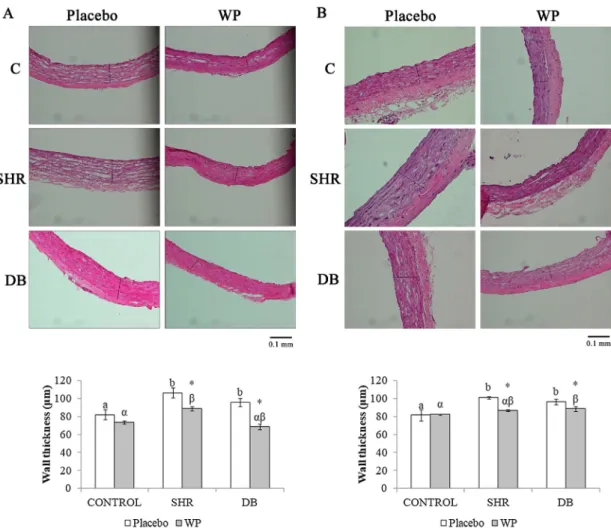
To investigate the effect of the wine pomace product (WP) in the hypertensive and diabetic vasculopathy, vascular wall thickness, cross sectional area (CSA) and wall/lumen (W/L) ratios were evaluated in thoracic and abdominal aortas of SHRs (hypertensive rats: SHR), STZ (diabetic rats: DB) and WKY (normal control rats: C) rats. Thoracic aortas were analyzed in sections stained with hematoxylin and eosin (Figs. 1 and 2).
Fig. 1 shows morphological appearance of the thoracic (A) and abdominal (B) aortic wall in normal rats (C), hypertensive rats (SHR) and diabetic rats (DB) treated (WP) or untreated (Placebo) with the wine pomace product. The values of wall thickness in all groups are shown in the graphs below the aortic wall images. Wall thickness of SHR and DB rats were significantly higher than that in control rats for both thoracic and abdominal aortas (Fig. 1). WP supplemented rats exhibited significant decreases in both aortic wall thicknesses when compared with the placebo group. The reduction of the wall thickness was about 16% (thoracic aortas) and 14% (abdominal aortas) for the SHR rats and 31% (thoracic aortas) and 8% (abdominal aortas) for DB rats comparing the supplemented rats with the correspondent placebo group.
Fig. 2illustrated the thoracic (A) and abdominal (B) aortic structure
in all groups treated (WP) or untreated (Placebo) with the wine pomace product. Cross sectional area (CSA) of aortas and wall/lumen (W/L) ratio are showed in the graphs below. A hypertrophy of thoracic and abdominal aortas was observed in SHR rats, as showed the 40% and 25% increment observed in the CSA, comparing with control group. WP reduced this CSA by 16% and 14% compared with the placebo SHR rats. Diabetic rats only experimented an elevation in the CSA of the ab-dominal aortas but not the thoracic. The ingestion of the WP reduced the CSA of DB rats by 16% and 10% for the thoracic and abdominal aortas, respectively. The W/L ratio of both section of aortas of the SHR rats were significantly greater than that of the control group, while no significant differences were observed between control and DB groups (Fig. 2). WP administration to SHR rats reduced the W/L ratio by 18% and 26% for the thoracic and abdominal aortas, respectively. In addi-tion, the W/L ratio of the thoracic aorta was reduced by 17% for the WP-treated DB rats.
3.2. Oxidative stress in the thoracic aorta
placebo.
3.3. Nitric oxide in thoracic aorta
Western blot analysis was conducted to measure the protein ex-pression levels of endothelial nitric oxide synthase (eNOS) in thoracic aortic tissue (Fig. 4). The protein phosphorylation of eNOS was sig-nificantly decreased in the SHR rats compared with the control group. The WP intake increased the eNOS phosphorylated levels of the SHR rats in about 2.3 fold times. No significant differences were observed between DB and control rats in the eNOS levels.
4. Discussion
The results show that the administration of wine pomace product (WP) modified some cardiovascular alterations associated to
hypertension and type 1 diabetes mellitus in two independent rat models. We previously reported that the administration of this WP ameliorates vascular complications of diabetes and hypertension by alleviating hyperglycemia, hipoinsulinemia, hypertension and their associated vascular oxidative damage (Del Pino-García, Rivero-Pérez, et al., 2016; Del Pino-García et al., 2017). Here we report the effect of the WP, rich in polyphenols, in the vascular structure and its association with the vascular oxidative status.
WP has high amounts of dimeric procyanthocyanidins, principally procyanidin B2. Some authors showed that dimeric, oligomeric and high weight proanthocyanidins improve the lipid profile of dyslipi-demic individuals (Pal, Naissides, & Mamo, 2004:Wong et al., 2016) and reduce the markers of oxidative stress and inflammation in in-dividuals with metabolic syndrome and diabetes (Cani, Osto, Geurts, &
Everard, 2012; Wong et al., 2016).
Cardiovascular risk factors such as hypertension and diabetes alter the redox state in the vessel and can led in endothelial dysfunction affecting vascular structure and function (Khoo et al., 2010). This vascular alteration is associated to oxidative stress and linked to an imbalance between nitric oxide (NO) and radical oxygen species (ROS) generation in the endothelial cells. Thus, the inhibition of oxidative stress becomes an important aspect to consider in explaining the ben-efits of the WP-polyphenols in the endothelial dysfunction.
The vascular system is exposed to alterations such as hypertension and hyperglycemia that induce arterial remodeling. In this process smooth muscle cells (SMCs) increase their proliferation which results in medial thickening of arteries and arterioles (Xu et al., 2001). Arterial remodeling is characterized by a low-grade inflammation of the arterial wall with inflammatory cell infiltration, enhanced oxidative stress and upregulation of inflammatory mediators including endothelial adhesion molecules (VCAM-1, ICAM-1), pro-inflammatory transcriptional factor NF-κB and other proinflammatory molecules (MCP-1, PAI-1). Arterial remodeling can be either hypertrophic (thickening of the vascular wall and increased cross sectional area), eutrophic (constant wall thickness) or hipotrophic (reduced cross sectional area), and can be outward (in-creased lumen diameter) or inward (reduced lumen diameter) (Renna et al., 2013). Although eutrophic remodeling has been described in primary hypertension in humans and in small arteries of SHRs rats; hypertrophic is found in secondary hypertension such as hypertension associated to diabetes mellitus, in hyperaldoresteronism, or in pheo-chromocytoma (Schiffrin, 2012; Van Varik et al., 2012). Large central arteries such as aorta undergo arteriosclerotic changes with outward hypertrophic remodeling characterized by increased cross sectional area (CSA) and lumen diameter (O’Rourke & Hashimoto, 2007). These changes were observed in the abdominal and thoracic aortas of the hypertensive rats evidencing the presence of hypertrophic changes with Fig. 3. Analysis of oxidative stress in rat thoracic aortic tissue.Dichlorodihydrofluorescein staining shows radical oxygen species (ROS) generation in thoracic (A) and abdominal (B) aorta. Fluorescence was quantified and results expressed as percentage of increase in relation to control, and they are showed in the graph below the images Significant differences (n = 5, ANOVA, p < 0.05) between control, hypertensive and diabetic rats are re-presented with Latin Letters for placebo groups and Greek Letters for supplemented group. Significant differences (n = 5, Student‘st-test, p < 0.05) be-tween placebo and WP for each group are re-presented with an asterisk (*). C: control rats; SHR: hypertensive rats; DB: diabetic rats; Placebo: rats not supplemented with the wine pomace product; WP: rats supplemented with 300 mg/kg/d of wine po-mace product.
an increase in the lumen and wall area of the aortas. The intake of WP by the hypertensive rats prevents the vascular remodeling by reducing the aortic CSA and wall thickness observed in the hypertensive rats. Furthermore, wine pomace ingestion also reduced the wall/lumen ratio. It was observed that hypertensive patients with the highest wall/ lumen ratio had increased incidence of cardiovascular events. Thus, the reduction of this alteration by the WP could contribute to a less in-cidence of cardiovascular complications. Some authors propose that antihypertensive agents may affect the vascular wall directly by redu-cing Angiotensin II (Ang II) response, and indirectly by blood pressure lowering. In previous studies we evidenced antihypertensive action of the WP with reduction in the blood pressure, thus the WP could have indirect action in the amelioration of the vascular remodeling. Poly-phenols of the WP such asflavonoids also reduce the gene expression of angiotensin converting enzyme (ACE) and this contribute to the low-ering of the Ang II effects (Balasuriya & Rupasinghe, 2011). By reducing pro-oxidant and proinflammatory actions of Ang II, NADPH oxidase activation decreases, with less generation of reactive oxygen species (ROS), decreased activity of the mitogen activated protein kinases (MAPK) and consequent reduction of the activation of proto-oncogenes c-fos, c-jun and c-myc that leads to reduced growth of SMCs and re-modeling. In addition, the activation of the pro-inflammatory tran-scriptional factor NF-κB and cell adhesion molecules are reduced after Ang II inhibition and this diminish the extravasation of leukocytes into the vascular wall. Furthermore, there is an important role of the per-oxisome proliferator activator receptor PPAR-γagainst the Ang II effect in the vascular remodeling (Schiffrin, 2012). Since resveratrol and other polyphenols of the WP exhibit a stimulatory effect in PPAR-γ, this could improve the Ang II-vascular remodeling (Abraham Domínguez-Avila, González-Aguilar, Alvarez-Parrilla, & de la Rosa, 2016). In this sense, it is important to highlight the importance of resveratrol in the prevention of vascular remodeling as other works demonstrated the effectiveness of this molecule in the prevention of aorta remodeling in different models of hypertension such as spontaneously and malignantly hypertensive rats (Grujic-Milanovic et al., 2017). Altogether, the antioxidant and anti-inflammatory action of the polyphenols in the WP contribute to regression of remodeling of the aortas.
Diabetes cause structural changes of large and small arteries and associated endothelial dysfunction that accelerated the process of ar-terial remodeling. Diabetes-associated vascular remodeling exhibits modifications of the extracellular matrix (ECM), dysfunction of en-dothelial cells (ECs) and SMCs, and changes in the circulating cyto-kines. These alterations conduce to thickening of the intima and media of the large vessels, increasing the risk for cardiovascular complications (Spinetti, Kraenkel, Emanueli, & Madeddu, 2010). Abdominal aortas of diabetic rats suffer hypertrophic outward remodeling with no changes in the wall/lumen ratio. The increase in the lumen diameter with the less thickening of the aortic wall compared with the hyperglycemic aortas explains the no modification in the wall/lumen ratio for the diabetic aortas. Additionally, thoracic aortas of diabetic rats showed similar thickening than the abdominal aortas but conserved cross sec-tional area and lower wall/lumen ratio. These observations suggest a different degree of remodeling in the large vessels as consequence of diabetes type 1 compared with the hypertensive aortas. In all cases, the treatment with the WP improves the structural of the aorta and reduces the vascular remodeling. Hyperglycemia also increased Ang II produc-tion and its related-vascular remodeling is accompanied by NAPDH oxidase and NF-κB activation. Reduction of Ang II signaling, inhibition of NF-κB and lowering of ROS production by polyphenols contribute to the anti-remodeling action of the WP in the diabetic aortas (Del Pino-García, Gerardi, et al., 2016; Gerardi et al., 2019). No changes were observed in the aortas of normal rats after WP consumption, indicating the absence of vascular changes by the WP in normal aortas.
Enhanced ROS production was found in the hypertensive aortas. Main sources of endothelial ROS production are the NADPH oxidases, principally the isoforms NOX2 and NOX4. NOX2 is responsible of ROS
production in response to Ang II and other pathological signals, whereas NOX4 is implicated in basal ROS production. NOX2 is upre-gulated in cardiovascular diseases such as hypertension and athero-sclerosis (Fan et al., 2014). Thus, the increased oxidative stress ob-served in the hypertensive aortas could be associated to increased NAPDH oxidase activity (Del Pino-García, Gerardi, et al., 2016). Fur-thermore, Maia et al. (2014) associated this oxidative stress to in-creased superoxide anion production. This is consistent with previous works in which the expression of superoxide dismutase 2, the sponsible enzyme that catalyzes dismutation of superoxide, was re-duced in hypertensive rats (Del Pino-García et al., 2017). Superoxide anion could react with NO and reduce its bioavailability. Furthermore, superoxide anion produces the uncoupling of eNOS and this generates more superoxide increasing the pro-oxidant status. However, the WP ingestion by the hypertensive rats reduced the aortic ROS levels pos-sible due the antioxidant effect of the polyphenols. Several mechanism are involved in the antioxidant activity of the polyphenols in the WP including superoxide scavenging action and NAPDH oxidase inhibition by catechin, epicatechin and epigallocatechin, reduced expression of NF-κB by gallic acid, and stimulation of SOD2 and other antioxidant enzymes through Akt and Sirt-1 activation by resveratrol and ferulic acid (Del Pino-García, Gerardi, et al., 2016; Gerardi et al., 2019; Serino & Salazar, 2019).
Endothelial dysfunction is characterized by a reduced NO produc-tion in aorta and this is associated with reduced phosphorylaproduc-tion of endothelial nitric oxide synthase (eNOS) (Maia et al., 2014) as observed for the hypertensive rats. However, WP significantly increases eNOS phosphorylation in the SHR. This observation was consistent with a previous study that showed a increased NO bioavailability in SHRs-hypertensive rats supplemented with WP (Del Pino-García et al., 2017). In this sense, WP reduce vascular remodeling by increased NO pro-duction that inhibit vascular smooth muscle cell proliferation (Spinetti et al., 2010). In another aspect, previous reports showed that diabetes mellitus increases peroxynitrite radicals and decreases constitutive ni-tric oxide production (López-Villodre et al., 2016). Furthermore, atherosclerosis of the diabetic vessel wall caused eNOS dysfunction in which eNOS phosphorylation is reduced due decreased Akt activity, and eNOS also is inactivated by DAG/PKC pathway (Spinetti et al., 2010). Type 1 diabetes mellitus did not significantly modify vascular eNOS protein phosphorylation in our experimental model but a slightly increase was observed for the administration of WP in the DB rats. WP polyphenols could increase eNOS phosphorylation by stimulation of kinases such as Akt (Gerardi et al., 2019). Likewise, it was demon-strated that grape polyphenols enhance eNOS activity and increase NO production in endothelial cells. Activation and phosphorylation of eNOS by polyphenols involves an estrogen receptorα-dependent (ERα), the PI3K-Akt and the MAPK-ERK1/2 pathways (Stangl, Dreger, Stangl, & Lorenz, 2007). Resveratrol also increase eNOS gene expression by activating PPARαin endothelial cells (Mokhtar & Rasool, 2017).
An important observation between hypertensive and diabetic vas-cular changes is the fact that diabetic aortas did not suffer alterations in their oxidative and nitrosative levels, reflected by the unaltered ROS levels and eNOS phosphorylation. The absence of these alterations could explain the less modification in the morphology of the diabetic aortas compared with the hyperglycemic.
5. Ethics statement
The authors of the manuscript“Wine pomace product ameliorates hypertensive and diabetic aorta vascular remodeling through anti-oxidant and anti-inflammatory actions”declare that the experimenta-tion with live animals was approved by the Ethics Committee for Experimental Animal Care at the University Hospital of Burgos and was carried out in accordance with the Spanish and European laws (Royal Decree 53/2013 of the Spanish Ministry of agriculture, Food and Environment and Ministry of Economy and Competitiveness, and European Directive 2010/63/EU) and following the ARRIVE guidelines.
CRediT authorship contribution statement
Gisela Gerardi:Conceptualization, Methodology, Formal analysis, Investigation, Writing - original draft. Mónica Cavia-Saiz:
Conceptualization, Methodology, Investigation. Raquel del Pino-García: Conceptualization, Methodology, Investigation. María D. Rivero-Pérez: Methodology, Investigation. María L. González-SanJosé: Conceptualization, Project administration, Funding acquisi-tion. Pilar Muñiz:Conceptualization, Methodology, Formal analysis, Writing - original draft, Project administration.
Declaration of Competing Interest
The authors declare that they have no known competingfinancial interests or personal relationships that could have appeared to infl u-ence the work reported in this paper.
Acknowledgments
The authors thank thefinancial support of Autonomous Government of Castilla y León (Research project BU282U13).
Appendix A. Supplementary material
Supplementary data to this article can be found online athttps:// doi.org/10.1016/j.jff.2020.103794.
References
Abraham Domínguez-Avila, J., González-Aguilar, G. A., Alvarez-Parrilla, E., & de la Rosa, L. A. (2016). Modulation of PPAR expression and activity in response to polyphenolic compounds in high fat diets.International Journal of Molecular Sciences, 17(7), 1–17. https://doi.org/10.3390/ijms17071002.
Balasuriya, B. W. N., & Rupasinghe, H. P. V. (2011). Plantflavonoids as angiotensin converting enzyme inhibitors in regulation of hypertension. Retrieved from Functional Foods in Health Disease, 5(5), 172–188.http://www.functionalfoodscenter. net/files/49461525.pdf.
Cao, Y. jun, Zhang, Y. min, Qi, J. peng, Liu, R., Zhang, H., & He, L. chong (2015). Ferulic acid inhibits H2O2-induced oxidative stress and inflammation in rat vascular smooth muscle cells via inhibition of the NADPH oxidase and NF-κB pathway.International Immunopharmacology, 28(2), 1018–1025.https://doi.org/10.1016/j.intimp.2015.07. 037.
Cani, P. D., Osto, M., Geurts, L., & Everard, A. (2012). Involvement of gut microbiota in the development of low-grade inflammation and type 2 diabetes associated with obesity.Gut microbes, 3(4), 279–288.https://doi.org/10.4161/gmic.19625. Del Pino-García, R., Gerardi, G., Rivero-Pérez, M. D., González-SanJosé, M. L.,
García-Lomillo, J., & Muñiz, P. (2016). Wine pomace seasoning attenuates hyperglycaemia-induced endothelial dysfunction and oxidative damage in endothelial cells.Journal of
Functional Foods, 22, 431–445.https://doi.org/10.1016/j.jff.2016.02.001. Del Pino-García, R., Rivero-Pérez, M. D., González-Sanjosé, M. L., Castilla-Camina, P.,
Croft, K. D., & Muñiz, P. (2016). Attenuation of oxidative stress in Type 1 diabetic rats supplemented with a seasoning obtained from winemaking by-products and its effect on endothelial function.Food and Function, 7(10), 4410–4421.https://doi.org/10. 1039/c6fo01071g.
Del Pino-García, R., Rivero-Pérez, M. D., González-SanJosé, M. L., Croft, K. D., & Muñiz, P. (2017). Antihypertensive and antioxidant effects of supplementation with red wine pomace in spontaneously hypertensive rats.Food & Function, 8(7), 2444–2454. https://doi.org/10.1039/C7FO00390K.
Fan, L. M., Douglas, G., Bendall, J. K., McNeil, E., Crabtree, M. J., Hale, A. B., ... Choudhury, R. P. (2014). Endothelial cell–specific reactive oxygen species pro-duction increases susceptibility to aortic dissection.Ciruculation, 129(25), 2661–2672.https://doi.org/10.1161/CIRCULATIONAHA.113.005062.Endothelial. Gerardi, G., Cavia-Saiz, M., Rivero-Pérez, M. D., González-SanJosé, M. L., & Muñiz, P. (2019). Modulation of Akt-p38-MAPK/Nrf2/SIRT1 and NF-κB pathways by wine pomace product in hyperglycemic endothelial cell line.Journal of Functional Foods, 58(May), 255–265.https://doi.org/10.1016/j.jff.2019.05.003.
Food & Function.https://doi.org/10.1039/C9FO01743G.
González-SanJosé, M. L., García-Lomillo, J., Del Pino-García, R., Muñiz-Rodríguez, P. and Rivero-Pérez, M. D. (2015). Sazonador de origen vegetal con propiedades con-servantes, sustitutivo de la sal, y procedimiento de obtención del mismo. Grujic-Milanovic, J., Miloradovic, Z., Jovovic, D., Jacevic, V., Milosavljevic, I., Milanovic,
S. D., & Mihailovic-Stanojevic, N. (2017). The red wine polyphenol, resveratrol im-proves hemodynamics, oxidative defence and aortal structure in essential and ma-lignant hypertension.Journal of Functional Foods, 34, 266–276.
Khoo, N. K. H., White, C. R., Pozzo-miller, L., Zhou, F., Constance, C., Inoue, T., ... Parks, D. A. (2010). Dietaryflavonoid quercetin stimulates vasorelaxation in aortic vessels. Free Radical Biology and Medicine, 49(3), 339–347.https://doi.org/10.1016/j. freeradbiomed.2010.04.022.
López-Villodre, J. A., Abdel-karim, M., De La Cruz, P., Rodríguez-Pérez, M. D., Reyes, J. J., Guzmán-Moscoso, R., ... González-Correa, J. A. (2016). Effects of hydroxytyrosol on cardiovascular biomarkers in experimental diabetes mellitus.Journal of Nutritional Biochemistry, 37, 94–100.
Maia, A. R., Batista, T. M., Victorio, J. A., Clerici, S. P., Delbin, M. A., Carneiro, E. M., & Davel, A. P. (2014). Taurine supplementation reduces blood pressure and prevents endothelial dysfunction and oxidative stress in post-weaning protein-restricted rats, 9(8). https://doi.org/10.1371/journal.pone.0105851.
Mokhtar, S. S., & Rasool, A. H. (2017). Plant-derived foods containing polyphenols with endothelial protective effects.International Food Research Journal, 24(2), 471–482. O’Rourke, M. F., & Hashimoto, J. (2007). Mechanical factors in arte- rial aging: A clinical
perspective.Journal of the American College of Cardiology, 50, 1–13.
Pal, S., Naissides, M., & Mamo, J. (2004). Polyphenolics and fat absorption.International Journal of Obesity, 28(2), 324.
Renna, N. F., De Las Heras, N., & Miatello, R. M. (2013). Pathophysiology of vascular remodeling in hypertension.International Journal of Hypertension, 2013.https://doi. org/10.1155/2013/808353.
Schiffrin, E. L. (2012). Vascular remodeling in hypertension: Mechanisms and treatment. Hypertension, 59(2 SUPPL. 1), 367–374.https://doi.org/10.1161/
HYPERTENSIONAHA.111.187021.
Serino, A., & Salazar, G. (2019). Protective role of polyphenols against vascular in-flammation, aging and cardiovascular disease.Nutrients, 11(53), 1–23.https://doi. org/10.3390/nu11010053.
Spinetti, G., Kraenkel, N., Emanueli, C., & Madeddu, P. (2010). UKPMC funders group diabetes and vessel wall remodelling: From mechanistic insights to regenerative therapies.Gaia Ecological Perspectives For Science And Society, 78(2), 1–19.https:// doi.org/10.1093/cvr/cvn039.Diabetes.
Stangl, V., Dreger, H., Stangl, K., & Lorenz, M. (2007). Molecular targets of tea poly-phenols in the cardiovascular system.Cardiovascular Research, 73(2), 348–358. https://doi.org/10.1016/j.cardiores.2006.08.022.
Van Varik, B. J., Rennenberg, R. J. M. W., Reutelingsperger, C. P., Kroon, A. A., De Leeuw, P. W., & Schurgers, L. J. (2012). Mechanisms of arterial remodeling: Lessons from genetic diseases.Frontiers in Genetics, 3(DEC), 1–10.https://doi.org/10.3389/fgene. 2012.00290.
Wong, X., Madrid, A. M., Tralma, K., Castillo, R., Carrasco-Pozo, C., Navarrete, P., ... Gotteland, M. (2016). Polyphenol extracts interfere with bacterial lipopolysaccharide in vitro and decrease postprandial endotoxemia in human volunteers.Journal of Functional Foods, 26, 406–417.https://doi.org/10.1016/j.jff.2016.08.011. Xu, C., Lee, S., Singh, T. M., Sho, E., Li, X., Sho, M., ... Zarins, C. K. (2001). Molecular