International Journal of
Molecular Sciences
Article
Homology Model and Docking-Based Virtual
Screening for Ligands of Human Dyskerin as
New Inhibitors of Telomerase for Cancer Treatment
Romina Gabriela Armando1,†, Diego Luis Mengual Gómez1,† , Ezequiel Ivan Juritz2,
Pablo Lorenzano Menna3and Daniel Eduardo Gomez1,*
1 Laboratory of Molecular Oncology, Department of Science and Technology, Quilmes National University,
Buenos Aires B1876BXD, Argentina; [email protected] (R.G.A.); [email protected] (D.L.M.G.)
2 Center for Bioinformatics and Integrative Biology, Facultad de Ciencias de la Vida,
Universidad Andrés Bello, Santiago 8370146, Chile; [email protected]
3 Laboratory of Molecular Pharmacology, Department of Science and Technology,
Quilmes National University, Buenos Aires B1876BXD, Argentina; [email protected] * Correspondence: [email protected]
† The authors have contributed equally to the work.
Received: 11 September 2018; Accepted: 8 October 2018; Published: 18 October 2018
Abstract: Immortality is one of the main features of cancer cells. Tumor cells have an unlimited replicative potential, principally due to the holoenzyme telomerase. Telomerase is composed mainly by dyskerin (DKC1), a catalytic retrotranscriptase (hTERT) and an RNA template (hTR). The aim of this work is to develop new inhibitors of telomerase, selecting the interaction between hTR–DKC1 as a target. We designed two models of the human protein DKC1: homology and ab initio. These models were evaluated by different procedures, revealing that the homology model parameters were the most accurate. We selected two hydrophobic pockets contained in the PUA (pseudouridine synthase and archaeosine transglycosylase) domain, using structural and stability analysis. We carried out a docking-based virtual screen on these pockets, using the reported mutation K314 as the center of the docking. The hDKC1 model was tested against a library of 450,000 drug-like molecules. We selected the first 10 molecules that showed the highest affinity values to test their inhibitory activity on the cell line MDA MB 231 (Monroe Dunaway Anderson Metastasis Breast cancer 231), obtaining three compounds that showed inhibitory effect. These results allowed us to validate our design and set the basis to continue with the study of telomerase inhibitors for cancer treatment.
Keywords:telomerase; DKC1; hTR; inhibitors; cancer
1. Introduction
The term “cancer” represents a large group of more than one hundred diseases that can affect almost any structure of an organism. Cancer is characterized as a complex disease involving different factors. Malignant tumors share certain biological features, including: sustained proliferative ability; lack of sensitivity to inhibitory growth signals; resistance to cell death; ability to induce angiogenesis;
activation of invasion and metastasis; and unlimited replicative immortality [1].
In more than 85% of malignant tumors, immortality of cancer cells is mainly due to the activity of the holoenzyme telomerase, which is not expressed in most somatic cells. The active human telomerase is a ribonucleoprotein, which is mainly composed of a catalytic subunit called hTERT (human
telomerase reverse transcriptase) and a template RNA called hTR (human telomerase RNA) [2,3].
However, several other proteins are necessary for in vivo assembly, subcellular trafficking and telomere
Int. J. Mol. Sci.2018,19, 3216 2 of 16
association of the functional telomerase holoenzyme. Among these proteins, we should highlight the role of dyskerin pseudouridine synthase (DKC1), which allows the correct assembly and stabilization
of mature hTR, which is essential for telomerase activity [4,5].
DKC1 is an evolutionarily conserved protein of 58 kDa that plays an active role in telomerase stabilization and maintenance, as well as recognition of snoRNAs (small nucleolar RNAs) containing H/ACA sequences (Motif H box/ ACA box sequences); a type of RNA that provides stability during biogenesis and assembly of H/ACA small nucleolar ribonucleoproteins (snoRNPs). Also, DKC1 may play additional roles in nucleo-cytoplasmic shuttling, the DNA damage response, and cell adhesion. Its sequence presents a high degree of phylogenetic conservation, evidencing its biological relevance. However, at present, there is no crystal structure available of human DKC1 (hDKC1). This protein is divided into at least three well preserved functional domains: the DKC-like domain (DKCLD; 48–106 amino acids (aa), typical of this protein family but still of unknown function; the TruB_N pseudouridine synthase catalytic domain (110–226 aa), involved in the pseudouridylation process; and finally, the PUA (pseudouridine synthase and archaeosine transglycosylase) RNA binding domain (297–370 aa), which is involved in the recognition of H/ACA RNAs, such as hTR. In addition, four low complexity regions (aa 11–20, 421–455, 467–480 and 498–507), rich in lysine and arginine, are identified
within the putative nuclear localization signals (NLSs) [6].
The study of DKC1 and its pathological role was originated several years ago due to the presence of irregularities on the structure or the expression level of this protein in different diseases. The most important one is dyskeratosis congenita (DC), a bone marrow failure syndrome where its clinical presentation exposes a variety of symptoms characterized by the classic triad of mucocutaneous features, followed by a large number of additional symptoms such as bone marrow
failure, defective stem cells, premature aging and increased tumor susceptibility [7].
Interest in DC has largely focused on the fact that some families with this disease have mutations in the gene encoding the RNA component of telomerase. However, the most widely recognized form of this disease is caused by mutations in hDKC1. These mutations are also related to Hoyeraal–Hreidarsson syndrome; characterized by developmental delay, immunodeficiency, aplastic anemia and early mortality, this syndrome is currently considered a severe variant of DC. It is essential to highlight that these diseases are characterized by the presence of short telomeres and dysfunction of telomerase activity, supporting the fundamental role of this protein in telomere
maintenance [8].
As mentioned above, several mutations associated with these diseases have been reported along the coding sequence of hDKC1. Interestingly, a vast majority are located in the PUA domain. Among them, the mutation positioned in the aa A353 is responsible for more of 40% of DC cases. Another reported mutation is in the aa K314; it causes an isoform of hDKC1 that is associated with the most severe variants of DC. Evidence suggests that these kind of mutations could be affecting the
binding of substrate RNAs [5,9].
Considering that the vast majority of cancer types rely on the holoenzyme telomerase for tumor progression, and that hDKC1 is highly relevant in telomere maintenance, this protein is an interesting
target for the development of anti-cancer therapies [10]. This work aims to study a novel strategy for
the rational development of new inhibitors of the assembly of the holoenzyme telomerase, based on the interruption of the interaction of hDKC1 with hTR. By in silico approaches, we obtained two models of hDKC1 with suitable characteristics for the search of candidate compounds by docking-based virtual screening (DBVS), achieving novel drugs with inhibitory effects on telomerase activity and potential clinical use for cancer treatment.
2. Results
Int. J. Mol. Sci.2018,19, 3216 3 of 16
2.1. Physicochemical Properties and the Abundance of aa in hDKC1 Protein
Results obtained by the use of ExPASy (Expert Protein Analysis System) showed that the hDKC1 protein sequence has 514 aa residues, resulting in a protein with an average molecular weight of 57.6 kDa. Furthermore, the most abundant aa in hDKC1 is lysine, with 12.06%, followed by leucine, glutamate and valine (8.95%, 8.37% and 7.98%, respectively). Phenylalanine and tryptophan were among the lowest abundant residues, with 0.97% and 1.17% respectively, followed by cysteine,
asparagine and methionine (Figure1).
The physicochemical parameters predicted a positively charged protein as a result of the high number of positively charged residues (arginine 5.84% and lysine 12.06%) in contrast with negatively charged ones (aspartic acid 4.86% and glutamic acid 8.37%).
The atomic composition of hDKC1 is 8255, with 2553 carbon (C), 4209 hydrogen (H), 723 nitrogen (N), 750 oxygen (O) and 20 sulfur (S). Furthermore, the protein is basic, with an isoelectric point of 9.46. The estimated half-life of this protein showed that it can remain intact without being degraded
for 30 h in humans, less than 20 h in yeast and less than 10 h inE. coli, and its extinction coefficient
is 54,360 M−1·cm−1. Finally, the generated aliphatic index was 89.14, with−0.483 GRAVY (grand
average of hydropathicity) and an instability index of 44.94.
Int. J. Mol. Sci. 2018, 19, x 3 of 17
2.1. Physicochemical Properties and the Abundance of aa in hDKC1 Protein
Results obtained by the use of ExPASy (Expert Protein Analysis System) showed that the hDKC1 protein sequence has 514 aa residues, resulting in a protein with an average molecular weight of 57.6 kDa. Furthermore, the most abundant aa in hDKC1 is lysine, with 12.06%, followed by leucine, glutamate and valine (8.95%, 8.37% and 7.98%, respectively). Phenylalanine and tryptophan were among the lowest abundant residues, with 0.97% and 1.17% respectively, followed by cysteine, asparagine and methionine (Figure 1).
The physicochemical parameters predicted a positively charged protein as a result of the high number of positively charged residues (arginine 5.84% and lysine 12.06%) in contrast with negatively charged ones (aspartic acid 4.86% and glutamic acid 8.37%).
The atomic composition of hDKC1 is 8255, with 2553 carbon (C), 4209 hydrogen (H), 723 nitrogen (N), 750 oxygen (O) and 20 sulfur (S). Furthermore, the protein is basic, with an isoelectric point of 9.46. The estimated half-life of this protein showed that it can remain intact without being degraded for 30 h in humans, less than 20 h in yeast and less than 10 h in E. coli, and its extinction coefficient is 54,360 M−1·cm−1. Finally, the generated aliphatic index was 89.14, with −0.483 GRAVY (grand average of hydropathicity) and an instability index of 44.94.
Figure 1. Graphical representation of the abundance of the 20 amino acids (aa) present in human dyskerin pseudouridine synthase (hDKC1). Lysine has the highest abundance, and phenylalanine has the lowest abundance.
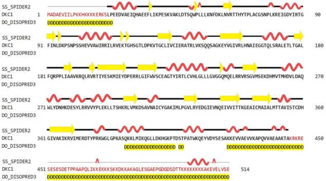
2.2. Prediction of the Two-Dimensional Structure of hDKC1
As shown in Figure 2, the predictions obtained using the HHPred server (Homology detection by Hidden Markov modes Prediction) revealed that the secondary structure of hDKC1 contains
26.85% α-helices, 10.51% β-strands, and 62.65% loops. Both N- and C-terminals showed a
disorganized structure. Regarding this feature, it was reported that the unique function of residues 1 to 21 is to act synergistically with the C-terminal nuclear localization sequence (NLS) to ensure dyskerin nucleolar localization. Likewise, the biological relevance of the C-terminal region (390–514 aa) is supported by the presence of a bipartite NLS and of several potentially modified residues [6]. These observations could explain the unstructured organization of this region, which is highlighted in yellow in Figure 2.
Figure 1. Graphical representation of the abundance of the 20 amino acids (aa) present in human dyskerin pseudouridine synthase (hDKC1). Lysine has the highest abundance, and phenylalanine has the lowest abundance.
2.2. Prediction of the Two-Dimensional Structure of hDKC1
As shown in Figure2, the predictions obtained using the HHPred server (Homology detection by
Hidden Markov modes Prediction) revealed that the secondary structure of hDKC1 contains 26.85%
α-helices, 10.51%β-strands, and 62.65% loops. Both N- and C-terminals showed a disorganized
structure. Regarding this feature, it was reported that the unique function of residues 1 to 21 is to act synergistically with the C-terminal nuclear localization sequence (NLS) to ensure dyskerin nucleolar localization. Likewise, the biological relevance of the C-terminal region (390–514 aa) is supported by
the presence of a bipartite NLS and of several potentially modified residues [6]. These observations
Int. J. Mol. Sci.2018,19, 3216 4 of 16
2.3. Sequence and Secondary Structure Analysis between hDKC1 and Saccharomyces Cerevisiae Dyskerin
As mentioned above, there is no crystal structure available for hDKC1, but it has a highly evolutionary conserved sequence. From all reported dyskerins in the Protein Data Bank (PDB), our studies showed that the highest value of sequence homology was present in the structures 3UAI
and 3U28 belonging to S. cerevisiae. In comparison, the 3UAI structure showed more structural
information than the 3U28 structure (data not shown). Taking advantage of this fact, we decided to
generate a three-dimensional (3D) structure model of hDKC1 by homology withS. cerevisiaedyskerin
(chain A from 3UAI). The initial step consisted of an analysis between the predicted secondary structure
of hDKC1 and the secondary structure obtained from 3UAI. As presented in Figure3, neither C- nor
N-terminal sequences are included in the crystal structure of 3UAI. This correlates with the results
observed in Figure2, where N- and C-terminal sequences had no secondary structure and they were
reported as cellular localization sequences. Based on these observations, we decided to model the sequence of hDKC1 comprising the residues from position 22 to 420, where a secondary structure was shown.
Int. J. Mol. Sci. 2018, 19, x 4 of 17
2.3. Sequence and Secondary Structure Analysis between hDKC1 and Saccharomyces Cerevisiae Dyskerin
As mentioned above, there is no crystal structure available for hDKC1, but it has a highly evolutionary conserved sequence. From all reported dyskerins in the Protein Data Bank (PDB), our studies showed that the highest value of sequence homology was present in the structures 3UAI and
3U28 belonging to S. cerevisiae. In comparison, the 3UAI structure showed more structural
information than the 3U28 structure (data not shown). Taking advantage of this fact, we decided to generate a three-dimensional (3D) structure model of hDKC1 by homology with S. cerevisiae dyskerin (chain A from 3UAI). The initial step consisted of an analysis between the predicted secondary structure of hDKC1 and the secondary structure obtained from 3UAI. As presented in Figure 3, neither C- nor N-terminal sequences are included in the crystal structure of 3UAI. This correlates with the results observed in Figure 2, where N- and C-terminal sequences had no secondary structure and they were reported as cellular localization sequences. Based on these observations, we decided to model the sequence of hDKC1 comprising the residues from position 22 to 420, where a secondary structure was shown.
Figure 2. Secondary structure prediction of hDKC1 using HHPred (SPIDER2 (Scoring Protein Interaction Decoys using Exposed Residues) for secondary structure prediction, and DISOPRED (Disorder Prediction Server) for unstructured or disordered region predictions). Yellow arrows represent beta sheets; alpha helixes are shown in red; disordered regions are highlighted in yellow.
Figure 3. Sequence and secondary structure of S. cerevisiae dyskerin obtained from the 3UAI Protein Data Bank (PDB) file. Yellow arrows represent beta sheets; alpha helixes are shown in red; turns are colored in green.
Figure 2. Secondary structure prediction of hDKC1 using HHPred (SPIDER2 (Scoring Protein Interaction Decoys using Exposed Residues) for secondary structure prediction, and DISOPRED (Disorder Prediction Server) for unstructured or disordered region predictions). Yellow arrows represent beta sheets; alpha helixes are shown in red; disordered regions are highlighted in yellow.
2.4. Predicted 3D Homology Model of hDKC1 by I-TASSER
Using I-TASSER (Iterative Threading Assembly Refinement), the 3D model structure of hDKC1 was carried out by two different strategies: the first one consisted of using the structure of 3UAI as template for modelling the hDKC1 sequence by homology. The second one was an ab initio model, where the software builds the 3D structure based on energy calculus. Both models are shown in
Figure4, visualized using MGLTools (Molecular Graphics Laboratory Tools).
I-TASSER evaluates the model using two parameters. The first one is the C-score, which is the confidence score to evaluate the quality of a predicted model. The C-score is typically in the range
of−5–2, where a C-score of higher value indicates a model with a high confidence and vice-versa.
Another important parameter to take into account is the TM-score (Template Modeling score), which is a proposed scale for measuring the structural similarity between two structures. A TM-score of
Int. J. Mol. Sci.2018,19, 3216 5 of 16
As shown in Table1, the C-score for both models is adequate, being the homology model the most
confident one. Although the TM-score and RMSD (Root-Mean-Square Deviation) values of both models are acceptable for a proper design, the homology one showed more robust results and was chosen for our analysis.
Int. J. Mol. Sci. 2018, 19, x 4 of 17
2.3. Sequence and Secondary Structure Analysis between hDKC1 and Saccharomyces Cerevisiae Dyskerin
As mentioned above, there is no crystal structure available for hDKC1, but it has a highly evolutionary conserved sequence. From all reported dyskerins in the Protein Data Bank (PDB), our studies showed that the highest value of sequence homology was present in the structures 3UAI and
3U28 belonging to S. cerevisiae. In comparison, the 3UAI structure showed more structural
information than the 3U28 structure (data not shown). Taking advantage of this fact, we decided to generate a three-dimensional (3D) structure model of hDKC1 by homology with S. cerevisiae dyskerin (chain A from 3UAI). The initial step consisted of an analysis between the predicted secondary structure of hDKC1 and the secondary structure obtained from 3UAI. As presented in Figure 3, neither C- nor N-terminal sequences are included in the crystal structure of 3UAI. This correlates with the results observed in Figure 2, where N- and C-terminal sequences had no secondary structure and they were reported as cellular localization sequences. Based on these observations, we decided to model the sequence of hDKC1 comprising the residues from position 22 to 420, where a secondary structure was shown.
Figure 2. Secondary structure prediction of hDKC1 using HHPred (SPIDER2 (Scoring Protein Interaction Decoys using Exposed Residues) for secondary structure prediction, and DISOPRED (Disorder Prediction Server) for unstructured or disordered region predictions). Yellow arrows represent beta sheets; alpha helixes are shown in red; disordered regions are highlighted in yellow.
Figure 3. Sequence and secondary structure of S. cerevisiae dyskerin obtained from the 3UAI Protein Data Bank (PDB) file. Yellow arrows represent beta sheets; alpha helixes are shown in red; turns are colored in green.
Figure 3.Sequence and secondary structure ofS. cerevisiaedyskerin obtained from the 3UAI Protein Data Bank (PDB) file. Yellow arrows represent beta sheets; alpha helixes are shown in red; turns are colored in green.
Int. J. Mol. Sci. 2018, 19, x 5 of 17
2.4. Predicted 3D Homology Model of hDKC1 by I-TASSER
Using I-TASSER (Iterative Threading Assembly Refinement), the 3D model structure of hDKC1
was carried out by two different strategies: the first one consisted of using the structure of 3UAI as
template for modelling the hDKC1 sequence by homology. The second one was an ab initio model,
where the software builds the 3D structure based on energy calculus. Both models are shown in
Figure 4, visualized using MGLTools (Molecular Graphics Laboratory Tools).
I-TASSER evaluates the model using two parameters. The first one is the C-score, which is the
confidence score to evaluate the quality of a predicted model. The C-score is typically in the range of
−
5–2, where a C-score of higher value indicates a model with a high confidence and vice-versa.
Another important parameter to take into account is the TM-score (Template Modeling score), which
is a proposed scale for measuring the structural similarity between two structures. A TM-score of >
0.5 indicates a model of correct topology and a TM-score < 0.17 indicates a random similarity [11]. As
shown in Table 1, the C-score for both models is adequate, being the homology model the most
confident one. Although the TM-score and RMSD (Root-Mean-Square Deviation) values of both
models are acceptable for a proper design, the homology one showed more robust results and was
chosen for our analysis.
Figure 4. The hDKC1 models obtained by I-TASSER (Iterative Threading Assembly Refinement). (A)
hDKC1 homology model; (B) hDKC1 ab initio model.
Table 1. Quality evaluation scores of the predicted 3D structures by I-TASSER.
hDKC1 Model
C-Score TM Score RMSD ( )
Homology Model
−
0.09
0.70 ± 0.12
7.1 ± 4.2
Ab initio
model
−
2.32
0.44 ± 0.14
13.0 ± 4.2
2.5. 3D Structure Validation
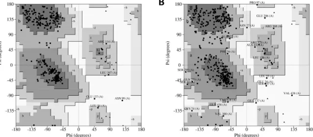
Subsequently, the quality of models was validated using PROCHECK (Program to check the
stereochemical quality of protein structures), a program that relies on Ramachandran plots for
structure verification. According to this software, a good quality model could be expected to have
over 90% of its residues in the most favored region [12]. As shown in Figure 5A, results from
PROCHECK determined that the homology model has 92.1% of its residues in the most favored
regions, 6.3% in the additional allowed regions, 0.9% in the generously allowed regions and 0.6% in
the disallowed regions. In contrast, results from the ab initio model showed 79.1% of residues in the
Figure 4. The hDKC1 models obtained by I-TASSER (Iterative Threading Assembly Refinement). (A) hDKC1 homology model; (B) hDKC1 ab initio model.
Table 1.Quality evaluation scores of the predicted 3D structures by I-TASSER.
hDKC1 Model C-Score TM Score RMSD (Å)
Homology Model −0.09 0.70±0.12 7.1±4.2
Int. J. Mol. Sci.2018,19, 3216 6 of 16
2.5. 3D Structure Validation
Subsequently, the quality of models was validated using PROCHECK (Program to check the stereochemical quality of protein structures), a program that relies on Ramachandran plots for structure verification. According to this software, a good quality model could be expected to have over 90%
of its residues in the most favored region [12]. As shown in Figure5A, results from PROCHECK
determined that the homology model has 92.1% of its residues in the most favored regions, 6.3% in the additional allowed regions, 0.9% in the generously allowed regions and 0.6% in the disallowed regions. In contrast, results from the ab initio model showed 79.1% of residues in the most favored regions, 18.8% in the additional allowed regions, 3.7% in the generously allowed regions and 1.6% in
the disallowed regions (Figure5B). Therefore, regarding the percentage distribution of the residues,
the homology model has a higher quality than the ab initio model.
Additionally, the G-factor for each model was determined using the same software. This value provides a measure of how unusual a determination of a given stereochemical property is using this
program. A G-factor of less than−0.5 is unusual and less than−1.0 is highly unusual [12]. The G-factor
obtained for the homology model was−0.27 for dihedral angles,−0.02 for main chain covalent forces
and−0.16 overall. Conversely, the values determined for the ab initio model were−0.92,−0.09 and
−0.57, respectively. This evidence strongly suggests that the homology model has more accurate
features than the ab initio model. Consequently, we continued this work using the hDKC1 homology model, since it was demonstrated to be a more robust model in all the parameters analyzed.
Int. J. Mol. Sci. 2018, 19, x 6 of 17
most favored regions, 18.8% in the additional allowed regions, 3.7% in the generously allowed regions and 1.6% in the disallowed regions (Figure 5B). Therefore, regarding the percentage distribution of the residues, the homology model has a higher quality than the ab initio model.
Additionally, the G-factor for each model was determined using the same software. This value provides a measure of how unusual a determination of a given stereochemical property is using this program. A G-factor of less than −0.5 is unusual and less than −1.0 is highly unusual [12]. The G-factor obtained for the homology model was −0.27 for dihedral angles, −0.02 for main chain covalent forces and −0.16 overall. Conversely, the values determined for the ab initio model were −0.92, −0.09 and
−0.57, respectively. This evidence strongly suggests that the homology model has more accurate
features than the ab initio model. Consequently, we continued this work using the hDKC1 homology model, since it was demonstrated to be a more robust model in all the parameters analyzed.
Figure 5. PROCHECK (Program to check the stereochemical quality of protein structures) results of modelled hDKC1 using the generated models from I-TASSER. (A) Ramachandran plot for hDKC1 homology model; (B) Ramachandran plot for hDKC1 ab initio model. Residues in most favored regions (A, B, L), residues in additional allowed regions (a, b, l, p) and residues in generously allowed regions (~a, ~b ~l, ~p).
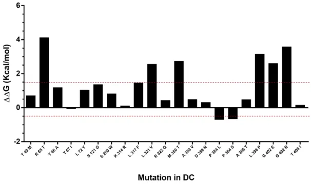
2.6. Study of Mutation Stability
The effect of mutations described in DC were predicted in the hDKC1 model. A comparison between the stability of all the reported mutations vs. the wild type protein was made using the Foldx suite. The predicted ΔΔG values of the mutations fell in a range of −0.7 to 4.1 kcal/mol (Figure 6). This analysis led us to the identification of eight destabilizing mutations, since the mutations with ΔΔG values under −0.5 or over 1.5 kcal/mol could significantly reduce protein stability [13].
Figure 5.PROCHECK (Program to check the stereochemical quality of protein structures) results of modelled hDKC1 using the generated models from I-TASSER. (A) Ramachandran plot for hDKC1 homology model; (B) Ramachandran plot for hDKC1 ab initio model. Residues in most favored regions (A, B, L), residues in additional allowed regions (a, b, l, p) and residues in generously allowed regions (~a, ~b ~l, ~p).
2.6. Study of Mutation Stability
The effect of mutations described in DC were predicted in the hDKC1 model. A comparison between the stability of all the reported mutations vs. the wild type protein was made using the Foldx
suite. The predicted∆∆G values of the mutations fell in a range of−0.7 to 4.1 kcal/mol (Figure6).
This analysis led us to the identification of eight destabilizing mutations, since the mutations with
Int. J. Mol. Sci.2018,19, 3216 7 of 16
Int. J. Mol. Sci. 2018, 19, x 7 of 17
Figure 6. Analysis of mutation stability in the hDKC1 model by Foldx software. Mutations comprised between red dashes lines (−0.5 Kcal/mol to 1.5 Kcal/mol) correlates with the mutations that are not destabilizing. DC = dyskeratosis congenita.
2.7. Evaluation and Recognition of Hydrophobic Pockets on hDKC1 Model
By using the FPocket software, we searched and analyzed all the hydrophobic pockets present in our model, with the aim of determining the best ones in order to set the parameters for DBVS. As a result, we found 21 pockets, ordered by a relevance parameter determined by the following formula: (pocket score/the highest score) × 100. The pockets, their relevance parameter, volume and the involved residues are presented in Table 2.
2.8. Study and Determination of the Hydrophobic Pocket Containing the Mutation K314
As we mentioned in the introduction, the aim of this work is to design a novel strategy for the rational development of new inhibitors of the assembly of the holoenzyme telomerase, based on the interruption of the interaction of hDKC1 with hTR. To carry out this work, we decided to emulate the phenotype showed and reported in DC, where a mutation on the residue K314 in the hDKC1 sequence leads to a loss in telomerase function caused by the inability of hDKC1 to join hTR. As
shown in Figure 6, the K314R site mutation exhibits a ΔΔG value of 0.104 kcal/mol, implying the
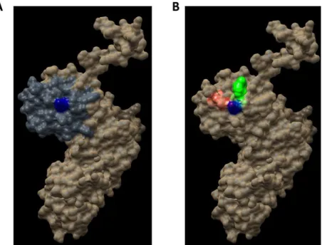
change does not reduce protein stability and thus it is an accurate site to direct the DBVS (Docking Based Virtual Screening). Consequently, from all the pockets found, we decided to work with those which contain the residue K314. Two pockets containing this residue were found: Pocket 2 and Pocket 13. Both are included in the PUA domain and are worthy of consideration due to their proper values of relevance, according to FPocket analysis. Figure 7 shows the DKC1 model, the PUA domain and the two pockets containing residue K314.
Figure 6.Analysis of mutation stability in the hDKC1 model by Foldx software. Mutations comprised between red dashes lines (−0.5 Kcal/mol to 1.5 Kcal/mol) correlates with the mutations that are not destabilizing. DC = dyskeratosis congenita.
2.7. Evaluation and Recognition of Hydrophobic Pockets on hDKC1 Model
By using the FPocket software, we searched and analyzed all the hydrophobic pockets present in our model, with the aim of determining the best ones in order to set the parameters for DBVS. As a result, we found 21 pockets, ordered by a relevance parameter determined by the following
formula: (pocket score/the highest score)×100. The pockets, their relevance parameter, volume and
the involved residues are presented in Table2.
2.8. Study and Determination of the Hydrophobic Pocket Containing the Mutation K314
As we mentioned in the introduction, the aim of this work is to design a novel strategy for the rational development of new inhibitors of the assembly of the holoenzyme telomerase, based on the interruption of the interaction of hDKC1 with hTR. To carry out this work, we decided to emulate the phenotype showed and reported in DC, where a mutation on the residue K314 in the hDKC1 sequence leads to a loss in telomerase function caused by the inability of hDKC1 to join hTR. As shown
in Figure6, the K314R site mutation exhibits a∆∆G value of 0.104 kcal/mol, implying the change does
not reduce protein stability and thus it is an accurate site to direct the DBVS (Docking Based Virtual Screening). Consequently, from all the pockets found, we decided to work with those which contain the residue K314. Two pockets containing this residue were found: Pocket 2 and Pocket 13. Both are included in the PUA domain and are worthy of consideration due to their proper values of relevance,
according to FPocket analysis. Figure7shows the DKC1 model, the PUA domain and the two pockets
containing residue K314.
2.9. Docking Based Virtual Screening on the hDKC1 Model by AutoDock Vina
Int. J. Mol. Sci.2018,19, 3216 8 of 16
Int. J. Mol. Sci. 2018, 19, x 8 of 17
Figure 7. Regions in the hDKC1 three-dimensional (3D) structure model. hDKC1 is colored in beige.
Residue K314 is indicated in blue (A) The PUA domain is colored in dark gray (B) Pocket 2 and Pocket 13 are highlighted in red and green, respectively.
Table 2. Analysis of hydrophobic pockets present in the hDKC1 model by FPocket software.
Pocket Relevance (%)
Volume
(Å3) Residue Number
1 100 615 96, 101, 102, 103, 122, 123, 124, 125, 126, 128, 129, 130, 131, 132, 153, 187, 188, 246, 248 2 94 203 299, 300, 301, 314, 315, 317, 320, 322, 323, 350, 354, 355, 358, 360, 361, 362, 363 3 79 284 81, 85, 88, 89, 138, 289, 290, 341, 342, 370, 371, 372, 375
4 69 926 259, 260, 261, 265, 272, 280, 281, 284, 285, 287
5 69 289 141, 142, 144, 145, 293, 296, 332, 333, 334, 344, 345, 346, 367, 368, 370
6 64 810 74, 75, 76, 80, 81, 82, 85, 88, 341, 372, 375
7 60 208 54, 91, 93, 254, 258, 259, 261, 284, 285, 287, 288, 289, 291, 292, 8 60 444 70, 71, 72, 303, 304, 307, 376, 377, 378, 379, 380, 9 58 100 157, 169, 170, 173, 204, 206, 207, 214, 215, 216, 233 10 57 367 98, 127, 129, 154, 155, 156, 215, 245, 246, 247, 249, 256 11 57 994 183, 184, 185, 186, 192, 227, 228, 231, 241, 242, 243
12 54 525 54, 55, 57, 58, 294, 295, 296, 297, 298, 324
13 47 580 301, 302, 304, 305, 308, 309, 313, 314, 315, 316, 318, 319, 378
14 47 158 53, 54, 55, 56, 77, 78, 79, 80, 290, 339, 340
15 44 92 83, 86, 87, 273, 278, 279, 282, 283
16 42 146 176, 180, 181, 182, 197, 199, 218, 220, 224, 225, 226, 228, 229, 232 17 40 295 103, 119, 120, 121, 122, 123, 124, 143, 147, 151, 222, 223 18 37 268 98, 125, 126, 127, 128, 187, 227, 243, 244, 245, 246
19 33 274 156, 206, 207, 208, 211, 213, 214, 215
20 32 425 83, 84, 87, 113, 114, 115, 137, 270, 273, 274, 282
21 28 374 138, 141, 310, 311, 369, 371, 372, 373, 374
2.9. Docking Based Virtual Screening on the hDKC1 Model by AutoDock Vina
Once the 3D structure model of hDKC1 was validated and the target pocket was defined, we set
the parameters to carry out the DBVS using AutoDock Vina Software. We established the alpha
carbon on residue K314 as the docking center. We defined a box of 25 Å to cover the two pockets
described above. Finally, we decided to use the library “Advanced Collection from Enamine”, since
it contains more than 400,000 compounds suitable for the development of new drugs for clinical use.
As result of DBVS, a ranking of candidate compounds was obtained. These compounds are
ordered by docking energy, being better candidates those with more negative values. The first 10
candidates obtained from the DBVS are listed in Table 3.
Figure 7.Regions in the hDKC1 three-dimensional (3D) structure model. hDKC1 is colored in beige. Residue K314 is indicated in blue (A) The PUA domain is colored in dark gray (B) Pocket 2 and Pocket 13 are highlighted in red and green, respectively.
Table 2.Analysis of hydrophobic pockets present in the hDKC1 model by FPocket software.
Pocket Relevance (%) Volume (Å3) Residue Number
1 100 615 96, 101, 102, 103, 122, 123, 124, 125, 126, 128, 129, 130, 131, 132, 153, 187, 188, 246, 248 2 94 203 299, 300, 301, 314, 315, 317, 320, 322, 323, 350, 354, 355, 358, 360, 361, 362, 363
3 79 284 81, 85, 88, 89, 138, 289, 290, 341, 342, 370, 371, 372, 375
4 69 926 259, 260, 261, 265, 272, 280, 281, 284, 285, 287
5 69 289 141, 142, 144, 145, 293, 296, 332, 333, 334, 344, 345, 346, 367, 368, 370
6 64 810 74, 75, 76, 80, 81, 82, 85, 88, 341, 372, 375
7 60 208 54, 91, 93, 254, 258, 259, 261, 284, 285, 287, 288, 289, 291, 292,
8 60 444 70, 71, 72, 303, 304, 307, 376, 377, 378, 379, 380,
9 58 100 157, 169, 170, 173, 204, 206, 207, 214, 215, 216, 233
10 57 367 98, 127, 129, 154, 155, 156, 215, 245, 246, 247, 249, 256
11 57 994 183, 184, 185, 186, 192, 227, 228, 231, 241, 242, 243
12 54 525 54, 55, 57, 58, 294, 295, 296, 297, 298, 324
13 47 580 301, 302, 304, 305, 308, 309, 313, 314, 315, 316, 318, 319, 378
14 47 158 53, 54, 55, 56, 77, 78, 79, 80, 290, 339, 340
15 44 92 83, 86, 87, 273, 278, 279, 282, 283
16 42 146 176, 180, 181, 182, 197, 199, 218, 220, 224, 225, 226, 228, 229, 232
17 40 295 103, 119, 120, 121, 122, 123, 124, 143, 147, 151, 222, 223
18 37 268 98, 125, 126, 127, 128, 187, 227, 243, 244, 245, 246
19 33 274 156, 206, 207, 208, 211, 213, 214, 215
20 32 425 83, 84, 87, 113, 114, 115, 137, 270, 273, 274, 282
21 28 374 138, 141, 310, 311, 369, 371, 372, 373, 374
As result of DBVS, a ranking of candidate compounds was obtained. These compounds are ordered by docking energy, being better candidates those with more negative values. The first
10 candidates obtained from the DBVS are listed in Table3.
2.10. In Vitro Screening of the Candidate Compounds by Telomerase Activity Assay
With the aim of determining if the candidate compounds show the desired inhibitory effect, we performed an assay for measuring telomerase activity. This assay was carried out in a tumor cell
line positive for telomerase activity. Results presented in Figure8show that at least three compounds
Int. J. Mol. Sci.2018,19, 3216 9 of 16
Table 3.Ranking of compounds obtained from DBVS by AutoDock Vina.
Name Compound Docking Energy
Table 3. Ranking of compounds obtained from DBVS by AutoDock Vina.
Name Compound Docking Energy
(kcal/mol) SD Name Compound
Docking Energy
2.10. In Vitro Screening of the Candidate Compounds by Telomerase Activity Assay
With the aim of determining if the candidate compounds show the desired inhibitory effect, we performed an assay for measuring telomerase activity. This assay was carried out in a tumor cell line positive for telomerase activity. Results presented in Figure 8 show that at least three compounds produced a significant decrease in telomerase activity after treatment, reaching an inhibitory effect of almost 40%. This evidence supports the rational design proposed in this work and sets the basis to continue investigating these compounds as potential anti-tumor drugs.
3. Discussion
Nowadays, drug design is increasingly reliant on computer modeling techniques. This type of strategy is often referred to as computer-aided drug design. More specifically, drug design that relies on the knowledge of the three-dimensional structure of the biomolecular target is known as structure-based drug design. In order to generate this type of drug design, an increasingly important number of computational methods for improving the affinity, selectivity and stability of these protein-based therapeutics have also been developed [14–16].
Regarding anti-tumor therapies, although effective cytotoxic compounds have been identified, treatments directed to a specific target still have ample room for improvement. Taking into account the experience of our group in the study of telomerase and our expertise on drug design using computational and molecular biology tools [17], we decided to carry out a DBVS on hDKC1, with the aim of generating new compounds with inhibitory effect on telomerase activity for cancer treatment.
−7.20 0.21 E6
Int. J. Mol. Sci. 2018, 19, x 9 of 17
Table 3. Ranking of compounds obtained from DBVS by AutoDock Vina.
Name Compound Docking Energy
(kcal/mol) SD Name Compound
Docking Energy
2.10. In Vitro Screening of the Candidate Compounds by Telomerase Activity Assay
With the aim of determining if the candidate compounds show the desired inhibitory effect, we performed an assay for measuring telomerase activity. This assay was carried out in a tumor cell line positive for telomerase activity. Results presented in Figure 8 show that at least three compounds produced a significant decrease in telomerase activity after treatment, reaching an inhibitory effect of almost 40%. This evidence supports the rational design proposed in this work and sets the basis to continue investigating these compounds as potential anti-tumor drugs.
3. Discussion
Nowadays, drug design is increasingly reliant on computer modeling techniques. This type of strategy is often referred to as computer-aided drug design. More specifically, drug design that relies on the knowledge of the three-dimensional structure of the biomolecular target is known as structure-based drug design. In order to generate this type of drug design, an increasingly important number of computational methods for improving the affinity, selectivity and stability of these protein-based therapeutics have also been developed [14–16].
Regarding anti-tumor therapies, although effective cytotoxic compounds have been identified, treatments directed to a specific target still have ample room for improvement. Taking into account the experience of our group in the study of telomerase and our expertise on drug design using computational and molecular biology tools [17], we decided to carry out a DBVS on hDKC1, with the aim of generating new compounds with inhibitory effect on telomerase activity for cancer treatment. −6.73 0.21
E2
Int. J. Mol. Sci. 2018, 19, x 9 of 17
Table 3. Ranking of compounds obtained from DBVS by AutoDock Vina.
Name Compound Docking Energy
(kcal/mol) SD Name Compound
Docking Energy
2.10. In Vitro Screening of the Candidate Compounds by Telomerase Activity Assay
With the aim of determining if the candidate compounds show the desired inhibitory effect, we performed an assay for measuring telomerase activity. This assay was carried out in a tumor cell line positive for telomerase activity. Results presented in Figure 8 show that at least three compounds produced a significant decrease in telomerase activity after treatment, reaching an inhibitory effect of almost 40%. This evidence supports the rational design proposed in this work and sets the basis to continue investigating these compounds as potential anti-tumor drugs.
3. Discussion
Nowadays, drug design is increasingly reliant on computer modeling techniques. This type of strategy is often referred to as computer-aided drug design. More specifically, drug design that relies on the knowledge of the three-dimensional structure of the biomolecular target is known as structure-based drug design. In order to generate this type of drug design, an increasingly important number of computational methods for improving the affinity, selectivity and stability of these protein-based therapeutics have also been developed [14–16].
Regarding anti-tumor therapies, although effective cytotoxic compounds have been identified, treatments directed to a specific target still have ample room for improvement. Taking into account the experience of our group in the study of telomerase and our expertise on drug design using computational and molecular biology tools [17], we decided to carry out a DBVS on hDKC1, with the aim of generating new compounds with inhibitory effect on telomerase activity for cancer treatment.
−7.11 0.08 E7
Int. J. Mol. Sci. 2018, 19, x 9 of 17
Table 3. Ranking of compounds obtained from DBVS by AutoDock Vina.
Name Compound Docking Energy
(kcal/mol) SD Name Compound
Docking Energy
2.10. In Vitro Screening of the Candidate Compounds by Telomerase Activity Assay
With the aim of determining if the candidate compounds show the desired inhibitory effect, we performed an assay for measuring telomerase activity. This assay was carried out in a tumor cell line positive for telomerase activity. Results presented in Figure 8 show that at least three compounds produced a significant decrease in telomerase activity after treatment, reaching an inhibitory effect of almost 40%. This evidence supports the rational design proposed in this work and sets the basis to continue investigating these compounds as potential anti-tumor drugs.
3. Discussion
Nowadays, drug design is increasingly reliant on computer modeling techniques. This type of strategy is often referred to as computer-aided drug design. More specifically, drug design that relies on the knowledge of the three-dimensional structure of the biomolecular target is known as structure-based drug design. In order to generate this type of drug design, an increasingly important number of computational methods for improving the affinity, selectivity and stability of these protein-based therapeutics have also been developed [14–16].
Regarding anti-tumor therapies, although effective cytotoxic compounds have been identified, treatments directed to a specific target still have ample room for improvement. Taking into account the experience of our group in the study of telomerase and our expertise on drug design using computational and molecular biology tools [17], we decided to carry out a DBVS on hDKC1, with the aim of generating new compounds with inhibitory effect on telomerase activity for cancer treatment. −6.69 0.11
E3
Int. J. Mol. Sci. 2018, 19, x 9 of 17
Table 3. Ranking of compounds obtained from DBVS by AutoDock Vina.
Name Compound Docking Energy
(kcal/mol) SD Name Compound
Docking Energy
2.10. In Vitro Screening of the Candidate Compounds by Telomerase Activity Assay
With the aim of determining if the candidate compounds show the desired inhibitory effect, we performed an assay for measuring telomerase activity. This assay was carried out in a tumor cell line positive for telomerase activity. Results presented in Figure 8 show that at least three compounds produced a significant decrease in telomerase activity after treatment, reaching an inhibitory effect of almost 40%. This evidence supports the rational design proposed in this work and sets the basis to continue investigating these compounds as potential anti-tumor drugs.
3. Discussion
Nowadays, drug design is increasingly reliant on computer modeling techniques. This type of strategy is often referred to as computer-aided drug design. More specifically, drug design that relies on the knowledge of the three-dimensional structure of the biomolecular target is known as structure-based drug design. In order to generate this type of drug design, an increasingly important number of computational methods for improving the affinity, selectivity and stability of these protein-based therapeutics have also been developed [14–16].
Regarding anti-tumor therapies, although effective cytotoxic compounds have been identified, treatments directed to a specific target still have ample room for improvement. Taking into account the experience of our group in the study of telomerase and our expertise on drug design using computational and molecular biology tools [17], we decided to carry out a DBVS on hDKC1, with the aim of generating new compounds with inhibitory effect on telomerase activity for cancer treatment.
7.04 0.06 E8
Int. J. Mol. Sci. 2018, 19, x 9 of 17
Table 3. Ranking of compounds obtained from DBVS by AutoDock Vina.
Name Compound Docking Energy
(kcal/mol) SD Name Compound
Docking Energy
2.10. In Vitro Screening of the Candidate Compounds by Telomerase Activity Assay
With the aim of determining if the candidate compounds show the desired inhibitory effect, we performed an assay for measuring telomerase activity. This assay was carried out in a tumor cell line positive for telomerase activity. Results presented in Figure 8 show that at least three compounds produced a significant decrease in telomerase activity after treatment, reaching an inhibitory effect of almost 40%. This evidence supports the rational design proposed in this work and sets the basis to continue investigating these compounds as potential anti-tumor drugs.
3. Discussion
Nowadays, drug design is increasingly reliant on computer modeling techniques. This type of strategy is often referred to as computer-aided drug design. More specifically, drug design that relies on the knowledge of the three-dimensional structure of the biomolecular target is known as structure-based drug design. In order to generate this type of drug design, an increasingly important number of computational methods for improving the affinity, selectivity and stability of these protein-based therapeutics have also been developed [14–16].
Regarding anti-tumor therapies, although effective cytotoxic compounds have been identified, treatments directed to a specific target still have ample room for improvement. Taking into account the experience of our group in the study of telomerase and our expertise on drug design using computational and molecular biology tools [17], we decided to carry out a DBVS on hDKC1, with the aim of generating new compounds with inhibitory effect on telomerase activity for cancer treatment. −6.59 0.25
E4
Int. J. Mol. Sci. 2018, 19, x 9 of 17
Table 3. Ranking of compounds obtained from DBVS by AutoDock Vina.
Name Compound Docking Energy
(kcal/mol) SD Name Compound
Docking Energy
2.10. In Vitro Screening of the Candidate Compounds by Telomerase Activity Assay
With the aim of determining if the candidate compounds show the desired inhibitory effect, we performed an assay for measuring telomerase activity. This assay was carried out in a tumor cell line positive for telomerase activity. Results presented in Figure 8 show that at least three compounds produced a significant decrease in telomerase activity after treatment, reaching an inhibitory effect of almost 40%. This evidence supports the rational design proposed in this work and sets the basis to continue investigating these compounds as potential anti-tumor drugs.
3. Discussion
Nowadays, drug design is increasingly reliant on computer modeling techniques. This type of strategy is often referred to as computer-aided drug design. More specifically, drug design that relies on the knowledge of the three-dimensional structure of the biomolecular target is known as structure-based drug design. In order to generate this type of drug design, an increasingly important number of computational methods for improving the affinity, selectivity and stability of these protein-based therapeutics have also been developed [14–16].
Regarding anti-tumor therapies, although effective cytotoxic compounds have been identified, treatments directed to a specific target still have ample room for improvement. Taking into account the experience of our group in the study of telomerase and our expertise on drug design using computational and molecular biology tools [17], we decided to carry out a DBVS on hDKC1, with the aim of generating new compounds with inhibitory effect on telomerase activity for cancer treatment.
−6.93 0.13 E9
Int. J. Mol. Sci. 2018, 19, x 9 of 17
Table 3. Ranking of compounds obtained from DBVS by AutoDock Vina.
Name Compound Docking Energy
(kcal/mol) SD Name Compound
Docking Energy
2.10. In Vitro Screening of the Candidate Compounds by Telomerase Activity Assay
With the aim of determining if the candidate compounds show the desired inhibitory effect, we performed an assay for measuring telomerase activity. This assay was carried out in a tumor cell line positive for telomerase activity. Results presented in Figure 8 show that at least three compounds produced a significant decrease in telomerase activity after treatment, reaching an inhibitory effect of almost 40%. This evidence supports the rational design proposed in this work and sets the basis to continue investigating these compounds as potential anti-tumor drugs.
3. Discussion
Nowadays, drug design is increasingly reliant on computer modeling techniques. This type of strategy is often referred to as computer-aided drug design. More specifically, drug design that relies on the knowledge of the three-dimensional structure of the biomolecular target is known as structure-based drug design. In order to generate this type of drug design, an increasingly important number of computational methods for improving the affinity, selectivity and stability of these protein-based therapeutics have also been developed [14–16].
Regarding anti-tumor therapies, although effective cytotoxic compounds have been identified, treatments directed to a specific target still have ample room for improvement. Taking into account the experience of our group in the study of telomerase and our expertise on drug design using computational and molecular biology tools [17], we decided to carry out a DBVS on hDKC1, with the aim of generating new compounds with inhibitory effect on telomerase activity for cancer treatment. −6.54 0.09
E5
Int. J. Mol. Sci. 2018, 19, x 9 of 17
Table 3. Ranking of compounds obtained from DBVS by AutoDock Vina.
Name Compound Docking Energy
(kcal/mol) SD Name Compound
Docking Energy
2.10. In Vitro Screening of the Candidate Compounds by Telomerase Activity Assay
With the aim of determining if the candidate compounds show the desired inhibitory effect, we performed an assay for measuring telomerase activity. This assay was carried out in a tumor cell line positive for telomerase activity. Results presented in Figure 8 show that at least three compounds produced a significant decrease in telomerase activity after treatment, reaching an inhibitory effect of almost 40%. This evidence supports the rational design proposed in this work and sets the basis to continue investigating these compounds as potential anti-tumor drugs.
3. Discussion
Nowadays, drug design is increasingly reliant on computer modeling techniques. This type of strategy is often referred to as computer-aided drug design. More specifically, drug design that relies on the knowledge of the three-dimensional structure of the biomolecular target is known as structure-based drug design. In order to generate this type of drug design, an increasingly important number of computational methods for improving the affinity, selectivity and stability of these protein-based therapeutics have also been developed [14–16].
Regarding anti-tumor therapies, although effective cytotoxic compounds have been identified, treatments directed to a specific target still have ample room for improvement. Taking into account the experience of our group in the study of telomerase and our expertise on drug design using computational and molecular biology tools [17], we decided to carry out a DBVS on hDKC1, with the aim of generating new compounds with inhibitory effect on telomerase activity for cancer treatment.
−6.81 0.17 E10
Int. J. Mol. Sci. 2018, 19, x 9 of 17
Table 3. Ranking of compounds obtained from DBVS by AutoDock Vina.
Name Compound Docking Energy
(kcal/mol) SD Name Compound
Docking Energy
2.10. In Vitro Screening of the Candidate Compounds by Telomerase Activity Assay
With the aim of determining if the candidate compounds show the desired inhibitory effect, we performed an assay for measuring telomerase activity. This assay was carried out in a tumor cell line positive for telomerase activity. Results presented in Figure 8 show that at least three compounds produced a significant decrease in telomerase activity after treatment, reaching an inhibitory effect of almost 40%. This evidence supports the rational design proposed in this work and sets the basis to continue investigating these compounds as potential anti-tumor drugs.
3. Discussion
Nowadays, drug design is increasingly reliant on computer modeling techniques. This type of strategy is often referred to as computer-aided drug design. More specifically, drug design that relies on the knowledge of the three-dimensional structure of the biomolecular target is known as structure-based drug design. In order to generate this type of drug design, an increasingly important number of computational methods for improving the affinity, selectivity and stability of these protein-based therapeutics have also been developed [14–16].
Regarding anti-tumor therapies, although effective cytotoxic compounds have been identified, treatments directed to a specific target still have ample room for improvement. Taking into account the experience of our group in the study of telomerase and our expertise on drug design using computational and molecular biology tools [17], we decided to carry out a DBVS on hDKC1, with the aim of generating new compounds with inhibitory effect on telomerase activity for cancer treatment. −6.52 0.45
SD = standard deviation.
Int. J. Mol. Sci. 2018, 19, x 12 of 17
Figure 8. Determination of telomerase activity by qPCR. Quantification of telomerase activity was carried out by real time PCR with specific primers, using a protein extract from treated and untreated telomerase positive cells as a template for 48 h. Dashes line indicates the 100% of the control. Values represent media ± SEM; n = 6, * p < 0.5 ** p < 0.01 vs. control (ANOVA followed by Dunnett).
Figure 9. Spatial arrangement of the resulting compounds in the hDKC1 3D structure model. hDKC1 is colored in beige Residue K314 is indicated in blue, the PUA domain is colored in dark gray and Pocket 13 is highlighted in red. (A) E1; (B) E5; (C) E8.
4. Materials and Methods
4.1. Sequence Obtaining and Physicochemical Parameters Analysis of hDKC1
The aa sequence was obtained from the Universe Protein Resource (UniProt) (available online: http://www.uniprot.org/). The physicochemical parameters of the protein were generated from the ProtParam tool (available online: http://web.expasy.org/protparam/) using the ExPAsy server.
Int. J. Mol. Sci.2018,19, 3216 10 of 16
3. Discussion
Nowadays, drug design is increasingly reliant on computer modeling techniques. This type of strategy is often referred to as computer-aided drug design. More specifically, drug design that relies on the knowledge of the three-dimensional structure of the biomolecular target is known as structure-based drug design. In order to generate this type of drug design, an increasingly important number of computational methods for improving the affinity, selectivity and stability
of these protein-based therapeutics have also been developed [14–16].
Regarding anti-tumor therapies, although effective cytotoxic compounds have been identified, treatments directed to a specific target still have ample room for improvement. Taking into account the experience of our group in the study of telomerase and our expertise on drug design using
computational and molecular biology tools [17], we decided to carry out a DBVS on hDKC1, with the
aim of generating new compounds with inhibitory effect on telomerase activity for cancer treatment. The basis for performing a DBVS is the availability of the crystallized structure of the target protein. There are several studies in which the crystallized structure of dyskerin is used to explain, in a very interesting way, the interactions and processes in which it is involved. However, the authors
utilized the structures coming from yeast [18,19] or archeas [20,21]. Although these structures are
useful for this kind of study, the aim of our work is directed towards the development of new drugs, therefore each step must be as accurate as possible in order to avoid the appearance of hurdles along the way. Due to the fact that hDKC1 has not yet been crystallized, the need to obtain a model as close as possible to its actual structure became one of the most relevant steps of this work. To carry out this goal, among the different bioinformatic tools in existence, we selected I-TASSER which has demonstrated significant advantages in protein structure prediction, combining various techniques from threading,
ab initio modeling and atomic-level structure refinement approaches [22]. Furthermore, the Critical
Assessment of protein Structure Prediction (CASP), a community-wide, worldwide experiment for
protein structure prediction taking place every two years since 1994 [23], has ranked I-TASSER as the
best method for automated protein structure prediction in many CASP experiments [24]. For example,
in the field of cancer research, it has been used to model the RAB38 protein (Ras related protein
38), relevant in melanoma [25] and RASSF2 (Ras Association Domain Family Member 2), studied in
different tumor types [26]. Similarly, in the study of malaria, a model of M17LAP (M17- Family Leucine
Aminopeptidase) was obtained and then used as a target protein in a DBVS assay [27]. In the same way,
the structure by homology of Sortase A, a protein involved in listeriosis disease, was achieved [28].
As mentioned above, we decided to use the hDKC1 sequence corresponding to aa 21 to 420, since
the first 20 aa and the last 96 aa were reported as signaling peptides with few relevant mutations [6] and,
as observed in the secondary structure analysis, they correlated with unstructured areas (Figures2and3).
From this sequence, and using the crystallized structure of theS. cerevisaedyskerin (3UAI) as a template,
we performed both ab initio and homology modeling of hDKC1 using I-TASSER. It is important to highlight that yeast dyskerin is crystallized together with SHQ1 (H/ACA ribonucleoprotein assembly factor), NOP10 (Nucleolar protein 10) and GAR1 (H/ACA ribonucleoprotein complex subunit 1), which
are proteins of the telomerase complex [18]. However, far from being a problem, this provides more
accurate information on the real conformation of the protein in a biological context. Both processes converged in two models that were later analyzed by PROCHECK software, in order to check the stereochemical quality of the protein structure and analyze its overall and residue-by-residue geometry. The obtained results indicated that homology modeling presented appropriate quality parameters
(Figure5) [12], with values similar to the ones reported in other very interesting works [29,30]. Given this,
studies were continued with the homology model of hDKC1.
Once the hDKC1 structure was obtained and validated, we set the parameters regarding the DBVS assay. The choice of the pocket and the docking center were based on several points: concerning the pocket site, the PUA domain has been recurrently reported as the domain that contains most of the mutations related to DC. Furthermore, the interaction with hTR occurs through this domain, so it
Int. J. Mol. Sci.2018,19, 3216 11 of 16
K314 was determined as the center of the docking. The choice of this residue as the center is due to the fact that it is reported as one of the most relevant mutations associated with the most severe
manifestation of DC and is related with the impediment to stabilize the hTR in the complex [9].
Moreover, the stability of the point mutations reported for DC was evaluated using the analysis carried
out using FoldX [6]. The results obtained showed∆∆G values lower than 0.5 for K314 (Figure6),
which allowed us to assume that the mutation reported for this aa generates a binding mutant suitable
for the DBVS assay [13].
It is pertinent to emphasize that the reasoning behind the choice of this site is the search for a molecule capable of interacting with this residue of hDKC1, preventing the stabilization of hTR and therefore, the formation of the telomerase complex. From the evaluation of all these parameters, modeling with appropriate characteristics was obtained to carry out the DBVS assay, setting the residue K314 as the center.
Regarding the use of DBVS as a search strategy for the development of new drugs, several examples can be found in the literature. Among them we can highlight inhibitors of the NS3
(Nonstructural protein 3) helicase of Dengue virus [33]; inhibitors of RAC1 (Ras-related C3 botulinum
toxin substrate 1) in highly aggressive breast cancer lines [34]; and inhibitors of human XPF
(Xeroderma pigmentosum, complementation group F) endonuclease for combination treatment with
chemotherapy [35]. In this work, the molecular docking was carried out using the AutoDock Vina
software, employing the homology model of hDKC1 obtained from the I-TASSER predictions as a target. The compounds’ library employed was the “Enamine Advanced Collection” which contains more than 450,000 drug-like compounds having led-like properties, with a molecular weight of less than 350, suitable lipophilicity values (logP < 3) and relevant pharmacophores groups, such as carboxyls, primary amines and amides. All these parameters are important in the development of a
new drug [36]. The Enamine library exhibits a large number and variety of compounds with useful
characteristics for performing high throughput screening assays, containing, in some cases, 10 times
more compounds than other libraries [37]. Furthermore, several studies have been carried out using
the libraries provided by Enamine. For example, inhibitors of tyrosine kinases [38]; STAT3 (Signal
transducer and activator of transcription 3) inhibitors [39]; and inhibitors ofMycobacterium tuberculosis
synthase [40], among others. This information represents great support in the selection of this library
for the development of our work, since it was reported in different works that concluded successfully. The employment of these methods for the development of new drugs for the treatment of cancer is already reported in the literature. Regarding the use of the modeled structure of a protein for a DBVS assay, a correlation between the success in modeling by software such as I-TASSER and the relative
success in the virtual screening process was demonstrated [41]. This suggests that the combination of
docking test and advanced structural modeling methods is a valuable approach in the study of the development of new drugs by DBVS, especially when there is a lack of crystallized structures of target proteins, as in our work. In addition to this evidence, molecules with anti-tumor action that are the result of strategies similar to the one proposed here already exist. This is the case for the work done on the DNA-dependent protein kinase (DNA-PK), which is involved in the process of recombination of non-homologous DNA and, therefore, is a relevant target for cancer research. In this work, the authors generated a model by homology of this enzyme, which was used in a DBVS assay. Once the compounds were obtained, they evaluated the in vitro effect, obtaining an inhibition of the kinase activity and a decrease in the proliferative ability in a combined treatment with doxorubicin and
cisplatin in breast and lung tumor lines [42]. Another case, which applies to cancer and other viral and
cardiovascular pathologies, is the search for inhibitors of human concentrative nucleoside transporters (hCNTs). hCNTs are not crystallized, so the authors generated models of homology of different
variants based on the similarity and conservation they maintain with those ofVibrio cholerae, which
Int. J. Mol. Sci.2018,19, 3216 12 of 16
commercial inhibitor [43]. Therefore, as shown by other authors, the bioinformatic path that we
applied here is able to select molecules with the activity mentioned in our hypothesis.
Finally, the first 10 compounds derived from the DBVS assay were acquired (Table3) and an
in vitro screening was carried out by a telomerase activity assay on MDA MB 231 tumor cells (Figure8).
In summary, we found three compounds that exhibit telomerase inhibitory activity. Their spatial
arrangement in the obtained model is shown in Figure9. Although more research needs to be carried
out, we have obtained results that validate our hypothesis, generating a basis to continue the study and development of inhibitors of telomerase activity that, for the first time, are aimed at the hTR–hDKC1 interaction, a crucial crossroad for the maintenance of telomerase activity in cancer cells.
Int. J. Mol. Sci. 2018, 19, x 12 of 17
Figure 8. Determination of telomerase activity by qPCR. Quantification of telomerase activity was carried out by real time PCR with specific primers, using a protein extract from treated and untreated telomerase positive cells as a template for 48 h. Dashes line indicates the 100% of the control. Values represent media ± SEM; n = 6, * p < 0.5 ** p < 0.01 vs. control (ANOVA followed by Dunnett).
Figure 9. Spatial arrangement of the resulting compounds in the hDKC1 3D structure model. hDKC1 is colored in beige Residue K314 is indicated in blue, the PUA domain is colored in dark gray and Pocket 13 is highlighted in red. (A) E1; (B) E5; (C) E8.
4. Materials and Methods
4.1. Sequence Obtaining and Physicochemical Parameters Analysis of hDKC1
The aa sequence was obtained from the Universe Protein Resource (UniProt) (available online: http://www.uniprot.org/). The physicochemical parameters of the protein were generated from the ProtParam tool (available online: http://web.expasy.org/protparam/) using the ExPAsy server.
Figure 9.Spatial arrangement of the resulting compounds in the hDKC1 3D structure model. hDKC1 is colored in beige Residue K314 is indicated in blue, the PUA domain is colored in dark gray and Pocket 13 is highlighted in red. (A) E1; (B) E5; (C) E8.
4. Materials and Methods
4.1. Sequence Obtaining and Physicochemical Parameters Analysis of hDKC1
The aa sequence was obtained from the Universe Protein Resource (UniProt) (available online:
http://www.uniprot.org/). The physicochemical parameters of the protein were generated from the
ProtParam tool (available online:http://web.expasy.org/protparam/) using the ExPAsy server.
4.2. Secondary Structure Prediction
Prediction of the secondary structure of hDKC1 was done using the SPIDER2 secondary structure
prediction server (available online:http://sparks-lab.org/server/SPIDER2). SPIDER2 applies deep
neural networks to secondary structure prediction, where deep neural networks refer to neural
networks with more than two hidden layers [44]. To study the disordered regions, the DISOPRED
server was used (http://bioinf.cs.ucl.ac.uk/web_servers/disopred/disopred_overview/). This server
allows the estimation of the probability of each residue in the sequence of being disordered [45].
4.3. Modelling of 3D Structure of hDKC1
The 3D structure predictions (by homology and ab initio) were carried out by the I-TASSER server