MAPT H1 haplotype is associated with late onset Alzheimer's disease risk in APOE epsilon 4 noncarriers: results from the dementia genetics Spanish consortium
10
0
0
Texto completo
(2) 344. P. Pastor et al. / MAPT H1 and Risk of AD in APOE 4 Noncarriers. m Neuroscience. Group, Instituto de Investigacion Hospital 12 de Octubre (i+12), Madrid, Spain disease and other cognitive disorders Unit, Neurology Department, Hospital Clı́nic, IDIBAPS, Barcelona, Spain o Molecular Genetics Laboratory, Genetics Unit, Hospital Universitario Central de Asturias, Oviedo, Spain p Universitat Oberta de Catalunya (UOC), Barcelona, Spain q Department of Neuropathology and Tissue Bank, Alzheimer Disease Research Unit, CIEN Foundation, Carlos III Institute of Health, Alzheimer Center Reina Sofia Foundation, Madrid, Spain r NeurologyService, Hospital Universitario La Paz (UAM), Madrid, Spain s Department of Neurology, Clı́nica Universidad de Navarra, University of Navarra School of Medicine, Pamplona, Spain t Neuroimaging Laboratory, Division of Neurosciences, Center for Applied Medical Research (CIMA), University of Navarra, Pamplona, Spain u Department of Psychiatry and Psychotherapy, University of Bonn, Bonn, Germany v German Center for Neurodegenerative Diseases (DZNE), Bonn, Germany w Institute of Human Genetics, University of Bonn, Bonn, Germany x Centro de Investigación Biomédica en Red de Diabetes y Enfermedades Metabólicas Asociadas (CIBERDEM) Spain, Hospital Clı́nico San Carlos, Madrid, Spain y Memory and Aging Center, Department of Neurology, University of California, San Francisco, CA, USA n Alzheimer’s. Accepted: 15 August 2015. Abstract. The MAPT H1 haplotype has been linked to several disorders, but its relationship with Alzheimer’s disease (AD) remains controversial. A rare variant in MAPT (p.A152T) has been linked with frontotemporal dementia (FTD) and AD. We genotyped H1/H2 and p.A152T MAPT in 11,572 subjects from Spain (4,327 AD, 563 FTD, 648 Parkinson’s disease (PD), 84 progressive supranuclear palsy (PSP), and 5,950 healthy controls). Additionally, we included 101 individuals from 21 families with genetic FTD. MAPT p.A152T was borderline significantly associated with FTD [odds ratio (OR) = 2.03; p = 0.063], but not with AD. MAPT H1 haplotype was associated with AD risk (OR = 1.12; p = 0.0005). Stratification analysis showed that this association was mainly driven by APOE 4 noncarriers (OR = 1.14; p = 0.0025). MAPT H1 was also associated with risk for PD (OR = 1.30; p = 0.0003) and PSP (OR = 3.18; p = 8.59 × 10⁻⁸) but not FTD. Our results suggest that the MAPT H1 haplotype increases the risk of PD, PSP, and non-APOE 4 AD. Keywords: A152T, Alzheimer’s disease, frontotemporal dementia, genetic association, H1H2, MAPT. INTRODUCTION Tau protein plays an essential role in the central nervous system by promoting microtubule assembly and stability in neuronal cells. Neurofibrillary tangles composed of truncated and hyperphosphorylated tau proteins are one of the hallmarks of Alzheimer’s disease (AD) pathology [1]. Neurofibrillary tangles are also present in a substantial subgroup of frontotemporal dementia patients (FTD), and in other FTD-spectrum tauopathies, such as progressive supranuclear palsy (PSP) and corticobasal degeneration (CBD). Tau deposits also colocalize with alpha-synuclein in Lewy bodies of Parkinson’s disease (PD) patients [1–4]. Tau protein is encoded by the MAPT gene (MAPT: OMIM: *157140), located at chromosome 17q21-22. There are two common MAPT extended haplotypes in Caucasians resulting from an ancestral inversion:. H1 and H2. The H1 haplotype has been linked with sporadic and familial neurodegenerative disorders like PSP [5–8], CBD [9], FTD [10], PD [11–13], and inconsistently with AD [14]. In fact, the last AlzGene meta-analysis including case-control data showed no significant association between MAPT H1 haplotype and AD [15] and, so far, available genome-wide association study found no MAPT risk variants in AD subjects [16], and only very recently the IGAP consortium has found a significant association with AD near MAPT in subjects not carrying APOE 4 [17]. Mutations in MAPT have been identified in familial FTD syndromes [18–24]; however, the role of rare genetic MAPT variants in sporadic neurodegenerative diseases is not well established. More recently, a rare variation in MAPT exon 7 (p.A152T) has been linked to both sporadic FTD and AD risk [25–27]; however, to date, p.A152T association has not been replicated in large independent populations..
(3) P. Pastor et al. / MAPT H1 and Risk of AD in APOE 4 Noncarriers. In the present study, we assessed the risk effect of the rare variant MAPT p.A152T and the common extended MAPT H1/H2 haplotypes in a large series of participants with sporadic and genetic neurodegenerative disorders from Spain. MATERIALS AND METHODS Ethics statement. 345. four centers using TaqMan SNP Genotyping Assays (Applied Biosystems, Foster City, CA). To minimize genotyping errors, a human DNA sample validated by Sanger sequencing, carrying the rare A-allele (rs143624519-A) or H1/H2 in a heterozygous state was distributed to all genotyping centers to be included as a positive control in all genotyping plates. Statistical analysis. A signed informed consent to participate in genetic research was obtained from all participants or patients’ relatives. The study protocols were approved by local ethical committees. Study subjects A total of 4,327 AD patients (mean age at onset 76.5 ± 9.3 years, 69.0% women), 563 FTD patients (mean age at onset 64.2 ± 10.3 years, 45.3% women), and 5,950 healthy controls (mean age at clinical assessment 64.1 ± 14.8 years, 62.1% women) were included through a collaborative effort involving 11 specialized centers across Spain belonging to the Dementia Genetics Spanish Consortium (DEGESCO). Additionally, we studied 21 families (101 individuals) with different genetic FTD mutations belonging to the Biodonostia Center (San Sebastian; Basque Country, Spain). All individuals were Spanish and of European origin. Patients were diagnosed using established clinical research criteria for AD [28], FTD [29], PSP [30], or PD [31]. The familial FTD sample included 15 families (n = 90 individuals) with a progranulin mutation (GRN IVS6-1G>A) that has only been reported in the Basque Country. The phenotypic profile associated with this mutation has been described elsewhere [32]. Additionally, we included six families with other FTD gene mutations: three families with the C9orf72 repeat expansion and three families with GRN mutations in Cys139Arg, Arg177His, and Pro357fs. Genotyping Genotyping of MAPT p.A152T (rs143624519) and H1/H2 (rs1800547) variants was performed in. Allelic and genotypic frequencies were compared using χ2 statistics. Adjusted analyses were performed using multiple logistic regression. Age, gender, and APOE 4 status were included in the model as covariates. Allelic frequencies, HWE analysis, and pair-wise LD D’ and r2 measurements were calculated using Haploview software [34]. Univariate and multivariate genotype assessments were performed using SPSS software version 19 (SPSS Inc., Chicago, IL). The student’s T test was performed to analyze the effect of MAPT p.A152T on age of disease onset. Power calculations were performed with PS software (version 2.1.30) RESULTS No deviation from Hardy Weinberg equilibrium (Pearson’s Chi-Square) was found in controls for both studied variants (p = 0.78 for MAPT p.A152T and p = 0.86 for MAPT H1/H2). Role of p.A152T in sporadic neurodegenerative diseases We found that 0.97% of AD, 1.42% of FTD, and 0.77% of patients with PD carried the MAPT p.A152T variant compared to 0.71% of controls. None of the PSP patients carried MAPT p.A152T. Comparing AD versus controls and PD versus controls for the variant showed no statistical difference between groups (Table 1). MAPT p.A152T frequency among FTD was double compared to controls showing a trend toward significance (OR = 2.03; 95% CI = 0.95–4.34; p = 0.06). Differences remained non-significant when. Table 1 MAPT p.A152T frequencies across groups. p.A152T carriers (%) OR (95%CI) P-value. AD (n = 4,327). FTD (n = 563). PSP (n = 84). PD (n = 648). Controls (n = 5,950). 42 (0.97) 1.38 (0.90–2.12) 0.14. 8 (1.42) 2.03 (0.95–4.34) 0.06. 0 (0.0) – –. 5 (0.77) 1.09 (0.43–2.77) 0.85. 42 (0.71) ref ref.
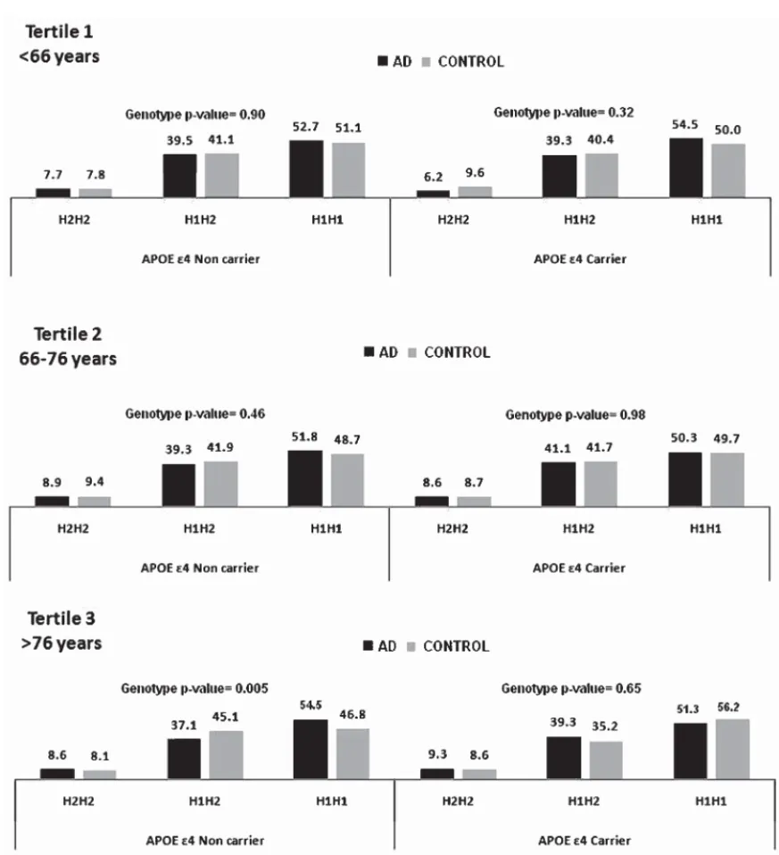
(4) P. Pastor et al. / MAPT H1 and Risk of AD in APOE 4 Noncarriers. 346. we adjusted these tests by age and gender in the entire sample, and for APOE 4 status in the AD group. Age of symptom onset was not modified by MAPT p.A152T for AD or FTD. Role of p.A152T in genetic FTD We found that MAPT p.A152T co-segregated, completely or partially, with GRN IVS6-1G>A, an intronic mutation carried by 15 FTD families from the Basque Country. MAPT p.A152T was also present in eight families, co-segregating in 70.5% of GRN IVS6-1G>A mutation carriers (Table 2). Linkage disequilibrium (LD) analysis in the families disclosed that p.A152T and GRN, both located in chromosome 17, were in partial LD (D’ = 0.78; r2 = 0.46). At the time of this study, none of the four MAPT p.A152T carriers negative for GRN IVS6-1G>A harbored a history of neurodegenerative or psychiatric disease: one individual passed away at 86 years of age, two other individuals remain healthy at 80 and 86 years of age, and the fourth individual is 52 years old who remains asymptomatic. Age of symptom onset was not associated with the MAPT p.A152T genetic variant in GRN IVS6-1G>A mutation carriers; mean age at onset was 60.9 ± 7.5 years in p.A152T-carriers and 61.4 ± 9.2 years in noncarriers (p = 0.87). We found no MAPT p.A152T carriers in three families with other GRN mutations (Cys139Arg, Arg177His, and Pro357fs), nor families with the C9orf72 expansion. Sanger sequencing of GRN in 97 MAPT p.A152T carriers from all participant centers did not reveal GRN mutations. Role of APOE 4 status and MAPT H1/H2 haplotype in neurodegenerative diseases APOE 4 status did not change the effect of MAPT p.A152T on AD risk. Table 3 shows the allelic and genotypic frequency distribution of the SNP rs1800547 Table 2 MAPT p.A152T in individuals belonging to 15 families with PGR VS6-1G mutation PGR+/A152T+ PGR+/A152T– PGR–/A152T+ PGR–/A152T– Total. Symptomatic (n). Asymptomatic (n). Total (n). 22 10 0 0 32. 9 3 4 42 58. 31 13 4 42 90. PGR+, carrier individual of PGR VS6-1G mutation; PGR-, noncarrier individual of PGR VS6-1G mutation; A152T+, carrier individual of MAPT p.A152T variant; A152T-, noncarrier individual of MAPT p.A152T variant.. tagging the MAPT H1/H2 haplotype. We found a statistically significant overrepresentation of MAPT H1 haplotype, present in 72.1% of AD compared to 69.8% of controls (p = 0.0005). When we stratified the sample by APOE 4 status, the association of H1 haplotype was driven by noncarriers of APOE 4 (p = 0.0025) (Table 3) and older subjects (genotype trend p = 0.005) (Fig. 1). As described for other European series, we also found a highly significant association between MAPT H1 and PD (OR = 1.30, 95% CI = 1.13–1.50; p = 0.0003) and PSP (OR = 3.18, 95% CI = 2.034–4.974 p = 8.59 × 10⁻⁸ ). FTD risk was not associated with the MAPT haplotype (p = 0.40).. DISCUSSION In our first analysis, we tested whether the MAPT p.A152T rare genetic variant was associated with risk for various neurodegenerative diseases (AD, FTD, PSP, and PD). We found that MAPT p.A152T occurs more frequently in Spanish patients with neurodegenerative disease compared with the study by Coppola et al. [25], whose cohort was primarily comprised of the US population (AD:0.97% versus 0.69%; FTD: 1.42% versus 0.89% and PD: 077% versus 0.48% respectively). Because the frequency of MAPT p.A152T was also significantly higher in our healthy controls than the healthy control cohort of Coppola et al. (0.71% versus 0.30%, respectively) [25], the association between AD risk and MAPT p.A152T was not significant in our population. Although our OR for AD risk associating with p.A152T occurred in same direction as in the previous study [25], our OR was considerably lower and thus did not reach statistical significance (OR = 1.4; 95% CI = 0.9–2.1 versus OR = 2.3; 95% CI = 1.3–4.2, respectively). Similarly, the OR we obtained for p.A152T in FTD risk trended toward significance, but was also lower than the OR for FTD risk in the previous study (OR = 2.0; 95% CI = 0.9–4.3 versus OR = 3.0; 95% CI = 1.6–5.6, respectively) [25]. Several factors may explain the lack of replication of previous results. Rare genetic variant frequencies can differ across populations, and MAPT p.A152T appears to occur more frequently in the general Spanish population than in the US. Another consideration is that the real ORs for diseases associated with the variant may be lower than the ORs in the discovery cohort due to the “winner’s course” effect, a common phenomenon observed in pioneer genetic epidemiological studies [35]. Another potential influence on the difference.
(5) P. Pastor et al. / MAPT H1 and Risk of AD in APOE 4 Noncarriers. 347. Fig. 1. Genotype frequency distribution of the rs1800547 SNP tagging MAPT H1/H2 haplotype stratified by APOE 4 status and Age tertile.. between our results and those of Coppola et al. [25] is the mean age at which controls were deemed healthy; for example, p.A152T carriers in one cohort may have been classified as controls at a younger age, prior to disease onset. Since the controls of Coppola et al. [25] were significantly younger (50 ± 16 years) than those analyzed in our study (64.1 ± 14.8 years), it is less likely that misclassification of our p.A152T carriers as controls who might manifest future degenerative disease could explain the higher p.A152T allelic frequency observed in our cohort.. A surprising finding of the present study was the co-segregation of MAPT p.A152T in 70% of carriers of the GRN mutation IVS6-1G>A (g.1872G>A) unique to the Basque Country [32]. This is a splicing mutation located at chromosome 17 (base pair position 139486) that causes truncated GRN protein due to mRNA degradation [33]. The fact that the MAPT p.A152T variant co-occurred with the GRN mutation only in families in a limited geographical region suggests that these individuals share the same haplotype, most likely from a common ancestor. However, MAPT.
(6) P. Pastor et al. / MAPT H1 and Risk of AD in APOE 4 Noncarriers. 348. Table 3 MAPT H1/H2 haplotype frequencies and AD risk ALL H2H2 H1H2 H1H1 H1 frequency APOE4+ H2H2 H1H2 H1H1 H1 frequency APOE4H2H2 H1H2 H1H1 H1 frequency. Control (%). AD (%). 532 (9.15) 2444 (42.03) 2839 (48.82) 0.698. 344 (8.34) 1614 (39.11) 2169 (52.56) 0.721. 78 (9.07) 345 (40.12) 437 (50.81) 0.709. 139 (8.50) 655 (40.04) 842 (51.47) 0.715. 343 (8.46) 1701 (41.97) 2009 (49.57) 0.706. 160 (8.16) 730 (37.24) 1070 (54.59) 0.732. Genotype P-value. Allelic -value. Allelic OR (95%CI). p = 0.001 [p = 0.016]∗. p = 0.00051. 1.12 (1.05–1.19). p = 0.88 [p = 0.86]. p = 0.65. 1.03 (0.91–1.18). p = 0.001 [p = 0.005]. p = 0.0025. 1.14 (1.05–1.24). In brackets p-values adjusted by age and gender . ∗ p-values adjusted by age, gender, and APOE status.. p.A152T variant in patients carrying the GRN IVS61G>A mutation did not influence age at onset. Future studies are necessary to probe the influence of the coocurrence of MAPT p.A152T and GRN IVS6-1G>A on the FTD clinical or neuropathological phenotype. Our last finding was that MAPT H1 haplotype is overrepresented in patients with AD, PD, and PSP compared to controls. Although the association of MAPT with PD and PSP risk is well documented [5–8, 11–13], its association with AD is much more controversial. To date, genome-wide association studies and case-control data meta-analyses such as AlzGene have not been able to link MAPT genetic variants to AD [15] despite numerous experimental evidence of the involvement of tau protein in AD pathogenesis [36]. In our study, we found a very significant overrepresentation of the MAPT H1 haplotype in patients with AD compared to controls. The mildly increased risk for AD conferred by the H1 haplotype emerged only in our subgroup of noncarriers for APOE 4, especially in the oldest subjects. This is in line with a recent publication re-analyzing the IGAP consortium data in APOE 4 carriers and non-carriers. That study reported genome-wide significant association with many SNPs across a region on chromosome 17 including MAPT and with the H1 haplotype, however, the association was accounted for by SNPs located between two genes (KANSL1 and LRRC37A) adjacent to MAPT [17]. Our results are consistent with the hypothesis that AD pathology could develop through different causal pathways with several genetic factors likely to be involved, APOE 4 being the strongest one. APOE 4 lowers the threshold for AD susceptibility, which 1) associates with an earlier age of disease onset, and 2) may decrease the number and magnitude of etiological factors that. would be necessary to start the disease’s pathological mechanisms. However, in the absence of APOE 4, the participation of an ensemble of alternative etiological factors, and for longer periods of time, might be necessary to elicit the disease. For instance, if MAPT H1 haplotype confers a modest risk for AD independent of APOE 4, we may be able to detect this association only in elderly individuals not carrying APOE 4; otherwise, APOE 4’s effect on AD risk might mask the ability to detect the effect of MAPT H1 on AD risk. The association between the MAPT haplotype and AD is consistent with studies suggesting that H1/H1 status is associated with an increased rate of conversion from mild cognitive impairment to AD [37]. Our results are also in line with a recent publication studying MAPT haplotypes in a large sample of late onset AD from the US that found that H2 haplotype carriers were protected from AD and had lower MAPT levels in brain [38]. An alternative explanation to our results could be that within the APOE 4 non-carriers group the number of subjects with dementia due to pure tauopathies (PSP, CBD, or FTDtau) misdiagnosed as AD might be higher than those among the APOE 4 carriers group. We suggest that in large population samples this phenomena is likely to occur to a certain degree, but we consider unlikely that these disorders with a low prevalence are contributing significantly to our results. Additionally, the fact by which most patients included in our study come from specialized memory units from academic hospitals increases the likelihood of a correct AD diagnosis. In summary, we did not find a significant association between the rare variant MAPT p.A152T and AD risk, although our findings trended toward significance for p.A152T being associated with FTD risk. Despite our large sample size, our results should be interpreted with.
(7) P. Pastor et al. / MAPT H1 and Risk of AD in APOE 4 Noncarriers. caution, as our study may be underpowered to detect the effect of such an infrequent genetic variant if the real OR is lower in our population than found in previous studies.OurfindingthatMAPTp.A152Tandtheprogranulin IVS6-1G>A mutation cosegregates in families from the Basque region raises interesting questions about the influence of multiple risk genetic variants coinciding in neurodegenerative diseases; future studies will address these questions [39]. Finally, we found a robust statistical association between MAPT H1 extended haplotypeandriskoflate-onsetADinAPOE4noncarriers. Our results, in a large sample of Spanish population, represent strong evidence supporting a link between common MAPT genetic variants and AD. The modest risk effect conferred by MAPT H1 haplotype and the fact that it is restricted to APOE 4 negative subjects might contribute to clarify controversial results in previous studies.. ACKNOWLEDGMENTS This work was made possible by the generous participation of the patients and their families, and the control subjects. The DEGESCO members would like to acknowledge Miguel Medina and CIBERNED support. We would like to thank Drs. P. Gil and P. Coria (Centro de Biologı́a Molecular Severo Ochoa, Madrid, Spain) for their cooperation in the generation of the case-control samples. We are indebted to Trinitat PortCarbó and her family for their support of the Fundació ACE (Barcelona, Spain) research programs. Fundació ACE collaborates with the Centro de Investigación Biomédica en Red sobre Enfermedades Neurodegenerativas (CIBERNED, Spain). We also would like to thank Drs. Berta Pascual-Sedano, Javier Pagonabarraga (Department of Neurology, Hospital Sant Pau, Barcelona, Spain), and Antonia Campolongo (Institut de Recerca-IIB Sant Pau, Barcelona, Spain) for their effort in clinical evaluation and sample recruitment. We thank Miren Zulaica Ijurco for her assistance. We thank Mrs. Ana Belen Pastor (CIEN Foundation, Madrid, Spain) for her technical support on human sample collection. We thank Giovanni Coppola (Semel Institute for Neuroscience and Human Behavior, University of California, Lo Angeles, CA, USA) for his support in sample genotyping and Bruce Miller (Memory and Aging Center, University of California, San Francisco, CA, USA) for his support and advice. This work was supported by grants from the Spanish Ministry of Science and Innovation SAF 2006-10126 (2006–2009) and SAF2010-22329-C02-. 349. 01 (2011–2013) to P.P and by the UTE project FIMA to P.P. Grants from the Ministry of Science (SAF2010-15558) and CIBERNED. Agustı́n Ruiz is supported by grant PI13/02434 (Acción Estratégica en Salud. Instituto de Salud Carlos III. Ministerio de Economı́a y Competitividad, Spain). Grant: Consolider (CSD2010-00045). This study was partially supported by grants from Instituto de Salud Carlos III (PI12/01311 and PI12/03005). CIBERNED, ILUNDAIN FUNDAZIOA. This work was partially supported by PI12/00045 grant. This work was supported by grants from Instituto de Salud Carlos III (FIS PI12/02288 and JPND-PI11/03028). Authors’ disclosures available online (http://j-alz. com/manuscript-disclosures/15-0555r1).. DEGESCO PROJECT MEMBERS Center for Applied Medical Research, University of Navarra (CIMA): Sara Ortega-Cubero, Oswaldo Lorenzo-Betancor, Elena Lorenzo, Maria A. Pastor, Pau Pastor Research Center and Memory Clinic of Fundació ACE: Isabel Hernández, Asunción Lafuente, Maitée Rosende-Roca, Ana Mauleón, Liliana Vargas, Octavio Rodrı́guez, Carla Abdelnour, Montserrat Alegret, Ana Espinosa, Gemma Ortega, Ángela Sanabria, Marta Ibarria, Susana Diego, Pilar Canyabate, Mariola Moreno, Mar Buendı́a, Ana Pancho, Marı́a Eugenia Palacio, Susana Ruiz, Natalia Tantinya, Marina Tarragona, América Morera, Marina Guitart, Oscar Sotolongo Grau, Elvira Martı́n, Victòria Fernández, Agustı́n Ruiz, Lluı́s Tárraga, Mercè Boada Hospital Clı́nic: Raquel Sánchez-Valle, José Luis Molinuevo, Albert Lladó, Anna Antonell, Lorena Rami Genética Molecular-HUCA: Eliecer Coto, Victoria Alvarez Biodonostia: Fermı́n Moreno, Begoña Indakoetxea, Myriam Barandiarán, Ana Gorostidi, Adolfo López de Munain FCIEN: Alberto Rábano, Miguel Calero HUMV: José Luis Vázquez-Higuera, Ana Pozueta, Andrea González Suarez, Eloy Rodriguez-Rodriguez, Ignacio Mateo, José Berciano, Pascual Sánchez-Juan, Onofre Combarros Centro de Biologı́a Molecular Severo Ochoa: Marı́a J. Bullido, Ana Frank-Garcı́a, Isabel Sastre Doce de Octubre: Eva Carro BIOMICs: Marian M de Pancorbo, Xabier Elcoroaristizabal.
(8) P. Pastor et al. / MAPT H1 and Risk of AD in APOE 4 Noncarriers. 350. Hospital de la Santa Creu i Sant Pau-Neurology Department-Memory Unit: Alberto Lleó, Rafael Blesa, Juan Fortea, Olivia Belbin, Daniel Alcolea, Marı́a Carmona-Iragui, Mª Belén Sánchez-Saudinós, Isabel Sala, Sofı́a Anton-Aguirre, Estrella Morenas, Roser Ribosa, Martı́ Colom-Cadena, Laura Cervera, Laia Muñoz, Oriol Dols-Icardo, Jordi Clarimón. [13]. REFERENCES [14] [1] [2]. [3]. [4]. [5]. [6]. [7]. [8]. [9]. [10]. [11]. [12]. Goedert M (2004) Tau protein and neurodegeneration. Semin Cell Dev Biol 15, 45-49. Galpern WR, Lang AE (2006) Interface between tauopathies and synucleinopathies, a tale of two proteins. Ann Neurol 59, 449-458. Rademakers R, Cruts M, van Broeckhoven C (2004) The role of tau (MAPT) in frontotemporal dementia and related tauopathies. Hum Mutat 24, 277-295. Ishizawa T, Mattila P, Davies P, Wang D, Dickson DW (2003) Colocalization of tau and alpha-synuclein epitopes in Lewy bodies. J Neuropathol Exp Neurol 62, 389-397. Baker M, Litvan I, Houlden H, Adamson J, Dickson D, Perez-Tur J, Hardy J, Lynch T, Bigio E, Hutton M (1999) Association of an extended haplotype in the tau gene with progressive supranuclear palsy. Hum Mol Genet 8, 711-715. Conrad C, Andreadis A, Trojanowski JQ, Dickson DW, Kang D, Chen X, Wiederholt W, Hansen L, Masliah E, Thal LJ, Katzman R, Xia Y, Saitoh T (1997) Genetic evidence for the involvement of tau in progressive supranuclear palsy. Ann Neurol 41, 277-281. Higgins JJ, Golbe LI, De Biase A, Jankovic J, Factor SA, Adler RL (2000) An extended 5’-tau susceptibility haplotype in progressive supranuclear palsy. Neurology 55, 1364-1367. Pastor P, Ezquerra M, Perez JC, Chakraverty S, Norton J, Racette BA, McKeel D, Perlmutter JS, Tolosa E, Goate AM (2004) Novel haplotypes in 17q21 are associated with progressive supranuclear palsy. Ann Neurol 56, 249-258. Houlden H, Baker M, Morris HR, MacDonald N, PickeringBrown S, Adamson J, Lees AJ, Rossor MN, Quinn NP, Kertesz A, Khan MN, Hardy J, Lantos PL, St George-Hyslop P, Munoz DG, Mann D, Lang AE, Bergeron C, Bigio EH, Litvan I, Bhatia KP, Dickson D, Wood NW, Hutton M (2001) Corticobasal degeneration and progressive supranuclear palsy share a common tau haplotype. Neurology 56, 1702-1706. Verpillat P, Camuzat A, Hannequin D, Thomas-Anterion C, Puel M, Belliard S, Dubois B, Didic M, Michel BF, Lacomblez L, Moreaud O, Sellal F, Golfier V, Campion D, ClergetDarpoux F, Brice A (2002) Association between the extended tau haplotype and frontotemporal dementia. Arch Neurol 59, 935-939. Martin ER, Scott WK, Nance MA, Watts RL, Hubble JP, Koller WC, Lyons K, Pahwa R, Stern MB, Colcher A, Hiner BC, Jankovic J, Ondo WG, Allen FH, Goetz CG, Small GW, Masterman D, Mastaglia F, Laing NG, Stajich JM, Ribble RC, Booze MW, Rogala A, Hauser MA, Zhang F, Gibson RA, Middleton LT, Roses AD, Haines JL, Scott BL, Pericak-Vance MA, Vance JM (2001) Association of single-nucleotide polymorphisms of the tau gene with late-onset Parkinson disease. JAMA 286, 2245-2250. Simón-Sánchez J, Schulte C, Bras JM, Sharma M, Gibbs JR, Berg D, Paisan-Ruiz C, Lichtner P, Scholz SW, Hernandez DG, Krüger R, Federoff M, Klein C, Goate A, Perlmutter J,. [15] [16]. [17]. Bonin M, Nalls MA, Illig T, Gieger C, Houlden H, Steffens M, Okun MS, Racette BA, Cookson MR, Foote KD, Fernandez HH, Traynor BJ, Schreiber S, Arepalli S, Zonozi R, Gwinn K, van der Brug M, Lopez G, Chanock SJ, Schatzkin A, Park Y, Hollenbeck A, Gao J, Huang X, Wood NW, Lorenz D, Deuschl G, Chen H, Riess O, Hardy JA, Singleton AB, Gasser T (2009) Genome-wide association study reveals genetic risk underlying Parkinson’s disease. Nat Genet 41, 1308-1312. Pastor P, Ezquerra M, Muñoz E, Martı́ MJ, Blesa R, Tolosa E, Oliva R (2000) Significant association between the tau gene A0/A0 genotype and Parkinson’s disease. Ann Neurol 47, 242245. Myers AJ, Kaleem M, Marlowe L, Pittman AM, Lees AJ, Fung HC, Duckworth J, Leung D, Gibson A, Morris CM, de Silva R, Hardy J (2005) The H1c haplotype at the MAPT locus is associated with Alzheimer’s disease. Hum Mol Genet 14, 2399-2404. AlzGene. http://www.alzgene.org/meta.asp?geneID=232. Last updated May 5, 2010, Accessed on August 12, 2014. Lambert J-C, Ibrahim-Verbaas CA, Harold D, Naj AC, Sims R, Bellenguez C, DeStafano AL, Bis JC, Beecham GW, Grenier-Boley B, Russo G, Thorton-Wells TA, Jones N, Smith AV, Chouraki V, Thomas C, Ikram MA, Zelenika D, Vardarajan BN, Kamatani Y, Lin CF, Gerrish A, Schmidt H, Kunkle B, Dunstan ML, Ruiz A, Bihoreau MT, Choi SH, Reitz C, Pasquier F, Cruchaga C, Craig D, Amin N, Berr C, Lopez OL, De Jager PL, Deramecourt V, Johnston JA, Evans D, Lovestone S, Letenneur L, Morón FJ, Rubinsztein DC, Eiriksdottir G, Sleegers K, Goate AM, Fiévet N, Huentelman MW, Gill M, Brown K, Kamboh MI, Keller L, Barberger-Gateau P, McGuiness B, Larson EB, Green R, Myers AJ, Dufouil C, Todd S, Wallon D, Love S, Rogaeva E, Gallacher J, St George-Hyslop P, Clarimon J, Lleo A, Bayer A, Tsuang DW, Yu L, Tsolaki M, Bossú P, Spalletta G, Proitsi P, Collinge J, Sorbi S, Sanchez-Garcia F, Fox NC, Hardy J, Deniz Naranjo MC, Bosco P, Clarke R, Brayne C, Galimberti D, Mancuso M, Matthews F, European Alzheimer’s Disease Initiative (EADI) Genetic, Environmental Risk in Alzheimer’s Disease Alzheimer’s Disease Genetic Consortium Cohorts for Heart, Aging Research in Genomic Epidemiology, Moebus S, Mecocci P, Del Zompo M, Maier W, Hampel H, Pilotto A, Bullido M, Panza F, Caffarra P, Nacmias B, Gilbert JR, Mayhaus M, Lannefelt L, Hakonarson H, Pichler S, Carrasquillo MM, Ingelsson M, Beekly D, Alvarez V, Zou F, Valladares O, Younkin SG, Coto E, Hamilton-Nelson KL, Gu W, Razquin C, Pastor P, Mateo I, Owen MJ, Faber KM, Jonsson PV, Combarros O, O’Donovan MC, Cantwell LB, Soininen H, Blacker D, Mead S, Mosley TH Jr, Bennett DA, Harris TB, Fratiglioni L, Holmes C, de Bruijn RF, Passmore P, Montine TJ, Bettens K, Rotter JI, Brice A, Morgan K, Foroud TM, Kukull WA, Hannequin D, Powell JF, Nalls MA, Ritchie K, Lunetta KL, Kauwe JS, Boerwinkle E, Riemenschneider M, Boada M, Hiltuenen M, Martin ER, Schmidt R, Rujescu D, Wang LS, Dartigues JF, Mayeux R, Tzourio C, Hofman A, Nöthen MM, Graff C, Psaty BM, Jones L, Haines JL, Holmans PA, Lathrop M, Pericak-Vance MA, Launer LJ, Farrer LA, van Duijn CM, Van Broeckhoven C, Moskvina V, Seshadri S, Williams J, Schellenberg GD, Amouyel P (2013) Extended meta-analysis of 74,538 individuals identifies 11 new susceptibility loci for Alzheimer’s disease. Nat Genet 45, 1452-1458. Jun G, Ibrahim-Verbaas CA, Vronskaya M, Lambert JC, Chung J, Naj AC, Kunkle BW, Wang LS, Bis JC, Bellenguez C, Harold D, Lunetta KL, Destefano AL, Grenier-Boley B, Sims R, Beecham GW, Smith AV, Chouraki V, HamiltonNelson KL, Ikram MA, Fievet N, Denning N, Martin ER,.
(9) P. Pastor et al. / MAPT H1 and Risk of AD in APOE 4 Noncarriers. [18]. [19]. [20]. [21]. [22]. [23]. [24]. Schmidt H, Kamatani Y, Dunstan ML, Valladares O, Laza AR, Zelenika D, Ramirez A, Foroud TM, Choi SH, Boland A, Becker T, Kukull WA, van der Lee SJ, Pasquier F, Cruchaga C, Beekly D, Fitzpatrick AL, Hanon O, Gill M, Barber R, Gudnason V, Campion D, Love S, Bennett DA, Amin N, Berr C, Tsolaki M, Buxbaum JD, Lopez OL, Deramecourt V, Fox NC, Cantwell LB, Tárraga L, Dufouil C, Hardy J, Crane PK, Eiriksdottir G, Hannequin D, Clarke R, Evans D, Mosley TH Jr, Letenneur L, Brayne C, Maier W, De Jager P, Emilsson V, Dartigues JF, Hampel H, Kamboh MI, de Bruijn RF, Tzourio C, Pastor P, Larson EB, Rotter JI, O’Donovan MC, Montine TJ, Nalls MA, Mead S, Reiman EM, Jonsson PV, Holmes C, St George-Hyslop PH, Boada M, Passmore P, Wendland JR, Schmidt R, Morgan K, Winslow AR, Powell JF, Carasquillo M, Younkin SG, Jakobsdóttir J, Kauwe JS, Wilhelmsen KC, Rujescu D, Nöthen MM, Hofman A, Jones L IGAP Consortium, Haines JL, Psaty BM, Van Broeckhoven C, Holmans P, Launer LJ, Mayeux R, Lathrop M, Goate AM, Escott-Price V, Seshadri S, Pericak-Vance MA, Amouyel P, Williams J, van Duijn CM, Schellenberg GD, Farrer LA (2015) A novel Alzheimer disease locus located near the gene encoding tau protein. Mol Psychiatry, doi: 10.1038/mp.2015.23 Heutink P, Stevens M, Rizzu P, Bakker E, Kros JM, Tibben A, Niermeijer MF, van Duijn CM, Oostra BA, van Swieten JC (1997) Hereditary frontotemporal dementia is linked to chromosome 17q21-q22, a genetic and clinicopathological study of three Dutch families. Ann Neurol 41, 150-159. Hutton M, Lendon CL, Rizzu P, Baker M, Froelich S, Houlden H, Pickering-Brown S, Chakraverty S, Isaacs A, Grover A, Hackett J, Adamson J, Lincoln S, Dickson D, Davies P, Petersen RC, Stevens M, de Graaff E, Wauters E, van Baren J, Hillebrand M, Joosse M, Kwon JM, Nowotny P, Che LK, Norton J, Morris JC, Reed LA, Trojanowski J, Basun H, Lannfelt L, Neystat M, Fahn S, Dark F, Tannenberg T, Dodd PR, Hayward N, Kwok JB, Schofield PR, Andreadis A, Snowden J, Craufurd D, Neary D, Owen F, Oostra BA, Hardy J, Goate A, van Swieten J, Mann D, Lynch T, Heutink P (1998) Association of missense and 5’-splice-site mutations in tau with the inherited dementia FTDP-17. Nature 393, 702-705. Hong M, Zhukareva V, Vogelsberg-Ragaglia V, Wszolek Z, Reed L, Miller BI, Geschwind DH, Bird TD, McKeel D, Goate A, Morris JC, Wilhelmsen KC, Schellenberg GD, Trojanowski JQ, Lee VM (1998) Mutation-specific functional impairments in distinct tau isoforms of hereditary FTDP-17. Science 282, 1914-1917. Murrell JR, Spillantini MG, Zolo P, Guazzelli M, Smith MJ, Hasegawa M, Redi F, Crowther RA, Pietrini P, Ghetti B, Goedert M (1999) Tau gene mutation G389R causes a tauopathy with abundant pick body-like inclusions and axonal deposits. J Neuropathol Exp Neurol 58, 1207-1226. Pickering-Brown S, Baker M, Yen SH, Liu WK, Hasegawa M, Cairns N, Lantos PL, Rossor M, Iwatsubo T, Davies Y, Allsop D, Furlong R, Owen F, Hardy J, Mann D, Hutton M (2000) Pick’s disease is associated with mutations in the tau gene. Ann Neurol 48, 859-867. Clark LN, Poorkaj P, Wszolek Z, Geschwind DH, Nasreddine ZS, Miller B, Li D, Payami H, Awert F, Markopoulou K, Andreadis A, D’Souza I, Lee VM, Reed L, Trojanowski JQ, Zhukareva V, Bird T, Schellenberg G, Wilhelmsen KC (1998) Pathogenic implications of mutations in the tau gene in pallido-ponto-nigral degeneration and related neurodegenerative disorders linked to chromosome 17. Proc Natl Acad Sci U S A 95, 13103-13107. Pastor P, Pastor E, Carnero C, Vela R, Garcı́a T, Amer G, Tolosa E, Oliva R (2001) Familial atypical progres-. [25]. [26]. [27]. [28]. [29]. [30]. [31]. 351. sive supranuclear palsy associated with homozigosity for the delN296 mutation in the tau gene. Ann Neurol 49, 263-267. Coppola G, Chinnathambi S, Lee JJ, Dombroski BA, Baker MC, Soto-Ortolaza AI, Lee SE, Klein E, Huang AY, Sears R, Lane JR, Karydas AM, Kenet RO, Biernat J, Wang LS, Cotman CW, Decarli CS, Levey AI, Ringman JM, Mendez MF, Chui HC, Le Ber I, Brice A, Lupton MK, Preza E, Lovestone S, Powell J, Graff-Radford N, Petersen RC, Boeve BF, Lippa CF, Bigio EH, Mackenzie I, Finger E, Kertesz A, Caselli RJ, Gearing M, Juncos JL, Ghetti B, Spina S, Bordelon YM, Tourtellotte WW, Frosch MP, Vonsattel JP, Zarow C, Beach TG, Albin RL, Lieberman AP, Lee VM, Trojanowski JQ, Van Deerlin VM, Bird TD, Galasko DR, Masliah E, White CL, Troncoso JC, Hannequin D, Boxer AL, Geschwind MD, Kumar S, Mandelkow EM, Wszolek ZK, Uitti RJ, Dickson DW, Haines JL, Mayeux R, Pericak-Vance MA, Farrer LA, Alzheimer’s Disease Genetics Consortium, Ross OA, Rademakers R, Schellenberg GD, Miller BL, Mandelkow E, Geschwind DH (2012) Evidence for a role of the rare p. A152T variant in MAPT in increasing the risk for FTD-spectrum and Alzheimer’s diseases. Hum Mol Genet 21, 3500-3512. Lee SE, Tartaglia MC, Yener G, Genç S, Seeley WW, Sanchez-Juan P, Moreno F, Mendez MF, Klein E, Rademakers R, Munain AL, Combarros O, Kramer JH, Kenet RO, Boxer AL, Geschwind MD, Gorno-Tempini ML, Karydas AM, Rabinovici GD, Coppola G, Geschwind DH, Miller BL (2013) Neurodegenerative disease phenotypes in carriers of MAPT p. A152T, a risk factor for frontotemporal dementia spectrum disorders and Alzheimer disease. Alzheimer Dis Assoc Disord 27, 302-309. Kara E, Ling H, Pittman AM, Shaw K, de Silva R, Simone R, Holton JL, Warren JD, Rohrer JD, Xiromerisiou G, Lees A, Hardy J, Houlden H, Revesz T (2012) The MAPT p. A152T variant is a risk factor associated with tauopathies with atypical clinical and neuropathological features. Neurobiol Aging 33, 2231.e7-2231.e14. McKhann G, Drachman D, Folstein M, Katzman R, Price D, Stadlan EM (1984) Clinical diagnosis of Alzheimer’s disease, report of the NINCDS-ADRDA Work Group under the auspices of Department of Health and Human Services Task Force on Alzheimer’s Disease. Neurology 34, 939-944. Rascovsky K, Hodges JR, Knopman D, Mendez MF, Kramer JH, Neuhaus J, van Swieten JC, Seelaar H, Dopper EG, Onyike CU, Hillis AE, Josephs KA, Boeve BF, Kertesz A, Seeley WW, Rankin KP, Johnson JK, Gorno-Tempini ML, Rosen H, Prioleau-Latham CE, Lee A, Kipps CM, Lillo P, Piguet O, Rohrer JD, Rossor MN, Warren JD, Fox NC, Galasko D, Salmon DP, Black SE, Mesulam M, Weintraub S, Dickerson BC, Diehl-Schmid J, Pasquier F, Deramecourt V, Lebert F, Pijnenburg Y, Chow TW, Manes F, Grafman J, Cappa SF, Freedman M, Grossman M, Miller BL (2011) Sensitivity of revised diagnostic criteria for the behavioural variant of frontotemporal dementia. Brain 134, 2456-2477. Litvan I, Agid Y, Calne D, Campbell G, Dubois B, Duvoisin RC, Goetz CG, Golbe LI, Grafman J, Growdon JH, Hallett M, Jankovic J, Quinn NP, Tolosa E, Zee DS (1996) Clinical research criteria for the diagnosis of progressive supranuclear palsy (Steele-Richardson-Olszewski syndrome), report of the NINDS-SPSP international workshop. Neurology 47, 1-9. Hughes AJ, Daniel SE, Kilford L, Lees AJ (1992) Accuracy of clinical diagnosis of idiopathic Parkinson’s disease, a clinico-pathological study of 100 cases. J Neurol Neurosurg Psychiatry 55, 181-184..
(10) 352 [32]. [33]. [34]. [35]. P. Pastor et al. / MAPT H1 and Risk of AD in APOE 4 Noncarriers Moreno F, Indakoetxea B, Barandiaran M, Alzualde A, Gabilondo A, Estanga A, Ruiz J, Ruibal M, Bergareche A, Martı́-Massó JF, López de Munain A (2009) “Frontotemporoparietal” dementia, clinical phenotype associated with the c.709-1G>A PGRN mutation. Neurology 73, 1367-1374. López de Munain A, Alzualde A, Gorostidi A, Otaegui D, Ruiz-Martı́nez J, Indakoetxea B, Ferrer I, Pérez-Tur J, Sáenz A, Bergareche A, Barandiarán M, Poza JJ, Zabalza R, Ruiz I, Urtasun M, Fernández-Manchola I, Olasagasti B, Espinal JB, Olaskoaga J, Ruibal M, Moreno F, Carrera N, Martı́ Massó JF (2008) Mutations in progranulin gene, clinical, pathological, and ribonucleic acid expression findings. Biol Psychiatry 63, 946-952. Barrett JC, Fry B, Maller J, Daly MJ (2005) Haploview, analysis and visualization of LD and haplotype maps. Bioinformatics 21, 263-265. Zollner S, Pritchard JK (2007) Overcoming the winner’s curse, estimating penetrance parameters from case-control data. Am J Hum Genet 80, 605-615.. [36] [37]. [38]. [39]. Krstic D, Knuesel I (2013) Deciphering the mechanism underlying late-onset Alzheimer disease. Nat Rev Neurol 9, 25-34. Samaranch L, Cervantes S, Barabash A, Alonso A, Cabranes JA, Lamet I, Ancı́n I, Lorenzo E, Martı́nez-Lage P, Marcos A, Clarimón J, Alcolea D, Lleó A, Blesa R, Gómez-Isla T, Pastor P (2010) The effect of MAPT H1 and APOE 4 on transition from mild cognitive impairment to dementia. J Alzheimers Dis 22, 1065-1071. Allen M, Kachadoorian M, Quicksall Z, Zou F, Chai HS, Younkin C, Crook JE, Pankratz VS, Carrasquillo MM, Krishnan S, Nguyen T, Ma L, Malphrus K, Lincoln S, Bisceglio G, Kolbert CP, Jen J, Mukherjee S, Kauwe JK, Crane PK, Haines JL, Mayeux R, Pericak-Vance MA, Farrer LA, Schellenberg GD, Parisi JE, Petersen RC, Graff-Radford NR, Dickson DW, Younkin SG, Ertekin-Taner N (2014) Association of MAPT haplotypes with Alzheimer’s disease risk and MAPT brain gene expression levels. Alzheimers Res Ther 6, 39. Pastor P (2013) Comment, double mutants of frontotemporal dementia genes–Simple co-occurrence? Neurology 81, 1338..
(11)
Figure
Documento similar
NSAIDs increase the risk of cardiovascular disease in the general population, and although there is little direct evidence for elevated cardiovascular risk with NSAIDs in people
1. Definition and measurement of parameters: The fundamental parameters, by which the risks associated with a project are assessed, are risk probability and risk
Even though the 1920s offered new employment opportunities in industries previously closed to women, often the women who took these jobs found themselves exploited.. No matter
Taking into account these results that suggested a potential influence of the TLR4 rs4986790 gene polymorphism in the risk of atherosclerosis, we conducted a study in a large series
The expansionary monetary policy measures have had a negative impact on net interest margins both via the reduction in interest rates and –less powerfully- the flattening of the
Jointly estimate this entry game with several outcome equations (fees/rates, credit limits) for bank accounts, credit cards and lines of credit. Use simulation methods to
In our sample, 2890 deals were issued by less reputable underwriters (i.e. a weighted syndication underwriting reputation share below the share of the 7 th largest underwriter
Incidence of dementia and probable Alzheimer´s disease in a general population: The Framingham study.. Hendiré H., Ogunniyi A.,
