Characterization of Campylobacter spp.
isolated from wild birds in the Antarctic and
Sub-Antarctic
Håkan Johansson1, Patrik Ellstro¨ mID2,3, Karin Artursson4, Charlotte Berg5, Jonas Bonnedahl1,6, Ingrid Hansson7, Jorge Hernandez2,8, Juana Lopez-Martı´n9, Gonzalo Medina-Vogel10, Lucila Moreno11, Bjo¨ rn Olsen2,3, Eva Olsson Engvall4, Hanna Skarin4, Karin Troell4, Jonas Waldenstro¨ m1
, JoakimÅgren4, Daniel Gonza´lez-AcuñaID12*
1 Centre for Ecology and Evolution in Microbial Model Systems, Linnaeus University, Kalmar, Sweden, 2 Zoonosis Science Center, Department of Medical Biochemistry and Microbiology, Uppsala University,
Uppsala, Sweden, 3 Department of Medical Sciences, Uppsala University, Uppsala, Sweden, 4 National Veterinary Institute, Uppsala, Sweden, 5 Department of Animal Environment and Health, Swedish University of Agricultural Sciences, Skara, Sweden, 6 Department of Infectious Diseases, Kalmar County Hospital, Kalmar, Sweden, 7 Department of Biomedical Sciences and Veterinary Public Health, Swedish University of Agricultural Sciences, Uppsala, Sweden, 8 Laboratory of Microbiology, Kalmar County Hospital, Kalmar, Sweden, 9 Departamento de Patologı´a y Medicina Preventiva, Facultad de Ciencias Veterinarias, Universidad de Concepcio´n, Chilla´n, Chile, 10 Centro de Investigacio´n para la Sustentabilidad, Universidad Andre´ s Bello, Santiago, Chile, 11 Facultad de Ciencias Naturales y Oceanogra´ficas, Universidad de Concepcio´n, Concepcio´ n, Chile, 12 Facultad de Ciencias Veterinarias, Universidad de Concepcio´n, Chilla´n, Chile
Abstract
A lack of knowledge of naturally occurring pathogens is limiting our ability to use the Antarc-tic to study the impact human-mediated introduction of infectious microorganisms have on this relatively uncontaminated environment. As no large-scale coordinated effort to remedy this lack of knowledge has taken place, we rely on smaller targeted efforts to both study present microorganisms and monitor the environment for introductions. In one such effort, we isolated Campylobacter species from fecal samples collected from wild birds in the Ant-arctic Peninsula and the sub-AntAnt-arctic island of South Georgia. Indeed, in South Georgia, we found Campylobacter lari and the closely related Campylobacter peloridis, but also dis-tantly related human-associated multilocus sequence types of Campylobacter jejuni. In con-trast, in the Antarctic Peninsula, we found C. lari and two closely related species,
Campylobacter subantarcticus and Campylobacter volucris, but no signs of human introduc-tion. In fact, our finding of human-associated sequence types of C. jejuni in South Georgia, but not in the Antarctic Peninsula, suggests that efforts to limit the spread of infectious microorganisms to the Antarctic have so far been successful in preventing the introduction of C. jejuni. However, we do not know how it came to South Georgia and whether the same mode of introduction could spread it from there to the Antarctic Peninsula.
a1111111111
Citation: Johansson H, Ellstro¨m P, Artursson K,
Berg C, Bonnedahl J, Hansson I, et al. (2018) Characterization of Campylobacter spp. isolated from wild birds in the Antarctic and Sub-Antarctic. PLoS ONE 13(11): e0206502.https://doi.org/ 10.1371/journal.pone.0206502
Editor: Patrick Jon Biggs, Massey University, NEW
ZEALAND
Received: February 1, 2018
Accepted: October 15, 2018
Published: November 9, 2018
Copyright:©2018 Johansson et al. This is an open access article distributed under the terms of the
Creative Commons Attribution License, which permits unrestricted use, distribution, and reproduction in any medium, provided the original author and source are credited.
Data Availability Statement: All relevant data are
within the paper and its Supporting Information files.
Funding: This study was funded by the Chilean
Antarctic Institute (INACH number T-12-13) and the Swedish Research Council Formas (2014-829). The funders had no role in study design, data collection and analysis, decision to publish, or preparation of the manuscript.
Competing interests: I have read the journal’s
Introduction
The Antarctic is among the most isolated places on Earth. By virtue of inhabiting such a remote location, Antarctic animals were long thought to be protected from disease introduc-tion from other regions. However, recent studies have reported the presence of human and animal pathogens previously believed to be absent from the region [1,2], includingSalmonella entericaserovar Enteriditis phage type 4 [3–5] and influenza A viruses [6]. In addition to find-ing pathogens with presumed non-Antarctic origin in Antarctic wildlife, it has been shown that penguins kept in captivity are susceptible to a range of infectious diseases not observed in the Antarctic (see [2], and references therein). Sustained transmission of some of these patho-gens are unlikely, due to the absence of suitable vectors in the Antarctic. Others may only be limited by geographical barriers. The breakdown of such barriers due to human activity may therefore pose a threat to the Antarctic ecosystem.
There has been no causal evidence of human-mediated pathogen introduction to the Antarctic [7]. However, due to a lack of knowledge concerning naturally occurring patho-gens in the region, it is difficult to determine whether a detected pathogen has been intro-duced by humans or not. Furthermore, any study of disease in the Antarctic faces several challenges, including the environment, which poses a major hurdle to longitudinal monitor-ing of individuals and populations, and limited access to sufficient laboratory infrastructure, which makes the study of fastidious microorganism difficult. Nevertheless, overcoming these obstacles and furthering our understanding of disease in the region is a priority for both conservation efforts and our ability to use the Antarctic to study human impact on a relatively uncontaminated environment [7–9].
In the present study, we focused onCampylobacter, a genus of bacteria that are often found in the gut microbiota of both wild and domestic animals, especially in avian species [10]. This genus includesCampylobacter jejuni, one of the leading causes of bacterial gastroenteritis in humans (e.g.[11–13]). At least five species ofCampylobacterhave been found in the Antarctic and the surrounding sub-Antarctic:Campylobacter insulaenigrae[14],Campylobacter jejuni [15,16],Campylobacter lari[14,17,18],Campylobacter subantarcticus[19] andCampylobacter volucris[18]. In addition, at least one unidentifiedC. lari-like bacterium has been reported [20]. So far, three isolates ofC. jejuniST-45 from Macaroni penguins (Eudyptes chrysolophus) on Bird Island, South Georgia, constitutes the only detection plausibly associated with human activity [15,16]. Therefore, the aim of our study was twofold:i) to look for potentially intro-ducedCampylobacter,i.e.human-associated strains of primarilyC. jejuni, andii) to further increase our knowledge ofCampylobacterspp. in the Antarctic and sub-Antarctic, particularly in light of recent characterizations of novelC. lari-likeCampylobacterspecies [19,21,22].
Materials and methods
Ethics statement
Samples were collected in accordance with the Wildlife and Protected Areas (WPA) Ordi-nance enacted by the Government of South Georgia and the South Sandwich Islands, and the Protocol on Environmental Protection to the Antarctic Treaty. Permission to collect samples were granted by the Government of South Georgia and the South Sandwich Islands (WPA/ 2012/034), the Swedish Polar Research Secretariat (2012-169) and the Chilean Antarctic Insti-tute (INACH 654/2014, 23/2015, 46/2016). Ethical consideration of sample methodology was approved by the Swedish animal ethics committee (Linko¨pings djurfo¨rso¨ksetiska na¨mnd, per-mits 112-11, 2-15).
Sampling
Fieldwork was conducted during the austral summer in the Antarctic and Sub-Antarctic in four years. In November 2012, we sampled birds at three locations in South Georgia: Strom-ness (-54.16˚, -36.71˚), Grytviken (-54.27˚, -36.51˚) and Gold Harbor (-54.63˚, -35.93˚); and six locations in the Antarctic Peninsula: Danco Harbor (-64.73˚, 62.59˚), Deception Island (-62.98˚, -60.65˚), Orne Harbor (-64.62˚, -62.53˚), Paradise Harbor (-64.82˚, -62.87˚), Peter-mann Island (-65.17˚, -64.14˚) and Yankee Harbor (-62.53˚, -59.77˚). In January and February 2014, we sampled birds at five locations in the Antarctic Peninsula: Ardley Island (-62.21˚, -58.93˚), base Gabriel Gonza´lez Videla (-64.82˚, -62.85˚), Cape Legoupil (-63.32˚, -57.90˚), Kopaitik Island (-63.32˚, -57.85˚) and Neko Harbor (-64.84˚, -62.53˚). In January and Febru-ary 2015, we sampled birds at three locations in the Antarctic Peninsula: Cape Shirreff (-62.46˚, -60.79˚), Kopaitik Island and Narebski Point (-62.24˚, -58.78˚). In January 2016, we sampled birds at four locations in the Antarctic Peninsula: Ardley Island, Cape Legoupil, Kopaitik Island and Rakusa Point (-62.16˚, 58.46˚).
In total, 2,278 samples were collected. Samples were predominantly collected from brush-tailed penguins (Pygoscelisspp.): Ade´lie penguins (Pygoscelis adeliae; n = 134), chinstrap pen-guins (Pygoscelis antarctica; n = 960) and gentoo penguins (Pygoscelis papua; n = 828). In addi-tion, samples were collected from giant petrels (Macronectesspp.; n = 43), kelp gulls (Larus dominicanus; n = 151), king penguins (Aptenodytes patagonicus; n = 27), skuas (Stercorarius spp.; n = 46) and snowy sheathbills (Chionis albus; n = 89).
Sampling strategy is one factor that can affect prevalence estimates. Bearing this in mind, samples were obtained from birds captured with hand nets or from fresh feces directly from the nest when possible; when not, fecal samples were obtained from the spots on the ground where the birds had been seen standing still for a while, either alone or in single-species groups. In the latter case—which was particularly common for king penguins, kelp gulls, skuas and snowy sheathbills—care was taken to avoid droppings involving material from more than one bird. Consequently, the risk of one sample containing bacteria from several birds was lim-ited, although occasional contamination cannot be ruled out.
Sampling methodology was similar in all years, and consisted of either fecal samples or clo-acal swabs. Collected samples were kept in Amies charcoal medium (Copan Diagnostics, Inc. Murrieta, CA, USA) at +4˚C. In 2012, the samples were kept refrigerated in Amies medium for about three weeks until they reached the Swedish National Veterinary Institute (SVA) where they were cultured immediately. In 2014, 2015 and 2016, the samples were kept in Amies charcoal medium for less than 24 h and then either cultured in a field-based laboratory (2015) or frozen to -70˚C in lysogeny broth (LB) with 5% glycerol and transported in an unbroken freeze chain to Linnaeus University, Sweden (2014 and 2016). In the latter cases, the time from sampling to culturing was no longer than 3 months.
Isolation and identification
Isolates from 2012 were identified to species using phenotypic tests [23], PCR [24], and MALDI-TOF mass spectrometry [25]. Five of the isolates could not be unambiguously identi-fied to species using MALDI-TOF. One of these isolates could not be analyzed further, but the remaining four were identified to species level by whole-genome sequencing and subsequent 16S rRNA gene analysis. Briefly, sequencing libraries were prepared using the Nextera XT kit (Illumina, San Diego, CA, USA) and 250 bp paired-end sequencing was performed on a MiSeq sequencer (Illumina). A partial (1,313 bp) 16S rRNA sequence that was shared between allCampylobacterspp. 16S rRNA gene sequences available in GenBank at the time (November, 2013) was identified and used as a reference sequence. For each isolate, the partial 16S rRNA gene sequence was determined by mapping the reads to the reference sequence using the crossmatch function of Consed [26]. The sequences were subsequently aligned with all Cam-pylobacterspp. 16S rRNA gene sequences available in GenBank at the time (November, 2013), and a phylogenetic analysis was performed using MrBayes [27]. The four isolates (74507, 74514, 74521 and 74521) grouped with theC. peloridisreference sequence (GenBank accession number: AM922331) (seeS1 Fig).
Isolates from 2014, 2015 and 2016 were identified to species following theatpA determina-tion scheme developed by Milleret al.[28], supplemented with additionalatpAreference sequences fromC. blaseri17S00004-5T(GenBank accession number: MG958595),C. ornitho-colaWBE38T(KX467979),C. pinnipediorumRM17260T(CP012546),C. hepaticusHV10T (LUKK01000000),C. iguaniorum1485ET(CP009043),C. geochelonisRC20T(FIZP01000001), C. corcagiensisCIT 045T(JFAP00000000). Briefly, theatpAgene was amplified and sequenced using a primer pair capable of targeting all known species ofCampylobacterat the time of the schemes development (March, 2014). The sequences were subsequently aligned with the refer-ence sequrefer-ences using MAFFT v. v7.313 [29], and a phylogenetic analysis was performed using RAxML v. 8.2.9 [30]. All species formed monophyletic clades with the exception ofC. lari which was paraphyletic with respect toC. subantarcticus(seeS2 Fig). However, as there was strong support for theC. subantarcticusdelimitation, samples falling within the largerC. lari -C. subantarcticusclade was treated asC. subantarcticusif they fell within theC. subantarcticus -clade and otherwise asC. lari.
AllC. jejunistrains and a subset of theC. laristrains were typed using multilocus sequence typing (MLST) and the PubMLST databases (http://pubmlst.org/campylobacter/) as previously described [31–33].
Results
We isolatedCampylobacterin samples from the majority of the sampling locations and from almost all of the sampled species (Table 1, with detailed information inS1 Table). Campylobac-tercolonization was modest in penguins, nowhere exceeding 8.5%. The colonization was simi-larly modest in giant petrels (14.0%) and kelp gulls (13.9%), although locally it reached as high as 30.6% in kelp gulls. The colonization was markedly higher in skuas (50%) and sheathbills (48.3%) and in some locations reached 100% for these species. However, sample sizes were generally small for the non-penguin species.
Table 1. Occurrence ofCampylobacterspp. in wild birds from South Georgia and the Antarctic Peninsula.
Year Region Location Species Positive (sampled)
2012 Antarctic Peninsula Danco Harbor Skua 0 (1)
Snowy sheathbill 3 (3)
Deception Island Giant petrel 0 (1)
Kelp gull 1 (63)�
Orne Harbor Kelp gull 0 (3)
Snowy sheathbill 1 (4)
Paradise Harbor Snowy sheathbill 0 (2) Petermann Island Kelp gull 0 (6)
Snowy sheathbill 0 (1)
Yankee Harbor Skua 1 (5)
South Georgia Gold Harbor Giant petrel 4 (22) Kelp gull 0 (1)
King penguin 0 (27)
Skua 4 (7)
Snowy sheathbill 8 (12)
Grytviken Kelp gull 11 (36)
Stromness Giant petrel 3 (20)
Kelp gull 3 (26)
Skua 2 (9)
2014 Antarctic Peninsula Ardley Island Gentoo penguin 1 (160) Base Gabriel Gonza´lez Videla Gentoo penguin 4 (92)
Skua 8 (10)
Snowy sheathbill 11 (17)
Cape Legoupil Gentoo penguin 6 (159)
Skua 1 (1)
Snowy sheathbill 13 (30)
Kopaitik Island Gentoo penguin 2 (342)
Snowy sheathbill 7 (17) Neko Harbor Gentoo penguin 0 (47)
Kelp gull 6 (16)
Skua 3 (6)
2015 Antarctic Peninsula Cape Shirreff Chinstrap penguin 2 (327) Kopaitik Island Chinstrap penguin 31 (371)
Narebski Point Chinstrap penguin 2 (258)
2016 Antarctic Peninsula Ardley Island Adelie penguin 0 (31)
Chinstrap penguin 0 (4) Gentoo penguin 0 (15)
Skua 4 (7)
Cape Legoupil Adelie penguin 0 (1)
Gentoo penguin 0 (13) Kopaitik Island Adelie penguin 0 (87)
Snowy sheathbill 0 (3)
Rakusa Point Adelie penguin 0 (15)
�The positive sample was identified asC. lari-like by MALDI-TOF, but could not be analyzed further.
Isolates recovered from South Georgia were identified asC. jejuni(18 isolates) or eitherC. peloridis(8 isolates) orC. lari-like bacteria (9 isolates). There were large overlaps between host species, with giant petrels and skuas carrying bothC. jejuniandC. lari-like bacteria, and snowy sheathbills carryingC. jejuni,C. peloridisandC. lari-like bacteria (Table 2).
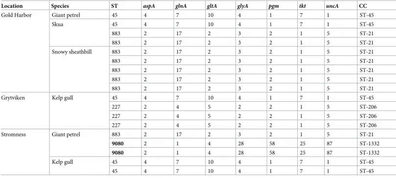
All but two of the 18C. jejuniisolates recovered belonged to known MLST sequence types (ST-45, ST-227 and ST-883) (Table 3). Sequence types ST-45 and ST-883 were found in multi-ple locations and in sammulti-ples from multimulti-ple host species. Sequence type ST-227 was only found in kelp gulls in Grytviken. The remaining two isolates belonged to a novel sequence type. Both isolates were from giant petrels in Stromness (Table 3).
Table 2. Number of samples positive for each of the five species ofCampylobacter. Numbers indicate samples for which species were determined byatpAsequencing; numbers in parentheses indicate additional samples for which species were determined by phenotypic tests, PCR and MALDI-TOF, but not byatpAsequencing. In the lat-ter case, the methods used do not distinguish betweenC. lariandC. subantarcticusorC. volucris; these samples should therefore be considered positive forC. lari-like bacteria.
Region Species C. jejuni C. lari C. peloridis C. subantarcticus C. volucris
Antarctic Peninsula Adelie penguin 0 0 0 0 0
Chinstrap penguin 0 12 0 23 0
Gentoo penguin 0 10 0 0 3
Giant petrel 0 0 0 0 0
Kelp gull 0 6 0 0 0
Skua 0 14 (1) 0 2 0
Snowy sheathbill 0 33 (2) 0 0 0
South Georgia Giant petrel 4 (3) 0 0 0
Kelp gull 6 (1) 4 (3) 0 0
King penguin 0 0 0 0 0
Skua 3 (3) 0 0 0
Snowy sheathbill 5 (2) (1) 0 0
https://doi.org/10.1371/journal.pone.0206502.t002
Table 3. Allele numbers, sequence types (STs) and clonal complexes (CCs) ofCampylobacter jejunifrom South Georgia. New STs are shown in bold.
Location Species ST aspA glnA gltA glyA pgm tkt uncA CC
Gold Harbor Giant petrel 45 4 7 10 4 1 7 1 ST-45
Skua 45 4 7 10 4 1 7 1 ST-45
883 2 17 2 3 2 1 5 ST-21
883 2 17 2 3 2 1 5 ST-21
Snowy sheathbill 883 2 17 2 3 2 1 5 ST-21
883 2 17 2 3 2 1 5 ST-21
883 2 17 2 3 2 1 5 ST-21
883 2 17 2 3 2 1 5 ST-21
883 2 17 2 3 2 1 5 ST-21
Grytviken Kelp gull 45 4 7 10 4 1 7 1 ST-45
227 2 4 5 2 2 1 5 ST-206
227 2 4 5 2 2 1 5 ST-206
227 2 4 5 2 2 1 5 ST-206
Stromness Giant petrel 883 2 17 2 3 2 1 5 ST-21
9080 2 1 4 28 58 25 87 ST-1332
9080 2 1 4 28 58 25 87 ST-1332
Kelp gull 45 4 7 10 4 1 7 1 ST-45
45 4 7 10 4 1 7 1 ST-45
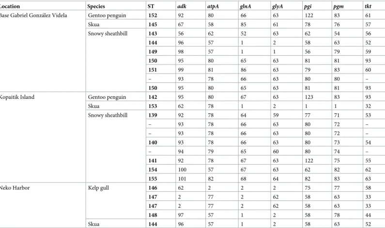
Of the 24C. lariisolates chosen for MLST analysis, 20 could be assigned to one of 17 novel sequence types (Table 4). Of the remaining four, thetktlocus could not be amplified and thus no sequence type assigned.
Discussion
In the worst-case scenario, the introduction of novel pathogens to an ecosystem may prelude an ecological catastrophe [34]. Nevertheless, in the absence of mass mortality, the establish-ment of a novel pathogen may impact reproductive investestablish-ment and success, which in turn may reduce the population size, disrupt the food web and increase the risk of species extinction [35,36]. Appropriately, the threat of such introductions to the Antarctic has been recognized [7,37]. However, whether the current measures put in place to mitigate the threat are suffi-cient, especially in the face of the predicted increase in human presence, has been called into question [9,38,39].
We isolatedCampylobacterspp. from apparently healthy birds, as was done in previous studies [18,40]. While the absence of overt signs of disease suggests commensal colonization rather than infection, clinical signs are rarely observed even in birds that mount an immune response to infection [41–43], and mild symptoms or opportunistic infections cannot be ruled out. Even if this is taken into account, it seems unlikely that the introduction ofCampylobacter spp. would have a substantial adverse impact on the Antarctic ecosystem. They may, however, be used as indicators for microbial pollution, signaling areas where care must be taken lest we cause outbreaks of more virulent pathogens.
Table 4. Allele numbers and sequence types (STs) of 24Campylobacter lariisolates from the Antarcitc Peninsula in 2014. New STs are shown in bold.
Location Species ST adk atpA glnA glyA pgi pgm tkt
Base Gabriel Gonza´lez Videla Gentoo penguin 152 92 80 66 63 122 83 61
Skua 145 67 58 85 61 78 76 57
Snowy sheathbill 143 56 62 52 63 62 54 56
144 96 57 1 2 58 63 52
149 98 57 1 1 56 79 59
150 95 80 65 63 81 81 93
151 99 81 86 63 79 83 60
– 93 78 66 63 80 80 –
150 95 80 65 63 81 81 93
Kopaitik Island Gentoo penguin 142 95 80 67 63 123 83 93
Skua 153 62 78 1 2 1 1 32
Snowy sheathbill 139 92 78 64 59 77 71 53
– 93 78 66 63 80 72 –
– 93 78 66 63 80 72 –
140 93 78 66 63 80 73 54
– 94 79 65 60 80 74 –
141 92 78 67 63 122 75 55
154 100 57 67 63 62 82 62
155 101 82 68 64 82 83 63
Neko Harbor Kelp gull 146 62 2 2 2 75 77 58
147 2 77 2 62 58 63 33
147 2 77 2 62 58 63 33
148 97 57 1 2 58 78 44
Skua 144 96 57 1 2 58 63 52
While the chosen culturing method generates the microaerobic atmosphere required for growth of most of theCampylobacterspecies previously observed in the Antarctic and sub-Antarctic, it does not generate hydrogen or formate. This excludes several species—C. conci-sus,C. curvus,C. rectus,C. mucosalis,C. showae,C. gracilis—that require hydrogen or for-mate as electron donors for microaerobic growth [10]. In addition, little is known about how different species ofCampylobacterrespond to prolonged storage in Amies medium or lysogeny broth. Barring these limitations, our findings corroborate earlier work suggesting that wild birds in the Antarctic are predominantly colonized byC. lariand closely related species [17–20]. Due to the limited number of studies ofC. lariin wild birds, it is difficult to draw conclusions as to whether the isolated strains are indigenous or if the Antarctic acts as a sink, repeatedly reseeded from an outside source. Some evidence favoring the former is provided by the MLST of the 24C. lariisolates yielding 17 novel sequence types, but without a clearer picture ofC. larihost association outside of the Antarctic this remains largely speculative.
Notably, to our knowledge, this is only the second time thatC. subantarcticushas been iso-lated in the wild.C. subantarcticus—initially described during a polyphasic taxonomic study of C. lari-like isolates from Bird Island, South Georgia [19]—responds well to isolation with rou-tine protocols used in studies of otherCampylobacterspecies. That it is largely absent in the lit-erature suggests that it may be geographically restricted to the Antarctic and sub-Antarctic, restricted to the host species that occur in the region, or both. However,Campylobacterspecies other thanC. jejuniandC. colihave generally received little attention and the apparent absence ofC. subantarcticusin other regions and in non-Antarctic species may be the result of such oversight.
While we found no evidence of introduction of human-associated strains ofCampylobacter to the Antarctic Peninsula, we did isolate such human-associated strains in South Georgia. Two of the three known sequence types recovered—ST-227 and ST-883—belong to clonal complexes frequently isolated from humans and domestic animals [44–46], but rarely from wild birds [47,48]. The third of the three known sequence types recovered—ST-45—has fre-quently been isolated from humans and domestic animals [44–46], but unlike the other two is also common in wild birds [47,49,50].
There are several routes by which human-associatedC. jejunimay have found its way to South Georgia. Some of the potential routes are historical and associated with the whaling era (1904–1965); alongside direct transmission from humans, these include the introduction of other known hosts forCampylobacter, including chickens, geese, pigeons, ducks, pigs and sheep [51]. Other potential routes may be more recent and include transmission from tourists or personnel, and yet another potential route is through transmission from remote areas by migrating birds. While the re-isolation ofC. jejuniST-45—the same sequence type isolated in 1998 on Bird Island, South Georgia, by Bromanet al.[15]—may reflect persistent circulation ofC. jejunifollowing a single introduction event, the presence of two additional human-associ-ated sequence types suggests repehuman-associ-ated introduction, but offers no further clues on the route of introduction.
In contrast to South Georgia,C. jejunihas never been found in the Antarctic, despite con-siderable monitoring effort [17,18,20]. The reason for this discrepancy remains unclear. Since the abandonment of the whaling stations in the 1960s, South Georgia houses no permanent residents, and personnel and tourist numbers are similar to comparable regions on the Penin-sula [52,53]. Furthermore, even though South Georgia is not encompassed by the Antarctic treaty regulations, similar management guidelines to limit the human impact are in place [52].
time, it is encouraging that we did not findC. jejunisouth of the 60˚S latitude—within the Antarctic Treaty Area and the pristine Antarctic—which suggests that current measures to reduce the risk of pathogen introduction may be paying off.
Supporting information
S1 Fig. Species identification ofCampylobacterstrains based on partial (1,313 bp) 16S rRNA gene sequences.
(PDF)
S2 Fig. Species identification ofCampylobacterstrains based onatpAgene sequences.
Ref-erence sequences are indicated by species names. Bootstrap values shown at nodes represent support in>95% (black),>85% (grey) and>75% (white) of 1,000 replicates, respectively. (EPS)
S1 Table. InferredCampylobacterspecies, host species, year, region, location, sample type and method ofCampylobacterspecies determination for all samples.
(HTML)
Acknowledgments
In carrying out the expeditions, we enjoyed the support of the Chilean Antarctic Institute, the Swedish Polar Research Secretariat, Quark Expeditions and the authorities of South Georgia and the South Sandwich Islands.
We thank Michele Thompson and Marı´a Fernanda Gonza´lez-Moraga, whose help during the fieldwork was indispensable. We also thank Birgitta Hellqvist and Mattias Myrenås, the captains and crews of the Ocean Diamond, Aquiles and Lautaro, as well as the officers, staff and personnel at the Antarctic bases Arctowski, Bernardo O’Higgins, Eduardo Frei, Escudero and Gabriel Gonza´lez Videla.
Our work was improved by the much appreciated input of two anonymous reviewers. This study was funded by the Chilean Antarctic Institute (INACH number T-12-13) and the Swedish Research Council Formas (2014-829). The study made use of theCampylobacter Multi Locus Sequence Typing website (https://pubmlst.org/campylobacter/) sited at the Uni-versity of Oxford [33], the development of which was funded by the Wellcome Trust.
Author Contributions
Conceptualization: Håkan Johansson, Patrik Ellstro¨m.
Data curation: Håkan Johansson.
Formal analysis: Håkan Johansson, Hanna Skarin, JoakimÅgren.
Funding acquisition: Jonas Waldenstro¨m, Daniel Gonza´lez-Acuña.
Investigation: Håkan Johansson, Patrik Ellstro¨m, Karin Artursson, Charlotte Berg, Jonas Bon-nedahl, Ingrid Hansson, Jorge Hernandez, Juana Lopez-Martı´n, Gonzalo Medina-Vogel, Lucila Moreno, Bjo¨rn Olsen, Eva Olsson Engvall, Hanna Skarin, Karin Troell, Jonas Wal-denstro¨m, JoakimÅgren.
Methodology: Håkan Johansson, Daniel Gonza´lez-Acuña.
Writing – original draft: Håkan Johansson, Patrik Ellstro¨m, Karin Artursson, Charlotte Berg, Jonas Bonnedahl, Ingrid Hansson, Jorge Hernandez, Bjo¨rn Olsen, Eva Olsson Engvall, Hanna Skarin, Karin Troell, Jonas Waldenstro¨m, JoakimÅgren.
Writing – review & editing: Håkan Johansson, Patrik Ellstro¨m, Karin Artursson, Charlotte Berg, Jonas Bonnedahl, Ingrid Hansson, Jorge Hernandez, Bjo¨rn Olsen, Eva Olsson Engvall, Hanna Skarin, Karin Troell, Jonas Waldenstro¨m, JoakimÅgren, Daniel Gonza´lez-Acuña.
References
1. Woods R, Jones HI, Watts J, Miller GD, Shellam GR. 2. In: Kerry KR, Riddle M, editors. Diseases of Antarctic seabirds. Berlin, Germany: Springer-Verlag; 2009. p. 35–55.
2. Grimaldi WW, Seddon PJ, Lyver PO’B, Nakagawa S, Tompkins DM. Infectious diseases of Antarctic penguins: current status and future threats. Polar Biology. 2015; 38(5):591–606.https://doi.org/10. 1007/s00300-014-1632-5
3. Olsen B, Bergstro¨ m S, McCafferty DJ, Sellin M, Wistro¨m J. Salmonella enteritidis in Antarctica: zoono-sis in man or humanozoono-sis in penguins? The Lancet. 1996; 348(9037):1319–1320.https://doi.org/10. 1016/S0140-6736(05)65807-2
4. Palmgren H, McCafferty D, Aspan A, Broman T, Sellin M, Wollin R, et al. Salmonella in sub-Antarctica: low heterogeneity in Salmonella serotypes in South Georgian seals and birds. Epidemiology and Infec-tion. 2000; 125(2):257–262.https://doi.org/10.1017/S0950268899004586PMID:11117947
5. Iveson JB, Shellam GR, Bradshaw SD, Smith DW, Mackenzie JS, Mofflin RG. Salmonella infections in Antarctic fauna and island populations of wildlife exposed to human activities in coastal areas of Austra-lia. Epidemiology and Infection. 2009; 137(6):858–870.https://doi.org/10.1017/S0950268808001222
PMID:18789175
6. Hurt AC, Su YCF, Aban M, Peck H, Lau H, Baas C, et al. Evidence for the introduction, reassortment, and persistence of diverse influenza A viruses in Antarctica. Journal of Virology. 2016; 90(21).https:// doi.org/10.1128/JVI.01404-16
7. Kerry KR, Riddle M. 1. In: Kerry KR, Riddle M, editors. Health of Antarctic wildlife: an introduction. Ber-lin, Germany: Springer-Verlag; 2009. p. 1–10.
8. S.C.A.R. Annex to S.C.A.R. bulletin no. 3: scientific investigations recommended by S.C.A.R. Polar Record. 1959; 9:596–603.
9. Woehler EJ, Ainley D, Jabour J. 2. In: Tin T, Liggett D, Maher PT, Lamers M, editors. Human Impacts to Antarctic Wildlife: Predictions and Speculations for 2060. Dordrecht: Springer Netherlands; 2014. p. 27–60.
10. Kaakoush NO, Castaño-Rodrı´guez N, Mitchell HM, Man SM. Global Epidemiology of Campylobacter Infection. Clinical Microbiology Reviews. 2015; 28(3):687–720.https://doi.org/10.1128/CMR.00006-15
PMID:26062576
11. Scallan E, Hoekstra RM, Angulo FJ, Tauxe RV, Widdowson MA, Roy SL, et al. Foodborne illness acquired in the United States—major pathogens. Emerging infectious diseases. 2011; 17(1).https:// doi.org/10.3201/eid1701.P11101
12. Platts-Mills JA, Kosek M. Update on the burden of Campylobacter in developing countries. Current opin-ion in infectious diseases. 2014; 27(5).https://doi.org/10.1097/QCO.0000000000000091PMID:
25023741
13. European Food Safety Authority (EFSA), European Centre for Disease Prevention and Control (ECDC). The European Union summary report on trends and sources of zoonoses, zoonotic agents and food-borne outbreaks in 2015. EFSA Journal. 2016; 14(12).
14. Garcı´a-Peña FJ, Pe´rez-Boto D, Jime´nez C, San Miguel E, Echeita A, Rengifo-Herrera C, et al. Isolation and Characterization of Campylobacter spp. from Antarctic Fur Seals (Arctocephalus gazella) at Decep-tion Island, Antarctica. Applied and Environmental Microbiology. 2010; 76(17):6013–6016.https://doi. org/10.1128/AEM.00316-10PMID:20639356
15. Broman T, Bergstro¨m S, On SLW, Palmgren H, McCafferty DJ, Sellin M, et al. Isolation and Characteri-zation of Campylobacter jejuni subsp. jejuni from Macaroni Penguins (Eudyptes chrysolophus) in the Subantarctic Region. Applied and Environmental Microbiology. 2000; 66(1):449–452.https://doi.org/10. 1128/AEM.66.1.449-452.2000PMID:10618265
17. Leotta G, Vigo G, Giacoboni G. Isolation of Campylobacter lari from seabirds in Hope Bay, Antarctica. Polish Polar Research. 2006; 27(4):303–308.
18. Garcı´a-Peña FJ, Llorente MT, Serrano T, Ruano MJ, Belliure J, Benzal J, et al. Isolation of
Campylo-bacter spp. from Three Species of Antarctic Penguins in Different Geographic Locations. EcoHealth.
2017; 14(1):78–87.https://doi.org/10.1007/s10393-016-1203-zPMID:28091764
19. Debruyne L, Broman T, Bergstro¨ m S, Olsen B, On SLW, Vandamme P. Campylobacter subantarcticus sp. nov., isolated from birds in the sub-Antarctic region. International Journal of Systematic and Evolu-tionary Microbiology. 2010; 60(4):815–819.https://doi.org/10.1099/ijs.0.011056-0PMID:19661523 20. Bonnedahl J, Broman T, Waldenstro¨m J, Palmgren H, Niskanen T, Olsen B. In search of
human-associ-ated bacterial pathogens in Antarctic wildlife: report from six penguin colonies regularly visited by tour-ists. Ambio. 2005; 34(6):430–432.https://doi.org/10.1579/0044-7447-34.6.430PMID:16201212 21. Debruyne L, On SLW, De Brandt E, Vandamme P. Novel Campylobacter lari-like bacteria from humans
and molluscs: description of Campylobacter peloridis sp. nov., Campylobacter lari subsp. concheus subsp. nov. and Campylobacter lari subsp. lari subsp. nov. International Journal of Systematic and Evo-lutionary Microbiology. 2009; 59(5):1126–1132.https://doi.org/10.1099/ijs.0.000851-0PMID:
19406805
22. Debruyne L, Broman T, Bergstro¨ m S, Olsen B, On SLW, Vandamme P. Campylobacter volucris sp. nov., isolated from black-headed gulls (Larus ridibundus). International Journal of Systematic and Evo-lutionary Microbiology. 2010; 60(8):1870–1875.https://doi.org/10.1099/ijs.0.013748-0PMID:
19767353
23. Nachamkin I. In: Murray PR, Baron EJ, Jorgensen JH, Landry ML, Pfaller MA, editors. Campylobacter and Arcobacter. 6th ed. Washington, D.C., USA: ASM Press; 1995. p. 113–117.
24. Wang G, Clark CG, Taylor TM, Pucknell C, Barton C, Price L, et al. Colony Multiplex PCR Assay for Identification and Differentiation of Campylobacter jejuni, C. coli, C. lari, C. upsaliensis, and C. fetus subsp. fetus. Journal of Clinical Microbiology. 2002; 40(12):4744–4747.https://doi.org/10.1128/JCM. 40.12.4744-4747.2002PMID:12454184
25. Mandrell RE, Harden LA, Bates A, Miller WG, Haddon WF, Fagerquist CK. Speciation of
Campylobac-ter coli, C. jejuni, C. helveticus, C. lari, C. sputorum, and C. upsaliensis by Matrix-Assisted Laser
Desorption Ionization-Time of Flight Mass Spectrometry. Applied and Environmental Microbiology. 2005; 71(10):6292–6307.https://doi.org/10.1128/AEM.71.10.6292-6307.2005PMID:16204551 26. Gordon D, Green P. Consed: a graphical editor for next-generation sequencing. Bioinformatics. 2013;
29(22):2936–2937.https://doi.org/10.1093/bioinformatics/btt515PMID:23995391
27. Ronquist F, Huelsenbeck JP. MrBayes 3: Bayesian phylogenetic inference under mixed models. Bioin-formatics. 2003; 19(12):1572–1574.https://doi.org/10.1093/bioinformatics/btg180PMID:12912839 28. Miller WG, Yee E, Jolley KA, Chapman MH. Use of an improved atpA amplification and sequencing
method to identify members of the Campylobacteraceae and Helicobacteraceae. Letters in Applied Microbiology. 2014; 58(6):582–590.https://doi.org/10.1111/lam.12228PMID:24517729
29. Kazutaka K, Standley DM. MAFFT multiple sequence alignment software version 7: improvements in performance and usability. Molecular biology and evolution. 2013; 30(4):772–780.https://doi.org/10. 1093/molbev/mst010
30. Stamatakis A. RAxML version 8: a tool for phylogenetic analysis and post-analysis of large phylogenies. Bioinformatics. 2014; 30(9):1312–1313.https://doi.org/10.1093/bioinformatics/btu033PMID:24451623 31. Dingle KE, Colles FM, Wareing DRA, Ure R, Fox AJ, Bolton FE, et al. Multilocus Sequence Typing
Sys-tem for Campylobacter jejuni. Journal of Clinical Microbiology. 2001; 39(1):14–23.https://doi.org/10. 1128/JCM.39.1.14-23.2001PMID:11136741
32. Miller WG, On SLW, Wang G, Fontanoz S, Lastovica AJ, Mandrell RE. Extended multilocus sequence typing system for Campylobacter coli, C. lari, C. upsaliensis, and C. helveticus. Journal of Clinical Micro-biology. 2005; 43(5):2315–2329.https://doi.org/10.1128/JCM.43.5.2315-2329.2005PMID:15872261 33. Jolley KA, Maiden MCJ. BIGSdb: Scalable analysis of bacterial genome variation at the population
level. BMC Bioinformatics. 2010; 11(1).https://doi.org/10.1186/1471-2105-11-595PMID:21143983 34. Daszak P, Cunningham AA, Hyatt AD. Emerging infectious diseases of wildlife–threats to biodiversity
and human health. Science. 2000; 287(5452):443–449.https://doi.org/10.1126/science.287.5452.443
PMID:10642539
35. Tompkins DM, Begon M. Parasites Can Regulate Wildlife Populations. Parasitology Today. 1999; 15 (8):311–313.https://doi.org/10.1016/S0169-4758(99)01484-2PMID:10407375
36. Smith KF, Acevedo-Whitehouse K, Pedersen AB. The role of infectious diseases in biological conserva-tion. Animal Conservaconserva-tion. 2009; 12(1).https://doi.org/10.1111/j.1469-1795.2008.00228.x
38. Convey P, Hughes KA, Tin T. Continental governance and environmental management mechanisms under the Antarctic Treaty System: sufficient for the biodiversity challenges of this century? Biodiversity. 2012; 13(3–4):234–248.https://doi.org/10.1080/14888386.2012.703551
39. Walton D. Keeping the aliens out. Antarctic Science. 2012; 24(4):321.https://doi.org/10.1017/ S0954102012000594
40. Gonza´ lez-Acuña D, Herna´ndez J, Moreno L, Herrmann B, Palma R, Latorre A, et al. Health evaluation of wild gentoo penguins (Pygoscelis papua) in the Antarctic Peninsula. Polar Biology. 2013; 36 (12):1749–1760.https://doi.org/10.1007/s00300-013-1394-5
41. Cawthraw S, Ayling R, Nuijten P, Wassenaar T, Newell DG. Isotype, Specificity, and Kinetics of Sys-temic and Mucosal Antibodies to Campylobacter jejuni Antigens, Including Flagellin, during Experimen-tal Oral Infections of Chickens. Avian Diseases. 1994; 38(2):341–349.https://doi.org/10.2307/1591960
PMID:7526839
42. Waldenstro¨m J, Axelsson-Olsson D, Olsen B, Hasselquist D, Griekspoor P, Jansson L, et al.
Campylo-bacter jejuni Colonization in Wild Birds: Results from an Infection Experiment. PLOS ONE. 2010; 5(2).
43. Humphrey S, Chaloner G, Kemmett K, Davidson N, Williams N, Kipar A, et al. Campylobacter jejuni Is Not Merely a Commensal in Commercial Broiler Chickens and Affects Bird Welfare. mBio. 2014; 5(4): e01364–14.https://doi.org/10.1128/mBio.01364-14PMID:24987092
44. E DK, Colles FM, Ure R, Wagenaar JA, Duim B, Bolton FJ, et al. Molecular characterization of
Campylo-bacter jejuni clones: a basis for epidemiologic investigation. Emerging infectious diseases. 2002; 8
(9):949–55.https://doi.org/10.3201/eid0809.020122
45. Manning G, Dowson CG, Bagnall MC, Ahmed IH, West M, Newell DG. Multilocus Sequence Typing for Comparison of Veterinary and Human Isolates of Campylobacter jejuni. Applied and Environmental Microbiology. 2003; 69(11):6370–6379.https://doi.org/10.1128/AEM.69.11.6370-6379.2003PMID:
14602588
46. Colles FM, Jones K, Harding RM, Maiden MCJ. Genetic Diversity of Campylobacter jejuni isolates from farm animals and the farm environment. Applied and Environmental Microbiology. 2003; 69(12):7409– 7413.https://doi.org/10.1128/AEM.69.12.7409-7413.2003PMID:14660392
47. Griekspoor P, Colles FM, McCarthy ND, Hansbro PM, Ashhurst-Smith C, Olsen B, et al. Marked host specificity and lack of phylogeographic population structure of Campylobacter jejuni in wild birds. Molec-ular Ecology. 2013; 22(5):1463–1472.https://doi.org/10.1111/mec.12144PMID:23356487
48. Waldenstro¨m J, Griekspoor P. In: Sheppard SK, editor. Ecology and host associations of
Campylobac-ter in wild birds. Norfolk: CaisCampylobac-ter Academic Press; 2014. p. 265–284.
49. Colles FM, McCarthy ND, Howe JC, Devereux CL, Gosler AG, Maiden MCJ. Dynamics of
Campylobac-ter colonization of a natural host, Sturnus vulgaris (European Starling). Environmental Microbiology.
2009; 11(1):258–267.https://doi.org/10.1111/j.1462-2920.2008.01773.xPMID:18826435
50. French NP, Midwinter A, Holland B, Collins-Emerson J, Pattison R, Colles F, et al. Molecular epidemiol-ogy of Campylobacter jejuni isolates from wild-bird fecal material in children’s playgrounds. Applied and Environmental Microbiology. 2009; 75(3):779–783.https://doi.org/10.1128/AEM.01979-08PMID:
19047378
51. Tønnessen JN, Johnsen AO. The history of modern whaling. Berkeley and Los Angeles, CA, USA: University of California Press; 1982.
52. Government of South Georgia and the South Sandwich Islands. Biodiversity action plan for South Geor-gia and the South Sandwich Islands 2016–2020; 2016.