3 Departamento de Neurologı´a, Facultad de Medicina, Pontificia Universidad Cato´lica de Chile, Santiago, Chile, 4 Instituto de Ciencias Biome´dicas, Facultad de Medicina, Universidad de Chile, Santiago, Chile, 5 Fundacio´n Ciencia & Vida, Santiago, Chile, 6 Facultad de Ciencias de la Salud, Universidad San Sebastia´ n, Santiago, Chile, 7 Facultad de Ciencia, Universidad San Sebastia´n, Santiago, Chile, 8 Regeneron Pharmaceuticals, New York, New York, United States of America, 9 Facultad de Ciencias Biolo´gicas, Departamento de Ciencias Biolo´gicas, Universidad Andres Bello, Santiago, Chile, 10 Facultad de Medicina, Universidad San Sebastia´ n, Santiago, Chile
☯These authors contributed equally to this work. *[email protected](AG);[email protected](AS)
Abstract
Galectin-8 (Gal-8) is a member of a glycan-binding protein family that regulates the immune system, among other functions, and is a target of antibodies in autoimmune disorders. How-ever, its role in multiple sclerosis (MS), an autoimmune inflammatory disease of the central nervous system (CNS), remains unknown. We study the consequences of Gal-8 silencing on lymphocyte subpopulations and the development of experimental autoimmune encepha-litis (EAE), to then assess the presence and clinical meaning of anti-Gal-8 antibodies in MS patients. Lgals8/Lac-Z knock-in mice lacking Gal-8 expression have higher polarization toward Th17 cells accompanied with decreased CCR6+and higher CXCR3+regulatory T cells (Tregs) frequency. These conditions result in exacerbated MOG35-55peptide-induced
EAE. Gal-8 eliminates activated Th17 but not Th1 cells by apoptosis and ameliorates EAE in C57BL/6 wild-type mice.β-gal histochemistry reflecting the activity of the Gal-8 promoter revealed Gal-8 expression in a wide range of CNS regions, including high expression in the choroid-plexus. Accordingly, we detected Gal-8 in human cerebrospinal fluid, suggesting a role in the CNS immune-surveillance circuit. In addition, we show that MS patients generate function-blocking anti-Gal-8 antibodies with pathogenic potential. Such antibodies block cell adhesion and Gal-8-induced Th17 apoptosis. Furthermore, circulating anti-Gal-8 antibodies associate with relapsing-remitting MS (RRMS), and not with progressive MS phenotypes, predicting clinical disability at diagnosis within the first year of follow-up. Our results reveal
OPEN ACCESS
Citation: Pardo E, Ca´rcamo C, Uribe-San Martı´n R, Ciampi E, Segovia-Miranda F, Curkovic-Peña C, et al. (2017) Galectin-8 as an immunosuppressor in experimental autoimmune encephalomyelitis and a target of human early prognostic antibodies in multiple sclerosis. PLoS ONE 12(6): e0177472. https://doi.org/10.1371/journal.pone.0177472
Editor: Robyn S Klein, Washington University, UNITED STATES
Received: November 22, 2016
Accepted: April 27, 2017
Published: June 26, 2017
Copyright:©2017 Pardo et al. This is an open access article distributed under the terms of the Creative Commons Attribution License, which permits unrestricted use, distribution, and reproduction in any medium, provided the original author and source are credited.
Data Availability Statement: All relevant data are within the paper and its Supporting Information files.
CAPES-that Gal-8 has an immunosuppressive protective role against autoimmune CNS inflamma-tion, modulating the balance of Th17 and Th1 polarization and their respective Tregs. Such a role can be counteracted during RRMS by anti-Gal-8 antibodies, worsening disease prog-nosis. Even though anti-Gal-8 antibodies are not specific for MS, our results suggest that they could be a potential early severity biomarker in RRMS.
Introduction
Multiple sclerosis (MS) is an autoimmune inflammatory disease of the central nervous system (CNS) that damages myelin and axons in the brain and spinal cord [1,2]. Most (80%) of patients are diagnosed with the relapsing-remitting form of MS (RRMS) and 60% of them evolve towards secondary progressive MS (SPMS) [1]. A subset of patients (20%) exhibits pri-mary progressive MS (PPMS) displaying worse evolution than RRMS patients from the begin-ning [1]. Such wide variations in the phenotype and aggressiveness of the disease are also reflected in the characteristic heterogeneity in presentation, evolution and treatment responses of RRMS [1,3,4]. Although the pathogenic mechanisms remain little understood, during RRMS an autoimmune-driven inflammation of variable intensity and dynamics predominates, underlying the severity of clinical evolution [1]. Observations in RRMS patients, as well as in the MS preclinical mouse model of experimental autoimmune encephalomyelitis (EAE), sug-gest that disease pathogenesis and resolution depends on a fine balance between the autoim-mune inflammation mediated by effector Th1 and Th17 lymphocytes [3,5] and the immune tolerance promoted by suppressive regulatory T cells (Tregs) [6,7]. Subpopulations of Tregs expressing either CXCR3 or CCR6 chemokine receptors, respectively, suppress Th1 or Th17-mediated inflammation [8–11], and are recruited to the CNS during neuroinflammatory pro-cesses [12,13], but their role in MS and EAE remains unclear [6]. An additional enigmatic aspect regards the pathogenic role of antibodies, which remains contentious and has mainly been explored focusing on neuronal and myelin elements as antigenic targets [3,14]. There-fore, modulators of Th1, Th17 and Treg cell homeostasis and their eventual neutralization by function-blocking antibodies entail great interest. Their study might not only reveal new determinants of worse disease evolution but also suggest new prognostic and/or therapeutic approaches.
Galectins are a family of glycan-binding proteins that has emerged as strong modulators of adaptive and innate immunity [15]. These lectins are secreted by an unconventional mecha-nism and have the potential to modify the function of a variety of cell surface glycoproteins, including signaling receptors and integrins involved in immune-related cellular processes [16–
18]. The conserved carbohydrate-recognition domains (CRDs) of galectins recognize β-tosides, displaying variations that result in unique fine specificities for more complex galac-tose-containing oligosaccharides and specific downstream effects [19–22]. In this way, different galectins can play redundant or complementary roles in autoimmune diseases by modulating the homeostasis of B and T cells [15], including Th1, Th17 cells and Tregs [15]. Among the 15 members of the galectin (Gal) family, only Gal-1, Gal-3, and Gal-9 have been systematically studied in EAE [20,23–26].
Gal-8 is a tandem-repeat galectin that, unlike other galectins, possesses unique high affinity forα-2,3-sialylated glycans in its N-terminal carbohydrate recognition domain (CRD), which is linked by a short peptide to a C-terminal CRD bearing another glycan specificity [27,28]. Gal-8 has been proposed to play immunosuppressive roles inducing apoptosis of activated T
CONICYT FB 0002 (Line 3) to FB (P.C.). FONDECYT N˚ 3120061 (E.P.). The funder provided support in the form of salaries for authors EP, CC, CH, CC, CH, PC, CO, RP, RN, AS and AG, but did not have any additional role in the study design, data collection and analysis, decision to publish, or preparation of the manuscript. The specific roles of these authors are articulated in the ‘author contributions’ section.
type mice; 3) The presence and clinical meaning of autoantibodies against Gal-8 in patients with MS. Our results indicate a protective immunosuppressive role of Gal-8 against CNS auto-immunity involving apoptotic elimination of activated Th17 cells and opposite dysbalances of CXCR3+and CCR6+Treg frequencies. Strikingly, MS patients generate neutralizing anti-Gal-8 antibodies that can block the Gal-anti-Gal-8 immunosuppressive function and impact upon RRMS prognostic. Patients recently diagnosed with RRMS and harboring anti-Gal-8 antibodies in serum, before establishing the treatment, develop a more aggressive disease within just one year of evolution, as shown by monitoring the Expanded Disability Status Scale (EDSS). Over-all, the evidence suggests that Gal-8 plays an immunosuppressive function that can be counter-acted by anti-Gal-8 antibodies in RRMS leading to worse evolution. Although Gal-8 function-blocking antibodies are not specific to MS, as they can be found in other autoimmune diseases [39], their presence in context of RRMS might be useful as an early biomarker of worse evolution.
Material and methods
Study approval
The Scientific Ethics Committee CEC-MED-UC and the Science Ethics Committee for Animal and Environmental Care of the Pontificia Universidad Cato´lica de Chile (PUC) approved the informed consent voluntarily given by MS patients and all animal procedures, respectively.
Patients and clinical variables
Antibodies and reagents
To determine expression levels of key surface and intracellular molecules associated with dif-ferent T-cell phenotypes, splenocytes were immunostained with difdif-ferent combinations of the following fluorochrome-conjugated monoclonal antibodies (mAbs) at room temperature for 30 min: allophycocyanin (Apc)-conjugated anti-FoxP3 (clone FJK-16S) and R-Phycoerythrin (PE)-conjugated anti-RORγt (clone BSD), both from eBioscience (San Diego, CA). Otherwise, PerCP/Cyanine dye 5.5 (PerCP/Cy5.5)-conjugated anti-CCR6 (clone 29-2L17), PE/Cyanine dye 7 (PE/Cy7)-conjugated anti-CD4 (clone GK1.5), Brilliant-Violet 421TM (Bv421)-conju-gated anti-CXCR3 (Clone CXCR3-173), PE-conju(Bv421)-conju-gated anti-CD44 (clone IM7), APC-conju-gated anti-CD25 (clone PC61) and APC/Cyanine dye 7 (APC/Cy7)-conjuAPC-conju-gated anti-CD62L (clone MEL14) were purchased from Biolegend (San Diego, CA). PerCP-conjugated anti-CD4 (clone RM4-5), PECy7-conjugated IFN-γ(clone XMG1.2) were purchased from eBioscience. Alexa 647-conjugated IL-17A (clone TC11-18H10.1), PE-conjugated anti-IFN-γand Alexa 488–conjugated anti-IL-17 were purchased from BioLegend. Alexa 488-conjugated anti-CD8a (clone 53–6.7), alexa 647-conjugated anti-CD19 (clone 1D3), PE-conjugated anti-CD11c (clone HL3), PE-conjugated anti-IL4 (11B11), Alexa 647-conjugated anti-IL17A (clone TC11-18H10), PE/Cy7-conjugated anti-IFN-γ(cloneXMG1.2) were purchased from BD Biosci-ences-Pharmingen. For T cell activation, anti-mouse CD28 (clone 37.51) and anti-mouse CD3 (clone 145-2C11) were purchased from BD Bioscience.
Ionomicina, brefeldin A and PMA (Phorbol Myristate Acetate) were purchased from Sigma-Aldrich. GST-Gal-8 expressed in bacteria was isolated by affinity chromatography and Gal-8 was released by thrombin treatment as described [37].
Animals
Mice were mantained under conditions of strict confinement, which included automatic con-trol of temperature (21˚C) and photoperiod (12 h light / 12 h dark).Lgals8/Lac-Z knock-in (here called Gal-8 KO orLgals8-/-) mice were generated fromC57BL/6NTacmice engineered in Regeneron Pharmaceuticals Inc., New York, using Velocigene technology for replacing the entire coding region of the mouseLgals8gene (18,427 bp) with LacZ lox-Ub1-EM7-Neo-lox Cassette containing the LacZ gene that encodesβ-galactosidase [40]. Details of theLgals8KO mice and PCR genotyping assay, including the predicted PCR products and the primers, are available at the Velocigene website (www.velocigene.com/komp/detail/14305). IL-17A-GFP reporter mice in the C57BL/6J background, which express EGFP under the control of the IL-17A promoter, were purchased from Jackson Laboratories (Bar Harbor, ME).
EAE induction, scoring and treatment
FACS. For polyclonal T cell activation, splenocytes were grown in the presence of 1μg/ml of anti-CD3 with or without anti-CD28 antibodies for 72 h. For Gal-8 effects on antigen-specific T cell activation, splenocytes were isolated after 10 days of EAE induction and re-stimulated with 10μg/ml MOGp in the presence or absence of 100μg/ml recombinant Gal-8 for 72 h. CD4+T cell subpopulations were analyzed in stimulated cells by incubating with 50 ng/ml PMA, 500 ng/ ml ionomycin, and 10μg/ml brefeldin A for the last 4 h of culture. Cells were stained with Zom-bie Aqua Fixable Viability kit (Biolegend) in PBS followed by staining for cell-surface markers using flurophore-conjugated monoclonal antibodies against mouse CD4, CD19, CD8, CD11c, CD25, CD44, CD62L, CXCR3 and CCR6. For intracellular staining, cells were washed, fixed, and permeabilized with Cytofix/Cytoperm kit (BD Biosciences-Pharmingen) and incubated simultaneously with fluorophore-conjugated antibodies against IFN-γ, IL-4, IL-17, Foxp3 and RORγt. Samples were acquired with BD FACSVerse or FACSCantoTM
II (BD) flow cytometers and data was analyzed with FlowJo V10 software or CellQuest Pro software (BD Biosciences).
Th17 cell death analysis
Detection of anti-Gal-8 autoantibodies
Western blot analysis was performed using recombinant Gal-8 to screen sera of patients with MS for anti-Gal-8 antibodies, as previously described in patients with SLE or AR [39]. The same positive and negative controls were included in each western blot and densitometric analysis of the bands was performed by ImageJ. Bands that duplicate the intensity of the nega-tive control in at least two independent experiments performed in triplicate and blinded with respect to the sample were considered positive for anti-Gal-8 reactivity.
Detection of Gal-8 in human cerebro spinal fluid (CSF)
CSF collected for diagnostic purposes from individuals studied for diplopia, vertiginous syn-drome (C7), cephalea, febrile synsyn-drome and meningitis was analyzed for the presence of Gal-8 by immunoblot, using a rabbit anti-Gal-8 polyclonal antibody produced in our laboratory [45]. CSF (1 ml) was centrifuged for 10 min at 1,000 g to eliminate debris and the supernatant was incubated with 50μl ofα-lactose-agarose beads for 3 h at 4˚C in presence of anti-proteases (2μg/ml leupeptin, 2μg/ml pepstatin and 2 mM PMSF). Lectins bound toα-lactose-agarose beads were pulled down at 1,000 rpm for 3 min, the beads were washed three times with PBS and suspended in 40μl of electrophoresis loading buffer. The samples were heated for 5 min at 100˚C and loaded in 10% SDS-PAGE, transferred to nitrocellulose membranes and analyzed with anti-Gal-8 antibodies.
Isolation of human peripheral blood mononuclear cells (PBMC)
Ten milliliters of peripheral blood was diluted with 10 ml of PBS, layered carefully over 7.5 ml of Histopaque-1077 (Sigma Chem. Co.) and centrifuged at 400 x g for 30 min at room temper-ature. Cells in the opaque interface were collected, diluted twice with 50 ml of PBS, centrifuged at 250 x g for 10 min and resuspended in RPMI 1640 medium containing 10% FBS.
Cell adhesion assay
Cover slips were coated overnight at 4˚C with 10μg/ml of Gal-8 in the absence or presence of anti-Gal-8 autoantibodies, as described [37,38,46]. Serum-starved PBMCs isolated from healthy people (200,000 cells/well) were plated on cover slips for 30 min at 37˚C. Adherent cells were stained with Hoechst and quantified analyzing at least three random fields on three cover slips.
Statistical analysis
The presence of anti-Gal-8 autoantibodies in RRMS, PPMS and SPMS patients was analyzed respect to various clinical, radiological and CSF variables using Mann-Whitney U test (with Tukey multiple comparisons) and SPSS Statistics 19.0 software. Yates chi-square corrected test and Odds ratio were calculated with VassarStats. EAE disease scores were analyzed with Mann–Whitney U test or one-way ANOVA Kruskal Wallis with Dunn post-test using Prism 5 software (GraphPad Software). Other analyses were made with two tailed non-paired Student’s t-test as indicated in figure legends.
Results
Endogenous Gal-8 protects against EAE
inLgals8-/-andLgals8+/+littermates immunized with MOG35-55peptide (MOGp). The two mouse groups had similar EAE incidence (85.2%Lgals8+/+littermates and 86.2%Lgals8-/-). However,Lgals8-/-mice displayed a faster disease onset and developed a more severe disease during the chronic phase (Fig 1AandTable 1). Spinal cord histological analysis showed enhanced inflammation (Fig 1B, i and ii) and demyelination (Fig 1B, ii and iv) in the affected areas ofLgals8-/-mice. Such EAE exacerbation under Gal-8 deficiency suggests a protective role of endogenous Gal-8 against CNS autoimmunity.
Gal-8 silencing leads to increased Th17 cells and Th1-like Tregs
To understand how the lack of Gal-8 predisposes the immune system to a more pronounced CNS autoimmunity we compared lymphocyte subpopulations and the responses of spleen cells toex vivoanti-CD3/anti-CD28 activation and to re-stimulation with MOGp. Splenocytes fromLgals8-/-andLgals8+/+littermates showed similar frequencies of B and T (CD4+and CD8+) lymphocytes, dendritic cells, and CD4+T-cells subsets, including naive (CD4+CD25-or
Fig 1. Lack of endogenous Gal-8 expression exacerbates EAE. Lgals8+/+(WT) and Lgals8-/-(Gal-8 KO) immunized with MOGp. (A) Clinical scores monitored daily for 25 days show an exacerbated EAE in Gal-8 KO mice (***p<0.001; Mann Whitney; day by day clinical Score comparisons; n = 20 WT; n = 22 Gal-8 KO). (B) Spinal cord histopathological analysis after 10 days of immunization show enhanced immune cell infiltration and demyelination in Gal-8 KO mice, shown by H&E staining (I and II) and luxol fast blue staining (III and IV). Scale bar = 50μm.
https://doi.org/10.1371/journal.pone.0177472.g001
Table 1. Clinical parameters of EAE progression.
Group of mice n Incidence (%)
Day of Onset (Mean±SD)
Maximum Score (Mean±SD)
Time to Peak (Mean±SD)
Accumulative Score (Mean±SD)
Lgals8+/+ 20 85.2 13.5±2.8 3.4±0.7 15.9±3.4 28.2±11.9
Lgals8-/- 22 86.2 12.5±2.6 3.6±0.6 15.5±3.2 40.7±23.2*
*P<0.05 comparing accumulative scores between WT and Gal-8 KO mice.
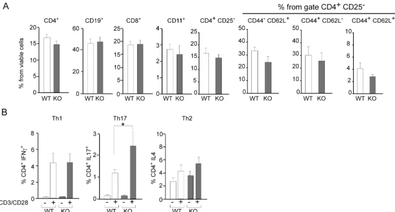
CD4+CD62L+CD44-), effector (CD4+CD62L-CD44+) and memory (CD4+CD62L+CD44+) T-cells (Fig 2A). However,Lgals8-/-splenocytes displayed higher polarization towards Th17 (CD4+IL17+) after anti-CD3/anti-CD28 activation (Fig 2B).
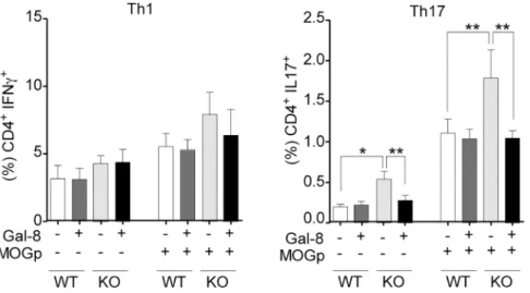
We then compared the systemic expansion of Th1 and Th17 cells during EAE induction by analyzing the splenocytes fromLgals8-/-andLgals8+/+mice after 10 days post-immunization andex vivore-stimulation with MOGp. We did not detect meaningful differences in the fre-quency of Th1 cells betweenLgals8-/-andLgals8+/+mice (Fig 3). Th1 cells also remained unaf-fected underex vivoGal-8 incubation (Fig 3). In contrast, splenocytes fromLgals8-/-mice unstimulated or re-stimulated with MOGpex vivoshowed an increased frequency of Th17 cells compared withLgals8+/+littermates (Fig 3). Incubation with exogenous Gal-8 reduced the frequency of Th17 cells inLgals8-/-splenocytes to the levels of control mice (Fig 3), thus contrasting with the lack of response seen inLgals8+/+splenocytes. Therefore, in the absence of endogenous Gal-8 expression there is increased Th17 polarization, while in the presence of endogenous Gal-8, the Th17 cells activated by MOGp immunization and likely involved in EAE seem to be eliminated.
As Tregs have been shown to control Th17 and Th1-mediated tolerance and inflammatory responses in MS and EAE [6], we analyzed total Tregs, as well as Tregs subpopulations that suppress responses mediated by either Th1 (CXCR3+CCR6-) [9,10] or Th17 (CXCR3 -CCR6+) Tregs [11] lymphocytes. Unexpectedly, we found an increased frequency of total Tregs (Foxp3+) inLgals8-/-mice splenocytes compared to control mice (Fig 4). We also found
Fig 2. Gal-8 deficit favors selective Th17 cell differentiation upon polyclonal activation. Splenocytes isolated from Lgals8+/+(WT) and Lgals8 -/-(KO) mice were analyzed by FACS: (A) Dendritic cells (CD11c+), B cells (CD19+), CD8+T cells and different CD4+T cells subsets, naïve (CD44-CD62L+), effector (CD44+CD62L+), memory (CD44+CD62L-) and total cells analyzed in the subset of viable CD4+CD25-T-cells show no differences between WT and KO mice. Graphics of frequency +/-SD (n = 5). (B) T cell activation by 72 h incubation with anti-CD3 (anti-CD3) and anti-CD28 (anti-CD28) antibodies show Th17 increased frequency in KO mice while Th1 and Th2 cells are similar in WT and KO mice. Graph shows frequency +/-SD (*p<0.05; ANOVA; n = 4).
a highly increased frequency of CXCR3+Tregs, whereas CCR6+Tregs tended to decrease in Lgals8-/-compared with controlLgals8+/+mice (Fig 4). Therefore, the absence of Gal-8 leads to an increase of Th17 polarization, as well as higher generation of CXCR3+Tregs and lower fre-quency of CCR6+Tregs, which respectively would impact upon Th1 and Th17 functions [8].
Gal-8 ameliorates EAE and induces apoptosis of activated Th17 cells
The key role of endogenous Gal-8 in regulating the functions of effectors and Tregs during EAE development prompted us to evaluate the impact of the exogenous Gal-8 treatment on EAE induction and Th17 survival in wild-type C57BL/6 mice. Daily treatment with Gal-8 starting from disease induction significantly delayed the progression of EAE clinical symptoms (Fig 5AandTable 2). To test the sensitivity of wild-type activated Th17 cells to Gal-8 treat-ment, we differentiated Th17 cellsin vitroand activated them with anti-CD3/anti-CD28.
Fig 4. Galectin-8 deficit increases the frequency of total Tregs and CXCR3+Tregs. Splenocytes isolated from Lgals8+/+(WT) and Lgals8-/-(KO) mice were analyzed at steady state for total Tregs (Foxp3+), CXCR3+ and CCR6+frequency in the Treg (Foxp3+CD4+) population. Graphs of frequency +/-SEM show increased total Tregs (Foxp3+) and CXCR3+
Tregs in KO mice (*p<0.05;**p<0.01; p***<0.001; Student’s t-test; n = 4).
https://doi.org/10.1371/journal.pone.0177472.g004
Fig 3. Gal-8 deficit favors Th17 polarization during MOGp-induced EAE and ex-vivo re-stimulation. Th17 and Th1 subpopulations in splenocytes from Lgals8-/-(KO) and Lgals8+/+(WT) mice obtained after 10 days of EAE induction were analyzed either immediately or after 72 h of ex vivo MOGp re-stimulation, in the absence or presence of Gal-8. Gal-8 KO mice show higher frequency of Th17 cells both at steady state and after MOGp re-stimulation. Incubation with Gal-8 reduced Th17 cells only in Gal-8 KO. Graph shows frequency +/-SD (*p<0.05; ANOVA; n = 4).
Annexin V/7-AAD staining showed that Gal-8 induced apoptosis of these Th17 activated cells (Fig 5B). These results indicate that exogenous Gal-8 exert immune-suppressive action against EAE induction involving apoptotic elimination of activated Th17 cells.
Fig 5. Gal-8 ameliorates EAE and induces Th17 cell death in vitro. (A) Gal-8 treatment ameliorates MOGp-induced EAE in C57BL/6 mice. The mice were injected daily by intraperitoneal injection of either PBS (Control) or Gal-8 100μg/ml. Gal-8-treated animals tend to start the disease later and show lower EAE scores during the acute and chronic phases of the disease (*p<0.05; Control, n = 7; Gal-8 treated, n = 5). (B) Gal-8-induced cell death in Th17 lymphocytes differentiated and activated in vitro. Naive (CD62L+CD44-) CD4+T cells were purified by cell sorting and differentiated to a Th17 phenotype. The differentiated Th17 cells were isolated with a commercial kit based on the cell surface expression of IL-17 and activated with anti-CD3/anti-CD28 in the presence or absence of Gal-8 (20μg/ml) for 72 h. Cell death was determined by cell staining with Annexin V and 7-AAD and analyzed by FACS. Representative dot plots show the frequency of Th17 cells before and after purification (upper panels), the selected gate in the forward scatter versus side scatter analysis (middle panels) and the associated contour-plots show the Annexin V versus 7-AAD analysis (lower panels). Numbers in quadrants indicate the percentage of cells in the respective quadrant.
https://doi.org/10.1371/journal.pone.0177472.g005
Table 2. Clinical parameters of EAE disease progression (day 0–20).
Group of mice n Day of Onset (Mean±SD)
Maximum Score (Mean±SD)
Time to Peak (Mean±SD)
Accumulative Score (Mean±SD)
Vehicle 7 12.7±1.7 4.1±0.8 16.4±2.4 20.8±8.0
Gal-8 treatment 5 13.6±0.9 2.8±1.6 17.8±1.4 10.1±7.0*
*P<0.05 comparing accumulative scores of mice injected with either vehicle or Gal-8.
Gal-8 expression in the brain
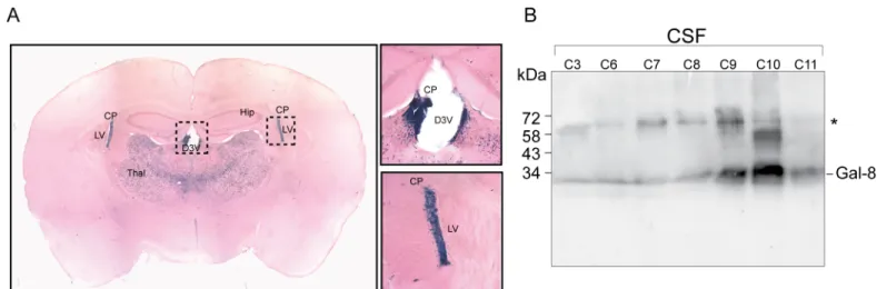
Although peripheral events tailoring the immune system contribute to MS and EAE pathology, most of the autoimmune pathogenic condition unfolds inside the CNS [3]. In our knock-in Lgals8mice theβ-galactosidase (β-gal) cassette reporter gene replaces the entire Gal-8 gene with LacZ, thus offering the possibility to assess the activity of the corresponding promoter by β-gal histochemistry [40–42]. This analysis revealed Gal-8 expression in several brain regions (Fig 6A;S1 Table). Interestingly, the choroid plexus, which generates CSF [47], displayed high expression levels, suggesting that Gal-8 might be secreted into the CSF. To test this possibility we analyzed CSF from patients studied for other pathologies, mainly cephalea, and included one patient with meningitis. We detected Gal-8 in all CSF samples with variable intensity. The highest levels corresponded to a patient studied for cephalea (Fig 6B). These results suggest that Gal-8 produced by the choroid-plexus is a component of the CSF.
Patients with MS generate function-blocking antibodies against Gal-8
The results showing an immunosuppressive and protective role of Gal-8 against EAE prompted us to assess whether patients with MS generate blocking-function Gal-8 anti-bodies, as previously reported in LES and AR patients [39,46]. Using recombinant human Gal-8 [37] and immunoblot analysis [39] we found clear evidence of anti-Gal-8 autoantibodies in a cohort of RRMS patients (Fig 7). We also found evidence of the presence of anti-Gal-8 antibodies in CSF, either coincident or independent of serum reactivity (Fig 7).
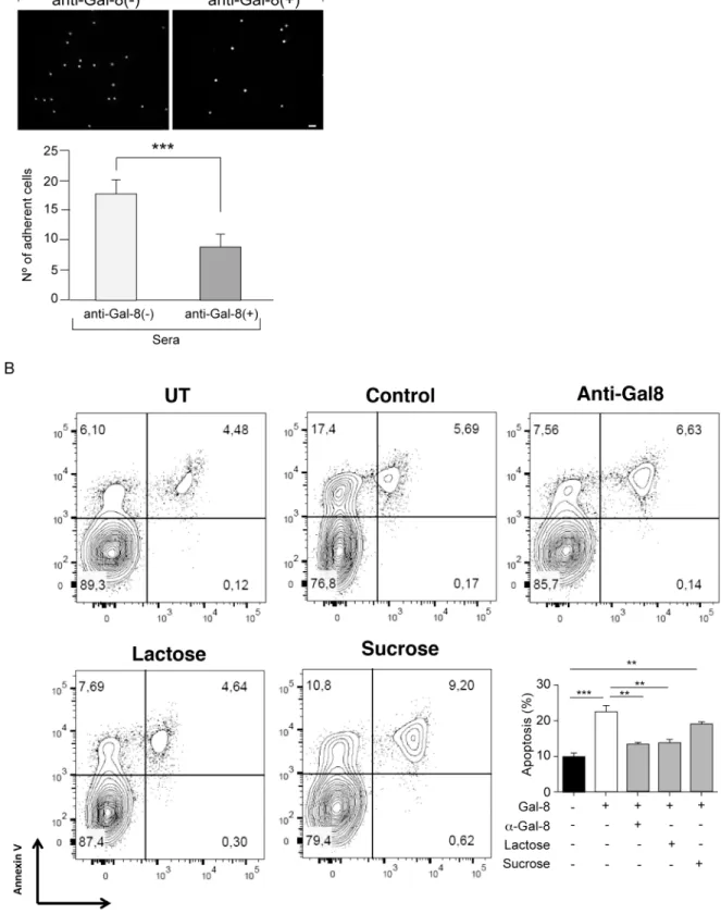
To evaluate whether MS patients generate function-blocking anti-Gal-8 autoantibodies we performed two assays. The established assay of cell adhesion to Gal-8-coated coverslips, which assesses glycan-mediated interaction of Gal-8 with integrins [37,38], showed that anti-Gal-8 (+) serum from RRMS patients decreases the adhesion of peripheral blood mononuclear cells (PBMC) (Fig 8A). In addition, we tested the potential of MS-generated anti-Gal-8 antibodies to counteract the apoptotic effect of Gal-8 on activated Th17 cells showed above inFig 5B. Affinity purified anti-Gal-8 antibodies from a pooled anti-Gal-8(+) sera effectively decreased
Fig 6. Gal-8 expression in mouse brain and presence in human CSF. (A) Histochemistry ofβ-gal staining reveals Gal-8 expression in several regions of the mouse brain (S1 Table). Brain slices depict high Gal-8 expression levels in the choroid plexus (CP) of the lateral ventricle (LV) and the dorsal 3rd ventricle (D3V), as well as in the ventrolateral thalamic nucleus. (B) Immunoblot with rabbit anti-Gal-8 antibody show Gal-8 reactivity in the CSF of individuals without MS. Samples C3-11 correspond to non-inflammatory CSF from individuals studied for diplopia (C3), vertiginous syndrome (C7), cephalea (C8 and C11) and febrile syndrome (C9), whereas C6 is an inflammatory CSF from a patient with meningitis. All samples show anti-Gal-8 reactivity, though with variable intensity.*Bands of unknown origin might include Gal-8 dimers or complexes with other proteins, not separable under SDS-PAGE conditions.
the apoptosis rate of activated Th17 cells incubated with Gal-8 (Fig 8B). These results indicate that patients with RRMS generate function-blocking anti-Gal-8 antibodies, which have the potential to neutralize the immunosuppressive role of Gal-8.
Circulating anti-Gal-8 antibodies are associated with worse prognosis in
patients with RRMS
As the presence of anti-Gal-8 neutralizing antibodies might mimic the condition of Gal-8 silencing that exacerbates EAE, we next explored the impact of these antibodies on the clinical course of MS. We studied 58 patients, 36 with recent diagnosis of RRMS and 22 with a pro-gressive disease (8 with SPMS and 14 with PPMS). The results show that 33% (19/58) of these patients have anti-Gal-8 antibodies. Interestingly, 90% of the patients bearing Gal-8(+) sera corresponded to the RRMS phenotype. Within the group of RRMS patients, 17 out of 36 had Gal-8 autoantibodies (47%). In contrast, only 9% of patients with progressive forms (2/22) had anti-Gal-8 autoantibodies (p = 0.006, Yates chi-square corrected p value = 0.006. Odds ratio = 8.947; 95% confidence interval = 1.817–44.054) comparing RRMS versus progressive MS) (Table 3). We previously reported a similar Gal-8 antibody frequency of about 10% in healthy individuals [39]. Therefore, even though anti-Gal-8 autoantibodies are not specific for MS [39], their presence associated with RRMS and not with progressive MS phenotypes.
We next analyzed whether anti-Gal-8 antibodies associate with worse prognosis in RRMS patients. Before starting the DMD treatment, RRMS patients with or without Gal-8 autoanti-bodies showed no differences in age, gender, age at onset, disease duration, EDSS, and pres-ence of baseline gadolinium-enhanced T1 lesions (S2 Table). However, clear differences appeared during an average of 12 months of follow-up. EDSS worsening occurred with higher frequency in anti-Gal-8(+) patients, independently of treatment and relapse number (Fig 9A;
Fig 7. Detection of Gal-8 autoantibodies in sera and CSF from MS patients. Immunoblot of sera and CSF from different MS patients (Pn) against Gal-8 indicating its (-) or (+) anti-Gal-8 reactivity compared with a negative control (C) from a healthy individual. In some patients (e.g. P2 and P10) anti-Gal-8 reactivity was detected in both sera and CSF, while in others (e.g. P3) was only detected in CSF. P14 is shown only in CSF but is also positive in serum (analyzed in other immunoblot), while P15 and P17 are negative both in CSF and serum (not shown). In most patients (e.g. P4-6) only sera could be analyzed. P9 was only analyzed in CSF.
Table 4; mean EDSS 1.5 vs 0, p = 0.02). Five out of 17 patients with anti-Gal-8(+) sera had clin-ically relevant EDSS worsening, while none of the anti-Gal-8(-) patients worsened during this follow-up period (Fig 9B; p = 0.016).
Considering our previous results in mice, the association of anti-Gal-8 antibodies with severity in RRMS patients might be due to an interference with the immunosuppressive role of Gal-8. Within the RRMS group of patients these antibodies constitute a potential early prog-nostic marker.
Discussion
Our results demonstrate a crucial role of Gal-8 and its function-blocking antibodies in the pathogenesis of MS. We first show that Gal-8 exerts an immunosuppressive protective influ-ence against EAE, as revealed by both the enhanced disease developed in Gal-8 KO mice and the ameliorating effect of Gal-8 treatment. The mechanism likely involves a modulating action on the balance of Th1 and Th17 cell polarization and differentiation of their respective CXCR3+and CCR6+Tregs. We then show that patients with MS generate function-neutraliz-ing Gal-8 antibodies that can counteract the immunosuppressive Gal-8 function. Our clinical analysis disclosed an association of circulating anti-Gal-8 antibodies with worse evolution in newly diagnosed and still untreated RRMS patients, showing EDSS worsening within the first year of follow up. Thus, anti-Gal-8 antibodies emerge as a potential early prognostic biomarker.
We show that Gal-8 KO mice at steady state display increased CXCR3+Tregs and decreased CCR6+Tregs frequencies, developing exacerbated EAE accompanied by an increased pola-rization toward Th17 cells after MOG immunization.In vitro, Gal-8 reduces the Th17 cell response to re-stimulation with MOG only in splenocytes from MOG-immunized Gal-8 KO but not from wild-type mice. Th1 cells are not affected. This suggests that Gal-8 eliminates activated Th17 but not Th1 cells duringin vivoimmunization. In congruency, we found that Gal-8 induces apoptosis of anti-CD3/anti-CD28 activated Th17 cellsin vitro. The relative con-tribution of Th1 versus Th17 cells in MS pathogenesis remains contentious [3]. The Th17/Th1 ratio likely influences the inflammatory regions in the CNS [48]. Dysregulation of Th17 cells seems to be a main driver of inflammation [49–51], while Th1 lymphocytes producing IFN-γ vitro differentiated Th17 cells from IL-17A-GFP reporter mice were purified based on IL-17A expression (GFP+) and incubated with Gal-8 (20μg/ml) in the presence of lactose, sucrose or anti-Gal-8 antibodies affinity purified from pooled serum of MS patients. The extent of apoptosis was quantified as the frequency of Annexin V+ 7AAD+ cells of the sample relative to the frequency of Annexin V+ 7AAD+ cells of the untreated control. Representative contour plots are shown in upper panels. Quantification of a representative experiment is shown in the lower panel. Values represent mean + SEM of triplicates. Data from a representative from four independent experiments is shown.**, p<0.01;***, p<.001 by one-way ANOVA followed by Tukey’s post-hoc test.
https://doi.org/10.1371/journal.pone.0177472.g008
Table 3. Frequency of anti-Gal-8 autoantibodies in 58 patients with multiple sclerosis according to relapsing-remitting (RRMS) or progressive forms.
Patients with RRMS had higher frequency of anti-Gal-8 antibodies than progressive forms. Yates chi-square corrected p value = 0.006. Odds ratio = 8.947 (95% confidence interval = 1.817–44.054).
can play either inflammatory or protective roles [52–54]. Although there is evidence indicating that Th17 cells predominate over Th1 cells in EAE [1,49,50,55], other studies suggest that both Th1 and Th17 cells can drive autoimmune-mediated CNS pathology [48,56]. Our results of Th17 polarization together with the alterations in CXCR3+and CCR6+Tregs, configuring
Fig 9. Anti-Gal-8 autoantibodies correlate with worse disability scores in RRMS patients. (A) RRMS patients with and without autoantibodies were followed during an average of 12 months. Patients (n = 17) with anti-Gal-8(+) sera have worse EDSS at the end of follow-up than patients (n = 19) without anti-Gal-8 autoantibodies (mean EDSS 1.5 vs 0,*p = 0.002, nonparametric Mann-Whitney U test), independent of the treatment received or number of relapses during this period. (B) At the end of follow-up, 5/17 patients with anti-Gal-8 autoantibodies developed confirmed EDSS worsening vs 0/19 of patients without anti-Gal-8 autoantibodies (*p = 0.016 by Fisher test).
an immunologic context prone to develop an exacerbated EAE, support the notion that both Th17 and Th1 functions are compromised in the pathogenesis of this disease.
An immunosuppressive role for Gal-8 has been originally suggested by its apoptotic effect on Jurkat T cells and CD3/CD28-activated human peripheral T cells [29]. Such an apoptotic effect involved a little known signaling pathway, in which Gal-8 increases phosphatidic acid leading to ERK activation and phosphodiesterase-4-mediated down regulation of protein kinase A [29]. More recently, Gal-8 was shown to killin vitrodifferentiated Th17 cells, as we corroborated here in a different setting, and to promote the differentiation of Tregs with ame-liorating effects on EAU [30]. In a mouse model of autoimmune uveitis involving exacerbated Th1 and Th17 activities and Treg dysfunction, Sampson et al. [30] showed that Gal-8 treat-ment increased the differentiation of Tregs at sites of ongoing inflammation, such as draining lymph nodes and retina, but not systemically, as the spleen CD4+T cell subpopulations remained unchanged. These Gal-8-polarized Tregs expressed CTL-4 and IL-10 markers of immune suppression activity and most of them lacked neurophilin expression, suggesting a peripheral rather than thymic origin [30]. Gal-8 also promoted the differentiation of Tregs with high immunosuppressive properties from splenic CD4+ T-cellsin vitro, modulating the known roles of IL-2 and TGF-βreceptors in this process [31]. These acute effects most likely contribute to ameliorate the severity of autoimmune uveitis in mice treated with Gal-8 [30], as well as EAE in our present experiments.
However, our results reveal a more complex role of Gal-8 on Treg homeostasis than previ-ously appreciated from studies showing Gal-8-promoted Treg differentiation [30,31]. At steady state, Gal-8 KO mice have lower frequency of CCR6+ Tregs, which are suppressors of Th17-mediated inflammation [8]. This condition, together with increased Th17 cells, probably provides an important background for the exacerbated EAE developed by these animals. Endogenous Gal-8 could be required for appropriate differentiation of these particular Tregs. However, the chronic deficit of Gal-8 function in KO mice clearly configures a more complex condition. Instead of resulting in a lower Treg polarization, Gal-8 KO mice have increased total Tregs, mostly composed of CXCR3+Tregs, which should compromise Th1-mediated functions [8]. Such Treg imbalance might originate at the thymus where Gal-8 has been shown to be expressed and can induce thymocyte apoptosis [57]. Indeed, a deficit in the
Table 4. Different EDSS outcome in RRMS patients according to the presence of anti-Gal-8 autoantibodies. Disease Duration (years) since first symptoms to baseline;
Median (range)
2.0 (1–16) 3.0 (1–9) 0.54
Follow-up (years) since baseline to last assessment Median (range)
Annualized relapse rate Median (range) 0.8 (0.1– 1.7)
0.6 (0.2– 1.9)
0.24
Baseline EDSS (Scale 0–10); Median (range) 0.0 (0.0– 1.5)
0.0 (0.0– 2.0)
0.93
Final EDSS (Scale 0–10); Median (range) 1.5 (0.0– 5.0)
0 (0.0–1.5) 0.02
RRMS: Relapsing-remitting multiple sclerosis; SD: Standard deviation. Mann-Whitney U test. Evolution is independent of relapse number and associates with the presence of anti-Gal-8 autoantibodies. Patients with anti-Gal-8 autoantibodies had worse EDSS.
and increases the frequency of Th17 suppressive Tregs [59]. Therefore, the exacerbated EAE developed by Gal-8 KO mice, similarly to RRMS, likely involves dysregulation of Th1-and Th17-suppressive Tregs. Gal-8 might be a master regulator of Treg subpopulations that sup-press Th1- and Th17-mediated functions and consequently might modulate the pathogenic effector functions of Th1 and Th17 cells in MS.
The high expression levels of Gal-8 found in the choroid plexus of mouse brain and the presence of Gal-8 in human CSF suggest a direct role of Gal-8 in the CNS immune-surveil-lance circuit. The immune-surveilimmune-surveil-lance circuit includes subarachnoid regions bathed in CSF and the deep cervical lymph nodes where dura lymphatic vessels drain CSF from the brain [3,
61,62]. Therefore, Gal-8 present in the CSF might modulate autoimmune inflammatory events at all these locations. In addition, the choroid plexus generates and regulates CSF and constitutes the main gate for T cells to cross the blood-CSF barrier during immune surveil-lance and the initial stages of EAE [3]. Th17 cells express the receptor CCR6 for the chemokine CCL20, which is constitutively expressed in the choroid plexus and is fundamental for EAE development [63]. Whether Gal-8 interacts with CCR6 remains unknown. However, CNS homing of MS- and EAE-pathogenic Th1 and Th17 cells also depends onα4β1 integrin and LFA-1 integrins, respectively [48,64–66]. Gal-8 binds poorly toα4β1 integrin [37], but effec-tively binds to LFA-1 and inhibits its interaction with ICAM [38]. It is thus possible that Gal-8 produced in the choroid plexus restricts CNS homing mainly of Th17 cells, the first T cell sub-set that migrates into the CNS during EAE [3].
disability progression within an evolution period of 12 months. Despite the small sample size and low EDSS scores, our data reached statistical significance associating clinical disability progression with anti-Gal-8 antibodies. The experimental evidence of an immunesuppressive role of Gal-8 in EAE provides robust mechanistic rational to these clinical findings. Therefore, MS patients can generate function-blocking anti-Gal-8 antibodies with pathogenic potential, as they can neutralize the immunosuppressive role of endogenous Gal-8 that would normally ameliorate CNS inflammation. Biomarkers that predict higher probability of developing dis-ability at the moment of MS diagnosis, thus helping to optimize a personalized therapy, have long been pursued and are still lacking [67,68]. Our results open a new possibility pointing to Gal-8 antibodies as suitable serum biomarkers for worse prognosis in recently diagnosed RRMS patients. Indeed, such an important possibility would require further validation in a larger cohort of patients.
A pathogenic role of antibodies in MS pathogenesis has long been debated [14]. Against such a role are the rapid responses elicited by B-cell depleting therapy almost without affecting antibody levels [3,14]. B-cell functions such as antigen presentation and immune regulation independent of antibody production have been involved in MS evolution [69]. On the other hand, intrathecal production of oligoclonal bands, clonal expansion of B cells coinciding in CSF and lesions, detection of antibodies and complement deposition in CNS lesions, the pres-ence of follicle-like aggregates in the meninges and the beneficial effects of plasmapheresis in some patients are all circumstantial evidence supporting a pathogenic role of antibodies [3,
14]. The identification of clinically relevant antigens is still a major problem [14]. MS patients generate autoantibodies against neurons, oligodendrocytes, astrocytes and immune cell-spe-cific antigens, encompassing a wide range of cell surface and intracellular molecules, including proteins, lipids, glycans, gangliosides and DNA [70]. Studies on relevant targets have been focused on myelin and axonal elements that might mediate demyelination and neurodegen-eration [14,71]. The most studied antibodies against MOG [72,73], two components of the node of Ranvier, neurofascin [74] and contactin [75] and the potassium channel KIR4.1, expressed in glial cells [76,77], have experimentally demonstrated pathogenic potential but so far do not correlate with clinical evolution [14]. Oligoclonal IgM bands against myelin lipids present in CSF predict aggressive evolution of RRMS [78–81], but their extensive use as bio-markers seems limited by methodological difficulties [71,82]. Our results point instead to a circulating antibody compromising the function of an immune suppressor factor, such as Gal-8, involved in the Th17, Th1 and Treg cell homeostasis. Neutralizing Gal-8 antibodies might underlie risk conditions for worse RRMS evolution counteracting Gal-8-mediated immune-modulating functions.
Several endogenous galectins probably contribute to the attenuation of autoimmune-inflammation in the CNS through both redundant and complementary pathways. Gal-1- and Gal-9-deficient mice develop more severe EAE due to selective expansion of antigen-specific Th1 and Th17 cells and to increased immunogenicity of dendritic cells [20,83]. In contrast, Gal-3 deficiency reduces the severity of EAE [26]. The fact that mice lacking Gal-1, Gal-9 or Gal-8 all develop more severe EAE suggests that the remaining galectins cannot compensate for the absence of any of the others. Fine variations in their CRD preference for galactose-con-taining oligosaccharides can underlie complementary roles of these lectins [19–22]. The N-terminal CRD of Gal-8 has a unique high affinity forα2,3-sialylated glycans [27,28]. There-fore, Gal-1, Gal-9 and Gal-8 can sustain immune-suppressive conditions sensitive to disrup-tion by the absence or decreased funcdisrup-tion of any of them.
tions of the heterocygous knock-inLgals8+/-mice with positive reaction forβ-gal histochemis-try are listed as regions considered to express Gal-8.
(PDF)
S2 Table. Baseline characteristics of RRMS patients with and without anti-Gal-8 bodies. RRMS patients before starting the DMD treatment, with or without Gal-8
autoanti-bodies in sera, share similar characteristics of age, gender, age at onset, disease duration, EDSS, and presence of baseline gadolinium-enhanced T1 lesions.
(PDF)
S1 File. Raw data ofFig 1andTable 1. Daily clinical score of EAE Lgals8+/+versus Lgals8 -/-mice, induced by immunization of an emulsion containing 150μg of MOG35-55 peptide (MOGp) in 8-12-week-old Lgals8-/-and Lgals8+/+mice. Daily monitored clinical scale of EAE symptoms: 0, no clinical signs; 1, loss of tail tone; 2, flaccid tail; 3, incomplete paralysis of one or two hind legs; 4, complete hind limb paralysis; 5, moribund 6, death. Data was used to estab-lish daily progression of EAE (Fig 1) and clinical parameters inTable 1.
(PDF)
S2 File. Raw data ofFig 2.Fig 2A: FACS-analyzed frequencies of immune cell subpopulations in splenocytes from 8-12-week-old female Lgals8+/+(WT) and Lgals8-/-(KO) mice.Table 2A: Frequencies of T cells (CD4+), B cells (CD19+), CD8+T cells and dendritic cells (CD11c+).
Table 2B: CD4+T cell subpopulations Th1, Th2 and Th17. For polyclonal T cell activation, splenocytes were grown in the presence of 1μg/ml ofαCD3 /μCD28 antibodies for 72 h. For the last 4 h of culture cell were stimulated incubating with 50 ng/ml PMA, 500 ng/ml ionomy-cin, and 10μg/ml brefeldin A. Tables show frequencies for Th1, Th2 and Th17 in Lgals8+/+ (WT) and Lgals8-/-(KO) mice in untreated condition (UN) or under polyclonal activation (aCD3/28).Fig 2B: Splenocytes isolated fromLgals8+/+ (WT) andLgals8-/-(KO) mice ana-lyzed by FACS: (A) Dendritic cells (CD11c+), B cells (CD19+), CD8+T cells and different CD4+T cells subsets, naïve (CD44-CD62L+), effector (CD44+CD62L+), memory (CD44+-CD62L-) and total cells analyzed in the subset of viable CD4+CD25-T cells. Results from 4–8 independent experiments show that Gal-8 deficit favors selective Th17 cell differentiation upon polyclonal activation.
(PDF)
S3 File. Raw data ofFig 3: Frequencies of Th1 and Th17 subpopulations in splenocytes from 10 day-induced EAE Lgals8-/-(KO) and Lgals8+/+(WT) mice re-stimulated for 72 h with MOGp in the absence (UN) or presence of Gal-8.
S4 File. Raw data ofFig 4. Splenocytes isolated fromLgals8+/+(WT) andLgals8-/-(KO) mice analyzed at steady state for total Tregs (Foxp3+), CXCR3+and CCR6+frequency in the Treg (Foxp3+CD4+) population. Upper Table: Total Tregs obtained from five WT and five Gal8-KO mice (upper table). Middle Table: CXCR3+CCR6-. Bottom Table: CXCR3-CCR6+. Analysis from three WT and three Gal8-KO mice. Data show that galectin-8 deficit increases the frequency of total Tregs and CXCR3+Tregs.
(PDF)
S5 File. Raw data ofFig 5andTable 2: Daily clinical score of EAE Gal-8-treated versus vehicle–treated WT mice. EAE was induced in wild-type C57BL/6J mice and simultaneously
treated (i.p.) with either 100μg recombinant Gal-8 or PBS (control group) during 20 consecu-tive days. EAE symptoms monitored daily using the following scale: 0, no clinical signs; 1, loss of tail tone; 2, flaccid tail; 3, incomplete paralysis of one or two hind legs; 4, complete hind limb paralysis; 5, moribund 6, death.
(PDF)
S6 File. Raw data ofFig 8A. Number of adherent cells counted in six randomly selected fields
after incubation with anti-Gal-8 autoantibodies from three positive (MS10, MS14 and MS20) and three negative patients (MS 21, MS 18 and MS27). Anti-Gal-8 autoantibodies inhibit cell adhesion.
(PDF)
S7 File. Raw data ofFig 8B.In vitrodifferentiated Th17 cells from IL-17A-GFP reporter mice were purified based on IL-17A expression (GFP+) and incubated with Gal-8 (20μg/ml) in the presence of lactose, sucrose or anti-Gal-8 antibodies affinity purified from pooled serum of MS patients. Numbers are the frequency of Annexin V+7AAD+cells of the treated sample rel-ative to the frequency of Annexin V+7AAD+cells of untreated control, representing apoptosis, from three independent experiments. Anti-Gal-8 autoantibodies inhibit Gal-8-induced apo-ptosis of Th17 cells.
(PDF)
S8 File. Raw data of Patients. Clinical characteristic of 17 RRMS patients anti-Gal-8(+) and
19 RRMS patients anti-Gal-8(-): N (number of patient); Gender (1 is man and 0 is woman); Age (is age at Anti-Gal-8 assay in years); Age at onset MS (age at first symptoms in years); Diagnostic delay (time between age of onset and age of MS diagnosis); Disease duration (years between onset of MS and age at sample assay); Basal EDSS (EDSS at moment of Anti-Gal assay); Brain MRI Gd+(number of Gadolinium enhancement T1 lesions at brain at moment of Anti-Gal assay); Spinal MRI Gd+ (number of Gadolinium enhancement T1 lesions at spinal cord at moment of Anti-Gal assay); DMD treatment (DMD treatment after Anti-Gal assay); Final EDSS (EDSS at follow-up); ARR (annual relapses rate in follow-up); Follow-up (years of follow-up after anti-Gal-8 assay).
(PDF)
Author Contributions
Conceptualization: CC AS AG.
Data curation: EP CC RUSM EC RP RN FSM CCP FM CH JET EA FOB MC PC CO.
Formal analysis: RUSM RN RP AS.
References
1. Compston A, Coles A. Multiple sclerosis. Lancet. 2008; 372(9648):1502–17.https://doi.org/10.1016/ S0140-6736(08)61620-7PMID:18970977.
2. Harrison DM. Multiple sclerosis. Annals of internal medicine. 2014; 160(7):2-18; quiz ITC4-6.https://doi.org/10.7326/0003-4819-160-7-201404010-01004PMID:24763702.
3. Dendrou CA, Fugger L, Friese MA. Immunopathology of multiple sclerosis. Nature reviews Immunol-ogy. 2015; 15(9):545–58.https://doi.org/10.1038/nri3871PMID:26250739.
4. Rieckmann P, Smith KJ. Multiple sclerosis: more than inflammation and demyelination. Trends in neu-rosciences. 2001; 24(8):435–7. PMID:11488295.
5. Fletcher JM, Lalor SJ, Sweeney CM, Tubridy N, Mills KH. T cells in multiple sclerosis and experimental autoimmune encephalomyelitis. Clinical and experimental immunology. 2010; 162(1):1–11.https://doi. org/10.1111/j.1365-2249.2010.04143.xPMID:20682002; PubMed Central PMCID:
PMCPMC2990924.
6. Kleinewietfeld M, Hafler DA. Regulatory T cells in autoimmune neuroinflammation. Immunol Rev. 2014; 259(1):231–44.https://doi.org/10.1111/imr.12169PMID:24712469; PubMed Central PMCID: PMCPMC3990868.
7. Dhaeze T, Stinissen P, Liston A, Hellings N. Humoral autoimmunity: a failure of regulatory T cells? Auto-immun Rev. 2015; 14(8):735–41.https://doi.org/10.1016/j.autrev.2015.04.006PMID:25913138. 8. Campbell DJ, Koch MA. Phenotypical and functional specialization of FOXP3+ regulatory T cells.
Nature reviews Immunology. 2011; 11(2):119–30.https://doi.org/10.1038/nri2916PMID:21267013; PubMed Central PMCID: PMCPMC3289970.
9. Koch MA, Tucker-Heard G, Perdue NR, Killebrew JR, Urdahl KB, Campbell DJ. The transcription factor T-bet controls regulatory T cell homeostasis and function during type 1 inflammation. Nature immunol-ogy. 2009; 10(6):595–602.https://doi.org/10.1038/ni.1731PMID:19412181; PubMed Central PMCID: PMCPMC2712126.
10. Hall AO, Beiting DP, Tato C, John B, Oldenhove G, Lombana CG, et al. The cytokines interleukin 27 and interferon-γpromote distinct Treg cell populations required to limit infection-induced pathology. Immunity. 2012; 37(3):511–23.https://doi.org/10.1016/j.immuni.2012.06.014PMID:22981537; PubMed Central PMCID: PMCPMC3477519.
11. Chaudhry A, Rudra D, Treuting P, Samstein RM, Liang Y, Kas A, et al. CD4+ regulatory T cells control TH17 responses in a Stat3-dependent manner. Science. 2009; 326(5955):986–91.https://doi.org/10. 1126/science.1172702PMID:19797626; PubMed Central PMCID: PMCPMC4408196.
12. Muller M, Carter SL, Hofer MJ, Manders P, Getts DR, Getts MT, et al. CXCR3 signaling reduces the severity of experimental autoimmune encephalomyelitis by controlling the parenchymal distribution of effector and regulatory T cells in the central nervous system. Journal of immunology. 2007; 179 (5):2774–86. PMID:17709491.
13. Yamazaki T, Yang XO, Chung Y, Fukunaga A, Nurieva R, Pappu B, et al. CCR6 regulates the migration of inflammatory and regulatory T cells. Journal of immunology. 2008; 181(12):8391–401. PMID: 19050256; PubMed Central PMCID: PMCPMC2752441.
15. Rabinovich GA, Croci DO. Regulatory circuits mediated by lectin-glycan interactions in autoimmunity and cancer. Immunity. 2012; 36(3):322–35. Epub 2012/03/27.https://doi.org/10.1016/j.immuni.2012. 03.004PMID:22444630.
16. Elola MT, Blidner AG, Ferragut F, Bracalente C, Rabinovich GA. Assembly, organization and regulation of cell-surface receptors by lectin-glycan complexes. The Biochemical journal. 2015; 469(1):1–16. https://doi.org/10.1042/BJ20150461PMID:26173257.
17. Kaltner H, Gabius HJ. A toolbox of lectins for translating the sugar code: the galectin network in phylo-genesis and tumors. Histol Histopathol. 2012; 27(4):397–416. Epub 2012/03/01. PMID:22374719. https://doi.org/10.14670/HH-27.397
18. Nabi IR, Shankar J, Dennis JW. The galectin lattice at a glance. J Cell Sci. 2015; 128(13):2213–9. https://doi.org/10.1242/jcs.151159PMID:26092931.
19. Hirabayashi J, Hashidate T, Arata Y, Nishi N, Nakamura T, Hirashima M, et al. Oligosaccharide specific-ity of galectins: a search by frontal affinspecific-ity chromatography. Biochim Biophys Acta. 2002; 1572(2– 3):232–54. Epub 2002/09/12. PMID:12223272.
20. Toscano MA, Bianco GA, Ilarregui JM, Croci DO, Correale J, Hernandez JD, et al. Differential glycosyla-tion of TH1, TH2 and TH-17 effector cells selectively regulates susceptibility to cell death. Nat Immunol. 2007; 8(8):825–34. Epub 2007/06/26.https://doi.org/10.1038/ni1482PMID:17589510.
21. Stowell SR, Arthur CM, Mehta P, Slanina KA, Blixt O, Leffler H, et al. Galectin-1, -2, and -3 exhibit differ-ential recognition of sialylated glycans and blood group antigens. J Biol Chem. 2008; 283(15):10109– 23. Epub 2008/01/25.https://doi.org/10.1074/jbc.M709545200PMID:18216021.
22. Zhuo Y, Chammas R, Bellis SL. Sialylation of beta1 integrins blocks cell adhesion to galectin-3 and pro-tects cells against galectin-3-induced apoptosis. J Biol Chem. 2008. Epub 2008/06/14.https://doi.org/ 10.1074/jbc.M800015200PMID:18550543.
23. Steelman AJ, Smith R, 3rd, Welsh CJ, Li J. Galectin-9 protein is up-regulated in astrocytes by tumor necrosis factor and promotes encephalitogenic T-cell apoptosis. J Biol Chem. 2013; 288(33):23776–87. https://doi.org/10.1074/jbc.M113.451658PMID:23836896; PubMed Central PMCID: PMC3745324. 24. Zhu C, Anderson AC, Schubart A, Xiong H, Imitola J, Khoury SJ, et al. The Tim-3 ligand galectin-9
neg-atively regulates T helper type 1 immunity. Nat Immunol. 2005; 6(12):1245–52. Epub 2005/11/16. https://doi.org/10.1038/ni1271PMID:16286920.
25. Starossom SC, Mascanfroni ID, Imitola J, Cao L, Raddassi K, Hernandez SF, et al. Galectin-1 deacti-vates classically activated microglia and protects from inflammation-induced neurodegeneration. Immu-nity. 2012; 37(2):249–63. Epub 2012/08/14.https://doi.org/10.1016/j.immuni.2012.05.023PMID: 22884314; PubMed Central PMCID: PMC3428471.
26. Jiang HR, Al Rasebi Z, Mensah-Brown E, Shahin A, Xu D, Goodyear CS, et al. Galectin-3 deficiency reduces the severity of experimental autoimmune encephalomyelitis. J Immunol. 2009; 182(2):1167– 73. Epub 2009/01/07. PMID:19124760.
27. Ideo H, Matsuzaka T, Nonaka T, Seko A, Yamashita K. Galectin-8-N-domain recognition mechanism for sialylated and sulfated glycans. J Biol Chem. 2011; 286(13):11346–55.https://doi.org/10.1074/jbc. M110.195925PMID:21288902; PubMed Central PMCID: PMCPMC3064191.
28. Ideo H, Seko A, Ishizuka I, Yamashita K. The N-terminal carbohydrate recognition domain of galectin-8 recognizes specific glycosphingolipids with high affinity. Glycobiology. 2003; 13(10):713–23. Epub 2003/07/10.https://doi.org/10.1093/glycob/cwg094PMID:12851289.
29. Norambuena A, Metz C, Vicuna L, Silva A, Pardo E, Oyanadel C, et al. Galectin-8 induces apoptosis in Jurkat T cells by phosphatidic acid-mediated ERK1/2 activation supported by protein kinase A down-regulation. J Biol Chem. 2009; 284(19):12670–9. Epub 2009/03/12.https://doi.org/10.1074/jbc. M808949200PMID:19276072.
30. Sampson JF, Hasegawa E, Mulki L, Suryawanshi A, Jiang S, Chen WS, et al. Galectin-8 Ameliorates Murine Autoimmune Ocular Pathology and Promotes a Regulatory T Cell Response. PLoS One. 2015; 10(6):e0130772. Epub 2015/07/01.https://doi.org/10.1371/journal.pone.0130772PMID:26126176; PubMed Central PMCID: PMC4488339.
31. Sampson JF, Suryawanshi A, Chen WS, Rabinovich GA, Panjwani N. Galectin-8 promotes regulatory T-cell differentiation by modulating IL-2 and TGFbeta signaling. Immunol Cell Biol. 2016; 94(2):213–9. https://doi.org/10.1038/icb.2015.72PMID:26282995; PubMed Central PMCID: PMCPMC4747822. 32. Nishi N, Shoji H, Seki M, Itoh A, Miyanaka H, Yuube K, et al. Galectin-8 modulates neutrophil function
via interaction with integrinαM. Glycobiology. 2003; 13(11):755–63. PMID:12881409.https://doi.org/ 10.1093/glycob/cwg102
38. Vicuna L, Pardo E, Curkovic C, Doger R, Oyanadel C, Metz C, et al. Galectin-8 binds to LFA-1, blocks its interaction with ICAM-1 and is counteracted by anti-Gal-8 autoantibodies isolated from lupus patients. Biological research. 2013; 46(3):275–80.https://doi.org/10.4067/S0716-97602013000300008 PMID:24346075.
39. Massardo L, Metz C, Pardo E, Mezzano V, Babul M, Jarpa E, et al. Autoantibodies against galectin-8: their specificity, association with lymphopenia in systemic lupus erythematosus and detection in rheu-matoid arthritis and acute inflammation. Lupus. 2009; 18(6):539–46. Epub 2009/04/28.https://doi.org/ 10.1177/0961203308099973PMID:19395456.
40. Valenzuela DM, Murphy AJ, Frendewey D, Gale NW, Economides AN, Auerbach W, et al. High-throughput engineering of the mouse genome coupled with high-resolution expression analysis. Nat Biotechnol. 2003; 21(6):652–9. Epub 2003/05/06.https://doi.org/10.1038/nbt822PMID:12730667. 41. Poueymirou WT, Auerbach W, Frendewey D, Hickey JF, Escaravage JM, Esau L, et al. F0 generation
mice fully derived from gene-targeted embryonic stem cells allowing immediate phenotypic analyses. Nat Biotechnol. 2007; 25(1):91–9. Epub 2006/12/26.https://doi.org/10.1038/nbt1263PMID:17187059. 42. Segovia-Miranda F, Serrano F, Dyrda A, Ampuero E, Retamal C, Bravo-Zehnder M, et al. Pathogenicity
of lupus anti-ribosomal p antibodies: Role of cross-reacting neuronal surface p-antigen in glutamatergic transmission and plasticity. Arthritis & rheumatology. 2015.https://doi.org/10.1002/art.39081PMID: 25709106.
43. Prado C, Contreras F, Gonzalez H, Diaz P, Elgueta D, Barrientos M, et al. Stimulation of dopamine receptor D5 expressed on dendritic cells potentiates Th17-mediated immunity. Journal of immunology. 2012; 188(7):3062–70.https://doi.org/10.4049/jimmunol.1103096PMID:22379034.
44. Rowse AL, Naves R, Cashman KS, McGuire DJ, Mbana T, Raman C, et al. Lithium controls central ner-vous system autoimmunity through modulation of IFN-γsignaling. PLoS One. 2012; 7(12):e52658. https://doi.org/10.1371/journal.pone.0052658PMID:23285134; PubMed Central PMCID: PMCPMC3532311.
45. Metz C, Doger R, Riquelme E, Cortes P, Holmes C, Shaughnessy R, et al. Galectin-8 promotes migra-tion and proliferamigra-tion and prevents apoptosis in U87 glioblastoma cells. Biological research. 2016; 49 (1):33.https://doi.org/10.1186/s40659-016-0091-6PMID:27459991; PubMed Central PMCID: PMCPMC4962418.
46. Pardo E, Carcamo C, Massardo L, Mezzano V, Jacobelli S, Gonzalez A, et al. [Antibodies against galectin-8 in patients with systemic lupus erythematosus]. Rev Med Chil. 2006; 134(2):159–66. Epub 2006/03/24. PMID:16554922.
47. Benarroch EE. Choroid plexus—CSF system: Recent developments and clinical correlations. Neurol-ogy. 2016; 86(3):286–96.https://doi.org/10.1212/WNL.0000000000002298PMID:26683646. 48. Stromnes IM, Cerretti LM, Liggitt D, Harris RA, Goverman JM. Differential regulation of central nervous
system autoimmunity by T(H)1 and T(H)17 cells. Nature medicine. 2008; 14(3):337–42.https://doi.org/ 10.1038/nm1715PMID:18278054; PubMed Central PMCID: PMC2813727.
49. Langrish CL, Chen Y, Blumenschein WM, Mattson J, Basham B, Sedgwick JD, et al. IL-23 drives a pathogenic T cell population that induces autoimmune inflammation. The Journal of experimental medi-cine. 2005; 201(2):233–40.https://doi.org/10.1084/jem.20041257PMID:15657292; PubMed Central PMCID: PMCPMC2212798.
Journal of clinical investigation. 2006; 116(5):1317–26.https://doi.org/10.1172/JCI25308PMID: 16670771; PubMed Central PMCID: PMCPMC1450386.
51. Huber AK, Wang L, Han P, Zhang X, Ekholm S, Srinivasan A, et al. Dysregulation of the IL-23/IL-17 axis and myeloid factors in secondary progressive MS. Neurology. 2014; 83(17):1500–7.https://doi. org/10.1212/WNL.0000000000000908PMID:25253754; PubMed Central PMCID: PMC4222856. 52. Arellano G, Ottum PA, Reyes LI, Burgos PI, Naves R. Stage-Specific Role of Interferon-γin
Experimen-tal Autoimmune Encephalomyelitis and Multiple Sclerosis. Frontiers in immunology. 2015; 6:492. https://doi.org/10.3389/fimmu.2015.00492PMID:26483787; PubMed Central PMCID:
PMCPMC4586507.
53. Naves R, Singh SP, Cashman KS, Rowse AL, Axtell RC, Steinman L, et al. The interdependent, over-lapping, and differential roles of type I and II IFNs in the pathogenesis of experimental autoimmune encephalomyelitis. Journal of immunology. 2013; 191(6):2967–77.https://doi.org/10.4049/jimmunol. 1300419PMID:23960239; PubMed Central PMCID: PMCPMC3779698.
54. Ottum PA, Arellano G, Reyes LI, Iruretagoyena M, Naves R. Opposing Roles of Interferon-Gamma on Cells of the Central Nervous System in Autoimmune Neuroinflammation. Frontiers in immunology. 2015; 6:539.https://doi.org/10.3389/fimmu.2015.00539PMID:26579119; PubMed Central PMCID: PMCPMC4626643.
55. Kebir H, Ifergan I, Alvarez JI, Bernard M, Poirier J, Arbour N, et al. Preferential recruitment of interferon-gamma-expressing TH17 cells in multiple sclerosis. Annals of neurology. 2009; 66(3):390–402.https:// doi.org/10.1002/ana.21748PMID:19810097.
56. Kroenke MA, Carlson TJ, Andjelkovic AV, Segal BM. IL-12- and IL-23-modulated T cells induce distinct types of EAE based on histology, CNS chemokine profile, and response to cytokine inhibition. The Jour-nal of experimental medicine. 2008; 205(7):1535–41.https://doi.org/10.1084/jem.20080159PMID: 18573909; PubMed Central PMCID: PMCPMC2442630.
57. Tribulatti MV, Mucci J, Cattaneo V, Aguero F, Gilmartin T, Head SR, et al. Galectin-8 induces apoptosis in the CD4(high)CD8(high) thymocyte subpopulation. Glycobiology. 2007; 17(12):1404–12. Epub 2007/ 09/26.https://doi.org/10.1093/glycob/cwm104PMID:17893094.
58. Josefowicz SZ, Lu LF, Rudensky AY. Regulatory T cells: mechanisms of differentiation and function. Annu Rev Immunol. 2012; 30:531–64.https://doi.org/10.1146/annurev.immunol.25.022106.141623 PMID:22224781.
59. Dominguez-Villar M, Baecher-Allan CM, Hafler DA. Identification of T helper type 1-like, Foxp3+ regula-tory T cells in human autoimmune disease. Nature medicine. 2011; 17(6):673–5.https://doi.org/10. 1038/nm.2389PMID:21540856; PubMed Central PMCID: PMCPMC3675886.
60. Koch MA, Thomas KR, Perdue NR, Smigiel KS, Srivastava S, Campbell DJ. T-bet(+) Treg cells undergo abortive Th1 cell differentiation due to impaired expression of IL-12 receptor beta2. Immunity. 2012; 37 (3):501–10.https://doi.org/10.1016/j.immuni.2012.05.031PMID:22960221; PubMed Central PMCID: PMCPMC3501343.
61. Aspelund A, Antila S, Proulx ST, Karlsen TV, Karaman S, Detmar M, et al. A dural lymphatic vascular system that drains brain interstitial fluid and macromolecules. J Exp Med. 2015; 212(7):991–9.https:// doi.org/10.1084/jem.20142290PMID:26077718; PubMed Central PMCID: PMCPMC4493418. 62. Louveau A, Smirnov I, Keyes TJ, Eccles JD, Rouhani SJ, Peske JD, et al. Structural and functional
fea-tures of central nervous system lymphatic vessels. Nature. 2015; 523(7560):337–41.https://doi.org/10. 1038/nature14432PMID:26030524; PubMed Central PMCID: PMCPMC4506234.
63. Reboldi A, Coisne C, Baumjohann D, Benvenuto F, Bottinelli D, Lira S, et al. C-C chemokine receptor 6-regulated entry of TH-17 cells into the CNS through the choroid plexus is required for the initiation of EAE. Nat Immunol. 2009; 10(5):514–23.https://doi.org/10.1038/ni.1716PMID:19305396.
64. Rothhammer V, Heink S, Petermann F, Srivastava R, Claussen MC, Hemmer B, et al. Th17 lympho-cytes traffic to the central nervous system independently ofα4 integrin expression during EAE. The Journal of experimental medicine. 2011; 208(12):2465–76.https://doi.org/10.1084/jem.20110434 PMID:22025301; PubMed Central PMCID: PMC3256959.
65. Glatigny S, Duhen R, Oukka M, Bettelli E. Cutting edge: loss ofα4 integrin expression differentially affects the homing of Th1 and Th17 cells. Journal of immunology. 2011; 187(12):6176–9.https://doi. org/10.4049/jimmunol.1102515PMID:22084440; PubMed Central PMCID: PMC3237912. 66. Sie C, Korn T, Mitsdoerffer M. Th17 cells in central nervous system autoimmunity. Exp Neurol. 2014;
262 Pt A:18–27.https://doi.org/10.1016/j.expneurol.2014.03.009PMID:24681001.
67. Ransohoff RM, Hafler DA, Lucchinetti CF. Multiple sclerosis-a quiet revolution. Nature reviews Neurol-ogy. 2015.https://doi.org/10.1038/nrneurol.2015.14PMID:25686758.
get for autoantibody-mediated axonal injury. The Journal of experimental medicine. 2007; 204 (10):2363–72.https://doi.org/10.1084/jem.20071053PMID:17846150; PubMed Central PMCID: PMCPMC2118456.
75. Derfuss T, Parikh K, Velhin S, Braun M, Mathey E, Krumbholz M, et al. Contactin-2/TAG-1-directed autoimmunity is identified in multiple sclerosis patients and mediates gray matter pathology in animals. Proceedings of the National Academy of Sciences of the United States of America. 2009; 106 (20):8302–7.https://doi.org/10.1073/pnas.0901496106PMID:19416878; PubMed Central PMCID: PMCPMC2688870.
76. Srivastava R, Aslam M, Kalluri SR, Schirmer L, Buck D, Tackenberg B, et al. Potassium channel KIR4.1 as an immune target in multiple sclerosis. The New England journal of medicine. 2012; 367(2):115–23. https://doi.org/10.1056/NEJMoa1110740PMID:22784115.
77. Brickshawana A, Hinson SR, Romero MF, Lucchinetti CF, Guo Y, Buttmann M, et al. Investigation of the KIR4.1 potassium channel as a putative antigen in patients with multiple sclerosis: a comparative study. The Lancet Neurology. 2014; 13(8):795–806.https://doi.org/10.1016/S1474-4422(14)70141-3 PMID:25008548; PubMed Central PMCID: PMC4144430.
78. Villar LM, Sadaba MC, Roldan E, Masjuan J, Gonzalez-Porque P, Villarrubia N, et al. Intrathecal synthe-sis of oligoclonal IgM against myelin lipids predicts an aggressive disease course in MS. The Journal of clinical investigation. 2005; 115(1):187–94.https://doi.org/10.1172/JCI22833PMID:15630459; PubMed Central PMCID: PMCPMC539201.
79. Villar LM, Masjuan J, Gonzalez-Porque P, Plaza J, Sadaba MC, Roldan E, et al. Intrathecal IgM synthe-sis is a prognostic factor in multiple sclerosynthe-sis. Annals of neurology. 2003; 53(2):222–6.https://doi.org/ 10.1002/ana.10441PMID:12557289.
80. Mandrioli J, Sola P, Bedin R, Gambini M, Merelli E. A multifactorial prognostic index in multiple sclero-sis. Cerebrospinal fluid IgM oligoclonal bands and clinical features to predict the evolution of the dis-ease. Journal of neurology. 2008; 255(7):1023–31.https://doi.org/10.1007/s00415-008-0827-5PMID: 18535872.
81. Garcia-Barragan N, Villar LM, Espino M, Sadaba MC, Gonzalez-Porque P, Alvarez-Cermeno JC. Multi-ple sclerosis patients with anti-lipid oligoclonal IgM show early favourable response to immunomodula-tory treatment. Eur J Neurol. 2009; 16(3):380–5.https://doi.org/10.1111/j.1468-1331.2008.02504.x PMID:19175382.
82. Villar LM, Alvarez-Cermeno JC. Comment on the article by Stauch et al. ’Intrathecal IgM synthesis in paediatric MS is not a negative prognostic marker of disease progression: quantitative versus qualita-tive IgM analysis’. Multiple sclerosis. 2012; 18(2):250–1; author reply 2–3.https://doi.org/10.1177/ 1352458511415890PMID:21865414.
83. Ilarregui JM, Croci DO, Bianco GA, Toscano MA, Salatino M, Vermeulen ME, et al. Tolerogenic signals delivered by dendritic cells to T cells through a galectin-1-driven immunoregulatory circuit involving interleukin 27 and interleukin 10. Nat Immunol. 2009; 10(9):981–91. Epub 2009/08/12.https://doi.org/ 10.1038/ni.1772PMID:19668220.
84. Cedeno-Laurent F, Opperman M, Barthel SR, Kuchroo VK, Dimitroff CJ. Galectin-1 triggers an immuno-regulatory signature in Th cells functionally defined by IL-10 expression. J Immunol. 2012; 188 (7):3127–37.https://doi.org/10.4049/jimmunol.1103433PMID:22345665; PubMed Central PMCID: PMCPMC3311782.
arthritis. Clin Immunol. 2008; 127(1):78–88.https://doi.org/10.1016/j.clim.2008.01.006PMID: 18282810.