The challenge of transfusion of patients infected with HIV/AIDS
6
0
0
Texto completo
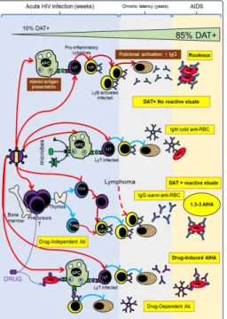
(2) This situation is to be expected, since it expressions the inability to recognize and present blood group antigens by the antigen-presenting cells (APC) and the decrease of lymphocytes T CD4+ type 2, a subpopulation that’s essential to this process [7]. In animal models, viral infections or viral-like inflammation can potentiate the alloimmunization, whereas Gramm negative bacterial infection or sepsis suppress or diminish alloimmune response [5]. On the other hand, the levels of antibodies of ABO system, anti-A and anti-B are usually normal, a situation that’s compatible with the fact that the immune response that produces this anti-carbohydrates and anti-glycolipids antibodies is T independent (Figure 2).. in RBC membrane Erythrocytes Problems The natural history of infections with hematogenous dissemination of microorganisms tends to count with the participation of RBC as receptors (Table 1), as “target” of their direct action or their products/enzymes. In fact, HIV can interact with glycolipid and glycoproteic structures of the erythrocyte membrane, although its clinical significance is still uncertain [8]. Table 1: Blood group antigens receptors to microbes Agent. Receptor. Clinical manifestations. M. Pneumoniae. I. Pneumonia. P. vivax/ P.knowlesi. DARC (Fy6). Malaria. P. Falciparum. A / B, GPA (MN), GPB (SsU), GPC (Ge), Banda3 (Di), CR1, CD36, ICAM-1, XK, Oka. Malaria. HIV. H, P, Lub, Oka. AIDS. H pilori. b. Le H. Pyloric ulcer. E. Coli. DAF (CROM); GPA (M); GLOB1 (P). Urinary infection. Echovirus / Coxsackie. DAF (CROM). Diarrhea, meningitis, pneumonia. M. Tuberculosis M Leprae. Figure 1: HIV effect and microbes of immunohematological importance Ly: Lymphocyte APC: Antigen present cell DAT: Direct antiglobulin test. CR1 (Knops). Tuberculosis Leprosy. H influenzae. AnWj. Severe infection. Parvovirus B19. Glob1 (P). RBC aplasia, Fifth disease. Poliovirus. CD44 (IN). Poliomyelitis. Pseudomona aer. B, P1, Pk,. Sepsis. Giardia. A. Parasitism. Streptococus suis. GLOB1 (P). Sepsis. Alteration in Blood Group Antigens From the microbe-erythrocyte interaction, arise alterations in the structures of the cell membrane surface, these alterations very rarely modify in a clinically significant way the half-life of RBC. The most affected structures tend to be the proteins or carbohydrates of blood group antigens and can potentiate, depress or acquire “de novo” blood group antigens (Table 2). The blood group antigens are products of the expression of one or more inherited genes (genotype), this phenomenon (modification of phenotype) are usually passengers and remit spontaneously or by the action of medical treatment.. Figure 2: HIV effect on alloimmune response to blood group antigens and effect of other germs over molecules carrying antigens J Clin Rev Case Rep, 2019. In some cases, the erythrocyte membrane can be modified by microbial enzymes (Neuraminidase, β-Galactosidase, Deacetylase) that can diminish the expression of blood group antigens. In the case of A and B antigens of ABO system, not always this diminution of expression may be detected by conventional agglutination test.. www.opastonline.com. Volume 4 | Issue 2 | 2 of 6.
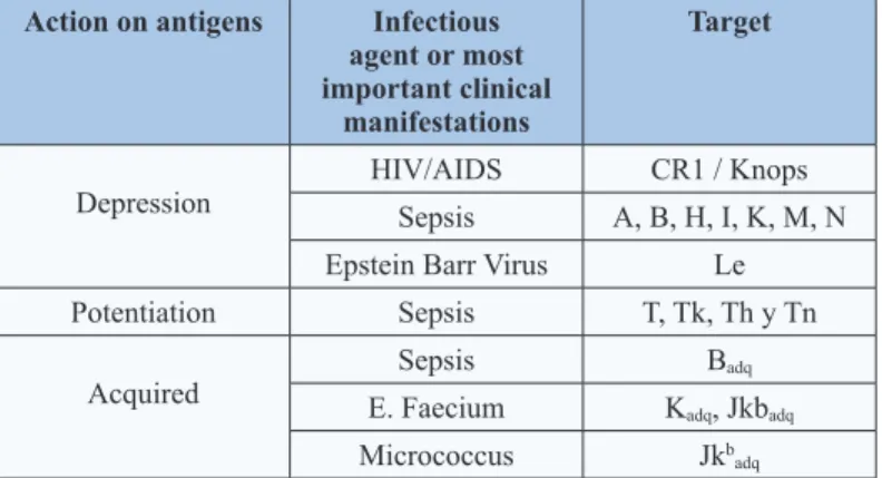
(3) Table 2: Blood group antigens affected by microbes Action on antigens. be ignored.. Infectious agent or most important clinical manifestations. Target. HIV/AIDS. CR1 / Knops. Sepsis. A, B, H, I, K, M, N. Depression. Epstein Barr Virus. Le. Potentiation. Sepsis. T, Tk, Th y Tn. Acquired. Sepsis. Badq. E. Faecium. Kadq, Jkbadq. Micrococcus. Jkbadq. In other cases and by a similar mechanism to the previously described (modification of membrane by bacterial enzymes) it may have the opposite effect that is to potentiate blood group antigens that are not generally detected by conventional test. These antigens are T, Tk, Th and Tn and responsible for the polyagglutination phenomenon. This problem is in general detected by “minor crossmatch”; resulting positive in saline medium at room temperature (patient’s erythrocytes vs donor’s plasma). Although in the case of “natural” antibodies not complement-activator IgM and non-reactive in anti-globulin medium, they usually do not have clinical significance and may. More rarely, the detection of unexpected antigens is usually the result of the direct action of microbial enzymes or the adsorption of bacterial products in the surface of erythrocytes. In these cases, they often produce discrepancies in ABO typing or other blood group systems (Figure 2). Positive Direct Antiglobulin Test and Non-Reactive Eluate Since the beginnings of the HIV/AIDS pandemic the high incidence of positive Direct Antiglobulin Test (DAT) and non-reactive eluate was described in HIV-infected individuals, without clinicallaboratorial evidence of immune hemolysis, This incidence at the beginning of the infection is usually 10% increasing progressively as the disease advances reaching 85% in the final stage of infection (AIDS) [3,9]. The most probable cause of DAT positive is polyclonal hypergammaglobulinemia (resolved with serum dosage of immunoglobulins); another cause may be the presence of circulating immune complexes and IgG autoantibodies. The differential diagnoses are ABO incompatibility, erythrocytes covered by IgA/IgM and anti-drug antibodies (Table 3) [10].. Table 3: Positive DAT differential diagnosis in HIV/AIDS Differential diagnosis. Antecedent Transfusion last 3 months. Postransfusional ABO incompatibility. Clinical signs of hemolysis. Resolution RBC. Mixed field. Elution vs A / B RBC and IAT. Platelet, plasma, hemoderivates. Elution vs RBC A / B and IAT. Acute anemia, shock, hemoglobinemia, hemoglobinuria, jaundice, renal insufficiency, thrombosis, disseminated intravascular coagulation. Laboratory indirect parameters. LDH, haptoglobin, reticulocytes, bilirubin. RBC coated by C3d /IgA or IgM Drug-induced antibodies. DAT with anti-C3d, -IgA, -IgM Recent drug administration and clinical/laboratorial parameters of hemolysis. Auto Agglutination Frequently, the HIV-infected patients have cold antibodies (mostly without clinical significance) during the disease [11]. Rarely, these cold antibodies (IgM) may be sufficiently potent to produce spontaneous agglutination of the blood sample that can cause discrepancies with ABO and Rh typing [12]. The resolution is to try that the cold antibody does not interfere with the RBC typing. So, initially let the sample incubates at 37ºC for 60 minutes and then washes RBC with physiological solution at 37ºC [13,14]. If the control (albumin at 6%) invalidates the typing by spontaneous agglutination, we must use a conservative elution of RBC (heat, chloroquine, etc.) or treatment with reducing agents (2-ME, DTT).. J Clin Rev Case Rep, 2019. Medical treatment. Drug dependent-antibodies detection in serum/ eluate. Finally, and if the cold autoantibody is very potent (very rare), must use genotype typing with nucleic acid amplification techniques. Serum Problems The main functions of the immune system are to recognize microbes and control/destroy the pathogenic ones; destroy tumoral cells and protect fetal and normal cells. Integrating the immune response to bacteria (T independent) the “natural” antibodies of blood group (ABO, H, Le) mostly IgM, IgG2/IgG4 of low affinity are generated and if they do not fix complement they are usually clinically not significant (apart of ABO antibodies). The previous exposure to pathogens carrying sequences of peptides similar to blood group antigens (T dependent) could explain the relationship between infection and detection of irregular antibodies [15] (Table 4).. www.opastonline.com. Volume 4 | Issue 2 | 3 of 6.
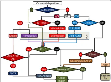
(4) Table 4: Blood group antibodies related to infections Infection Anti-M. Proteus mirabilis. Anti-P1. Hydatidosis, liver flukes. Anti-K. E. coli, Campylobacter jejuni, M. tuberculosis, E. faecalis. Anti-Jkb Micrococcus; Proteus Mirabilis Anti-P. E. Coli, measles, mumps, chickenpox, adenovirus, Cytomegalovirus, Epstein Barr Virus, syphilis, Haemophilus influenzae, M. pneumoniae. Anti-Pr. Viral infections. Anti-I. M. pneumoniae. Anti-i. Epstein-Barr, Virus HIV. Anti-Rx viral Infection. As mentioned previously, there is a strong relationship between infections and development of anti-RBC autoantibodies (with or without hemolysis); the prevalence in HIV-infected patients is higher [16]. There are several proposed mechanisms, not mutually exclusive, that explain these findings [17]: a). Molecular mimicry [18]. The structural similarity between HIV and RBC structures could induce the production of autoantibodies associated with the development of autoimmune hemolytic anemia (AIHA). b) Escape to thymic deletion of auto reactive clones [17]. c) Disfunction T-B which decrease of Tregs lymphocytes and increase of Th2; it may even generate polyclonal B-cell activation [19]. d) Predisposition to develop drug-induced antibodies [20]. This mechanism may be the consequence of a rearrangement of the homeostasis of the immune system, which leads to exacerbate functions and express pre-existing autoreactive elements. When the immune system is exposed to polymerization (which is often in our institution), patients may develop anti-drug antibodies (via primary RBC-microbe-drug interaction).. a mild form, moderate to fulminant hemolysis (infrequent) [23]. Most of the well-documented cases of drug-induced AIHA in patients infected with HIV/AIDS are diagnosed in our Hospital and it certainly reflects a higher level of suspicion compared to other institutions [20]. The diagnosis of AIHA in patients infected with HIV/AIDS follows the same clinical-laboratory steps of the non-infected [24]. That is, clinical evidence (acute anemia, shock, hemoglobinemia, jaundice, renal insufficiency, coagulopathy) and indirect parameters of hemolysis (increase of LDH and indirect bilirubin, decrease of haptoglobin). Clearly the finding of positive DAT + in patients infected with HIV/ AIDS (in the absence of clinical and laboratory signs of hemolysis) is not sufficient evidence for the diagnosis of AHAI. Synthesis: The HIV infected patients may develop clinically significant alloantibodies (less frequently that HIV-negative); autoantibodies (more frequently that HIV-negative) and DrugInduced antibodies (more frequently that HIV-negative). Selection of Blood to be transfused The indication of transfusing patients infected with HIV/AIDS does not differ substantially compared with HIV-negative [25]. However, the main immunohematological problem is the positivity of pretransfusional crossmatch test (Figure 3) [10,26]. Prior to any attempt at resolution, always consider the patient’s antecedents (transfusion, pregnancy, drugs, etc.) since they can guide to the possible causes of the positive crossmatch, positive DAT and positive Indirect Antiglobulin Test (IAT).. The antibodies produced can be cold (IgM) or warm (IgG). Cold Autoantibodies The pathological cold autoantibodies are defined by presenting a positive DAT by anti-C3d (negative for anti-IgG), title at 4º C > 64 and evidence of clinical hemolysis. Any cold antibody that does not meet these characteristics is generally considered non-pathological. The clinically significant antibodies are detected by pre-heated 37°C the samples; in cases of very potent antibodies (with high thermal range), must use the cold autologous adsorption supernatant. Warm Autoantibodies Different publications have documented that patients infected with HIV/AIDS have AIHA incidence of 3%; being their risk 28 times higher than the controls even in those patients treated with antiretroviral therapy, the prevalence may be the same or higher [21,22]. The clinical presentation of AIHA associated with HIV varies from J Clin Rev Case Rep, 2019. Figure 3: Basic resolution scheme of positive crossmatch Ads: Adsorption Autol: Autologous Ab: antibody * Clinically significant DD: Drug Dependent NIPA: No immune protein adsorption AIHA: autoimmune hemolytic anemia IAT: indirect antiglobulin test. www.opastonline.com. Volume 4 | Issue 2 | 4 of 6.
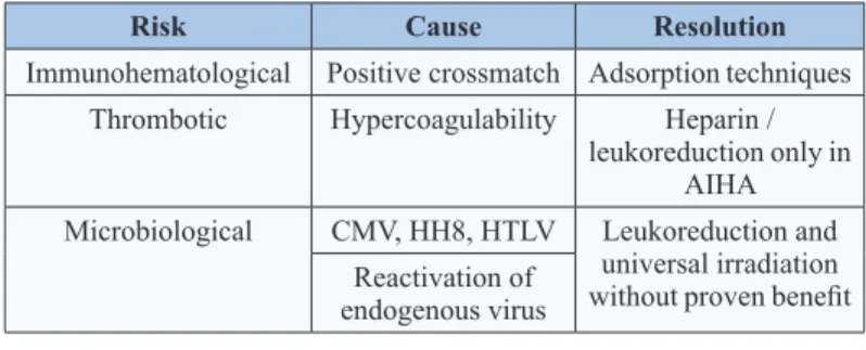
(5) Conclusion. Tx: transfusion RBC: red blood cells Ag: antigen AHTR: Acute hemolytic transfusión reaction If the patient was transfused in less than 3 months, may suspect postransfusional hemolytic reaction by performing eluate and homologous adsorption to detect clinically significant antibodies/ perform crossmatch. If there is not an antecedent of recent transfusion, the best choice is autologous adsorption to detect clinically significant antibodies and to perform cross-match. Another important antecedent is the previous detection of clinically significant antibodies. In such case, select antigen negative RBC (even if they are not actually detected). If the patient had a recent administration of drugs associated with positive DAT/AIHA (α-Methyl Dopa, Cefotetan, Ceftriaxone, Piperacillin, diclofenac, etc.), the problem is solved by performing drug- dependent antibody detection tests in serum/eluate [20]. Transfusional Risk Patients infected with HIV/AIDS are up to 4 times more likely to present thrombosis compared to the general population due to their hypercoagulable state/ pre-disseminated intravascular coagulation (DIC) and, since transfusion is a potentially thrombogenic factor, thromboprophylaxis has been suggested [27,28].. The world incidence of HIV infection/AIDS is relevant; therefore, it is important to recognize the transfusional risks and problems that can be produced and how they can be solved. This publication aims to contribute to its resolution.. Acknowledgment. I express my gratitude to my medical guide Prof. Dr. Enrique Rewald (1926-2016+), a brilliant mind that Argentina did not know how to value. Special thanks to Ana Maria Ahumada FUHESA for support.. References 1. 2. 3. 4. 5. 6.. Initially retrospective studies suggested that RBC transfusion to HIV-infected patients may re-activate endogenous Cytomegalovirus, Herpes Virus 8 and HIV, therefore proposing universal leukoreduction, subsequently three prospective multicentric controlled trials did not prove beneficial effect of universal leukoreduction in patient’s HIVinfected, being the clinical indication to leukoreduction the same as for HIV-negative patients [29-34].. 7.. Regarding the universal irradiation of blood components to transfuse to HIV-infected patients, there is only one case of postransfusional GVHD [35]. This is probably because postransfusional microchimerism in HIV-infected patients is rarely detected, so the universal irradiation of blood components is not recommended. The indications to irradiation are the same as for HIV-negative patients [36,37].. 10.. In summary, the immunohematological risk in HIV positive patients detected by positive crossmatch is solved by adsorption techniques; the thrombotic risk (due to hypercoagulable state) is solved by pretransfusional heparin administration and leukoreduction only in cases of AIHA; and the presumed microbiological risk is similar to HIV-negative patients (Table 5).. 13.. Table 5: Transfusional risk in HIV positive patients Risk Immunohematological. Cause. Resolution. Positive crossmatch Adsorption techniques. Thrombotic. Hypercoagulability. Heparin / leukoreduction only in AIHA. Microbiological. CMV, HH8, HTLV. Leukoreduction and universal irradiation without proven benefit. Reactivation of endogenous virus. 8. 9.. 11. 12.. 14. 15.. 16. 17. 18.. 19. J Clin Rev Case Rep, 2019. https://www.who.int/es/news-room/fact-sheets/detail/the-top10-causes-of-death https://www.who.int/es/news-room/fact-sheets/detail/hiv-aids Zon LI, Arkin C, Groopman JE (1987) Hematologic manifestations of the human immune deficiency virus (HIV). Br J Haematol 66: 251-256. Boctor FN, Ali NM, Mohandas K, Uehlinger J (2003) Absence of D-alloimmunization in AIDS patients receiving D-mismatched RBCs. Transfusion 43: 173-176. Hendrickson JE, Tormey CA (2016) Understanding red blood cell alloimmunization triggers. Hematology 1: 446-451. Körmöczi GF, Mayr WR (2014) Responder individuality in red blood cell alloimmunization. Transfus Med Hemother 41: 446-451. Parker DC (1993) T Cell-dependent B cell activation. Annu Rev Immunol 11: 331. Cooling L (2015) Blood Groups in Infection and Host Susceptibility. Clin Microbiol Rev 28: 801-870. Lai M, d’Onofrio G, Visconti E, Tamburrini E, Cauda R, et al. (2006) Aetiological factors related to a positive direct antiglobulin test result in human immunodeficiency virusinfected patients. Vos Sanguinis 90: 325-330. González CA (2010) Prueba Cruzada Positiva. Rev Arg Transfusion 36: 147-153. González CA (1997) Semiología Inmunohematológica en la Infección por el VIH-1. Revista Argentina de Infectología X: 11-20. Endoh T, Kobayashi D, Tsuji N, Tanabe H, Koshida S, et al. (2006) Optimal prewarming conditions for Rh antibody testing. Transfusion 46: 1521-1525. Judd WJ (2006) How I manage cold agglutinins. Transfusion 46: 324-326. Berentsen S (2018) How I manage patients with cold agglutinin disease. Br J Haematol 181: 320-330. Hudson KE, Lin E, Hendrickson JE, Lukacher AE, Zimring JC, et al. (2010) Regulation of primary alloantibody response through antecedent exposure to a microbial T-cell epitope. Blood 115: 3989-3996. Christen U (2018) Pathogen infection and autoimmune disease. Clinical and experimental immunology 195: 10-14. Barcellini W (2015) New Insights in the pathogenesis of autoimmune hemolytic anemia. Transfus Med Hemother 42: 287-293. Tsiakalos A, Routsias JG, Kordossis T, Moutsopoulos HM, Tzioufas AG, et al. (2011) Fine epitope specificity of antierythropoietin antibodies reveals molecular mimicry with HIV1 p17 protein: a pathogenetic mechanism for HIV-1-related anemia. J Infect Dis 204: 902-911. Okoye AA, Picker LJ (2013) CD4 (+) T-cell depletion in HIV. www.opastonline.com. Volume 4 | Issue 2 | 5 of 6.
(6) 20. 21. 22.. 23. 24. 25.. 26. 27. 28. 29.. infection: mechanisms of immunological failure. Immunol Rev 254: 54-64. González CA, Guzmán L, Nocetti G (2003) Drug-dependent antibodies with immune hemolytic anemia in AIDS patients Immunohematology 19: 10-15. Olayemi E, Awodu OA, Bazuaye GN (2008) Autoimmune hemolytic anemia in HIV-infected patients: a hospital based study. Annals of African Medicine 7: 72-76. Yen YF, Lan YC, Huang CT, Jen IA, Chen M, et al. (2017) Human Immunodeficiency Virus Infection Increases the Risk of Incident Autoimmune Hemolytic Anemia: A Population-Based Cohort Study in Taiwan. J Infect Dis 216: 1000-1007. Gonzalez CA (1998) Successful treatment of AIHA with IVIG in a patient with AIDS. Transplant Proc 30: 4151-4152. González CA (2000) Diagnóstico inmunohematológico de anemia hemolítica autoinmune. Revista Argentina de Transfusión XXVI: 21-40. Van den Berg K, James van Hasselt, Evan Bloch, Robert Crookes, James Kelley, et al. (2012) A review of the use of blood and blood products in HIV-infected patients. South Afr J HIV Med 13: 87-104. Chiaroni J, Gouvitsos J, Dettori I, Ferrera V (2009) How we evaluate panagglutinating sera Transfusion 49: 1540-1545. Crum-Cianflone NF, Weekes J, Bavaro M (2008) Thromboses among HIV-infected patients during the highly active antiretroviral therapy era. AIDS Patient Care STDS 22: 771-778. Kato GJ, Taylor JG (2010) Pleiotropic effects of intravascular haemolysis on vascular homeostasis. Br J Haematol 148: 690701. Lumadue JA, Shirey RS, Kickler TS, Ness PM (1996) Leukocyte reduction of red cells when transfusing patients with autoimmune. 30.. 31. 32.. 33. 34.. 35. 36. 37.. hemolytic anemia: a strategy to decrease the incidence of confounding transfusion reactions. Immunohematology 12: 84-86. Busch MP, Lee TH, Heitman J (1992) Allogeneic leukocytes but not therapeutic blood elements induce reactivation and dissemination of latent human immunodeficiency virus type 1 infection: implications for transfusion support of infected patients. Blood 80: 2128-2135. Preiksaitis JK (2000) the cytomegalovirus-”safe” blood product: is leukoreduction equivalent to antibody screening? Transfus Med Rev 14: 112-136. Collier AC, Kalish LA, Busch MP, Gernsheimer T, Assmann SF, et al. (2001) Leukocyte-reduced red blood cell transfusions in patients with anemia and human immunodeficiency virus infection: the Viral Activation Transfusion Study: a randomized controlled trial. JAMA 285: 1592-1601. Drew WL (2003) Absence of activation of CMV by blood transfusion to HIV-infected, CMV-seropositive patients. Transfusion 43: 1351-1357. Asmuth DM (2003) Absence of HBV and HCV, HTLV-I and -II, and human herpes virus-8 activation after allogeneic RBC transfusion in patients with advanced HIV-1 infection. Transfusion 43: 451-458. Klein C (1996) Moderate and transient transfusion-associated cutaneous graft-versus-host disease in a child infected by human immunodeficiency virus. Am J Med 101: 445-446. Kruskall MS (2001) Survival of transfused donor white blood cells in HIV-infected recipients. Blood 98: 272-279. HW Reesink, S Panzer, CA Gonzalez, Lena N, Muntaabski P, et al. (2010) Haemovigilance for the optimal use of blood products in the hospital. Vox sanguinis 99: 278-293.. Copyright: ©2019 Carlos A Gonzalez. This is an open-access article distributed under the terms of the Creative Commons Attribution License, which permits unrestricted use, distribution, and reproduction in any medium, provided the original author and source are credited. J Clin Rev Case Rep, 2019. www.opastonline.com. Volume 4 | Issue 2 | 6 of 6.
(7)
Figure
Documento similar