Physiotherapeutic intervention in peripheral arterial disease by functional hyperemia in diabetic patients
10
0
0
Texto completo
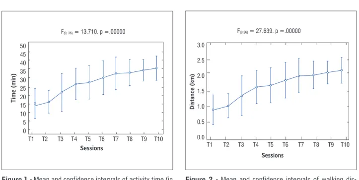
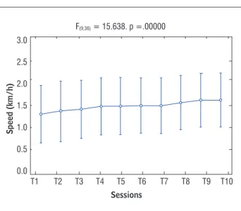
(2) 732. Wichnieski C, Kirchhof SFN, Beraldo PC, Bertassoni Neto L, Jara CC.. programmed exercise therapy that consisted of walking on a treadmill. Results: There was a significant increase in mean activity time (F9,36 = 13.710; p < 0.001 ), mean walking distance (F9,36 = 27.689 ; p < .001 ), and mean speed (F9,36 = 15.638 ; p < .001 ). No statistically significant differences in the ankle-brachial index were noted. Conclusion: There was a significant increase in walking distance, time, and speed for diabetic subjects. Our findings indicate the importance of physical therapists in the supervised treatment of peripheral vascular disorders in diabetic patients.. [P]. Keywords: Diabetes Complications. Intermittent claudication. Exercise therapy. Collateral circulation. Physical Therapy. [B]. Resumo. Introdução: A Diabetes Mellitus (DM) caracteriza-se como um problema de saúde pública, sendo responsável, inclusive, por variados graus de morbidade ao organismo humano. Nas complicações circulatórias da patologia, como na claudicação intermitente, as alternativas físico-funcionais de tratamento são pouco exploradas, o que mostra a necessidade de buscar técnicas auxiliares para o tratamento fisioterapêutico em sujeitos diabéticos. Objetivo: Verificar os efeitos do procedimento de hiperemia funcional no tratamento da insuficiência arterial periférica em sujeitos portadores da Diabetes Mellitus. Materiais e Métodos: Estudo realizado com grupo de 5 voluntários diabéticos da Associação Paranaense do Diabético (APAD), com manifestação de distúrbios vasculares periféricos em membros inferiores. Foram realizados 10 atendimentos duas vezes na semana. O procedimento de hiperemia funcional foi aplicado através da terapia por exercício programado composto por caminhada sobre a esteira ergométrica. Resultados: Foi possível verificar que houve um acréscimo significativo das médias de tempo de atividade (F9,36 = 13,710; p = 0,000), distância do percurso (F9,36 = 27,689; p = 0,000) e velocidade (F9,36 = 15,638; p = 0,000). Na avaliação do índice tornozelo/braquial não foram observadas diferenças estatísticas significativas. Conclusão: Observou-se um aumento significativo na distância, tempo e velocidade de caminhada dos sujeitos diabéticos. O presente estudo evidenciou a importância do fisioterapeuta nos tratamentos supervisionados nas disfunções vasculares periféricas em diabéticos. [K]. Palavras-chave: Complicações do Diabetes. Claudicação intermitente. Terapia por exercício. Circulação colateral. Fisioterapia.. Introduction Diabetes Mellitus (DM) is a complex, chronic metabolic disorder characterized by elevated levels of glucose in the blood, causing hyperglycemia (1). This increase occurs because the insulin, the hormone responsible for the absorption of glucose into cells, is either not produced by the pancreas, produced in insufficient quantities or does not work properly (2). Besides being characterized by impaired glucose metabolism, diabetes is also characterized by impairment of other energy producing substances and is associated with a variety of complications in organs that are essential to survival (3). DM is a major public health problem worldwide due to its high prevalence and morbidity rates, the associated risk of developing disabling chronic Fisioter Mov. 2015 Oct/Dec;28(4):731-40. complications, and its high economic costs generated by treatment and reduced work capacity of individuals of working age (3). The main symptoms of diabetes are caused by an excess of glucose in the blood and a lack of glucose in the cells (4). Vascular diseases are among the most important chronic complications of diabetes (5). Two types of vascular disease are seen in patients with diabetes: non-occlusive microcirculatory dysfunction and macroangiopathy (6). Microcirculatory dysfunction affects capillaries and arterioles in the kidneys, retina and peripheral nerves. Macroangiopathy is characterized by atherosclerotic lesions in the coronaries and peripheral blood circulation (6). Peripheral arterial disease (PAD) is characterized by a narrowing or occlusion of the arteries, resulting.
(3) Physiotherapeutic intervention in peripheral arterial disease by functional hyperemia in diabetic patients. in a gradual decrease of blood flow to the extremities (7, 8). Injury to peripheral arteries thus leads to vascular insufficiency, which, according to Assumpção (9), occurs early in diabetics. Thus, DM is a cardiovascular risk factor that is different from other pathologies (10). Arteriosclerosis is one of the most common peripheral obstructive arterial diseases found in diabetic patients (6). Long-term atherosclerosis may result in narrowing of the main arteries (11) and arteries of the infrapatellar segment are more frequently affected (9). This condition causes functional limitation as consequence of ischemia (12). Thus, one of the clinical features that may be found in individuals with peripheral artery disease is intermittent claudication (IC) (7, 13, 14), which is often the first clinical manifestation of arterial ischemia of the lower limbs (15). However, some patients may be asymptomatic (7), not necessarily presenting the classic symptoms of IC (16, 17). Intermittent claudication is a term used to define a change in locomotion in which the individual is forced to stop walking (6) due exercise induced cramping or aching in the calves, thighs or buttocks (14). IC occurs when the peripheral circulation is inadequate to meet the metabolic requirement of the active leg musculature (18). It limits physical activity and reduces quality of life (8, 13, 19, 20). One way to investigate the presence of PAD is using the ankle-brachial index (ABI). It is noninvasive, inexpensive and easy to perform, and enables the detection of disease in symptomatic and asymptomatic individuals (21, 22, 39). In addition to the ABI, (23) the severity of peripheral arterial disease can also be assessed by means of the degree of functional impairment of individuals with intermittent claudication. Exercise has been shown to be of value in the management of peripheral vascular disease (PVD) (11). Supervised exercise programs have been recommended (22) as first line therapy for patients with peripheral arterial disease (PAD) by several sources, including: the American College of Cardiology/ American Heart Association 2005 Practice Guidelines for the Management of Patients With Peripheral Arterial Disease; the American Association of Cardiovascular and Pulmonary Rehabilitation 2004 Guidelines for Cardiac Rehabilitation and Secondary Prevention Programs; the Intersociety Consensus for. the Management of PAD (TASC II); and the American College of Sports Medicine 2010 Guidelines for Exercise Testing and Prescription. Active/functional hyperemia is characterized by an increased blood flow to the tissues. This can occur in cases of increased functional requirement, when a tissue becomes very active, as would be the case for a muscle (24). In this way, functional hyperemia could contribute to the treatment of peripheral arterial disease, because during this process the increase in blood flow is accompanied by an increase in metabolic activity (24, 25, 26). In addition, (27) during exercise, the muscles need more oxygen. As a consequence of arterial obstruction and due to ischemia, the oxygen supply to the tissues is not sufficient. Consequently, physical exercise could also lead to collateral circulation. Thus, the aim of this study was to investigate the effects of functional hyperemia on peripheral arterial disease in patients with Diabetes Mellitus.. Materials and Methods This study was approved by the Ethics Committee of the Pontifical Catholic University of Parana, opinion number 0005699/12. All volunteers who agreed to participate in the study signed consent forms. This is an exploratory qualitative case study. This study used a stratified sampling design. A group of 10 diabetic volunteers from the Diabetics Association of Parana (Associação Paranaense do Diabético, APAD) participated in this study. There was an overall sample loss of 50% due to changes in the clinical state of diabetic volunteers, which made it impossible for them to travel to the association. The APAD was contacted prior to the commencement of the study to secure agreement for their organization's participation. Inclusion criteria were: being a volunteer and an associate in the APAD; having type I or II diabetes; being aged over 18 years; reporting pain in the calf during walking; and being considered by the medical service of the APAD as being able to participate in this study. Subjects currently receiving physical therapy treatments or practicing regular physical activity during the experimental stages of the study; subjects with other associated disabling disorders such as musculoskeletal changes, Fisioter Mov. 2015 Oct/Dec;28(4):731-40. 733.
(4) 734. Wichnieski C, Kirchhof SFN, Beraldo PC, Bertassoni Neto L, Jara CC.. decompensated heart disease, labyrinthitis, or dizziness of any kind, who did not feel safe enough to walk on the treadmill; and subjects using painkillers or medicines to improve blood flow were excluded from the study in order not to interfere with the study results. To ensure the safety of participants — even though they had already received medical release to participate in the study — they were subjected to physical and functional evaluation of muscle strength of the lower limbs and range of motion, Fukuda stepping test and Tinetti balance test. These tests and assessments aimed to provide greater security in the use of the treadmill. Physical and functional evaluation done according to a final term paper based on a study conducted at the APAD in 2009: "Mapping of peripheral arterial disease in subjects from a specialized institution in the city of Curitiba, and a proposal for therapeutic intervention by functional and reactive hyperemia" (28). In addition, we administered a questionnaire to assess signs and symptoms of peripheral vascular disorders. The questionnaire had been previously validated with regard to construct, content and clarity. Next, we assessed the ankle/brachial index using continuous Doppler examination, a noninvasive transcutaneous method for detecting blood flow with the use of ultrasound and the Doppler effect. The exercise therapy program consisted of walking on a treadmill. This exercise machine was chosen because it allows the simulation of a "walk on the street" and, at the same time, the assessment of exertion variables such as heart rate, blood pressure and subjective manifestations of discomfort reported by the participant during the exercise. Participants were then explained how to use the treadmill. They were instructed about the correct way to stand on it and were informed that, during the test, the treadmill would be activated only after their permission had been given. In addition to the treadmill, we also used a heart monitor, a sphygmomanometer and a stethoscope to measure heart rate and blood pressure before, during and after the exercise. After explaining how to use the treadmill, we performed a test based on the Ellestad protocol (29). During the test, one physical therapist measured the heart rate and a second physical therapist measured the blood pressure of patients every time the speed was increased. This test allowed to identify the ideal initial velocity for each patient, according to their Fisioter Mov. 2015 Oct/Dec;28(4):731-40. heart rate values (which should not exceed 80% of the maximum value), and to reports of pain or fatigue. After the test, patients received physical therapy care. Physical therapy care was delivered at the APAD and at the Clinic of the School of Physical Therapy of PUCPR, according to the availability of the subjects. All subjects attended 10 sessions (twice weekly). During the exercise, the patient was placed on the treadmill and monitored with a heart monitor and a sphygmomanometer. The treadmill was activated at an initial adaptation speed of 0.9 km/h. Then the speed was increased the fastest walking speed was achieved for each patient (according to the test results). This speed was maintained until the patient reported aching or fatigue in the legs. He/she was then encouraged to keep walking a bit more (this varied according to each subject), and soon afterward the speed was lowered until no pain or decreased fatigue was reported. This procedure was repeated for five cycles. Patients' blood pressure and heart rate were measured in every cycle. After 10 sessions, another continuous Doppler examination was performed. After examination, the ABI was assessed. The ABI was calculated as the ratio the highest systolic blood pressure (SBP) of the ankle of each leg and the highest SBP of the brachial artery of the arm, according to the formula: ABI = ankle SBP/ brachial SBP (21). Values ranging between 0.90 and 1.30 are considered normal. Patients with claudication usually have an ABI of about 0.50; patients who have ischemic pain during rest have an ABI between 0.20 and 0.30; and an ABI smaller than 0.20 is associated with ischemic ulcers (8). The following variables were analyzed in this study: walking distance, speed and time, and anklebrachial index. Thus, the data regarding activity time (in minutes), walking distance (in km), and walking speed (in km/h), and the ankle/brachial index measured with the use of the continuous Doppler examination were examined for standard distribution by means of the Shapiro-Wilk test. Since the data were in a normal range, the first three variables were analyzed using the ANOVA test for repeated measures. In case of statistical significance, the Tukey follow-up test was used to compare the differences between the means of all 10 sessions. Ankle/brachial index was analyzed using the t test for paired samples. The acceptable level of significance for all statistical tests performed was P ≤ 0.05..
(5) Physiotherapeutic intervention in peripheral arterial disease by functional hyperemia in diabetic patients. Results As a result of the intervention, there was a significant increase in mean activity time (F9,36 = 13.710; p = 0.000), mean walking distance (F9,36 = 27.689 ; p = 0.000) and mean speed (F9, 36 = 15.638 ; p = 0.000). Already in the fourth session, there was a significant increase in mean activity time and speed, when compared to baseline. This increase (significantly) continued throughout the period of the study (Table 1; Figures 1 and 3). There was a significant increase in. mean walking distance after the third session, when compared to baseline. This increase also continued throughout the period of the study (Table 1; Figure 2). With regard to the right ankle/brachial index, found that there was a trend towards higher means in the post-treatment period (t = 2.55, p = 0.06). The mean values changed from 0.99 + 0.12 at baseline to 1.04 + 0.12 at post-treatment (Table 2). No statistically significant differences were observed between pretreatment and post-treatment for the left anklebrachial index (t = -1.00; p = 0.37) (Table 2).. Table 1 - Mean ± Standard Deviation of time, distance and speed over the course of the 10-session physical therapy program Sessions. Time. Distance. Speed. T1. 15.4 ± 5.9. 0.70 ± 0.29. 2.96 ± 1.06. T2. 17.4 ± 4.8. 0.86 ± 0.38. 3.10 ± 1.10. T3. 23.0 ± 8.0. 1.18 ± 0.64. 3.18 ± 1.07. T4. 27.2 ± 6.6. 1.42 ± 0.56. 3.32 ± 1.06. T5. 28.0 ± 7.0. 1.46 ± 0.54. 3.32 ± 1.06. T6. 30.6 ± 7.1. 1.62 ± 0.58. 3.34 ± 1.02. T7. 33.0 ± 6.8. 1.74 ± 0.55. 3.34 ± 1.02. T8. 33.4 ± 5.5. 1.80 ± 0.45. 3.48 ± 1.01. T9. 34.8 ± 4.5. 1.92 ± 0.42. 3.60 ± 0.99. T10. 35.8 ± 5.1. 1.96 ± 0.49. 3.60 ± 0.99. p. p = 0.000. p = 0.000. p = 0.000. Note: T= Therapy.. F(9,36) = 27.639. p =.00000 3.0. 50 45 40 35 30 25 20 15 10 5 0. 2.5 Distance (km). Time (min). F(9, 36) = 13.710. p =.00000. 2.0 1.5 1.0 0.5. T1. T2. T3. T4. T5. T6. T7. T8. T9. T10. Sessions. 0.0. T1. T2. T3. T4. T5. T6. T7. T8. T9. T10. Sessions. Figure 1 - Mean and confidence intervals of activity time (in. minutes) over the course of the 10 sessions. Figure 2 - Mean and confidence intervals of walking dis-. tance (in km) over the course of the 10 sessions Fisioter Mov. 2015 Oct/Dec;28(4):731-40. 735.
(6) Wichnieski C, Kirchhof SFN, Beraldo PC, Bertassoni Neto L, Jara CC.. Discussion F(9,36) = 15.638. p =.00000 3.0 2.5 Speed (km/h). 736. 2.0 1.5 1.0 0.5 0.0. T1. T2. T3. T4. T5. T6. T7. T8. T9. T10. Sessions. Figure 3 - Mean and confidence intervals of walking speed. (in km/h) over the course of the 10 sessions. In a systematic review of studies on Peripheral Artery Disease (PAD), it has been found that the level of physical activity is lower in individuals with PAD, when compared to healthy individuals, and that individuals with higher disease severity generally show the lowest levels of physical activity (19). These individuals show symptoms of IC earlier than others (5). Thus, their walking speed is reduced and the IC distance may result in progressive loss of function and long-term disability and lead to higher functional limitation. Studies also show that individuals with PAD who have higher levels of physical activity also have better health indicators (30) and this influences several risk factors for people with DM, including macrovascular disease.. Table 2 - Mean±Standard Deviation of ankle/brachial index pretreatment and post-treatment Ankle/brachial Index. Pretreatment. Post-treatment. p. Right. 0.99 ± 0.12. 1.04 ± 0.12. p = 0.06. Left. 1.04 ± 0.04. 1.04 ± 0.04. p = 0.37. Supervised exercise programs are currently considered as first-line therapy for patients with peripheral arterial disease (20), and aerobic exercise programs are also widely recommended (13, 32, 33). Rehabilitation exercises including supervised treadmill walking result in a significant improvement in walking performance in people with IC (16). Exercise training induces vascular adaptations that improve blood flow to the skeletal muscles. It is effective in improving the maximum pain-free walking distance, reducing the symptoms in the extremities, increasing functional capacity and reducing cardiovascular events in patients with PAD (8, 22 , 35). Thus, functional hyperemia may contribute to the treatment of peripheral arterial insufficiency, since the initial event that triggers local vasodilation during functional hyperemia is increased organ or tissue metabolic activity. If, for example, aerobic metabolism increases, the level of O2 in the tissues decreases and CO2 production increases. Any intervention that results in insufficient oxygen supply to the tissues induces the formation of vasodilator metabolites, which Fisioter Mov. 2015 Oct/Dec;28(4):731-40. are released by the tissue and act locally by dilating the vessels. This vasodilation increases blood flow to the tissue, supplying additional oxygen to meet the increased metabolic demand and remove CO2. Thus, it contributes to the treatment of peripheral arterial disease in patients with diabetes. (25, 26). Moreover, the performance of exercise would lead to a neovascularization, in order to meet the oxygen demand of the muscle. This would satisfy the hemodynamic needs created and result in an improved gait performance and physical fitness, reducing the consumption of O2, improving symptoms, claudication distance and quality of life (27, 35). In addition, (27) treatment based on exercise training not only improves walking distance, but also improves the ability to walk predefined distances and at predefined speeds. Direct supervision is a secondary factor that influences the achievement of good results, because its goal is to keep individuals motivated during exercise (27). A literature review (13) describes several studies on exercise in patients with peripheral obstructive.
(7) Physiotherapeutic intervention in peripheral arterial disease by functional hyperemia in diabetic patients. arterial disease and intermittent claudication. These studies used walking (on a treadmill or not) as the primary mode of exercise and some of them combined aerobic and endurance exercises. However, in this study, we only analyzed those studies that used only walking as their exercise program (8, 14, 35, 36, 37, 38). Of note, the studies presented different methodologies, e.g., different walking times, different speeds, different variables. In addition, participants already presented PAD and IC. Some studies have shown a significant improvement in maximum walking distance for patients with IC (35, 38, 39). The studies also found increases in claudication onset distance (35-38), peak walking time (8, 14, 37), time of pain-free walking (14-37), maximum walking speed and pain-free walking speed (35). These results demonstrate that a physical exercise program may significantly improve the walking ability of patients with IC (13). The results of these studies are in line with this paper, since our subjects showed significant increases in walking distance, walking speed and walking time. Most of the subjects, however, did not have ABI values indicative of intermittent claudication. There is recent evidence that supervised exercise programs are also beneficial for patients with Peripheral Arterial Disease who do not have claudication (22). With regard to the blood flow, two out of seven studies (36, 37) measured venous blood flow by using venous occlusion plethysmography of the symptomatic leg. The evaluation was performed before and after the aerobic exercise program. Both studies showed a significant increase in the blood flow. Only one study (35) has found a significant result when comparing ABI values. Some studies (35, 36, 37, 38) found no significant changes. The lack of significant improvement in ABI is probably explained by the fact that exercise increases blood flow due to improved collateral circulation. This does not necessarily affect tibial artery systolic pressure, because the occurring mechanism seems to induce a local redistribution of blood flow rather than an absolute increase of blood flow (13). This corroborates our study, which found no significant increase in ABI. There are controversial data in the literature regarding the duration and frequency in which exercises should be performed in people with PAD.. The best results are achieved with a walking exercise program performed three times a week and close to the patient's peak pain level (13). The frequency should be three to four 20- to 60-minute-sessions per week at an exercise intensity requiring no more than 85% of VO2max (maximum oxygen volume). Exercise intensity may be also estimated based on perceived exertion (31). At least 150 minutes of moderate-intensity aerobic exercise (50% to 70% maximum heart rate) or 90 minutes of high-intensity aerobic exercise is recommended per week. Moreover, symptomatic PVD patients should perform supervised exercise by walking on a treadmill at least three times/week (17). Walking at least one hour, three times a week is enough (34) to produce a significant improvement in claudication distance. Nevertheless, the exercise program should be performed for at least six months. Although the time and weekly frequency used in this study is below the values found in the literature, we still found increases in time, distance and speed. Another study (18) shows that patients with IC who engage in regular physical activity have lower mortality rates. The study evaluated patients with IC symptoms from 1994 through 2002, and was continued in 2004 using the "The Social Security Death Index". 434 patients participated in the study. Of these, 299 were classified as sedentary and 135 were classified as physically active. Of the subjects who were followed up, 108 died: 86 belonged to the sedentary group (28.8%) and 22 belonged to the physically active group (16.3%). The study concluded that patients functionally limited by intermittent claudication who engaged in any amount of weekly physical activity beyond light intensity at baseline had a lower mortality rate than their more sedentary counterparts. Therefore, the data suggest that the prognosis worsens with more severe PAD, as the relative risk for mortality progressively increases with a decrease in the ankle-brachial index (ABI). Exercise also brings other benefits to patients, such as improved quality of life and prognosis, reduced stress levels, increased pain threshold and improved functional capacity, facilitating the performance of occupational and daily living activities. Although not the main objective of this paper, this theoretical base supports the findings to this study, because participants reported a decrease in pain that. Fisioter Mov. 2015 Oct/Dec;28(4):731-40. 737.
(8) 738. Wichnieski C, Kirchhof SFN, Beraldo PC, Bertassoni Neto L, Jara CC.. improved their quality of life and improved their ability to perform activities of daily living (13).. 2.. Molena-Fernanes M, Juniro N, Tasca RS, Pelloso SM, Cuman KN. A importância da associação de dieta e de atividade física na prevenção e controle do Diabetes mellitus tipo 2. Acta Sci. Health Sci. 2005; 27(2)195-205.. 3.. Brasileiro JL, Oliveira WP, Monteiro B, Chen J, Junior EM, Santos A. Pé diabético: aspectos clínicos. Jornal Vascular Brasileiro. 2005;4(1). [Cited in 2011 Oct 25]. Available from: http://tinyurl.com/pd8hrvg.. 4.. Bonetti E, Schaly D, Rover C, Fiedler M. Atividade física em indivíduos portadores de diabetes mellitus do município de Joaçaba, SC. Evidência. 2012 Jan/ Jun;12(1):41-50.. 5.. Marso SP, Hiatti WR. Peripheral arterial disease in patients with diabetes. Hiatt, MD. Journal of the American College of Cardiology. 2006;47(5).. 6.. Silva RG, Colombo-Consolim M. Aspectos relevantes para identificação da claudicação intermitente. Acta Paul Enferm. 2011;24(3):426-9.. 7.. Jude EB, Eleftheriadou I, Tentolouris N. Peripheral arterial disease in diabetes: a review. Journal compilation Diabetic Medicine. 2009;27,4-14.. 8.. Hodges LD, Sandercock GH, Das SK, Brodie D. Randomized controlled trial of supervised exercise to evaluate changes in cardiac function in patients with peripheral atherosclerotic disease. Clin Physiol Funct Imaging. 2008;28,32-7.. 9.. Assumpção EC, Pitta GB, Macedo CL, Mendonça G, Albuquerque A, Lyra BC, et al. Comparação dos fatores de risco para amputações maiores e menores em pacientes diabéticos de um Programa de Saúde da Família. Jornal Vascular Brasileiro. 2009;8(2). [Cited in 2011 Oct 05]. Available from: http://tinyurl.com/pwhbh3x.. Conclusion The results obtained with the use of functional hyperemia show a significant increase in the walking distance, time and speed of subjects and thus an improvement in quality of life. Based on the evidence presented above, we conclude that the performance of kinesiotherapy with controlled aerobic exercise was enough to produce local changes in circulation, and that these changes might have been mediated by metabolic responses and the need to increase local blood supply. The small variations in the ankle-brachial index can be explained by the development of neovascularization, known as collateral circulation. It led to a reduction of symptoms and, thus, to a better walking performance and physical fitness of participants. This study stresses the importance of the preventive role of physical therapy in peripheral vascular disorders in diabetic patients, and offers an important alternative for the complementary treatment of this population. The major limitations experienced during the conduction of this study were associated to the fact that some subjects had complications triggered by diabetes, such as hypoglycemic attacks, which resulted in a reduction of our final sample. Other factors that limited the participation of subjects were: patient's unavailability to perform the treatment twice a week; the distance between the patient's home and the place where the study was conducted; travel costs; and lack of time. Thus, we suggest that this study be continued with a larger sample size, an increased number of physical therapy sessions and the use of plethysmography to check blood flow in the lower limbs before and after the application of the technique described here.. References 1.. Bar-Shalom D, Poulsen K, Rasmussen M, Diederichsen CP, Sand PR, Heriksen E, et al. Plasma copeptin as marker of cardiovascular disease in asymptomatic type 2 diabetes patients. Diabetes & Vascular Disease Research. 2014;11(6)448–50.. Fisioter Mov. 2015 Oct/Dec;28(4):731-40. 10. Nandy D, Johnson C, Basu R, Joyner M, Brett J, Svendsen CB, et la. The effect of liraglutide on endothelial function in patients with type 2 diabetes. Diabetes & Vascular Disease Research. 2014;11(6) 419-30. 11. Kolt GS, Snynder-Mackler, L. Fisioterapia no esporte e no exercício. Rio de Janeiro: Revinter, 2008. 619 p. 12. Durazzo AS, JR Cid J, Presti C, Silva ES, Luccia N. Doença arterial obstrutiva periférica: que atenção temos dispensado à abordagem clínica dos pacientes? Jornal Vascular Brasileiro. 2005;4(3). [Cited in 2011 Oct 25]. Available from: http://tinyurl.com/ogfbf5j..
(9) Physiotherapeutic intervention in peripheral arterial disease by functional hyperemia in diabetic patients. 13. Locatelli EC, Pelizzari S, Scapini KB, Leguisamo CP, Silva AB. Exercícios físicos na doença arterial obstrutiva periférica. Jornal Vascular Brasileiro. 2009;8(3). [Cited in 2011 Nov 01]. Available from: http://tinyurl. com/ogyortg. 14. Crowther RG, Crowther WL, Spinks L S, Sangla K, Quigley F, Golledge J. Effects of a long-term exercise program on lower limb mobility, physiological responses, walking performance, and physical activity levels in patients with peripheral arterial disease. J Vasc Surg. 2008;47:303-9. 15. Filho MS, Rodoky A, Costa F, Ferreira B, Wolosker N, Puech-Leão P. Comparação entre o resultado do tratamento clínico de pacientes com claudicação intermitente por obstrução femoropoplítea bilateral versus obstrução aórtica. Jornal Vascular Brasileiro.2005;4(2). [Cited in 2011 Oct 30]. Available from: http://tinyurl.com/py7xh8f. 16. Mc Dermott, Liu K, Ferrucci L, Criqui M, Greenland P, Guralnik M, et al. Physical performance in peripheral arterial disease: a slower rate of decline in patients who walk more. Annals of Internal Medicine. 2006; 144:10-20. 17. Triches C, Schaan B, Gross J, Azevedo J. Complicações macrovasculares do diabetes melito: peculiaridades clínicas, de diagnóstico e manejo. Arq Bras Endocrinol Metab. 2009;53-6. [Cited in 2012 Oct 25]. Available from: http://dx.doi.org/10.1590/ S0004-27302009000600002. 18. Gardner W, Montgomery S, Parker E. Physical activity is a predictor of all-cause mortality in patients with intermittent claudication. J Vasc Surg. 2008; 47(1):117122. 19. Barbosa S, Henriques M, Barros G, Wolosker N, RittiDias M. Nível de atividade física em indivíduos com doença arterial periférica: uma revisão sistemática. Jornal Vascular Brasileiro. 2012;11(1). [Cited in 2012 Oct 25]. Available from: http://dx.doi.org/10.1590/ S0004-27302008000200013. 20. Milani V, Lavie J. The role of exercise training in peripheral arterial disease. Vascular Medicine. 2007; 12:351-8. [Cited in 2012 Nov 02]. Available from: http://tinyurl.com/nel2fo6.. 22. Hamburg M, Balady J. Exercise rehabilitation in peripheral artery disease functional impact and mechanisms of benefits. Circulation. 2011;123:87-97. 23. Cunha-Filho T, Pereira G, Carvalho B, Campedeli L, Soares M, Freitas JS. Confiabilidade de testes de caminhada em pacientes claudicantes: estudo piloto. Jornal Vascular Brasileiro. 2008;7(2). [Cited in 2011 Dec 01]. Avalaible from: http://dx.doi.org/10.1590/ S1677-54492008000200004. 24. Ripka WL, Dutra G, Ulbricht L, Martelli C, Gewehr PM. Sensor de fibra óptica para detecção de hiperemia ativa fisiológica funcional em teste ergométrico. XXIV Congresso Brasileiro de Engenharia Biomédica – CBEB; 2014. 25. Garofolo L, Ferreira G, Junior FM. Biomarcadores inflamatórios circulantes podem ser úteis para identificar doença arterial obstrutiva periférica mais grave. J Vasc Bras. 2014;13(3),182-91. 26. Pereira C, Miname M, Makdisse M, Filho RK, Santos D. Associação das doenças arterial periférica e cardiovascular na hipercolesterolemia familiar. Arq Bras Cardiol. 2014;103(2):118-23. 27. Sudbrack AC, Sarmento-Leite R. Efetividade do exercício na claudicação. Rev. Bras. Cardiol. Invas. 2007,15(3):261-266. [Cited in 2011 Nov 20]. Available from: http://tinyurl.com/p7yzwd9. 28. Silva F, Pohren I, Pereira JM, Bertassoni NT, Beraldo PC. Mapeamento da insuficiência arterial periférica em sujeitos, em instituição especializada na cidade de Curitiba, com proposta de intervenção terapêutica por meio da hiperemia reativa e funcional. 2010 79 f. TCC (Curso Fisioterapia) - Pontifícia Universidade Católica do Paraná, Curitiba, 2010. 29. DIRETRIZES do ACSM para os testes de esforço e sua prescrição. 18ed. Rio de Janeiro: Guanabara Koogan;2007;xviii 266 p. 30. Ramalho R, Soares S. O Papel do exercício no tratamento do diabetes melito tipo 1. Arq Bras Endrocrinol Metab 2008; 52-2. [Cited in 2012 Oct 24]. Available from: http://dx.doi.org/10.1590/ S1677-54492012000100005.. 21. Nunes F, Leão S, Exel AL, Diniz C. Índice TornozeloBraquial em pacientes de alto risco cardiovascular. Rev Bras Cardiol. 2012;25(2):94-101. Fisioter Mov. 2015 Oct/Dec;28(4):731-40. 739.
(10) 740. Wichnieski C, Kirchhof SFN, Beraldo PC, Bertassoni Neto L, Jara CC.. 31. Arsa G, Lima L, Almeida S, Moreira R, Campbell G, Simoes G. Diabetes mellitus tipo 2: aspectos fisiológicos, genéticos e formas de exercício físico para seu controle. Rev Bras Cineantropom Desempenho Hum. 2009;11(1):103-11. 32. Murayana R, Carraro D, Galvanin T, Izukawa M, Umeda I, Oliveira F. Insuficiência vascular periférica compromete a capacidade funcional no paciente com insuficiência cardíaca. J Vasc Bras. 2014;13(2):101-7. 33. Carvalho C, Maux X, Tashiro T, Moraes A. The effect of endurance training on the neovascularization of skeletal musculature. Acta Cirúrgica Brasileira. 2006;21(6). 34. Yoshida A, Matida K, Sobreira M, Moura R, Rollo A, et al. Comparative study of evolution and survival of patients with intermittent claudication, with or without limitation for exercises, followed in a specific outpatient setting. J Vasc Bras. 2008;7(2):112-22. 35. Manfredini F, Malagoni AM, Mascoli F, et al. Training rather than walking the test in-train out program for homebased rehabilitation in peripheral arteriopathy. Circ J. 2008;72:946-52.. Fisioter Mov. 2015 Oct/Dec;28(4):731-40. 36. Wang J, Zhou S, Bronks R, Graham J, Myers S. Effects of supervised treadmill walking training on calf muscle capillarization in patients with intermittent claudication. Angiology. 2009;60:36-41. 37. Roberts AJ, Roberts EB, Sykes K, De Cossart L, Edwards P, Cotterrrell D. Physiological and functional impact of an unsupervised but supported exercise programme for claudicants. Eur J Vasc Endovasc Surg. 2008;36:319-24. 38. Gardner W, Killewich A, Montgomery S, Katzel I. Response to exercise rehabilitation in smoking and nonsmoking patients with intermittent claudication. J Vasc Surg. 2004;39:531-8. 39. Galvão C. O Índice Tornozelo-Braquial. Rev Soc Bras Med Trop. 2012; Ano XX(24).. Received: 06/22/2013 Recebido: 22/06/2013 Approved: 05/20/2015 Aprovado: 20/05/2015.
(11)
Figure
Documento similar
Recent reports of long-term outcomes for radial shortening osteotomy in osteonecrosis early stage patients and for proximal row carpectomy (PRC) in advanced Kienböck’s disease
Effects of resistance training on muscle strength, exercise capacity, and mobility in middle-aged and elderly patients with coronary artery disease: A meta-analysis.
Effects of cardiac resynchronization on disease progression in patients with left ventricular systolic dysfunction, an indication for an implantable
Changes in serum IL-15 levels at 4 and 8 weeks as compared with baseline values were directly associated with changes in body weight, BMI, fat-free mass, and muscle mass
A Study on the Relationship between Serum Beta 2-Microglobulin Levels, Underlying Chronic Kidney Disease, and Peripheral Arterial Disease in High-Vascular-Risk
Persistent diarrhea in patients with Crohn’s disease after mucosal healing is associated with lower diversity of the intestinal microbiome and increased dysbiosis. Molecular
Finally, other well- recognized factors with an impact on physical function, such as age, disease activity, and radiographic damage in patients with AS (3), as well as
We know that patients with kidney disease have elevated serum levels of BPA and that some of the effects of BPA in laboratory ani- mals also appear in uraemic patients on
