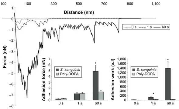
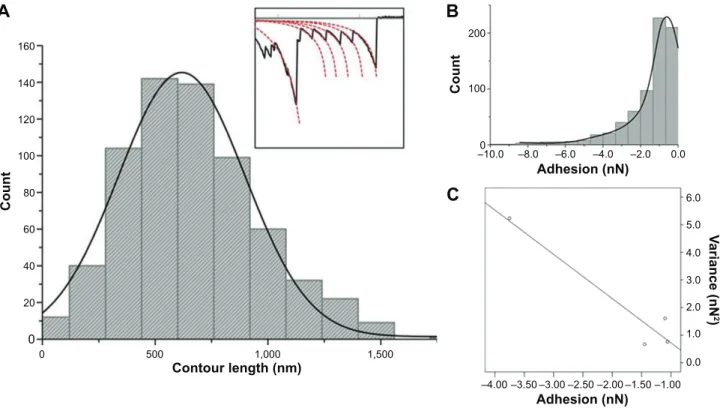
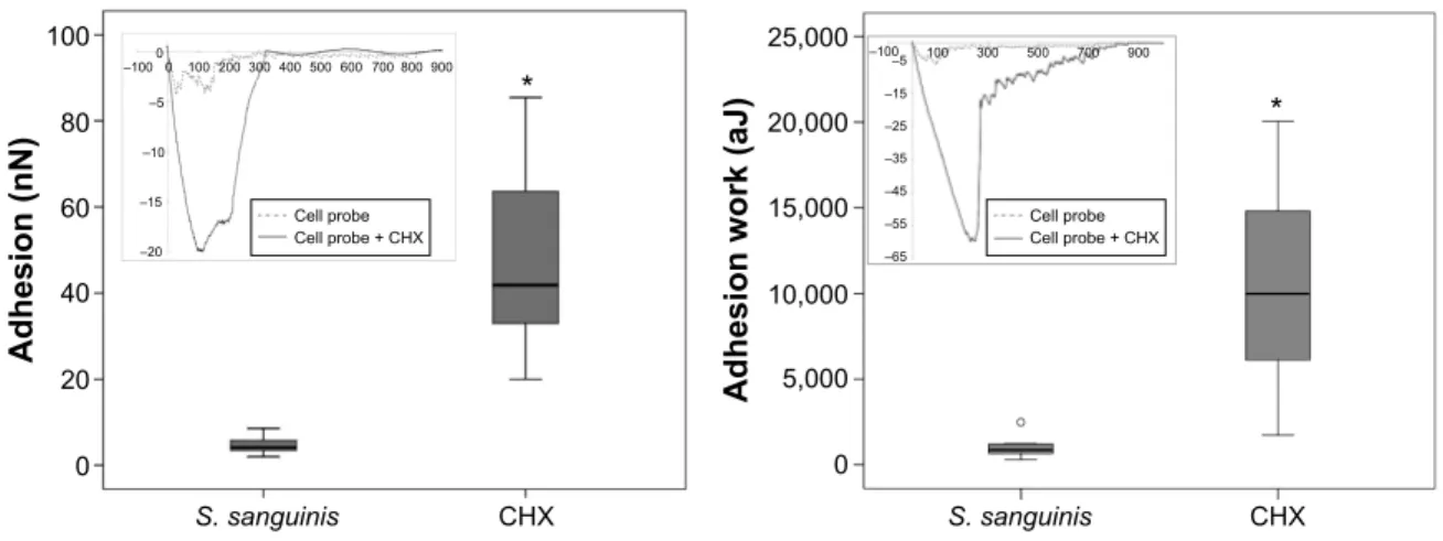
Probing the nanoadhesion of Streptococcus sanguinis to titanium implant surfaces by atomic force microscopy
Texto completo
Figure


Documento similar
The Court, referring to its above findings as to the lawfulness of the interference with the applicant’s right to freedom of expression under Article 10 of the Convention,
The Commission notes the applicant's conviction for having edited, published and distributed various articles and finds, therefore, that there has been an interference with
In the preparation of this report, the Venice Commission has relied on the comments of its rapporteurs; its recently adopted Report on Respect for Democracy, Human Rights and the Rule
In this chapter, we will study the deformation that a single force ring produces on a cylindrical membrane limited by a rigid wall, reflecting the inner bacterial membrane surrounded
Astrometric and photometric star cata- logues derived from the ESA HIPPARCOS Space Astrometry Mission.
To achieve such goal, the Electrostatic Force Microscope was used to measure the electrical properties of bacterial vegetative cells and bacterial endospores at the single cell
1) Correlative microscopy provides a powerful tool for studying biological systems in real time. Several technical challenges were found and overcome to successfully
Sigma54 dependent promoters Small molecule binding. Oligomerization