Functional convergence of microbes associated with temperate marine sponges
17
0
0
Texto completo
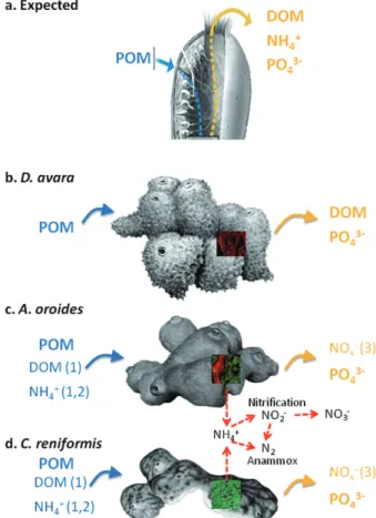
(2) Functional role of microbial associations in marine sponges. Fig. 1. Expected (A) and observed (B–D) nutrient fluxes in the three sponge species studied. Blue arrows indicate removal and yellow arrows indicate excretion. Bolded compounds names indicate that the flux is due to sponge metabolism. Un-bolded compound names indicate process related to microbial symbionts. Numbers indicate potential processes involved in the observed fluxes: (1) microbial heterotrophy, (2) microbial photoautotrophy, (3) microbial chemoautotrophy (nitrification and anammox indicated by red arrows). Pictures are from confocal microscopy of the sponge tissue using FISH with universal bacteria probes (bright green area) to visualize the relative abundance of symbiotic bacteria in each sponge. POM, Particulate organic matter; DOM, Dissolved organic matter.. Most marine sponges establish a persistent association with microbes including archaea, bacteria and protists. Microbial associates are hypothesized to contribute to the health and nutrition of sponges in different ways, such as by producing protective antibiotics, acquiring limiting nutrients and processing metabolic waste (Hoffmann et al., 2005; Taylor et al., 2007; Siegl et al., 2008). Based on the density of the microbial communities they host, sponges can be divided into ‘highmicrobial-abundance’ (HMA; formerly ‘bacteriosponges’, e.g. Reiswig, 1981) which are sponges that contain dense and host-specific microbial populations that exceed the microbial density of the surrounding water by two to four orders of magnitude (e.g. Weisz et al., 2008).. 1225. HMA species have dense tissues and low pumping rates (Siegl et al., 2008; Weisz et al., 2008 and references therein). In ‘low-microbial-abundance’ (LMA) sponges, the microbial community resembles the nearby seawater, both in concentration (Vacelet and Donadey, 1977; Reiswig, 1981; Wilkinson, 1983; Hentschel et al., 2006) and in phylogenetic composition (Schmitt et al., 2007). LMA species have well-irrigated tissues and high specific pumping rates. Most of our current knowledge about the microbes associated with marine sponges is based on 16S rRNA gene library construction, functional gene surveys and metagenomics, which are used to infer alternative metabolic routes for sponge metabolism. These metabolic routes include a variety of processes with alternative energy (photo- or chemotrophic) and carbon (hetero- or autotrophic) sources under different oxygen conditions (see Hoffmann et al., 2005; 2009; Hentschel et al., 2006; Taylor et al., 2007). An outcome of each of these processes is the growth of microbial biomass in the consortium, but the adaptive value of these processes to the host sponge and/or the consortium is not well understood. Moreover, while a phylogenetically and physiologically diverse array of microbes associate with sponges, their roles in nutrient cycles remains largely unknown (Hentschel et al., 2006; Taylor et al., 2007; Moya et al., 2008; Hoffmann et al., 2009). To elucidate the relationships between sponges, their microbial symbionts and nutrient cycles, we use molecular tools to quantify the microbial community composition and nutrient uptake and excretions, for three Mediterranean desmosponges Dysidea avara, Agelas oroides and Chondrosia reniformis. The selected species were formerly classified as either LMA (D. avara) or HMA (A. oroides and C. reniformis) on the basis of earlier qualitative observations (Turon et al., 1997; Vacelet and Donadey, 1977; Bayer et al., 2008). In a previous study using incubation chambers, we found contrasting dissolved nitrogen (DN) fluxes and a lack of balance between nitrogen released and removed from particulate food (Jiménez and Ribes, 2007). We suggested that alternative sources of organic nitrogen, such as dissolved organic nitrogen, might have been used by the consortium. In this study, we used a direct sampling method to compare the dissolved and particulate content of the water inhaled and exhaled by the sponge. This method allowed us to reliably examine the uptake and excretion of DON and DOC and address the hypothesis that the ability of sponges to handle dissolved compounds and the resulting nutrient flux is related to particular metabolic processes mediated by specific microbial associations. Thus, marked differences in the metabolic output should exist between species hosting contrasting microbial associates.. © 2012 Society for Applied Microbiology and Blackwell Publishing Ltd, Environmental Microbiology, 14, 1224–1239.
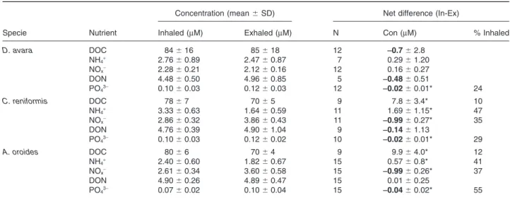
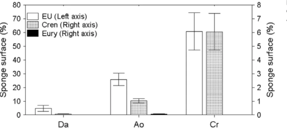
(3) 1226 M. Ribes et al. Results Nutrient removal and excretion The nutrient budget of the LMA sponge D. avara was markedly different from the budgets of the HMA sponges (C. reniformis and A. oroides, Fig. 1). While HMA sponges removed large amounts of dissolved organic carbon (8–10 mmol l-1 DOC) from the inhaled water, DOC was not significantly excreted or removed by D. avara (Table 1). DON was significantly excreted by D. avara but no differences were found between inhaled and exhaled concentrations in the HMA species. Similarly, NH4+ was removed by both C. reniformis (47%) and A. oroides (41%) but not by D. avara. The NOx- (NO2-+NO3-) budget also highlighted differences between D. avara and the other two species. C. reniformis and A. oroides had significant net excretion of NOx-, increasing ambient concentrations > 35%, whereas D. avara did not show significant differences between inhaled and exhaled concentrations (Table 1). Significant amounts of phosphate (PO43-, 24–55%) were excreted by all three sponges (Table 1). Microbial abundance Each of the three sponge species support a specific microbial community containing both bacteria and archaea (Fig. 2) but in very different proportions. Bacteria dominated the microbial community in A. oroides and C. reniformis forming a compact layer, so dense, that single-cell enumeration was impossible in most of the sponge tissue (Figs 2A and 3). Among archaea, only A. oroides hosted both Crenarchaea and Euryarchaea (Figs 2B and 3). Dysidea avara had the lowest microbial abundance per tissue volume, with only 6% of the sponge. volume occupied by microbes (Figs 2 and 3). In contrast, ~ 30% of the tissue volume of A. oroides was occupied by microbes (28% bacteria, 2% Crenarchaea and 0.1% Euryarchaea). The highest abundance of microbes was found in C. reniformis, with ~ 70% of its tissue occupied (60% bacteria and 6% Crenarchaea) (Figs 2 and 3). Using a non-EUB probe as negative control excluded the possibility of false non-specific or auto fluorescence in our measurements (Fig. S2). Ammonium-oxidizing groups Ammonium uptake and excretion of NOx- (nitrification) was detected in C. reniformis and A. oroides but not in D. avara. The presence and diversity of potential nitrifiers, including bacteria and archaea, was analysed by targeting amoA genes using PCR-based techniques. Surprisingly, a unique population of ammonia oxidizers microbes was present in each of the three sponges and these belong to different and remote taxonomic groups. Dysidea avara samples yielded positive PCR products only with b-proteobacteria primers (44 clones sequenced); A. oroides samples yielded positive PCR products only with archaeal primers (65 clones sequenced); C. reniformis samples yielded positive PCR product only with g-proteobacteria primers (83 clones sequenced). Water column samples were positive only with archaeal primers (50 clones sequenced). Four different sets of primers were used to amplify the 16S rRNA genes of putative anammox bacteria, including Planctomycetes and Scalindua (Table 2). All of the PCRs were compared with a positive control culture of Brocardia sp. (Sànchez-Melsió et al., 2009). Neither the sponge samples nor water column samples showed. Table 1. Average concentration (⫾ SD) and average net differences of dissolved nutrients at the inhaled and exhaled water for each species. Concentration (mean ⫾ SD). Net difference (In-Ex). Specie. Nutrient. Inhaled (mM). Exhaled (mM). N. Con (mM). % Inhaled. D. avara. DOC NH4+ NOxDON PO43-. 84 ⫾ 16 2.76 ⫾ 0.89 2.28 ⫾ 0.21 4.48 ⫾ 0.50 0.10 ⫾ 0.03. 85 ⫾ 18 2.47 ⫾ 0.87 2.12 ⫾ 0.16 4.96 ⫾ 0.85 0.12 ⫾ 0.03. 12 7 12 5 12. -0.7 ⫾ 2.8 0.29 ⫾ 1.20 0.16 ⫾ 0.27 -0.48 ⫾ 0.51 -0.02 ⫾ 0.01*. C. reniformis. DOC NH4+ NOxDON PO43-. 78 ⫾ 7 3.33 ⫾ 0.63 2.86 ⫾ 0.32 4.76 ⫾ 0.39 0.10 ⫾ 0.03. 70 ⫾ 5 1.64 ⫾ 0.59 3.86 ⫾ 0.43 4.90 ⫾ 1.04 0.12 ⫾ 0.02. 9 11 11 9 10. 7.8 ⫾ 3.4* 1.69 ⫾ 1.15* -0.99 ⫾ 0.27* -0.14 ⫾ 1.13 -0.02 ⫾ 0.01*. 10 47 35. A. oroides. DOC NH4+ NOxDON PO43-. 80 ⫾ 6 2.40 ⫾ 0.60 2.61 ⫾ 0.34 4.90 ⫾ 0.26 0.07 ⫾ 0.02. 70 ⫾ 4 1.82 ⫾ 0.67 3.60 ⫾ 0.58 4.89 ⫾ 0.47 0.10 ⫾ 0.04. 9 15 15 15 15. 9.9 ⫾ 4.0* 0.57 ⫾ 0.8* -0.99 ⫾ 0.26* 0.01 ⫾ 0.25 -0.04 ⫾ 0.02*. 12 41 37. 24. 29. 55. N: number of InEx pairs. The net nutrient difference is presented as the mean of paired differences and means of the percentage of the difference from the inhaled concentration (standard deviations were omitted for clarity of presentation). Negative values denote excretion (bolded). The asterisk (*) denotes significant differences between inhaled and exhaled concentrations (P < 0.05, two-tailed Wilcoxon test).. © 2012 Society for Applied Microbiology and Blackwell Publishing Ltd, Environmental Microbiology, 14, 1224–1239.
(4) Functional role of microbial associations in marine sponges. 1227. Fig. 2. Representative confocal microscopy (CLSM) images of the tissue of the three sponge species (Da, Dysidea avara; Ao, Agelas oroides; Cr, Chondrosia reniformis). CARD-FISH was used to visualize different microbial populations (upper panels in A and B) in combination with nucleic acid and sponge cells staining (lower panels in A and B). Different probes had different colours as follow: (A) Eubacteria: green for Eubacteria (EU), red for sponge cells (Spo) and blue for nucleic acids (NA); (B) Archaea: green for Euryarchaea (Eury), red for Crenarchaea (Cren) and blue for sponge cells (Spo).. positive PCRs products for 16S rRNA genes from anammox bacteria. Analysis of photoautotrophic microbes The presence and diversity of phototrophic microbes, potential consumers of NH4+, NOx- and PO4- and producers of DOC, was analysed by small subunit-rRNA and DGGE fingerprinting and sequence identification using both. general bacterial primers and specific cyanobacterial primers. To insure that DGGE results were representative we use several replicates per sample for each of the molecular markers analysed (bacterial and cyanobacterial 16S rRNA genes). The different replicates were highly reproducible yielding nearly identical DGGE patterns. To confirm the identity of the DGGE bands, we sequenced DNA retrieved from the same positions in several replicates and sponge samples. All three sponges seem to Fig. 3. Percentage of the sponge tissue occupied by different microbial populations. Abbreviations as in Fig. 2.. © 2012 Society for Applied Microbiology and Blackwell Publishing Ltd, Environmental Microbiology, 14, 1224–1239.
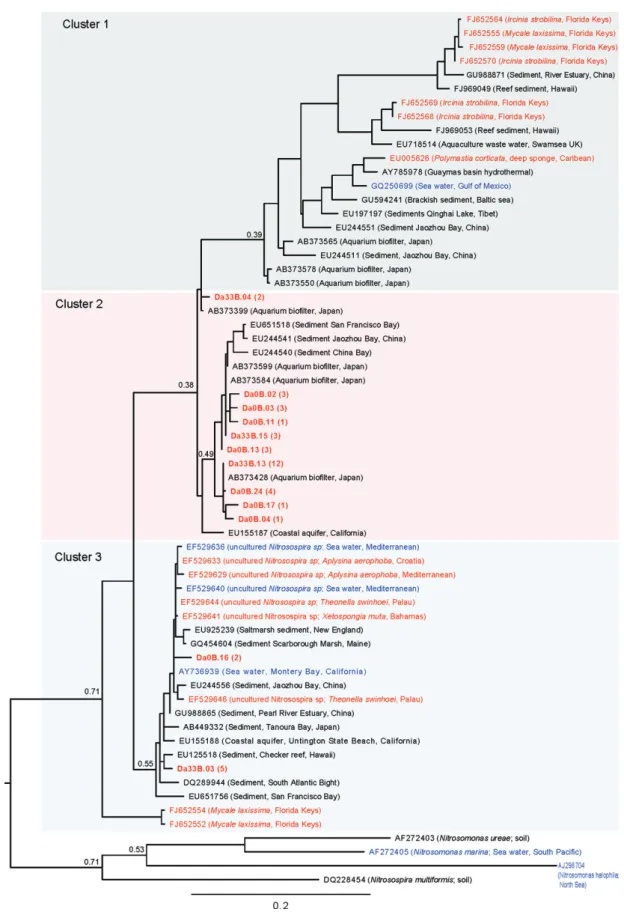
(5) 1228 M. Ribes et al. Table 2. Primers used for PCR amplification of amoA and 16S rRNA genes. Primer sets. Target group. Reference. Nitrifiers amoA-1F/amoA-2R amoA-3F/amoB-4R Arch-amoAR/Arch-amoAF. amoA b-Proteobacteria g-Proteobacteria Crenarchaeota. Rotthauwe et al. (1997) Purkhold et al. (2000) Francis et al. (2005), de la Torre et al. (2008). Anammox Brod541F/Brod1260R Pla46F/Amx368R Amx368F/BS820R Amx368F/1392R. 16S rRNA Candidatus Scalindula Anammox bacteria Anammox bacteria Anammox bacteria. Penton et al. (2006) Sànchez-Melsió et al. (2009) Amano et al. (2007) Mohamed et al. (2010). Bacteria 357F/907RM CYA106F/CYA781 Ra/b. Universal Cyanobacteria. Muyzer et al. (1997), Sánchez et al. (2007) Nübel et al. (1997). PCR conditions are indicated in the cited reference.. harbour both photoautotrophic and photoheterotrophic microbial components. DGGE results showed significant differences between the three sponge species investigated (Table 3). As shown in Table 3 only DGGE bands SJ1, 3, 5 and 9 (most affiliated to chloroplasts of eukaryotes) were shared by more than one species of sponge. Sequences obtained from those bands and the ones at the same position in the gel were found identical (99–100% similarity). Sequencing specific bands from our 16S rRNADGGE profiles indicate the presence of several bands that shared some similarity to the photoheterotrophic members of Chloroflexi, a green non-sulfur group of bacteria (Table 3). These phylotypes were well represented in C. reniformis (DGGE bands B2 and Sj15) and A. oroides (DGGE bands B5, B13 and B15, and Sj12) but were absent in D. avara. In addition, bands related to other bacteria, such as Acidobacteria (DGGE bands B11) and Proteobacteria (DGGE bands B1 and Sj13–14), were also present in A. oroides and C. reniformis respectively (Table 3). Using 16S rRNA primers specific to cyanobacteria, our analysis revealed a low level of cyanobacteria diversity with a distinct pattern for each of the three sponge species. Analysis of the most dominant DGGE bands revealed several photoautotrophic cyanobacteria closely related to unicellular Chroococcales of the genus Synechococcus (Table 3). These Synechococcus-like representatives may be an important part of the phototrophic microbial community associated with D. avara (DGGE band Sj6) and A. oroides (DGGE band Sj5). Indeed, Synechococcus-like chroococcales and phototrophic eukaryotes were the only phototrophic organisms associated with D. avara. In both A. oroides and D. avara sponges, the DGGE bands Sj1, 3, 9 and 10 were affiliated with chloroplasts of diatoms and pelagophytes (Table 3). Phylogenetic analysis of ammonia oxidizers Clone sequences of b-proteobacteria amoA in D. avara and archaea in A. oroides and water column samples. were used for phylogenetic analyses. Rarefaction curves for the three clone libraries approached an asymptote when clustering sequences at a 97% similarity threshold (Fig. S1), indicating that we were close to sample the total diversity of each functional group. From the 83 clone sequences of g-proteobacteria amoA in C. reniformis, seven showed a similarity of 100% with Nitrosococcus oneani amoA gene. The remaining 76 clone sequences presented a similarity of 99–100% between them but no significant similarity with amoA sequences from the Gene bank. The b-proteobacteria amoA genes from D. avara fell into three different clusters (Fig. 4). Cluster 1 was dominated by D. avara sequences, which shared ~ 99% amino acid similarity with uncultured b-proteobacteria ammonia oxidizers bacteria from an aquarium sand biofilter in Tokyo [Cluster B2 (AB373584, AB373599) and cluster B3 (AB373428), respectively, in Urakawa et al., 2008]. Sediment sequences from three different sites also fell into cluster 1. Cluster 2 included sponge sequences from the Florida Keys and the deep Caribbean that were unrelated to D. avara sequences. Cluster 3 included seven D. avara clones that were 98–99% identical to uncultured Nitrosospira spp. clones from Mediterranean, Caribbean and Pacific Ocean marine sponges (sequences with Accession No. EF5296XX; Bayer et al., 2008). Dysidea avara clones in cluster 3 were also 98% identical to uncultured bacteria from sediments and sequences from the sponge Mycale laxissima (Florida Keys) reported by Mohamed and colleagues (2010). Crenarchaeal ammonia oxidizers from A. oroides and water column samples primarily fell into three of the five different clusters (Fig. 5). Cluster 1 contained most of our water column samples, as well as seawater samples from the Pacific, Atlantic and Arctic oceans. Cluster 1 also included a few sequences of archaea associated with benthic invertebrates (three sequences from A. oroides, five sequences from sponges from Sydney (Australia) and. © 2012 Society for Applied Microbiology and Blackwell Publishing Ltd, Environmental Microbiology, 14, 1224–1239.
(6) 16S rRNA gene/primer set. Universal Bacteria. Universal Bacteria. Universal Bacteria. Universal Bacteria Universal Bacteria. Universal Bacteria. Specific Cyanobacteria. Specific Cyanobacteria. Specific Cyanobacteria. Specific Cyanobacteria. Specific Cyanobacteria. Specific Cyanobacteria. Specific Cyanobacteria. Specific Cyanobacteria Specific Cyanobacteria. Specific Cyanobacteria. DGGE band accession No.. B1-JN314396. B2-JN314397. B5-JN314398. B11-JN314399 B13-JN314400. B15-JN314401. Sj1-JN314402. Sj3-JN314403. Sj5-JN314404. Sj6-JN314405. Sj9-JN314406. Sj10-JN314407. Sj12-JN314408. Sj13-JN314409 Sj14-JN314410. Sj15-JN314411. C. reniformis. C. reniformis C. reniformis. A. oroides. D. avara and A. oroides A. oroides. D. avara. D. avara and A. oroides D. avara and A. oroides D. avara and A. oroides. A. oroides. A. oroides A. oroides. A. oroides. C. reniformis. C. reniformis. Sponge. Uncultured Chloroflexi clone E117; FJ529342. Plastid Aureococcus anophagefferens strain CCMP Plastid uncultured phototrophic eukaryote clone HF770_25L02; EU361155 Uncultured Chloroflexi bacterium clone CC10; DQ247942 Uncultured bacterium clone i153; FM160905 Uncultured bacterium clone 27H6; EU 183804. Uncultured sponge symbiont clone Hg5a2D10; EU817117. Uncultured sponge symbiont clone Hg5a2D10; EU817117. Plastid Skeletonema pseudocostatum; X82155. Plastid Skeletonema pseudocostatum; X82155. Uncultured Chloroflexi clone AD020; EF076163. Uncultured sponge symbiont clone PAWS52F; AF186417 Uncultured Chloroflexi clone W04IS5E12; EF629745 Uncultured bacterial clone CC13; DQ247947 Uncultured Chloroflexi clone E29; FJ529321. Uncultured alphaproteobacteria clone SHFH749; FJ203650. Nearest relative/GenBank accession No.. Table 3. 16S rRNA gene sequence identities of bacterial DGGE bands from sponge samples.. Aplysina fulva Brazil Rhopaloeides odorabile Pelorus Isl, Australia Svenzea zeai Salvador Isl, Bahamas. Corticium embryo Palau. Montastraea faveolata San Cristobal, Panamá Theonella sp. Caroline Isl, Palau Ircinia strobilina Conch reef Key Largo, Fl Corticium embryo Palau Svenea zeai San Salvador Is, Bahamas Antho chartacea Southeastern Australia Skeletonema pseudocostatum Skeletonema pseudocostatum Haliclona sp. northern Pacific, Monterey harbour Haliclona sp. Northern Pacific, Monterey harbour Aureococcus anophagefferens North Pacific Subtropical Gyre. Source location. Cyanobacteria. 99. 95. 97 97. 90. 94. Chloroflexi. Deltaproteobacteria Deltaproteobacteria. Chloroflexi. Pelagophyte chloroplast Eukaryotic chloroplast. Cyanobacteria. 94. 97. Diatom chloroplast. Diatom chloroplast. Chloroflexi. Acidobacteria Chloroflexi. Chloroflexi. 98. 99. 83. 85 79. 90. Chlorofexi. Alphaproteoacteria. 86. 94. Phylogenetic affiliation. % identity. Lee et al. (2009). Hardoim et al. (2009) Webster et al. (2008). Sharp et al. (2007). Pham et al. (2008). Ong et al. (2010). Sipkema et al. (2009). Sipkema et al. (2009). Medlin et al. (1995). Medlin et al. (1995). Taylor et al. (2007). Sharp et al. (2007) Lee et al. (2009). Mohamed et al. (2008). Hentschel et al. (2002). Sunagawa et al. (2009). Reference. Functional role of microbial associations in marine sponges. © 2012 Society for Applied Microbiology and Blackwell Publishing Ltd, Environmental Microbiology, 14, 1224–1239. 1229.
(7) 1230 M. Ribes et al.. Fig. 4. Maximum likelihood phylogenetic tree based on b-proteobacteria amoA DNA sequences (420 informative positions). Sequences for D. avara are in red and boldface and database sequences for other marine sponges are in red. Database water column sequences are in blue. Sequences are identified by GenBank accession numbers. Bootstrap values for important nodes are displayed. Brackets refer to number of sequences.. © 2012 Society for Applied Microbiology and Blackwell Publishing Ltd, Environmental Microbiology, 14, 1224–1239.
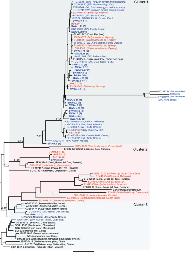
(8) Functional role of microbial associations in marine sponges two sequences from a Red Sea coral). Cluster 3 included most of the A. oroides sequences that were related to archaea from the sponge Luffariella and from a coral from Panama. None of our sequences fell in cluster 4 with Cenarchaeum symbiosum; only two of our water column samples fell into cluster 4 with Nitrosopumilus maritimus, which also included sediment, aquaculture and aquaria biofilter samples, as well as ground and freshwater. Discussion Observed nutrient fluxes and related microbial types The nutrient fluxes we observed for the three studied sponges can be related to at least three different metabolic pathways: heterotrophy, photoautotrophy and chemoautotrophy (Fig. 1). Dissolved organic carbon taken up by the sponge is transferred to and metabolized by heterotrophic bacteria and archaea (Wilkinson and Garrone, 1980; De Goeij et al., 2008a,b), therefore a high abundance of heterotrophic microbes is expected to result in high DOC removal. Ammonium (NH4+) can support both photoautotrophy and chemoautotrophy. While phototrophs use ammonium as the preferred nitrogen source, chemoautotrophic ammonia oxidizers use ammonium as an energy source. In the later case, the decrease in ammonium excretion should be linked with enhanced NOx- excretion (Fig. 1). Other chemoautotrophic processes such anammox may also contribute to NH4+ removal (Fig. 1), nevertheless annamox bacteria were not detected in any of the three sponge species. Removal of DOC and microbial abundance Removal of dissolved organic carbon was associated with high abundance of bacteria and archaea. Dysidea avara has the lowest abundance of microbes (6% of the sponge volume occupied by microbes) and accordingly showed no significant DOC removal. In contrast, A. oroides and C. reniformis had much higher densities of associated microbes (~ 30 and 70% of sponge volume respectively) and consequently, high DOC uptake (average > 10%) was evident in 93% of the In-Ex pairs in A. oroides and 100% of the In-Ex pairs in C. reniformis. The ability of sponges to remove large quantities of DOC from the water they pump has been reported for coral reef sponges (Yahel et al., 2003; De Goeij and van Duyl, 2007; De Goeij et al., 2008a,b), indicating that the removal of DOC by reef sponges may play a major role in the trophic dynamics of coral reefs. In other tropical systems, Southwell and colleagues (2008a) found contrasting levels of DOC uptake for LMA versus HMA, suggesting that total respiration in HMA sponges is largely fuelled by DOM uptake as previously suggested by Reiswig (1981).. 1231. While marine sponges are now routinely categorized into the LMA and HMA groups depending on their microbial abundance, these classifications are mostly based on visual assessments either with electron microscopy (Vacelet and Donadey, 1977; Friedrich et al., 1999) or by fluorescent in situ hybridization (FISH) and polarized/ epifluorescent light microscopy (Friedrich et al., 1999; Pape et al., 2006; Bayer et al., 2008). In the HMA species we studied, the microbial populations were arranged in compact and dense structures resembling microbial biofilms. Quantification of the individual cell number in such dense communities is nearly impossible by standard microscopy. Therefore, previous workers, relying on epifluorescent light microscopy and related techniques (Schläppy et al., 2010), were forced to select sparse tissue regions which likely result in underestimate of the real microbial densities. We conclude that confocal microscopy is required for reliable quantification of similarly structured microbial communities in HMA sponges. This is the first attempt to quantify the percentage of sponge tissue being occupied by microbial cells in marine sponges using confocal laser scanning microscopy. Presence of photoautotrophs Photoautotrophs are potential consumers of DIN and producers of DOC. The unique DGGE profile of each sponge species provides a clear representation of the specific microbial communities that it harbours. Using universal bacterial primers and specific cyanobacterial primers, we identified bacterial phylotypes that are closely related to Chloroflexi in C. reniformis and A. oroides and Chroococcales unicellular cyanobacteria in A. oroides and D. avara. Both groups resemble previously reported photosynthetic symbionts from other sponges worldwide (Hentschel et al., 2002; Sharp et al., 2007; Taylor et al., 2007; Hardoim et al., 2009; Lee et al., 2009; Sunagawa et al., 2009). Only a few bands were related to cyanobacteria and only in A. oroides and D. avara and none was recovered with a bacterial universal primer, suggesting a low relative abundance for these organisms in our samples. The low abundance of photoautotrophs present in D. avara, was apparently insufficient to elicit net ammonia removal. In contrast, high diversity of Chloroflexi-like phylotypes recovered by our bacterial and cyanobacterial 16S rRNA gene analysis suggests that this group of potentially photosynthetic organisms is an important symbiont in the investigated species. However, not all Chloroflexi bacteria are phototrophs, and thus a phototrophic function of the Chloroflexi-like bacteria within the sponge we examined is yet to be confirmed. The overall contribution of phototrophic components to nutrient fluxes in the sponge consortium needs to be further studied.. © 2012 Society for Applied Microbiology and Blackwell Publishing Ltd, Environmental Microbiology, 14, 1224–1239.
(9) 1232 M. Ribes et al.. Fig. 5. Maximum likelihood phylogenetic tree based on archaeal amoA DNA sequences (570 informative positions). Sequences for A. oroides are in red and boldface, sequences for other sponges are in red. Database water column sequences are in blue and water column sequences from our sampling site are in blue and boldface. Database sequences are identified by GenBank accession numbers. Bootstrap values for important nodes are displayed. Brackets refer to number of sequences.. © 2012 Society for Applied Microbiology and Blackwell Publishing Ltd, Environmental Microbiology, 14, 1224–1239.
(10) Functional role of microbial associations in marine sponges -. Release of NOx and presence of ammonia oxidizers Large amounts of excess NOx- (> 35% or 1 mmol l-1 per litre pumped) were excreted by the two HMA sponges A. oroides and C. reniformis but not by the LMA sponge D. avara (Fig. 1, Table 1). Ammonia oxidation, the first and limiting step in nitrification, can be performed by b- or g-proteobacteria and by crenarchaeota from Marine Group 1 (Francis et al., 2005; 2007). Both groups of microorganisms have been previously reported in sponges (Taylor et al., 2007). Each of the three sponge species we studied appears to harbour its own nitrifier group: g-proteobacteria (putative) in C. reniformis, b-proteobacteria in D. avara and archaea in A. oroides. The phylogenetic tree based on b-proteobacteria ammonia oxidizers shows that only few of the amoA sequences derived from D. avara are related to amoA sequences of the Nitrosospira lineage. Clusters 1 and 2 were dominated by sequences that shared ~ 99% amino acid similarity with uncultured b-proteobacteria ammonia oxidizers from an aquarium biofilter in Tokyo (Clusters B2 and B3, respectively, in Urakawa et al., 2008). The crenarchaeal ammonia oxidizers phylogenetic tree shows that A. oroides-derived sequences are clearly separated from their environmental water sample sequences, as well as from seawater samples from different oceans and sediments. Coral-derived amoA sequences from Panama and the sponge Luffariella sp. from Queensland, Australia, were the most closely related to the amoA sequences obtained from A. oroides whereas amoA sponge sequences reported in Steger and colleagues (2008) [cluster 4] were not related to A. oroides amoA sequences. It was not possible to generate a phylogenetic tree with g-proteobacteria ammonia oxidizers derived from C. reniformis as only 7 sequences were found to be related to a known GenBank sequences (Nitrosococcus oceani). Further studies will be performed with the remaining 76 sequences as they can be pointing out the presence of an unknown group of ammonia oxidizers. A relationship between the amount of nitrification and specific pumping rates in marine sponges has previously been suggested (Weisz et al., 2008). Southwell (2007) and Southwell and colleagues (2008b) related nitrification rates to the amount of microbes associated with sponges and pumping rates. They found that 12 of 13 HMA sponges (with low specific pumping rates) showed nitrification, whereas nitrification was not detected in LMA sponges (with high specific pumping rates). Our results are consistent with this interpretation. The similar specific pumping rates of C. reniformis and A. oroides (both showing high nitrification rates) were three times lower than those of D. avara that showed no evidence for significant nitrification (M. Ribes unpubl. data). Active nitrifi-. 1233. cation in HMA sponges with low specific pumping rate may be preventing the toxic effect of ammonia on sponge tissues. Differences between ammonia oxidation in bacteria and archaea can depend on rates, carbon fixation mechanisms, environmental controls and competitive ability in the biogeochemical process (Francis et al., 2007). Crenarchaeal ammonia oxidizers appear to greatly outnumber their bacterial counterparts in soil systems (Leininger et al., 2006) and in the marine water column (Francis et al., 2005). However, it remains unclear whether archaea or bacteria are the main ammonia oxidizers in sponges (Taylor et al., 2007). Our results indicate that both bacteria and archaea may act as the main ammonia oxidizers group depending on the sponge species. The particular case of A. oroides shows that even though the bacterial population dominates in microbial abundance, the high nitrification rate of the species is likely attributed to its small archaeal population. NOx- concentration did not change significantly in the water inhaled and exhaled by D. avara despite the presence of small populations of ammonia oxidizer microbes. In contrast, Schläppy and colleagues (2010) did measure net NOx- excretion by the D. avara specimens they studied. The differences between the two studies may be related to methodological biases or to seasonal differences in sponge metabolism. Schläppy and colleagues (2010) used closed vessels and long incubation times (24 h). A comparison of the direct In-Ex method we used with closed vessel incubations indicate that assessing dissolved compound fluxes in sponges is consistently biased by prolonged incubation times (M. Ribes, unpubl. data). Nevertheless, temporal variation in activity and bacterial symbiont composition are also possible because Schläppy and colleagues (2010) worked in early spring (March) whereas our experiments were performed in May–July. Removal of ammonia Ammonia uptake is an important source of nitrogen for autotrophic and heterotrophic microorganisms (Kirchman et al., 1990, Hoch and Kirchman, 1995), but also a potential energy source for ammonia-oxidizing microorganisms. Data about the ability of sponges to remove DIN (NH4+, NOx-) from the water column is still scarce and only reported for coral reef sponge (Schubauer, 1988; Díaz and Ward, 1997; Yahel et al., 2003; Southwell et al., 2008a). While all three sponges we studied contained ammonia oxidizer associate microorganisms, considerable ammonium removal was found only in HMA A. oroides and C. reniformis (41% and 52% respectively) but not in LMA D. avara. HMA sponges also showed high nitrification rates. While it is tempting. © 2012 Society for Applied Microbiology and Blackwell Publishing Ltd, Environmental Microbiology, 14, 1224–1239.
(11) 1234 M. Ribes et al. to associate nitrification with ammonia removal, since representative of all three potential ammonium consumers were present in each of the studied sponges, a more complex nitrogen pathways may exists. These findings call for attention to DIN consumption that appears as an important mechanism of nutrient acquisition in temperate sponges.. Release of PO43The three sponges studied showed high and significant net excretion of PO43- (~ 20%, 30% and 60% for D. avara, C. reniformis and A. oroides respectively) without any apparent relationship to microbial abundance or composition. To the best of our knowledge, phosphorus metabolism has not previously been studied in marine sponges (Taylor et al., 2007). Our results suggest that sponges are important recyclers of phosphorus in the coralligenous communities. It is also evident that P is not a limiting nutrient for the microbial symbionts of the studied sponges, and that the already know particulate feeding by sponges seems to provide an excess amount of this nutrient for the consortium during the study period. The composition of microbial components of the studied sponges and the surrounding water was focused on the most abundant groups, which we assumed were the most likely to account for gross metabolic processes. Nevertheless, more detailed analysis (e.g. pyrosequencing) may reveal rare microbial groups unaccounted for here and may bring new insights on specificity of microbial components in marine sponges. Analysis of the microbial communities associated with the studied sponges indicated that each sponge species harboured a unique microbial community, different from those of the water column, and with a specific set of associated microbes. Surprisingly, these distinct communities converged to similar metabolic pathways in the HMA sponges, exhibiting high nitrification and high DOC and NH4+ uptake, and similar net nutrient fluxes. In each consortium, these similar metabolic activities and nutrient dynamic processes were accomplished by contrasting species or microbial communities. The results points to a functional convergence of microbial partners in these sympatric sponge species suggesting that these metabolic processes are of special relevance to the sponge consortium success.. Experimental procedures Specimen collection Six specimens from each of the three sponge species (D. avara, A. oroides and C. reniformis) were collected by scuba divers from the coralligenous community of the Montgri. Coast (NW Mediterranean Sea, 42°3′N, 3°13′E) at a 15 m depth. Whole specimens were transported within a few hours to the Experimental Aquaria Zone (ZAE) of the Institute of Marine Sciences (ICM-CSIC) and maintained in 125 l tanks with a flow-through system of fresh seawater that renewed the total volume every 15 min. Parallel to specimen collection, water samples (4 l) were collected from the water column near to the sponge community. Three replicates of 300–500 ml were filtered in the laboratory through 0.2 mm polycarbonate filters. Filters were frozen in liquid nitrogen and stored at -80°C until microbial DNA extraction was performed (see below).. Quantification of nutrient removal and excretion Nutrient removal and excretion by each sponge specimen was measured using a direct method (Yahel et al., 2005). In each experiment, two 0.55 mm ID Teflon tubes were used to sample simultaneously the water inhaled and exhaled (In-Ex) by the sponge at rates of < 1 ml min-1. One tube was positioned a few millimetres inside the osculum to sample exhaled water, and the other was placed next to the sponge to sample inhaled water. Sampled water was collected in darkened glass bottles kept in ice during the sampling (6–10 h). For each sponge species we collected at least three In-Ex pairs from each of the five collected specimens. Experiments were conducted between May and June 2007. The temperature was recorded at the beginning and end of the experiments. To avoid contamination from microbes in the water, after the experiments, small tissue samples (1 cm3) of each individual were positioned in sterile 2 l containers filled with filtered seawater (0.22 mm) for 5 h. Each piece was then rinsed with filtered seawater before being frozen with liquid nitrogen and stored at -80°C for further microscopy observations and molecular analysis.. Analysis of dissolved compounds Water samples were collected in acid-rinsed, 50 ml plastic bottles, frozen and analysed for inorganic nutrients with an Alliance autoanalyser following the method of Grasshoff and colleagues (1983). The following dissolved compounds were measured: NH4+ (ammonium), NOx- [NO3-+NO2-] and PO43(phosphate). The DN (including DIN and DON) concentration was analysed using a Bran-Luebbe AA3 autoanalyser after a double oxidation with UV light and persulfate (Grasshoff et al., 1983). DON concentration was calculated by subtracting the DIN concentration (NH4++NOx-) from the DN. Samples for DOC determination were filtered through pre-combusted (450°C for at least 2 h) GFF filters with a baked glass filtration system. Water for DOC analysis was collected in 10 ml precombusted glass ampoules and acidified with concentrated, trace-metal grade, hydrochloric acid (0.1% final concentration). Ampoules were heat-sealed and stored at -20°C until analysis. DOC concentrations were determined using hightemperature catalytic oxidation method on a Shimadzu TOC-V analyser (Sharp et al., 2002). The accuracy of the DOC determination was evaluated by daily comparisons with DOC reference materials (Yahel et al., 2003).. © 2012 Society for Applied Microbiology and Blackwell Publishing Ltd, Environmental Microbiology, 14, 1224–1239.
(12) Functional role of microbial associations in marine sponges. 1235. FISH and confocal microscopy. Cloning and sequencing. We used catalysed reporter deposition (CARD)-FISH and confocal laser-scanning microscopy (CLSM) to quantify the microbial community associated with each sponge specimen. Tissue blocks of the sponges fixed in 1% paraformaldehyde were embedded in paraffin, and 10 mm slices were obtained. Three specimen per specie and three slices from each specimen were hybridized using CARD-FISH with probes for Bacteria (Eub 338 I+II+III; Amann et al., 1990; Daims et al., 1999) and Archaea (Cren 537 and Eury 806; Teira et al., 2004), following the protocol described in Pernthalerand collaborators (2002). Non-EUB probe was used as negative control following the same procedures (Non338 5′-ACT CCTACGGGAGGCAGC-3′, Wallner et al., 1993). The structure of the sponge was stained with CellMask™ Deep Red (Molecular Probes, Invitrogen, Carlsbad, CA, USA), and the nucleic acids were labelled with Hoechst 33342 (Molecular Probes, Eugene, OR, USA). Samples were observed with a Leica TCS-SP5 confocal spectral microscope (Leica Microsystems Heidelberg GmbH; Mannheim, Germany) using a Plan-Apochromatic 63 ¥ 1.4 (oil HC ¥ PL APO lambda blue objective). A series of images (xyz) was taken in four randomly selected areas of each sample to visualize the emission signal of Alexa 488 (for Eub 338 and Cren 537), Alexa 546 (for Eury 806), Hoechst and CellMask. The resulting images (11 images per area) were processed with Metamorph imaging software (Universal Imaging Corporation, West Chester, PA, USA) in order to calculate the percentage of sponge tissue volume occupied by microbes.. Amplified PCR products for b-proteobacteria (~ 420 bp) and for archaea (~ 570 bp) from two or three individuals were pooled and purified with a PCR Purification kit (Qiagen) and cloned using the TOPO-TA Cloning kit (Invitrogen) according to the manufacturer’s instructions. Putative positive colonies were picked and transferred to a multiwell plate with LB-7% glycerol medium plus kanamycin. After an overnight incubation at 37°C the plates were stored at -80°. The presence of the insert in the colonies was checked by PCR reamplification with the original primer set. Positive clones were sent to Macrogen Sequencing Service (Korea) to be purified and sequenced. All chromatograms were visually inspected to minimize the sequencing errors. After this first examination, sequences were submitted to the BLAST search (Altschul et al., 1997) for a first phylogenetic affiliation and to remove putative chimeras. Forty-four clones were sequenced for b-proteobacteria ammonia oxidizers from D. avara, four of them were chimeric sequences. Sixty-five clones were sequenced for crenarchaeal ammonia oxidizers from A. oroides and 50 clones for crenarchaeal ammonia oxidizers from water samples. No chimeric sequences were detected in A. oroides or water samples.. DNA extraction and PCR amplification Tissue samples (~ 2 mm3) from each specimen were dissected into small sections using a sterile scalpel. Total genomic DNA was extracted from sponge samples using a DNeasy Tissue Kit (Qiagen) following the manufacturer’s protocol with the following modifications: removal of water excess before the DNA extraction; incubation with lysis buffer overnight instead of few hours; final elution in 100 ml of Buffer AE instead of 200 ml (A. Blanquer and S. LópezLegentil, pers. comm.). Putative amoA gene fragments from the total microbial DNA of proteobacteria and archaea were amplified by PCR with the primers listed in Table 2 and using GoTaq Flexi Dna Polymerase (Promega). For PCR amplification we followed the conditions described in the reference papers with the following modifications: for g-proteobacteria (Purkhold et al., 2000) we ran 35′ cycles and a final elongation step of 10′ was added. For archaea amoA (de la Torre et al., 2008) we used an annealing temperature of 56°C. Specific 16S rRNA genes for different groups of annamox organisms were amplified by PCR with the primers listed in Table 2 and following original references conditions. Reactions of 25 ml were carried out in an automated thermocycler (Bio-Rad) in parallel for the three sponge species and water column samples. A positive control was used to validate the PCR methodology in each run. An agarose gel was run in order to check PCR products with a molecular weight standard (Low DNA Mass Ladder, Invitrogen). The gel was visualized with UV in the Quantity One-Chemidoc software (Bio-Rad).. Phylogenetic analysis DNA sequences were aligned with MAFFTusing the slow and iterative refinement method FFT-NS-i (Katoh et al., 2002). The alignment was checked manually and edited using Seaview 3.2 (Galtier et al., 1996), to retain the longest region that was common to most sequences. Maximum likelihood (ML) phylogenetic trees were constructed with RAxML (Stamatakis, 2006) using the evolutionary model GTR+G + I″ that best fits our data in the freely available University of Oslo Bioportal (http://www.bioportal.uio.no). Repeated runs on distinct starting trees were carried out to select the tree with the best topology (the one having the best Likelihood of 1000 alternative trees). Bootstrap ML analysis was carried out using 1000 pseudo-replicates. Trees were edited with the FigTree v1.3.1. Rarefaction analysis estimates were performed in the program MOTHUR (Schloss et al., 2009) after grouping sequences at 97%, 98% and 99% similarity thresholds.. 16S rRNA-DGGE fingerprinting Denaturing gradient gel electrophoresis (DGGE) was used to compare bacterial and cyanobacterial communities associated with the different investigated sponge species. PCR amplifications were performed using the bacterial universal and cyanobacterial-specific 16S rDNA oligonucleotide primers (Table 2). DGGE was performed with a DGGE-2000 system (CBS Scientific) as described by Díez and colleagues (2001). Electrophoresis was performed using 0.75-mm-thick, 6% polyacrylamide gels (37.5:1 acrylamide: bisacrylamide) submerged in 1¥ TAE buffer (40 mM Tris, 40 mM acetic acid and 1 mM EDTA, pH 7.4) at 60°C with a linear gradient of denaturing agents from 45% to 75% for both cyanobacteria and bacteria. After electrophoresis, the gel was stained in 1¥. © 2012 Society for Applied Microbiology and Blackwell Publishing Ltd, Environmental Microbiology, 14, 1224–1239.
(13) 1236 M. Ribes et al. TAE buffer containing SYBRGold Nucleic Acid Stain (1:10 000 dilution; Molecular Probes) to reveal the band patterns. The results were recorded using a molecular imager (Chemi Doc XRS system, Bio-Rad™). To obtain the gene sequences of the DGGE bands, polyacrylamide fragments were excised from the gel using sterilized razor blades, resuspended in 20 ml of MilliQ water and stored at 4°C overnight. An aliquot of the eluted DNA was re-amplified using PCR with the same primers and conditions as before. The re-amplified PCR products were sequenced (with the corresponding forward primer) at the Macrogen Sequencing Service (Korea).. Nucleotide sequence accession numbers Sequences obtained in this study were submitted to GenBank under Accession No. JN409473–JN409512 (for D. avara bacterial amoA genes), JN409513–JN409554 (for A. oroides archaea amoA genes), JN409555–JN409596 (for seawater archaea amoA genes), JQ280408–JQ280414 (for C. reniformis bacterial amoA genes) (JN314396– JN314401 (for bacterial 16S rRNA genes) and JN314402– JN314411 (for cyanobacterial 16S rRNA genes).. Acknowledgements We thank Dr Mònica Roldán and the Microscopy Service, Autonomous University of Barcelona (UAB) for their assistance on confocal microscopy. We also thank Irene Forn for her participation on the CARD-FISH methodology. The study was funded by the Spanish ‘Ministerio de Ciencia e Innovación’ through Grants CGL2010-18466 CGL2007-66757C02-01/BOSand CTM2006-01463. This is a contribution from the Marine Biogeochemistry and Global Change research group, funded by the ‘Generalitat de Catalunya’ (Catalan Government) through Grant 2009SGR142.. References Altschul, S.F., Madden, T.L., Schäfer, A.A., Zhang, J., Zhang, Z., Miller, W., and Lipman, D.J. (1997) Gapped BLAST and PSI-BLAST: a new generation of protein database search programs. Nucleic Acids Res 25: 3389–3402. Amann, R.I., Binder, F.J., Olson, R.J., Chisholm, S.W., Devereux, R., and Stahl, D.A. (1990) Combination of 16S rRNA-targeted oligonucleotide probes with flow cytometry for analyzing mixed microbial populations. Appl Environ Microbiol 56: 1919–1925. Amano, T., Yoshinaga, I., Okada, K., Yamagishi, T., Ueda, S., Obuchi, A., et al. (2007) Detection of anammox activity and diversity of anammox related 16S rRNA genes in coastal marine sediments in Japan. Microbes Environ 22: 232– 242. Bayer, K., Schmitt, S., and Hentschel, U. (2008) Physiology, phylogeny and in situ evidence for bacterial and archaeal nitrifiers in the marine sponge Aplysina aerophoba. Environ Microbiol 10: 2942–2955. Brusca, R., and Brusca, G. (1990) Phylum Porifera: the sponges. In The Invertebrates. Sinauer, A. (ed.). Sunderland, MA, USA: Sinauer Press, pp. 181–210.. Coma, R., Ribes, M., Gili, J.M., and Hughes, R.N. (2001) The ultimate opportunists: consumers of seston. Mar Ecol Prog Ser 219: 305–308. Daims, H., Brühl, A., Amann, R., Schleifer, K.H., and Wagner, M. (1999) The domain-specific probe EUB338 is insufficient for the detection of all Bacteria: development and evaluation of a more comprehensive probe set. Syst Appl Microbiol 22: 434–444. De Goeij, J.M., and van Duyl, F.C. (2007) Coral cavities are sinks of dissolved organic carbon (DOC). Limnol Oceanogr 52: 2608–2617. De Goeij, J.M., Moodley, L., Houtekamer, M., Carballeira, N.M., and van Duyl, F.C. (2008a) Tracing 13C-enriched dissolved and particulate organic carbon in the bacteriacontaining coral reef sponge Halisarca caerulea: evidence for DOM-feeding. Limnol Oceanogr 53: 1376–1386. De Goeij, J.M., van den Berg, H., van Oostveen, M.M., Epping, E.H.G., and Van Duyl, F.C. (2008b) Major bulk dissolved organic carbon (DOC) removal by encrusting coral reef cavity sponges. Mar Ecol Prog Ser 357: 139– 151. De La Torre, J.R., Walker, C.B., Ingalls, A.E., Könneke, M., and Stahl, D.A. (2008) Cultivation of a thermophilic ammonia oxidizing archaeon synthesizing crenarchaeol. Environ Microbiol 10: 810–818. Díaz, M.C., and Rützler, K. (2001) Sponges: an essential component of Caribbean coral reefs. Bull Mar Sci 69: 535– 546. Díaz, M.C., and Ward, B.B. (1997) Sponge-mediated nitrification in tropical benthic communities. Mar Ecol Prog Ser 156: 97–109. Díez, B., Pedrós-Alió, C., Marsh, T.L., and Massana, R. (2001) Application of Denaturing Gradient Gel Electrophoresis (DGGE) to study the diversity of marine picoeukaryotic assemblages and comparison of DGGE with other molecular techniques. Appl Environ Microbiol 67: 2942– 2951. Falkowski, P.G., Fenchel, T., and Delong, E.F. (2008) The microbial engines that drive earth’s biogeochemical cycles. Science 320: 1034–1039. Francis, C.A., Roberts, K.J., Berman, J.M., Santoro, A.E., and Oakley, B.B. (2005) Ubiquity and diversity of ammonium-oxidizing archaea in water columns and sediments of the ocean. Proc Natl Acad Sci USA 102: 14683– 14688. Francis, C.A., Berman, J.M., and Kuypres, M.M.M. (2007) New processes and players in the nitrogen cycle: microbial ecology of anaerobic and archaeal ammonia oxidation. ISME J 1: 19–27. Friedrich, A.B., Merkert, H., Fendert, T., Hacker, J., Proksch, P., and Hentschel, U. (1999) Microbial diversity in the marine sponge Aplysina cavernicola (formerly Verongia cavernicola) analyzed by fluorescence in situ hybridization (FISH). Mar Biol 134: 461–470. Galtier, N., Gouy, M., and Gautier, C. (1996) SEAVIEW and PHYLO_WIN: two graphic tools for sequence alignment and molecular phylogeny. Comput Appl Biosci 12: 543– 548. Gili, J.M., and Coma, R. (1998) Benthic suspension feeders: their paramount role in littoral marine food webs. Trends Ecol Evol 13: 316–321.. © 2012 Society for Applied Microbiology and Blackwell Publishing Ltd, Environmental Microbiology, 14, 1224–1239.
(14) Functional role of microbial associations in marine sponges Grasshoff, K., Ehrhardt, M., and Kremling, K. (1983) Methods of Seawater Analysis, 2nd edn. Weinheim, Germany: Verl. Chem. Hadas, E., Marie, D., Shpigel, M., and Ilan, M. (2006) Virus predation by sponges is a new nutrient-flow pathway in coral reef food webs. Limnol Oceanogr 51: 1548–1550. Hardoim, C.C., Costa, R., Araujo, F.V., Hajdu, E., Peixoto, R., Lins, U., et al. (2009) Diversity of bacteria in the marine sponge Aplysina fulva in Brazilian coastal Waters. Appl Environ Microbiol 75: 3331–3343. Hentschel, U., Hopke, J., Friedrich, A.B., Wagner, M., Hacker, J., and Moore, B.S. (2002) Molecular evidence for a uniform microbial community in sponges from diVerent oceans. Appl Environ Microbiol 68: 4431–4440. Hentschel, U., Usher, K.M., and Taylor, M.W. (2006) Marine sponges as microbial fermenters. FEMS Microbiol Ecol 55: 167–177. Hoch, M.P., and Kirchman, D.L. (1995) Ammonium uptake by heterotrophic bacteria in the Delaware estuadry and adjacent coastal waters. Limnol Oceanogr 40: 886–897. Hoffmann, F., Larsen, O., Thiel, V., Rapp, H.T., and Reitner, J. (2005) An anaerobic world in sponges. Geomicrobiol J 22: 1–10. Hoffmann, F., Radax, R., Woebken, D., Holtappels, M., Lavik, G., Rapp, H.T., et al. (2009) Complex nitrogen cycling in the sponge Geodia barretti. Environ Microbiol 11: 2228– 2243. Hooper, J.M.A., and van Soest, R.W.M. (2002) Systema Porifera: A Guide to the Classification of Sponges. New York, NY, USA: Kluwer Academic/Plenum Publishers. Jiménez, E., and Ribes, M. (2007) Sponges as a source of dissolved inorganic nitrogen: nitrification mediated by temperate sponges. Limnol Oceanogr 52: 948–958. Katoh, K., Misawa, K., Kuma, K., and Miyata, T. (2002) MAFFT: a novel method for rapid multiple sequence alignment based on fast Fourier transform. Nucleic Acids Res 30: 3059–3066. Kirchman, D.L., Keil, R.G., and Wheeler, P.A. (1990) Carbon limitation of ammonium uptake by heterotrophic bacteria in the subarctic Pacific. Limnol Oceanogr 35: 1258–1266. Lee, O.O., Chui, P.Y., Wong, Y.H., Pawlik, J.R., and Qian, P.Y. (2009) Evidence for vertical transmission of bacterial symbionts from adult to embryo in the Caribbean sponge Svenzea zeai. Appl Environ Microbiol 75: 6147–6156. Leininger, S., Urich, T., Schloter, M., Schwark, L., Qi, J., Nicol, G.W., et al. (2006) Archaea predominate among ammonia-oxidizing prokaryotes in soils. Nature 442: 806– 809. Madigan, M.T., Martinko, J.M., and Parker, J. (2003) Metabolic diversity. In Brock Biology of Microorganisms. Madigan, M.T., Martinko, J.M., and Parker, J. (eds). Upper Saddle River, NJ, USA: Pearson Education, pp. 552–597. Medlin, L.K., Cooper, A., Hill, C., Wrieden, S., and Wellbrock, U. (1995) Phylogenetic position of the Chromista plastids based on small subunit rRNA coding regions. Curr Genet 28: 560–565. Mohamed, N.M., Colman, A.S., Tal, Y., and Hill, R.T. (2008) Diversity and expression of nitrogen fixation genes in. 1237. bacterial symbionts of marine sponges. Environ Microbiol 10: 2910–2921. Mohamed, N.M., Saito, K., Tal, Y., and Hill, R.T. (2010) Diversity of aerobic and anaerobic ammonia-oxidizing bacteria in marine sponges. ISME J 4: 38–48. Moya, A., Peretó, J., Gil, R., and Latorre, A. (2008) Learning how to live together: genomic insights into prokaryote– animal symbioses. Nature 9: 218–229. Muyzer, G., Brinkhoff, T., Nübel, U., Santegoeds, C., Schaäfer, H., and Wawer, C. (1997) Denaturing gradient gel electrophoresis (DGGE) in microbial ecology. In Molecular Microbial Ecology Manual. Akkermans, D.L., van Elsas, J.D., and de Bruijn, F.J. (eds). Dordrecht, the Netherlands: Kluwer Academic, pp. 1–27. Nübel, U., Garcia-Pichel, F., and Muyzer, G. (1997) PCR primers to amplify 16S rRNA genes from cyanobacteria. Appl Environ Microbiol 63: 3327–3332. Ong, H.C., Wilhelm, S.W., Gobler, C.J., Bullerjahn, G., Jacobs, M.A., McKay, J., et al. (2010) Analyses of the complete chloroplast genome sequences of two members of the Pelagophyceae: Aureococcus anophagefferens CCMP1984 and Aureoumbra lagunensis CCMP1507. J Phycol 46: 602–615. Pape, T., Hoffmann, F., Queric, N.V., von Juterzenka, K., Reitner, J., and Michaelis, W. (2006) Dense populations of archaea associated with the desmosponge Tentorium semisuberites Schmidt, 1870 from Arctic deep-waters. Polar Biol 29: 662–667. Penton, C.R., Devol, A.H., and Tiedje, J.M. (2006) Molecular evidence for the broad distribution of anaerobic ammonium-oxidizing bacteria in freshwater and marine sediments. Appl Environ Microbiol 72: 6829–6832. Pham, V.D., Konstantinidis, K.T., Palden, T., and DeLong, E.F. (2008) Phylogenetic analyses of ribosomal DNAcontaining bacterioplankton genome fragments from a 4000 m vertical profile in the North Pacific Subtropical Gyre. Environ Microbiol 10: 2313–2330. Pile, A.J. (1997) Finding Reiswig’s missing carbon: quantification of sponge feeding using dual-beam flow cytometry. Proceedings of the 8th International Coral Reef Symposium 2: 1403–1410. Purkhold, U., Pommering-Röser, A., Juretschko, S., Schmid, M.C., Koops, H.P., and Wagner, M. (2000) Phylogeny of all recognized species of ammonia oxidizers based on comparative 16S rRNA and amoA sequence analysis: implications for molecular diversity surveys. Appl Environ Microbiol 66: 5368–5538. Reiswig, H.M. (1981) Partial carbon and energy budgets of the bacteriosponge Verongia fistularis (Porifera: Demospongiae) in Barbados. PSZNI Mar Ecol 2: 273–263. Ribes, M., Coma, R., and Gili, J.M. (1999) Natural diet and grazing rate of the temperate sponge Dysidea avara (Demospongiae, Dendroceratida) throughout an annual cycle. Mar Ecol Prog Ser 176: 179–190. Rotthauwe, J.H., Witzel, K.P., and Liesack, W. (1997) The ammonia monooxygenase structural gene amoA as a functional marker: molecular fine-scale analysis of natural ammonia-oxidizing populations. Appl Environ Microbiol 63: 4704–4712. Sánchez, O., Gasol, J.M., Massana, R., Mas, J., and PedrósAlió, C. (2007) Comparison of different denaturing gradient. © 2012 Society for Applied Microbiology and Blackwell Publishing Ltd, Environmental Microbiology, 14, 1224–1239.
(15) 1238 M. Ribes et al. gel electrophoresis primer sets for the study of marine bacterioplankton communities. Appl Environ Microbiol 73: 5962–5967. Sànchez-Melsió, A., Cáliz, J., Balaguer, M.D., Colprim, J., and Vila, X. (2009) Development of batch-culture enrichment coupled to molecular detection for screening of natural and man-made environments in search of anammox bacteria for N-removal bioreactors systems. Chemosphere 75: 169–179. Schläppy, M.L., Schöttner, S.I., Lavik, G., Kuypers, M.M., de Beer, D., and Hoffmann, F. (2010) Evidence for nitrification and denitrification in high and low microbial abundance sponges. Mar Biol 157: 593–602. Schloss, P.D., Westcott, S.L., Ryabin, T., Hall, J.R., Hartmann, M., Hollister, E.B., et al. (2009) Introducing MOTHUR: open-source, platform-independent, community-supported software for describing and comparing microbial communities. Appl Environ Microbiol 75: 7537– 7541. Schmitt, S., Wehrl, M., Bayer, K., Siegl, A., and Hentschel, U. (2007) Marine sponges as models for commensal microbe–host interactions. Symbiosis 44: 43–50. Schubauer, J.P. (1988) Metabolism and Nutrient Cycling by Marine Sponges. Athens, GA, USA: University of Georgia. Sharp, J.H., Carlson, C.A., Peltzer, E.T., Castle-Ward, D.M., Savidge, K.B., and Rinker, K.R. (2002) Final dissolved organic carbon broad community intercalibration and preliminary use of DOC reference materials. Mar Chem 77: 239–253. Sharp, K.H., Eam, B., Faulkner, D.J., and Haygood, M.G. (2007) Vertical transmission of diverse microbes in the tropical sponge Corticium sp. Appl Environ Microbiol 73: 622–629. Siegl, A., Bayer, K., Kozytska, S., Hentschel, U., and Schmitt, S. (2008) Sponges and microbes – new frontiers in an ancient symbiosis. Vie et Milieu Life and Environment 58: 165–174. Sipkema, D., Holmes, B., Nichols, S.A., and Blanch, H.W. (2009) Biological characterisation of Haliclona (?gellius) sp.: sponge and microorganisms. Microb Ecol 58: 903– 920. Southwell, M.W. (2007) Sponges Impacts on Coral Reef Nitrogen Cycling, Key Largo, Florida. North Carolina: Chapel Hill. Southwell, M.W., Popp, B.N., and Martens, C.S. (2008a) Nitrification controls on fluxes and isotopic composition of nitrate from Florida Keys sponges. Mar Chem 108: 96–108. Southwell, M.W., Weisz, J.B., Martens, C.S., and Lindquist, N. (2008b) In situ fluxes of dissolved inorganic nitrogen from the sponge community on Conch Reef, Key Largo, Florida. Limnol Oceanogr 53: 986–996. Stamatakis, A. (2006) RAxML-VI-HPC: maximum likelihoodbased phylogenetic analyses with thousands of taxa and mixed models. Bioinformatics 22: 2688–2690. Steger, D., Ettinger-Epstein, P., Whalan, S., Hentschel, U., de Nys, R., Wagner, M., and Taylor, M.W. (2008) Diversity and mode of transmission of ammonia-oxidizing archaea in marine sponges. Environ Microbiol 10: 1087–1094. Sunagawa, S., DeSantis, T.Z., Piceno, Y.M., Brodie, E.L.,. DeSalvo, M.K., Voolstra, C.R., et al. (2009) Bacterial diversity and White Plague Disease-associated community changes in the Caribbean coral Montastraea faveolata. ISME J 3: 512–521. Taylor, M.W., Radax, R., Steger, D., and Wagner, M. (2007) Sponge associated microorganisms: evolution, ecology and biotechnological potential. Microbiol Mol Biol Rev 71: 295–347. Teira, E., Reinthaler, T., Pernthaler, A., Pernthaler, J., and Herndl, G.J. (2004) Combining catalyzed reporter deposition-fluorescence in situ hybridization and microautoradiography to detect substrate utilization by bacteria and archaea in the deep ocean. Appl Environ Microbiol 70: 4411–4414. Turon, X., Galera, J., and Uriz, M.J. (1997) Clearance rates and aquiferous systems in two sponges with contrasting life-history strategies. J Exp Zool 278: 22–36. Urakawa, H., Tajima, Y., Numata, Y., and Tsuneda, S. (2008) Low temperature decreases the phylogenetic diversity of ammonia-oxidizing archaea and bacteria in aquarium biofiltration systems. Appl Environ Microbiol 74: 894–900. Vacelet, J., and Donadey, C. (1977) Electron microscope study of the association between some sponges and bacteria. J Exp Mar Biol Ecol 30: 301–314. Wallner, G., Amann, R., and Beisker, W. (1993) Optimizing fluorescent in situ hybridization with rRNA-targeted oligonucleotide probes for flow cytometric identification of microorganisms. Cytometry 14: 136–143. Webster, N.S., Cobb, R.E., and Negri, A.P. (2008) Temperature thresholds for bacterial symbiosis with a sponge. ISME J 2: 830–842. Weisz, J.B., Lindquist, N., and Martens, C.S. (2008) Do associated microbial abundances impact marine desmosponge pumping rates and tissue densities? Oecologia 155: 367– 376. Wilkinson, C.R. (1983) Net primary productivity in coral reef sponges. Science 219: 410–412. Wilkinson, C.R., and Garrone, R. (1980) Nutrition in marine sponges. Involvement of symbiotic bacteria in the uptake of dissolved carbon. In Nutrition in the Lower Metazoa. Smith, D.C., and Tiffon, Y. (eds). Oxford, UK: Pergamon Press, pp. 157–161. Yahel, G., Sharp, J.H., Marie, D., Hase, C., and Genin, A. (2003) In situ feeding and element removal in the symbiont-bearing sponge Theonella swinhoei: bulk DOC is the major source for carbon. Limnol Oceanogr 48: 141– 149. Yahel, G., Marie, D., and Genin, A. (2005) InEx – a direct in situ method to measure filtration rates, nutrition, and metabolism of active suspension feeders. Limnol Oceanogr Methods 3: 46–58.. Supporting information Additional Supporting Information may be found in the online version of this article: Fig. S1. Rarefaction curves for ammonia-oxidizers groups. (A) b-Ammonia-oxidizing bacteria from D. avara, (B) ammonia-oxidizing archaea amoA clone libraries for A. oroi-. © 2012 Society for Applied Microbiology and Blackwell Publishing Ltd, Environmental Microbiology, 14, 1224–1239.
(16) Functional role of microbial associations in marine sponges des and (C) ammonia-oxidizing archaea amoA clone libraries for water samples. OTUs were defined at a 99, 98 and 97 sequence similarity thresholds. Fig. S2. Representative confocal microscopy (CLSM) images of the tissue of the three sponges species (Da, Dysidea avara; Ao, Agelas oroides; Cr, Chondrosia reniformis).. 1239. A. Negative controls using a non-EUB probe. B. Autofluorescence controls without probe (see Fig. 2). Please note: Wiley-Blackwell are not responsible for the content or functionality of any supporting materials supplied by the authors. Any queries (other than missing material) should be directed to the corresponding author for the article.. © 2012 Society for Applied Microbiology and Blackwell Publishing Ltd, Environmental Microbiology, 14, 1224–1239.
(17) Copyright of Environmental Microbiology is the property of Wiley-Blackwell and its content may not be copied or emailed to multiple sites or posted to a listserv without the copyright holder's express written permission. However, users may print, download, or email articles for individual use..
(18)
Figure
+2
Documento similar
It is generally believed the recitation of the seven or the ten reciters of the first, second and third century of Islam are valid and the Muslims are allowed to adopt either of
One might expect that there is a smooth transition between a small sample regime, in which finite size effects such as those observed in sums of heavy-tailed random variables,
Nutrients derived from penguin colonies and other marine vertebrates altered the trophic interactions of communities within microbial mats, as well as the relative abundance and
Comparing pairs of groups shows that, with regard to the analysis of negative impacts, the highest levels of conflict are associated with disagreements between the joint group of
In the preparation of this report, the Venice Commission has relied on the comments of its rapporteurs; its recently adopted Report on Respect for Democracy, Human Rights and the Rule
This study reports the comparative microbial ecology of the water column of Nuestra Señora del Carmen acid pit lake with the extreme acidic Río Tinto basin.. The
On the other hand, SEM and XPS clearly indicated that the enhancement of the activity and selectivity to benzaldehyde was associated with the concentration of the
The concentrations of microorganisms in the digester effluent were determined for each HRT tested. The analysis of the microbial popula- tion was performed at the end of each
