Circadian rhythms synchronize mitosis in Neurospora crassa
6
0
0
Texto completo
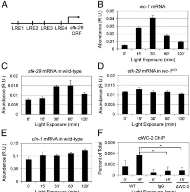
(2) The cell cycle regulation of Neurospora has yet to be investigated thoroughly because of some technical limitations, such as adequate methods to synchronize, image, and measure doubling times of nuclear divisions in growing mycelia. We explored the Neurospora genome (25) to find the homologs of key cell cycle regulators and found that Neurospora has a low number of predicted cyclins and CDKs. Neurospora has a single Cdk1 homolog (cdc-2, NCU09778), one G1 cyclin that resembles the sequence of the G1/S regulating budding yeast Clns (cln-1, NCU02114), and two B-type cyclins (clb-1, NCU02758, and clb-3, NCU01242) (26). There also exists a homolog of the CDK1 inhibitor WEE1 kinase (stk-29, NCU04326), which is regulated in a circadian manner in the mouse liver (5). Interactions between the above homologous proteins in budding and fission yeast have been well characterized, and their conservation among eukaryotes (27, 28) suggests they may be wired in a similar fashion in N. crassa. Here, we investigate the molecular connection between the cell cycle and the circadian clock and functional consequences of this coupling in N. crassa. First, we show that there is a conserved connection between the cell cycle and the circadian clock in Neurospora as in mammals via STK-29, which is the Neurospora homolog of WEE1. Based on this finding and on the hypothesis of conserved cell cycle regulatory interactions, we use mathematical modeling to investigate molecular profiles of both cell cycle and circadian clock components. Our computational simulations predict circadian oscillations of cell cycle components, such as CLN-1 and CLB-1. We experimentally validate this prediction with luciferase bioluminescence reporters to track both cell cycle and circadian clock components in real time in vivo. Moreover, we demonstrate circadian clock-induced phase shifts of cell cycle components, which may alter the timing of divisions. The circadian oscillations of key cell cycle components suggest circadian clock-gated synchronized nuclear divisions. By observing nuclear morphology over time at 25 °C in DD, we indicate that most divisions occur in the evening. We propose that there is a significant coupling between the cell cycle and the circadian clock, which might result in immediate changes in the dynamics of cell cycle regulation upon alterations in circadian rhythms. Results There Is a Conserved Coupling Between the Cell Cycle and the Circadian Clock in N. crassa as in Mus musculus. A heterodimeric. circadian transcription factor, WCC, recognizes light-responsive elements (LREs) to activate target genes (13, 29, 30). We found four putative LREs (GAGATCC, CCGATCC, CCGATCG, and TCGATCT) within 1.75 kb of the stk-29 gene 5′ upstream region (Fig. 1A). To test WCC-dependent activation of stk-29, we performed a light induction experiment. WC-1 is also a photoreceptor that undergoes a light induction response, which is described by a sharp increase in its expression followed by a decrease to its basal level of expression when Neurospora is transferred from dark to light conditions (Fig. 1B). Light-induced WC-1 activates many downstream target genes by recognizing LREs (31). We observe light response from stk-29 mRNA in the wild type, which is abolished in the wc-1ko (Fig. 1 C and D). In contrast, we do not observe a light response of cln-1 mRNA in wild-type strains (Fig. 1E). The WC-1–dependent light response of stk-29 indicates that stk-29 is activated by WCC and that it is a potential target for circadian regulation. To verify direct binding of WCC to the promoter of stk-29, we performed a WC-2 ChIP experiment and show that the WC-2 binds to the region close to LRE1 (Fig. 1F). Based on the finding that stk-29 is activated by WCC, we tested a mathematical model of the Neurospora circadian clock and cell cycle as a coupled oscillator and explored coupled dynamics (Figs. S1–S4 and Tables S1–S5). 1398 | www.pnas.org/cgi/doi/10.1073/pnas.1319399111. Fig. 1. stk-29 mRNA shows WC-1–dependent light response, and WC-2 directly binds to the stk-29 promoter. (A) There are four LREs within 1.75 kb of the stk-29 gene 5′ upstream region. The first LRE, GAGATCC, is located ∼1.75 kb upstream (LRE1); the second LRE, CCGATCC, is located ∼1.2 kb upstream (LRE2); the third LRE, CCGATCG, is located ∼0.8 kb upstream (LRE3); and the fourth LRE, TCGATCT, is located ∼0.25 kb upstream (LRE4) of the stk-29 gene. (B) wc-1 mRNA undergoes light response when Neurospora is moved from dark to light conditions. (C and D) stk-29 mRNA shows light response in the wild type (C), which is abolished in wc-1ko (D). (E) cln-1 mRNA does not show light response in the wild type. The above data are relative units (R.U.) normalized with actin mRNA. The average ± SD is shown. The above data are representative of two or more independent experiments. (F) WC-2 directly binds to the promoter of stk-29. ChIP assay was performed on a wildtype strain (FGSC2489), with samples grown in the dark (0′) or in response to a 15-min light pulse (15′) using a polyclonal antibody that recognizes WC-2 protein and oligos specific for a region of the stk-29 promoter. A nonspecific IgG and a strain lacking the wc-2 gene (Δwc-2) were used as controls. The results are an average of five experiments, and the error bars represent the SDs. The asterisks indicate a P value <0.001.. cln-1 and clb-1 Gene Expression and Protein Abundance Show Circadian Clock-Dependent Oscillations. Our mathematical model predicts. circadian oscillations of cell cycle components such as CLN-1 and CLB-1 proteins if intermediate to strong coupling exists between the circadian clock and the cell cycle (Figs. S1–S4). To validate circadian-dependent oscillations of cell cycle factors, we constructed bioluminescence reporters to track in vivo gene expression of cln-1 (NCU02114), clb-1 (NCU02758), stk-29 (NCU04326), and cdc-2 (NCU09778) in real time. Bioluminescence reporters were constructed by fusing the fully codon-optimized luciferase from firefly with a promoter of interest (32). Our data indicate that expression of cln-1, clb-1, and stk-29 from populations of Neurospora nuclei show circadian oscillations (Fig. 2A). We also observe circadian oscillations of cln-1 and clb-1 mRNA expressions (Fig. S5). Expression of cdc-2, however, does not follow circadian regulation (Fig. 2A). This is in accord with the cell cycle model that we adapted (33), which assumes constitutive expression of cdc-2. We then constructed translational bioluminescence reporters of CLN-1luc, CLB-1luc, and CDC-2luc by fusing luciferase to genes of interest as previously described for FRQluc (34), and followed protein abundances of CLN-1, CLB-1, and CDC-2. The abundance of both CLN-1 and CLB-1 Hong et al..
(3) shows circadian oscillations with phase information similar to that of their gene expression profiles (Fig. 2B). The observed phase relationship between CLN-1 and CLB-1 is expected based on their cell cycle functions in G1 and G2/M phases, respectively. In contrast, the abundance of CDC-2 increases continuously over time, corresponding to the growth in mass of Neurospora, and does not exhibit circadian oscillations (Fig. 2B). The data suggest that CDC-2 is stable with a constant rate of expression, consistent with findings in budding yeast (35). Importantly, circadian oscillations of CLN-1 protein and clb-1 and stk-29 gene expression are lost in the frqko strain, an arrhythmic mutant in which the circadian clock is nonfunctional (Fig. 2C). This indicates that the. synchronized oscillations of cell cycle elements are under the influence of circadian rhythms. Based on the above data, we hypothesized that the expression of cell cycle genes such as clb-1 might be altered in a circadian manner. We performed light-pulse experiments to phase-shift circadian rhythms and investigated the circadian-dependent phase shifts of cell cycle components. We tracked bioluminescence of frq, clb-1, and stk-29 gene expression after a 90-min light pulse at specific time points in DD. We observed ∼3–5-h phase advances and delays in the expression of frq, clb-1, and stk-29 when light pulses were given at DD32 [circadian time 23 (CT23)] and DD48 (CT16), respectively (Fig. 3). This demonstrates that the phases of clb-1 and stk-29 gene expression are influenced by phase. Fig. 3. clb-1 and stk-29 gene expressions indicate circadian clock-dependent phase shifts. (A–C) A 90-min light pulse is given at either DD32 (dashed black) or DD48 (solid black), and the phases of peak expressions of frq, clb-1, and stk-29 genes are compared with unperturbed data (frq, orange; clb-1, blue; stk-29, maroon) at the fourth peak of unperturbed data (dashed straight line). Corresponding peaks are labeled in each figure. The data shown represent three independent experiments. (D) A 90-min light stimulus at DD32 and DD48 creates ∼3–5-h phase advances and delays, respectively. The data are from three independent experiments (Fig. S6), and the average ± SD is shown. Arbitrary units (AU) are shown.. Hong et al.. PNAS | January 28, 2014 | vol. 111 | no. 4 | 1399. CELL BIOLOGY. Fig. 2. cln-1, clb-1, and stk-29 demonstrate circadian oscillations. (A) cln-1, clb-1, stk-29, and cdc-2 promoters are fused to the codon-optimized firefly luciferase (32) for real-time analyses of their gene expressions in vivo. A strain carrying frq-luciferase reporter, an established core circadian component, is used as a positive control. (B) cln-1, clb-1, and cdc-2 genes are fused with the codon-optimized firefly luciferase for real-time observation of CLN-1, CLB-1, and CDC-2 protein abundances. (C) A strain housing clb-1–luciferase or stk-29–luciferase reporter is crossed with frqko mutant resulting in clb-1–luciferase and stk-29– luciferase reporters in frqko background that show loss of circadian oscillations of clb-1 and stk-29 gene expression. Similarly, the CLN-1luc translational reporter is crossed with frqko mutant, resulting in a CLN-1luc reporter strain in frqko background, which shows an arrhythmic phenotype. The above data are representative of three or more independent experiments. Arbitrary units (AU) are shown..
(4) changes of the circadian clock that are similar in degree and direction, which may alter the timing of nuclear divisions in N. crassa. Circadian Clock-Dependent Synchronized Nuclear Divisions Occur in the Middle of the Night. The lack of circadian oscillations of clb-1. gene expression in frqko does not necessarily indicate altered mitosis (Fig. 2C). Rather, it suggests asynchronous mitotic divisions uncoupled from circadian rhythms. To verify this, we investigated circadian clock-dependent synchronized nuclear divisions. In Neurospora, nuclei are visualized readily by using an hH1-sgfp strain in which histone H1 is fused to GFP (21, 24). By using this strain, the stages of the cell cycle can be visualized and categorized. We performed a time-course experiment under circadian conditions (i.e., DD at 25 °C) and classified the populations of nuclei into two categories: interphase and mitotic phase (Fig. 4A). At CT4, or during the subjective day, most nuclei are in interphase, as shown by round nuclear morphology (Fig. 4B). In contrast, many nuclei undergo mitosis at around CT17, which corresponds to late subjective evening (Fig. 4C). Although there is variability in mitotic stage, around 60% of nuclei are actively dividing in the evening (Fig. 4D). These data clearly demonstrate circadian oscillations in Neurospora mitotic divisions. The synchronized nuclear divisions are not observed in the frqko strain (Fig. 4E), which indicates that circadian rhythms are necessary for this daily synchronization of cell cycles. These observations are in accord with the arrhythmic clb-1 and stk-29 gene expression in frqko (Fig. 2C). We also used an established mitosis marker, phospho-histone H3 (pH3) antibody, as an independent. Fig. 4. Circadian clock-gated synchronized nuclear divisions are observed in Neurospora. (A) Different stages of mitotic cycles can be visualized with the hH1-sgfp strain and categorized based on the morphology of nuclei. (B and C) Microscopy data showing strands of hyphae at two different time points: CT4 and CT17. CT denotes circadian time in a free-running period in DD, in which subjective day begins at CT0 and subjective night begins at CT12. (D) Percentages of nuclei in mitosis are calculated as a time course with 2-h resolution. The average ± SD is shown. DD27 is statistically different from DD37 (*P = 0.021). The values are obtained from a time-course experiment with four to six samples from each time point. (E) Percentages of nuclei in mitosis are calculated and compared between the wild type (black) and frqko (gray) at four different time points (CT15, CT17, CT23, and CT4). The average ± SEM is shown. The data are from three or more independent experiments. Two-way ANOVA indicates there is a significant difference between DD27 and DD37 in the wild type (*P = 0.012) but not in the frqko strain (**P = 0.33). There is a significant difference between the wild type and the frqko at DD27 (***P = 0.005) but not at DD37 (****P = 0.609). Similar data are shown from live cell imaging (Fig. S8 and Movies S1–S4).. 1400 | www.pnas.org/cgi/doi/10.1073/pnas.1319399111. measurement of mitosis (36, 37). We observed more pH3-positive nuclei at DD25 (CT15) than at DD35 (CT2) (Fig. S7 A and B). The above experiments are performed by harvesting Neurospora from liquid culture media in DD and counting the number of nuclei present in fixed cells. It is important to note that we observe similar results via live cell imaging from Neurospora grown in defined solid agar media, in which we observe a second cycle of increased and decreased mitosis at DD47 and DD57, respectively (Fig. S8 and Movies S1–S4). Discussion In silico, we investigated various scenarios of coupled dynamics between the circadian clock and the cell cycle, which demonstrated circadian oscillations of cell cycle components if significant coupling exists between the two oscillators (Figs. S2–S4). We have demonstrated experimentally that elements of the cell cycle (e.g., cln-1 and clb-1) undergo circadian oscillations, which manifest a circadian clock-dependent synchronized mitotic division in Neurospora. We also show that both clb-1 and stk-29 gene expression undergo light-dependent phase shifts in a length and direction similar to those of frq gene expression. This suggests circadian clock-dependent phase shifts of cell cycle components, which might be used to alter the timing of mitotic divisions. The fundamental molecular regulatory architecture of circadian rhythms that highlight the time-delayed negative feedback mechanism is conserved from N. crassa to M. musculus (38). Coupling between circadian rhythms and the DNA damage response pathway is also conserved between Neurospora and mammals. Checkpoint kinase 2 (CHK2) is activated upon DNA damage and phosphorylates one of the core clock components (i.e., PER1 in mammals and FRQ in Neurospora), resulting in a subsequent degradation of PER1 or FRQ that leads to predominantly phase advances in circadian rhythms (39–43). We demonstrate that WC-2 binds to the promoter of stk-29 (NCU04326) and that stk-29 undergoes WC-1–dependent light-response and circadian oscillations, which shows conserved coupling between the cell cycle and circadian rhythms. The binding of WC-2 to the stk-29 promoter was not reported in the recent WC-2 ChIP-sequencing data (44). This is probably a result of the low expression of stk-29 and the less dramatic light response of stk-29 compared with other targets. Further investigations are needed to understand the detailed dynamics of these connections as well as other possible coupling factors. We have shown circadian oscillations in a few cell cycle regulators. However, it is unclear whether these cycling components are genuine coupling components or mere reflections of the circadian-gated cell cycle determined by the currently known coupling factor STK-29. Recently, microarray data have suggested that several genes in cell cycle control show oscillatory behavior in Neurospora (45). Identification of other factors that couple the cell cycle and circadian rhythms will elucidate distinct points of interactions in which the circadian clock influences the cell cycle. Identified conserved coupling components (i.e., CHK2 and WEE1) among the circadian clock, DNA damage response, and cell cycle mechanisms pose Neurospora as an ideal model organism to investigate the fundamental wiring of this network. However, one of the main disadvantages of Neurospora is that it is technically difficult to assess the doubling time of mitotic cycles in Neurospora mycelium grown on solid agar media. Previous measurements in liquid culture media showed a range in doubling time from 72 to 239 min, depending on growth conditions from young germinating conidia (46), which is in good agreement with our measurements in noncircadian conditions (e.g., LL) (Fig. S9). However, the doubling time in mature mycelium in circadian conditions (i.e., DD) might be different because of the presence of the circadian clock. Therefore, measuring the doubling time of nuclear divisions in DD for an extended period will be critical for future experiments. Real-time fluorescence and bioluminescence reporters, in addition to the use of Hong et al..
(5) Materials and Methods Strains. Strains used for the experiments are a clock wild-type ras-1bd;a (328-4) and three arrhythmic mutants from the laboratories of Drs. J. Dunlap and J. Loros (Dartmouth Medical School, Hanover, NH) [ras-1bd; frqko;a (358-6), ras-1bd;wc-1ko (S38), and ras-1bd;wc-2ko (Δwc-2)]. Wild-type strain FGSC#2489 (Mat A) was used for the ChIP experiment. Strain hH1-sgfp (FGSC#9518) was obtained from the Fungal Genetics Stock Center (FGSC, University of Missouri–Kansas City) (48). cln-1-luc, clb-1-luc, and cdc-2-luc strains were made by integrating these reporter constructs into the csr-1 locus as previously described (49). CLN-1luc, CLB-1luc, and CDC-2luc translational fusion strains were made by knock-in strategies as previously described (50). The strain clb1-luc;frqko is a progeny from a cross between clb-1-luc;ras-1bd;A and 358–6 (ras-1bd; frqko; a). The strain stk-29-luc;frqko is a progeny from a cross between stk-29-luc;ras-1bd;A and 358–6 (ras-1bd; frqko;a). The strain CLN-1luc; frqko is a progeny from a cross between CLN-1luc;ras-1bd;A and 358–6 (ras1bd; frqko;a). The strain hH1-sgfp;frqko is a cross-progeny between hH1-sgfp (FGSC#9518) and 358–6 (ras-1bd; frqko;a).. Neurospora mycelia were cross-linked with 1% formaldehyde for 15 min and then quenched with 0.1 M glycine for an additional 15 min. The Neurospora was harvested by filtration and ground with a mortar and pestle, and the tissue was added to 10 mL FA lysis buffer (0.05 M Hepes, pH 7.4/0.15 M NaCl/ 0.001 M EDTA/1% Triton TX-100/0.1% SDS) containing protease inhibitors (0.002 mg/mL leupeptin, 0.002 mg/mL pepstatin A, 0.001 M PMSF). To improve cell disruptions, the tissue was subjected to a single sonication at 50% power and the cellular debris was removed by centrifugation at 2500 × g for 10 min. A chromatin-enriched fraction then was obtained by a high-speed spin at 60,000 × g for 30 min. The pellet was suspended in the lysis buffer plus protease inhibitors and sonicated to an average size of 500 bp. Equal amounts of sheared chromatin were incubated with WC-2 antibody (53) plus protein A Dynabeads overnight at 4 °C with constant mixing. The beads were washed with the lysis buffer and eluted two times with 50 mL 0.1 M sodium bicarbonate and 1.0% SDS. The cross-links were reversed by incubating for 4 h at 65 °C in the presence of 0.1 M NaCl. The DNA was recovered by treatment with proteinase K for 1 h followed by a phenol/ chloroform extraction, then suspended in 10 mM Tris, pH 7.5/1.0 mM EDTA. Two milliliters of the purified DNA was used in a quantitative PCR with primers specific to the stk-29 promoter. Bioluminescence Assay. In all experiments, Neurospora was grown at 25 °C in constant white fluorescent light (LL) overnight before being transferred into constant darkness (DD) for time-course experiments. For bioluminescence assays, we used standard race tubes containing Vogel’s medium (pH 5.8) with 0.1% glucose, 0.17% arginine, 50 ng/mL biotin, 1.5% (wt/vol) agar, and 12.5 μM luciferin (Fig. 2). In vivo luciferase activity was collected for 10 min every hour with a PIXIS CCD camera from Princeton Instruments controlled by WinView/32 software from Roper Scientific. A 90-min pulse of white fluorescent light (80 μmol photons·m−2·s−1) was given at indicated time points for phase-shift experiments, and in vivo luciferase activity was collected for 10 min every 2 h. Microscopy. For microscopy experiments, Neurospora conidia suspensions were grown in 500-mL baffled flasks in liquid culture media containing Vogel’s medium (pH 5.8) with 2% (wt/vol) glucose, 0.5% arginine, and 50 ng/mL biotin. Neurospora was grown at 25 °C in constant white fluorescent light (LL) overnight before being transferred into constant dark (DD) for time-course experiments. Samples were grown on a shaker at 125 rpm. Random samples of mycelia were collected and fixed in 2% (wt/vol) paraformaldehyde/PBS at indicated time points and observed under a confocal fluorescence microscope (Zeiss LSM710). Two to three slides were prepared from each time point, and four to six images of mycelia were captured from each slide. Nuclei from each image were analyzed to calculate the average number of nuclei undergoing mitosis.. ChIP. ChIP was performed in a manner similar to methods previously described, with slight modifications (51, 52). One hundred-milliliter cultures of. ACKNOWLEDGMENTS. We thank P. Stambrook, M. Montrose, N. Horsemen, K. Lee, and C. H. Chen for discussions. We thank S. Yoo, S. Moon, D. Ruter, and S. Kim for technical assistance. We thank C. Closson at the Live Microscopy Core for his help with confocal microscopy, and the Fungal Genetics Stock Center for providing the hH1-sgfp strain (FGSC#9518) (48). We are pleased to acknowledge use of materials generated by Grant P01 GM0668087, “Functional Analysis of a Model Filamentous Fungus” (54). C.I.H. was supported by Department of Interior Grant D12AP00005 and startup funds from the Department of Molecular and Cellular Physiology, University of Cincinnati. L.F.L. was supported by Fondo Nacional de Desarrollo Cientifico y Tecnologico 1090513. W.J.B. is supported by National Institutes of Health Grant R01 GM101378 and is a member of the National Institute on Environmental Health Sciences Center for Environmental Exposure and Disease (P30 ES005022).. 1. Fu L, Pelicano H, Liu J, Huang P, Lee C (2002) The circadian gene Period2 plays an important role in tumor suppression and DNA damage response in vivo. Cell 111(1):41–50. 2. Green CB, Takahashi JS, Bass J (2008) The meter of metabolism. Cell 134(5):728–742. 3. Panda S, et al. (2002) Coordinated transcription of key pathways in the mouse by the circadian clock. Cell 109(3):307–320. 4. Edmunds LN, Jr., Funch RR (1969) Circadian rhythm of cell division in Euglena: Effects of random illumination regimen. Science 165(3892):500–503. 5. Matsuo T, et al. (2003) Control mechanism of the circadian clock for timing of cell division in vivo. Science 302(5643):255–259. 6. Sweeney BM, Hastings JW (1958) Rhythmic cell division in populations of Gonyaulax polyedra. J Eukaryot Microbiol 5:217–224. 7. Yang Q, Pando BF, Dong G, Golden SS, van Oudenaarden A (2010) Circadian gating of the cell cycle revealed in single cyanobacterial cells. Science 327(5972):1522–1526. 8. Zámborszky J, Hong CI, Csikász Nagy A (2007) Computational analysis of mammalian cell division gated by a circadian clock: Quantized cell cycles and cell size control. J Biol Rhythms 22(6):542–553.. 9. Yeom M, Pendergast JS, Ohmiya Y, Yamazaki S (2010) Circadian-independent cell mitosis in immortalized fibroblasts. Proc Natl Acad Sci USA 107(21):9665–9670. 10. Dunlap JC, et al. (2007) A circadian clock in Neurospora: How genes and proteins cooperate to produce a sustained, entrainable, and compensated biological oscillator with a period of about a day. Cold Spring Harb Symp Quant Biol 72:57–68. 11. Liu Y, Bell-Pedersen D (2006) Circadian rhythms in Neurospora crassa and other filamentous fungi. Eukaryot Cell 5(8):1184–1193. 12. Aronson BD, Johnson KA, Loros JJ, Dunlap JC (1994) Negative feedback defining a circadian clock: Autoregulation of the clock gene frequency. Science 263(5153): 1578–1584. 13. Cheng P, He Q, He Q, Wang L, Liu Y (2005) Regulation of the Neurospora circadian clock by an RNA helicase. Genes Dev 19(2):234–241. 14. Cha J, Chang SS, Huang G, Cheng P, Liu Y (2008) Control of WHITE COLLAR localization by phosphorylation is a critical step in the circadian negative feedback process. EMBO J 27(24):3246–3255.. Quantitative RT-PCR. Neurospora was grown in liquid culture media containing Vogel’s medium (pH 5.8) with 2% (wt/vol) glucose, 0.5% arginine, and 50 ng/mL biotin, and harvested as previously described (31). Total RNA was isolated using Tri Reagent (Molecular Research Center, Inc.), and quantitative RT-PCR (qRT-PCR) was performed as previously described (31). The actin mRNA is used to normalize real-time qRT-PCR data.. Hong et al.. PNAS | January 28, 2014 | vol. 111 | no. 4 | 1401. CELL BIOLOGY. microfluidic devices for single-nucleus imaging, may facilitate measurement of doubling times for Neurospora growing in both liquid culture and solid agar media. In this report, we demonstrate that many nuclear divisions occur during a specific window of circadian time. However, our experimental data also show that synchronized divisions are spread out over 6 h, with less frequent nuclear divisions also occurring at other times of the day (Fig. 4D). This does not imply that a single nucleus spends 6 h in the mitotic state; rather, our data suggest an increase in mitosis as a population within that 6-h window. It is also possible to hypothesize that a weak coupling might exist that enables the circadian clock to modulate the total abundance of CLB-1, which might allow more divisions during the evening in a threshold-dependent manner while keeping the cell cycle time short (Figs. S2B and S3B). Another hypothesis that might result in the observed phenotype is context-dependent (e.g., aging, nutrient conditions) weak to strong coupling. Our modeling work and other mathematical models predict quasiperiodic multimodal doubling times depending on the strength of the coupling and the frequency of the two oscillators (8, 47). In our future work, we plan to assess the strength of the coupling and the doubling time by using both computational simulations and experiments observing both cell cycle and circadian components with bioluminescence assays. Our discovery of circadian clock-dependent synchronized mitotic cycles in Neurospora will serve as a stepping-stone for further investigations to uncover conserved principles of coupled mechanisms between the cell cycle and circadian rhythms..
(6) 15. Hong CI, Ruoff P, Loros JJ, Dunlap JC (2008) Closing the circadian negative feedback loop: FRQ-dependent clearance of WC-1 from the nucleus. Genes Dev 22(22): 3196–3204. 16. Schafmeier T, et al. (2008) Circadian activity and abundance rhythms of the Neurospora clock transcription factor WCC associated with rapid nucleo-cytoplasmic shuttling. Genes Dev 22(24):3397–3402. 17. Baker CL, Kettenbach AN, Loros JJ, Gerber SA, Dunlap JC (2009) Quantitative proteomics reveals a dynamic interactome and phase-specific phosphorylation in the Neurospora circadian clock. Mol Cell 34(3):354–363. 18. Querfurth C, et al. (2011) Circadian conformational change of the Neurospora clock protein FREQUENCY triggered by clustered hyperphosphorylation of a basic domain. Mol Cell 43(5):713–722. 19. Tang CT, et al. (2009) Setting the pace of the Neurospora circadian clock by multiple independent FRQ phosphorylation events. Proc Natl Acad Sci USA 106(26): 10722–10727. 20. Gladfelter AS (2006) Nuclear anarchy: Asynchronous mitosis in multinucleated fungal hyphae. Curr Opin Microbiol 9(6):547–552. 21. Roca MG, Kuo HC, Lichius A, Freitag M, Read ND (2010) Nuclear dynamics, mitosis, and the cytoskeleton during the early stages of colony initiation in Neurospora crassa. Eukaryot Cell 9(8):1171–1183. 22. Serna L, Stadler D (1978) Nuclear division cycle in germinating conidia of Neurospora crassa. J Bacteriol 136(1):341–351. 23. Rosenberger RF, Kessel M (1967) Synchrony of nuclear replication in individual hyphae of Aspergillus nidulans. J Bacteriol 94(5):1464–1469. 24. Freitag M, Hickey PC, Raju NB, Selker EU, Read ND (2004) GFP as a tool to analyze the organization, dynamics and function of nuclei and microtubules in Neurospora crassa. Fungal Genet Biol 41(10):897–910. 25. Galagan JE, et al. (2003) The genome sequence of the filamentous fungus Neurospora crassa. Nature 422(6934):859–868. 26. Borkovich KA, et al. (2004) Lessons from the genome sequence of Neurospora crassa: Tracing the path from genomic blueprint to multicellular organism. Microbiol Mol Biol Rev 68(1):1–108. 27. Nurse P (1990) Universal control mechanism regulating onset of M-phase. Nature 344(6266):503–508. 28. Csikász-Nagy A, Battogtokh D, Chen KC, Novák B, Tyson JJ (2006) Analysis of a generic model of eukaryotic cell-cycle regulation. Biophys J 90(12):4361–4379. 29. Froehlich AC, Liu Y, Loros JJ, Dunlap JC (2002) White Collar-1, a circadian blue light photoreceptor, binding to the frequency promoter. Science 297(5582):815–819. 30. He Q, et al. (2002) White collar-1, a DNA binding transcription factor and a light sensor. Science 297(5582):840–843. 31. Chen CH, Ringelberg CS, Gross RH, Dunlap JC, Loros JJ (2009) Genome-wide analysis of light-inducible responses reveals hierarchical light signalling in Neurospora. EMBO J 28(8):1029–1042. 32. Gooch VD, et al. (2008) Fully codon-optimized luciferase uncovers novel temperature characteristics of the Neurospora clock. Eukaryot Cell 7(1):28–37. 33. Tyson JJ, Novak B (2001) Regulation of the eukaryotic cell cycle: Molecular antagonism, hysteresis, and irreversible transitions. J Theor Biol 210(2):249–263. 34. Larrondo LF, Loros JJ, Dunlap JC (2012) High-resolution spatiotemporal analysis of gene expression in real time: In vivo analysis of circadian rhythms in Neurospora crassa using a FREQUENCY-luciferase translational reporter. Fungal Genet Biol 49(9): 681–683.. 1402 | www.pnas.org/cgi/doi/10.1073/pnas.1319399111. 35. Spellman PT, et al. (1998) Comprehensive identification of cell cycle-regulated genes of the yeast Saccharomyces cerevisiae by microarray hybridization. Mol Biol Cell 9(12): 3273–3297. 36. Hendzel MJ, et al. (1997) Mitosis-specific phosphorylation of histone H3 initiates primarily within pericentromeric heterochromatin during G2 and spreads in an ordered fashion coincident with mitotic chromosome condensation. Chromosoma 106(6):348–360. 37. Plikus MV, et al. (2013) Local circadian clock gates cell cycle progression of transient amplifying cells during regenerative hair cycling. Proc Natl Acad Sci USA 110(23): E2106–E2115. 38. Dunlap JC (1999) Molecular bases for circadian clocks. Cell 96(2):271–290. 39. Gamsby JJ, Loros JJ, Dunlap JC (2009) A phylogenetically conserved DNA damage response resets the circadian clock. J Biol Rhythms 24(3):193–202. 40. Gery S, et al. (2006) The circadian gene per1 plays an important role in cell growth and DNA damage control in human cancer cells. Mol Cell 22(3):375–382. 41. Hong CI, Zámborszky J, Csikász-Nagy A (2009) Minimum criteria for DNA damageinduced phase advances in circadian rhythms. PLOS Comput Biol 5(5):e1000384. 42. Oklejewicz M, et al. (2008) Phase resetting of the mammalian circadian clock by DNA damage. Curr Biol 18(4):286–291. 43. Pregueiro AM, Liu Q, Baker CL, Dunlap JC, Loros JJ (2006) The Neurospora checkpoint kinase 2: A regulatory link between the circadian and cell cycles. Science 313(5787): 644–649. 44. Smith KM, et al. (2010) Transcription factors in light and circadian clock signaling networks revealed by genomewide mapping of direct targets for neurospora white collar complex. Eukaryot Cell 9(10):1549–1556. 45. Dong W, et al. (2008) Systems biology of the clock in Neurospora crassa. PLoS One 3(8):e3105. 46. Martegani E, Levi M, Trezzi F, Alberghina L (1980) Nuclear division cycle in Neurospora crassa hyphae under different growth conditions. J Bacteriol 142(1):268–275. 47. Gérard C, Goldbeter A (2012) Entrainment of the mammalian cell cycle by the circadian clock: Modeling two coupled cellular rhythms. PLOS Comput Biol 8(5):e1002516. 48. McCluskey K, Wiest A, Plamann M (2010) The Fungal Genetics Stock Center: A repository for 50 years of fungal genetics research. J Biosci 35(1):119–126. 49. Chen CH, DeMay BS, Gladfelter AS, Dunlap JC, Loros JJ (2010) Physical interaction between VIVID and white collar complex regulates photoadaptation in Neurospora. Proc Natl Acad Sci USA 107(38):16715–16720. 50. Larrondo LF, Colot HV, Baker CL, Loros JJ, Dunlap JC (2009) Fungal functional genomics: Tunable knockout-knock-in expression and tagging strategies. Eukaryot Cell 8(5):800–804. 51. Belden WJ, Lewis ZA, Selker EU, Loros JJ, Dunlap JC (2011) CHD1 remodels chromatin and influences transient DNA methylation at the clock gene frequency. PLoS Genet 7(7):e1002166. 52. Belden WJ, Loros JJ, Dunlap JC (2007) Execution of the circadian negative feedback loop in Neurospora requires the ATP-dependent chromatin-remodeling enzyme CLOCKSWITCH. Mol Cell 25(4):587–600. 53. Denault DL, Loros JJ, Dunlap JC (2001) WC-2 mediates WC-1-FRQ interaction within the PAS protein-linked circadian feedback loop of Neurospora. EMBO J 20(1-2): 109–117. 54. Colot HV, et al. (2006) A high-throughput gene knockout procedure for Neurospora reveals functions for multiple transcription factors. Proc Natl Acad Sci USA 103(27): 10352–10357.. Hong et al..
(7)
Figure


Documento similar