Building functional surfaces for biosensors development
Carlos Morón , Alfonso Garcia , Enrique Tremps
Jose Andrés Somolinos
Keywords: covalent binding, biosensor.
Abstract. Pol y electrolyte multilayers (PEM) built by layer-by-layer technique have been extensively studied over the last years, resulting in a wide variety of current and potential applications. This technique can be used to construct thin films with different functionalities, or to functionalize surfaces with substantial different properties of those of the underlying substrates. The multilayering process is achieved by the alternate adsorption of oppositely charged polyelectrolytes. In this work we get advantage of the protein resistant property of the Poly(l-lysine)-graft-(polyethyleneglycol) to create protein patterns. Proteins can be immobilized on a surface by unspecific physical adsorption, covalent binding or through specific interactions. The first protein used in this work was lacease, a copper-containing redox enzyme that catalyse the oxidation of a broad range of polyphenols and aromatic substrates, coupled to the reduction of O2 to H2O without need of cofactors. Applications of laceases have been reported in food, pulp, paper, and textile industry, and also in biosensor development. Some uses require the immobilization of the enzyme on solid supports by adsorption, covalent attachment, entrapment, etc, on several substrates. Especially for biosensor development, highly active, stable and reproducible immobilization of lacease is required.
Introduction
Different parameters influence the building-up of the polyelectrolyte multilayer, such as polymer molecular weight and concentration, ionic strength, surface and polymer charge density [1; 5]. But once the building conditions are set, the PEM assembly is highly reproducible. One of the biggest advantages of polyelectrolytes (PEs) technology is the versatility. PE can be applied on a wide variety of substrates and different surfaces characteristics can be obtained depending on the PEs used [2; 3]. Integrating proteins on/or in the PE multilayer, different functionalities ranging from DNA to specific recognition entities, can be incorporated to the surface [4; 6].
Besides, by means of a soft-lithographic technique such as microcontact printing, it is possible to obtain surfaces where a PE is deposited following a desired feature with micrometers dimensions on a opposite charge PE surface [7].
Covalent protein immobilisation often begins with surface modification and/or an activation step. Two activation steps commonly used are based on carboxylic acid or amine moieties that through the reaction with coupling agents such as glutaradehyde or carbodiimide, chemically anchors the biomolecule though its amine groups [8].
Also, the use of biotin-strept(avidin) system is an example of specific interaction systems that can be used as bridge between a surface and the biomolecule of interest. The highly specific interaction between biotin and strept(avidin), and feasibility to introduce biotin in a target biomolecule without affecting the activity, lead to other possible method to modify surfaces through biotynalated molecules.
provide strong stable attachment. However, the adsorption typically perturb the protein structure much less depending on its rigidity [8].
The protein used in this work was lacease, a copper-containing redox enzyme that catalyse the oxidation of a broad range of polyphenols and aromatic substrates, coupled to the reduction of O2 to H2O without need of cofactors. Especially for biosensor development, highly active, stable and reproducible immobilisation of lacease is required. Different configuration for lacease immobilization on polyelectrolyte multilayers, self assembly monolayers have been proposed.
Experimental and results
Polyelectrolyte multilayer assembly
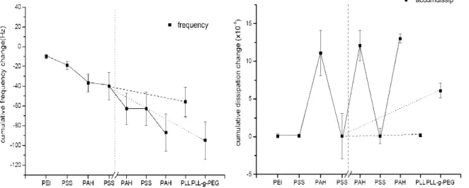
The assembly of the polyelectrolyte multilayers was monitored by Quartz crystal microbalance with dissipation monitoring (QCM-D). The change in frequency after each layer is showed in figure 1. Poly(ethylenimine) (PEI) was the first layer with a Af = 10 ± 2 Hz and AD = 0.1 ± 0.3x10⁻⁶ after adsorption. Lately, PSS (Poly(sodium 4-styrenesulfonate)) and PAH (Poly(allylamine hydrochloride)) were adsorbed until a total of 4 layers.
frequency
77/—1 1 1 1 1 1 PSS PAH PSS PAH PLLPLL-g-PEG
accumdissip
- 1 — PSS
—f—
PAH
-V/-T-PSS PAH
-1 1 1 1 1 PSS PAH PLLPLL-g-PEG
Fig. 1. Frequency and dissipation chance during the layer-by-layer deposition on gold crystals. A continuous decrease in frequency was observed after deposition of every PE layer. The dissipation increased after PAH adsorption, but always decreased after PSS adsorption.
The change in frequency for PSS after PEI was of 9 ± 2 Hz and the dissipation remained close to 0. When the first PAH layer was adsorbed on PSS, the change of frequency was of 18 ± 5 Hz, and its deposition always led to an increase in dissipation of 10 ± 3x10" . After the second layer of PSS, the frequency remained almost constant, with a Af = 3 ± 2 Hz and a AD = -10 ± 3xl0-6. This
behaviour repeats for the next PAH/PSS/PAH adsorption. After the second PSS layer, other 2 positive polyelectrolytes were adsorbed in different experiments, PLL and PLL-g-PEG (Poly(l-lysine)-graft-(polyethyleneglycol)). After PLL, the frequency drop was of 16 ± 1 Hz and the dissipation increase 0.1 ± 0.2xl0-6. The PLL-g-PEG adsorption led to the biggest change in
frequency for only one layer, 55 ± 5 Hz, and the dissipation increase was 6 ± lxlO-6.
Contact angle
lower when measured on glass. The PLL presented a contact angle of 31±3° on glass, and as expected the PLL-g-PEG surface presented a most hydrophilic surface, with a contact angle of 24 ±3°.
Table 1. PE contact angle measurements on SiOx.
Table 2. PE contact angle measurements on glass.
Surface
SiOx
PEI
PSS
PAH
PLL-g-PEG
Contact angle
12 ± 6
48 ±22
44 ± 5
44 ± 9
24 ± 3
Surface
PSS
PAH
PLL
Contact angle
33 ± 5
3 8 ± 2
31 ± 3
Polyelectrolyte patterning
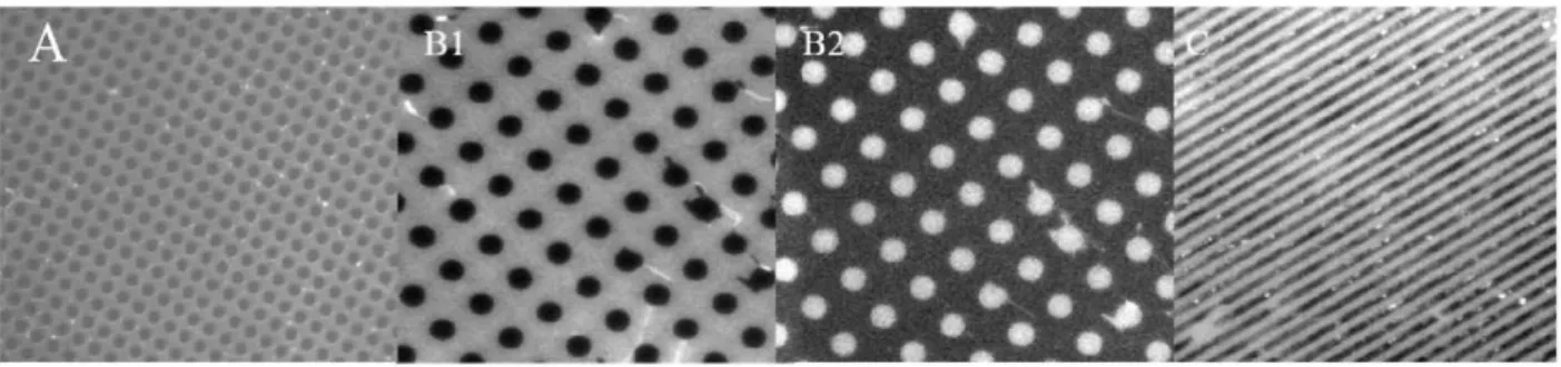
The patterning technique used was the micro-contact printing. It was possible to obtain different patterning features with different polyelectrolytes. Figure 2 shows some examples obtained with fluorescently labelled PAH (PAH-FITC). Besides, PLL-g-PEG patterns were obtained. Backfilling of the pattern for PLL-g-PEG pattern with PAH and PLL were accomplished. In figure 2, RBITC-PAH was used to backfill the free PSS areas between the PLL-g-PEG stripes.
Fig. 2. Fluorescence microscope images of patterned surfaces. A) PAH-FITC is the complementary area of circles D=10 urn, Bl) RBITC-PAH stamped pattern and B2) FITC-PAH pattern obtained after backfilling the RLBTC-PAH free areas, C) PLL-g-PEG pattern backfilled with RBITC-PAH..
Lacease immobilization and interaction with polyelectrolytes
Lacease was covalently immobilized with glutaraldehyde (GA). GA first reacted with the amines of poly(ethyleneimine) (PEI) adsorbed on gold, and later with lacease. The enzyme immobilization process was followed by QCM-D as shown in figure 3. The adsorption of PEI and the reaction with GA led to a compact layer with Af and AD change of 10±2 Hz and 0.1 ± 0.3x10"6,
and 6±1 Hz and 0 ± 0.3xl0"6, respectively.
Due to the fact that succinate buffer was used to dilute the lacease, prior to lacease immobilization, this buffer was pumped through the QCM chamber. Before dilution, the crude extract was centrifuged and filtered as explained in Materials and Methods. The protein concentration and activity of the diluted lacease were 270±20 mgl"1 and 570±80 Ul"1. Lacease
N
cr CD
1 0 - 1
0 - —
1 0
2 0
3 0
4 0
5 0
6 0
T -2
T " 4
T 8
- -20
-25
10
time (h)
Fig.3. QCM-D measurement of the lacease immobilization. A decrease in frequency without significant change in dissipation was observed after adsorption of PEI and the covalent attachment of GA. The immobilization of lacease led to a decrease in frequency of 24 Hz with dissipation increase of 1.2x10" .
In order to check whether the change in frequency was due to immobilization of active lacease, 2,2'-azino-di-[3-ethyl-benzo-thiazolin-sulphonate] (ABTS) was injected into the chamber and while recording frequency and dissipation change, the effluent was collected. After injecting succinate buffer solution to remove ABTS, the frequency and dissipation returned to their original values within the experimental error. Since lacease oxidises ABTS to a dark green product, the presence of active lacease was confirmed by measuring an increase in the absorbance of the effluent. When the flow increased, the effluent absorbance decreased. After lacease immobilization, polyelectrolytes were adsorbed. After the adsorption of PAH, PAH/PSS/PAH, and (PAH/PSS)2, ABTS was pumped
into the chamber to test if the enzyme was still active. We have observed that the PE on the lacease changed the shape of the adsorption curve.
References
[1] G. Decher, J.D. Hong, and J. Schmitt, Thin Solid Films 210, 831 (1992).
[2] G. Decher, and J.B. Schlenoff, (Eds.), Multilayer Thin Films, Wiley-VCH, 1 (2003).
[3] D.L. Elbert, C.B. Herbert, and J.A. Hubbell, Langmuir 15, 5355 (1999).
[4] R. Heuberger, G. Sukhorukov, J. Voros, M. Textor, and H. Mohwald, Advanced Functional Materials 15,357(2005).
[5] M. Schonhoff, Current Opinion in Colloid and Interface Science 8, 86 (2003).
[6] GB. Sukhorukov, H. Mohwald, G. Decher, and Y.M. Lvov, Thin Solid Films 284-285, 220 (1996).
[7] P. Schwinte, V. Ball, B. Szalontaii, Y. Haikel, J.C. Voegel, and P. Schaaf, Biomacromolecules 3, 1135(2002).
Materials and Applications for Sensors and Transducers II
10.4028/www.scientific.net/KEM.543
Building Functional Surfaces for Biosensors Development