In vitro antiatherogenicity of extracts from Halimeda incrassata seaweed: antioxidant activity and smooth muscle cell migration studies
8
0
0
Texto completo
(2) Ars Pharmaceutica In vitro antiatherogenicity of extracts from Halimeda incrassata seaweed: antioxidant activity and smooth muscle cell migration studies. Costa-Mugica A1, Batista-González AE1, Mondejar D1, Soto Y2, Brito V2, Vázquez AM2, Brömme D3, ZaldívarMuñoz C1, Mancini-Filho J4, Vidal-Novoa A1 1. Department of Biochemistry, Faculty of Biology, University of Havana, Cuba; 2. Center for Molecular Immunology, Havana City, Cuba; 3. Department of Oral Biological and Medical Sciences, Faculty of Dentistry, University of British Columbia, Canada; 4.Department of Food and Experimental Nutrition, Faculty of Pharmaceutical Sciences, University of Sao Paulo, Brazil.. Original Paper Artículo Original Correspondence/Correspondencia: Alexis Vidal Novoa Facultad de Biología. Departamento de Bioquímica. Universidad de La Habana Calle 25 No. 455 entre J e I, Vedado, CP 10 400, La Habana, Cuba [email protected] [email protected] Phone: 53-7-8309821, Fax: 53-7-8321321 Competing interest / Conflicto de interes: Authors declared that there was no conflict of interest associated with this research work. Fundings / Financiación: The research was funded by IFS grant F/4897-1. Partial funding was also provided by CIHR grant MOP24447, the Canadian Research Chair award (D.B.) and a personal grant from GSEP, offered by the Canadian Bureau for International Education (A.C) and CNPq- (Brasil). Received: 23.04.2012 Accepted: 03.02.2013. RESUMEN Objetivos: El objetivo de este trabajo fue evaluar el potencial ateroprotector in vitro del alga Halimeda incrassata en la migración de células de músculo liso de ratón y la oxidación de lipoproteínas en relación con su actividad antioxidante. Material y métodos: La actividad antioxidante fue determinada mediante los métodos de inhibición de radicales DPPH y la Capacidad antioxidante total (ORAC). La actividad inhibitoria de la oxidación de LDL mediada por iones Cu2+ se determinó por la cuantificación de TBARS y dienos conjugados. El efecto del extracto acuoso sobre la migración de las células de músculo liso se evaluó en la línea de células de músculo liso aórtica de ratón MOVAS-1. Resultados: Se demostró el efecto inhibidor del extracto sobre la oxidación de LDL mediada por Cu2+. El extracto del alga causa inhibición dosis-dependiente de la formación de TBARS (IC50 = 0,8 mg/mL) y dienos conjugados. Las algas tuvieron una alta actividad antioxidante en los ensayos realizados y podría estar relacionada con el contenido de compuestos fenólicos. Conclusiones: Los resultados de este trabajo representan un paso más en la caracterización de la acción ateroprotectora de Halimeda incrassata y evidencian sus posibles aplicaciones como nutracéutico y/o fitofármaco. PALABRAS CLAVES: antiaterogenicidad, H. incrassata, actividad antioxidante, células de musculatura lisa.. ABSTRACT Aim: The aim of this work was to evaluate the in vitro atheroprotective potential of the seaweed Halimeda incrassata in smooth muscle cell migration and lipoprotein oxidation in relation to its antioxidant activity. Material and methods: Antioxidant activity was determinate by DPPH• radical scavenging assay and ORAC method. The inhibitory effect of the aqueous extract on LDL oxidation mediated by Cu2+ ions was determinate by TBARS and conjugated diene quantification. The effect of the seaweed aqueous extract on smooth muscle cell migration was evaluated in MOVAS-1 mouse aortic smooth muscle cell. Results: The inhibitory effect of the aqueous extract on lipoprotein oxidation mediated by Cu2+ was demonstrated. Seaweed extract caused dose-dependent inhibition of TBARS (IC50 = 0.8 mg/mL) and conjugated dienes formation. The seaweed had a high antioxidant activity in the assays performed. The activity could be related to the phenolic content of Halimeda incrassata. Conclusions: In summary, the results of this study represent a further step in the characterization of the atheroprotective action of Halimeda incrassata and indicate the seaweed could be used for a nutraceutical and/or phytoterapeutic application. KEYWORDS: antiatherogenicity, Halimeda incrassata, antioxidant activity, smooth muscle cell.. Ars Pharm. 2013; 54(2): 04-11.. 5.
(3) Costa-Mugica A, Batista-González AE, Mondejar D, Soto Y, Brito V, Vázquez AM, Brömme D, Zaldívar-Muñoz C, Mancini-Filho J, Vidal-Novoa A. INTRODUCTION Atherosclerosis is characterized by lipoprotein retention and oxidative modification in the artery wall with subsequent oxidized LDL uptake and foam cell formation. As lesion progresses smooth muscle cells migrate from the tunica media to the arterial intima marking lesion advancement1. Oxidative stress is considered a causal factor in all stages of atherosclerosis development, from oxidative modification of lipoproteins to platelet activation and atherothrombosis2. Atherosclerosis is also considered a common factor of other diseases related to oxidative stress such as ischemic heart disease and in this field antioxidants have been attractive to decrease oxidative stress associated with plaque formation2. Seaweeds are marine organisms that have been consumed as an integral part of the traditional diet in Asian countries and they are becoming increasingly popular in the western world in association to their health boosting properties3,4. Seaweeds have relatively high amounts of bioactive phytochemicals such as polyphenols that are consistent with antioxidative properties3. Interestingly, the main effect of algae consumption has been associated with cardiovascular and intestinal health with the epidemiological data available indicating a correlation between seaweed consumption and low atherosclerosis-related morbidity and mortality4. The mechanisms for cardiovascular protection are beginning to be elucidated and have been so far mainly related to antioxidative, antihypertensive, anti-inflammatory and hypocholesterolemic effects4. In a previous study we showed that Halimeda incrassata was one of the most active specimens presenting antioxidant activity in an initial screening study using a lipoperoxidation model5. The seaweed has also shown antioxidant activity in the mouse hypothalamic cell6 and exerted neuroprotection5 and hepatoprotection7 in vivo. In the present study we investigated the antiatherogenic potential of Halimeda incrassata in two key events of atherogenesis: lipoprotein oxidation and smooth muscle cell migration in relation to its antioxidant activity. MATERIALS AND METHODS Seaweed collection and hydrophilic extracts preparation The seaweed Halimeda incrassata (Ellis) Lamouroux was collected in December 2010 in the Bajo de Santa Ana, La Habana (Cuba). Voucher specimens were authenticated by Dr. A.M. Suárez from Seaweeds Laboratory, at the Marine Research Center. Freshly collected specimens were washed with distilled water and dried at room temperature for 3-5 6. days. Dry seaweed powder was extracted with distilled water (1:5 w/v) and centrifuged at 800 g and at 4ºC for 20 minutes. Supernatant was recovered, lyophilized and kept at -20ºC until use. Weight yield of the final extract in terms of dry seaweeds was 5.7%. A polyphenol-rich fraction containing free phenolic acids (FPA) was obtained according to Krygier et al.8 Dry seaweed was extracted with tetrahydrofuran for 3 min, and evaporated to dryness under vacuum at 30ºC and the pellet was resuspended in ethanol. Total phenolic content was determined as in Vidal et al.9 and expressed as mg of gallic acid (GAE)/g of seaweed. DPPH• radical scavenging assay The DPPH• radical scavenging method was realized according Goupy et al.10 and the results are expressed as percent decrease from the initial DPPH• radical absorption by the test samples and calculation of the value of 50% inhibitory concentration (IC50). Oxygen Radical Absorbance Capacity (ORAC) The ORAC assay was determined according to Gillespie et al.11 using AAPH as a peroxyl radical generator. ORAC values were given as trolox equivalents. Inhibitory activity on LDL oxidation mediated by Cu2+ ions LDL was isolated from human plasma by sequential ultracentrifugation as in Frostegard et al.12. LDL was oxidized with 10 µM CuSO4 and degree of oxidation was determined by TBARS and expressed as nmoles MDA equivalents/mg protein. Conjugated diene formation was also monitored as in Esterbauer et al.13. Kinetic parameters lag phase, Vmax and Dienemax were determined according to Pinchuk et al.14. Inhibition of smooth muscle cell migration Smooth muscle cell (SMC) migration experiments were done with mouse aortic smooth muscle cells MOVAS-1, generously provided by Dr. Mansoor Hussain from the Heart and Stroke Richard Lewar Center of Excellence in Cardiovascular Research (Canada). Cells were cultured in DMEM containing FBS, 2 mM L-glutamine and streptomycin/penicillin in 5% CO2 atmosphere. All experiments were performed with MOVAS-1 SMC between passages 5 to 15. In the wound healing assay smooth muscle cells were grown to 90% confluence in DMEM + 10% FBS. After damaging the monolayers with a sterile tip, cells were treated with 5 ng/mL Platelet derived growth factor and seaweed extract Ars Pharm. 2013; 54(2): 04-11..
(4) In vitro antiatherogenicity of extracts from Halimeda incrassata seaweed: antioxidant activity and smooth muscle cell migration studies.. Transmigration experiments were done as in Goncharova et al.16 with some modifications. Cells were incubated for 16 hours and after swabbing the upper part, membranes were fixed with methanol and then stained with crystal violet. Transmigrated cells were counted under a microscope at 100x; 10 fields were counted/insert. Statistical analysis Values are given as mean + standard deviation (s.d.). For statistical significance an unpaired student´s t test was performed. Data was processed using Microcal Origin 5.0 and GraphPad Prism software (version 3.01 for Windows). p < 0.05 was considered statistically significant. RESULTS Phenolic content H.incrassata had a high total phenolic content with 2.8 µg GAE/mg for the lyophilized aqueous extract and 14.1 µg GAE/g of dry seaweed for the FPA fraction. DPPH• scavenging capacity of the extract As shown in Figure 1, H. incrassata extract presents a concentration dependent free radical scavenging activity, with an IC50 of 0.27 + 0.011 mg/mL. ORAC Antioxidant activity by the ORAC assay is shown in Figure 2. The total antioxidant capacity was higher with increasing concentrations of seaweed with an ORAC value of 3960 trolox equivalents/g of dry seaweed.. Figure 1. Antioxidant activity of the aqueous extract of Hi in the DPPH• radical scavenging assay. The assay was performed according to Goupy et al.10.Data are given as mean ± s.d (n=3).. Figure 2. Total antioxidant capacity by ORAC assay as previously described in methods. Data is presented as average RFU where s.d < 10 % of mean RFU. 1.2. control Hi 2 ug Hi 1 ug Hi 0.5 ug FPA 8 ug FPA 0.8 ug. 0.8. RFU. and allowed to migrate for 16 hours. Cells were stained with crystal violet and after taking photographs distance migrated were calculated as described by Ho et al.15.. 0.4. 0.0. 0. 20. 40. 60. 80. 100. 120. Time (minutes). 140. 160. 180. Inhibition of LDL oxidation The dose response for inhibition of lipoperoxidation during LDL oxidation mediated by Cu2 is shown in Figure 3.A with IC50 of 0.8 mg/mL while as shown in Figure 3.B conjugated. Figure 3. Effect of Hi on LDL oxidation induced by Cu2+ ions. A. Inhibition of TBARS formation during LDL oxidation. B. Conjugated dienes formation during LDL oxidation. Results are presented as mean ± s.d. (n=3).. Ars Pharm. 2013; 54(2): 04-11.. 7.
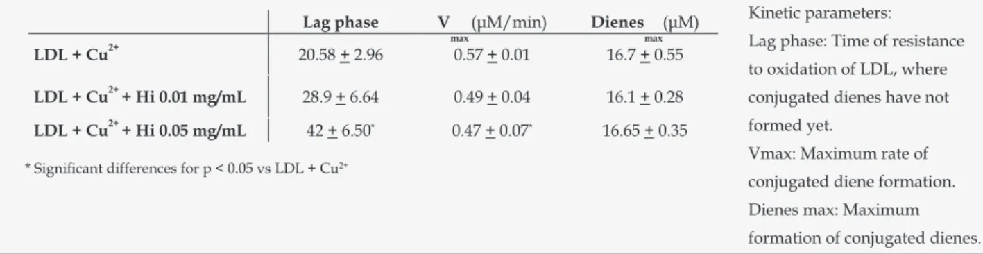
(5) Costa-Mugica A, Batista-González AE, Mondejar D, Soto Y, Brito V, Vázquez AM, Brömme D, Zaldívar-Muñoz C, Mancini-Filho J, Vidal-Novoa A. Table 1. Effect of Hi on kinetics of LDL oxidation mediated by Cu2+ ions. Results are presented as mean ± s.d. (n=3). Lag phase 2+. 20.58 + 2.96. 2+ 2+. LDL + Cu. LDL + Cu + Hi 0.01 mg/mL LDL + Cu + Hi 0.05 mg/mL. V. max. (μM/min). Dienes. max. (μM). Kinetic parameters: Lag phase: Time of resistance. 0.57 + 0.01. 16.7 + 0.55. 28.9 + 6.64. 0.49 + 0.04. 16.1 + 0.28. conjugated dienes have not. 42 + 6.50*. 0.47 + 0.07*. 16.65 + 0.35. formed yet.. to oxidation of LDL, where. Vmax: Maximum rate of. * Significant differences for p < 0.05 vs LDL + Cu2+. conjugated diene formation. Dienes max: Maximum formation of conjugated dienes.. diene formation. The kinetic parameters lag phase, Vmax and Diene max were presented in Table 1. Antimigratory activity The effect of H. incrassata on PDGF-BB-induced MOVAS-1 SMC migration is shown in Table 2. DISCUSSION Oxidative stress has been critically involved in the progression of atherosclerosis with natural antioxidant extracts being presently studied for potential disease modulation2. In this work we further evaluated the in vitro antiatherogenic properties of H. incrassata and its antioxidant activity in cell free systems. A high phenolic content was found for hydrophilic fractions from H. incrassata, which is similar to the one reported by our group for other seaweeds from the Halimeda spp9. Likewise Yoshie et al.17 showed that there was a high polyphenols content in the seaweeds H. opuntia and H. macroloba and identified a variety of phenolic compounds that could be relevant to the antioxidant properties.. lyophilized is in the range of the one reported for other seaweeds in the literature in different regions. Heo et al.18 obtained a 70 % DPPH• scavenging in hydrophilic extracts from E. cava; an activity that correlated to phenolic content. Additionally our group informed an IC50 of 1,18 mg/mL for the seaweed Bryothamnion triquetrum19 whereas H. incrassata - with a significantly higher phenolic content - is about 4 times more effective in DPPH• scavenging. Likewise Serevinathne et al.20 found an association between antioxidant capacity in DPPH• scavenging and solvent polarity; which also was related to a higher phenolic content in these fractions. The authors found a similar antioxidant activity pattern for other assays such as inhibition of lipoperoxidation. Together with DPPH• radical scavenging and inhibitory action on lipoprotein oxidation H. incrassata also exhibited a high total antioxidant capacity in the range found for other natural extracts21. In hydrophilic extracts the activity was similar for the aqueous extract and the polyphenol rich FPA fraction indicating that phenolic compounds are important in the effect observed. Likewise Price et al.22 found significant antioxidant activity by ORAC assay in. • The high DPPH scavenging capacity for H. Table 2. Effect of Halimeda incrassata seaweed on incrassata smooth muscle cell migration in MOVAS-1 mouse aortic smooth muscle cell. line. The experiment was performed as previously described by Zargham et al.17 Results are expressed as mean + s.d of each experiment performed in triplicate. Significant differences for p < 0.05 compared to PDGF-BB induced migration are indicated in the graphs (*). In transwell experiment 10 fields were counted per insert membrane. Wound migration assay Migrated area (% zero hour) + PDGF 100 30.2 + 2.55 * + PDGF + H. incrassata 1 mg/mL 40.3 + 4.56 * + PDGF + H. incrassata 0.5 mg/mL 46.9 + 1.12 * + PDGF + H. incrassata 0.1 mg/mL. control PDGF PDGF + H. incrassata 1 mg/mL PDGF + H. incrassata 0.1 mg/mL. 8. Transwell assay Transmigrated cells (% of control) 100 + 7.78 460.3 + 37.06 227.5 + 21.51 * 262.5 + 17.68 *. Ars Pharm. 2013; 54(2): 04-11..
(6) In vitro antiatherogenicity of extracts from Halimeda incrassata seaweed: antioxidant activity and smooth muscle cell migration studies.. seaweed extracts; where the highest activity correlated to the presence of hydrophilic compounds. Previous work has shown that the main phenolic compounds in H. incrassata are phenolic acids with a majoritary composition of salicylic and lower quantities of ferulic acid that might also contribute to the antioxidant action23. Salicylic acid is relevant in plant defence mechanisms and has antinflammatory and antioxidant properties related to lypoxygenase inhibition and to decrease of endothelial cells adhesion molecules expression24. LDL oxidation is a key therapeutic target as it is a main emerging risk factor for cardiovascular diseases. Indeed from fatty streak formation to thrombosis in advanced plaques oxidized LDL has a relevant role in atherosclerosis related events such as migration and proliferation of smooth muscle cells and endothelial dysfunction2. To help dilucidate the mechanism behind antioxidant action in inhibition of LDL oxidation conjugated diene formation was monitored in presence of seaweed aqueous extract. During LDL oxidation by Cu2+ the metal binds to LDL and catalyses the constant formation of free radicals at the expense of the reduction of antioxidants bound to the particle14. The variation in oxidation kinetics with increase in the lag phase and decrease in Vmax on addition of aqueous extract could be indicative of a chelating effect of Cu2+ or result from the blockage of the sites of binding of cupper to the lipoprotein14. Polyphenolic compounds found in the extract could be relevant in this effect since it is known that polyphenols that act through free radical scavenging can also efficiently bind transition metals. The in vitro antioxidant activity assays indicate that inhibition of LDL oxidation could be attained potentially by both Cu2+ chelating and free radical scavenging, where the phenolic content and antioxidant activity could contribute to this effect. LDL oxidation in vivo is a complex process not fully understood and Cu2+ mediated oxidation is a model frequently used to study lipoperoxidation reactions that could take place in the vascular wall14. The protective effect in TBARS formation found for H. incrassata extract could be associated to the presence of significant amounts of hydrophilic antioxidants involved in scavenging of free radicals formed in the aqueous phase. Several groups have evaluated the antiatherogenicity of natural extracts against LDL oxidation finding an activity that correlates to phenolic content. The antilipoperoxidative activity found in our study is quite promising as compared to other previously reported natural extracts. For instance a 37 % inhibition of TBARS formation was found in AAPH. Ars Pharm. 2013; 54(2): 04-11.. and 74 % in Cu2+ peroxidation by Hseu et al.25. In that study T. sinensis extracts had 6.5 μg GAE; a phenolic content higher than the one needed in our study to reach 50 % inhibition of TBARS formation in both oxidation systems used, adding therefore further evidence to the antioxidant potential of H. incrassata. Antiatherogenic properties of natural extracts in smooth muscle cell biology is another field of relevance in cardioprotection as these are key cell types in the pathogenesis of vascular disease26. Their migration from tunica media to subendothelial space marks the transit from the fatty streak to more advanced lesions; since they produce most of extracellular matrix generated during the plaque fibroproliferative response. PDGF secreted locally is the most potent stimulus for smooth muscle cell migration and signalling through its receptor is associated with ROS production resulting from NADPH oxidase activation27. Thereafter antioxidants have been of interest for targeting smooth muscle cell migration by several authors. Polyphenol rich natural extracts such as those from tea and cocoa28 have shown inhibition of smooth muscle cell migration whereas several pure phenolic compounds like the phenolic acid derivative avenanthramide also had antioxidant activity in smooth muscle cells increasing NO production by upregulation of eNOS mRNA29. The 43% inhibition of migration in the transwell assay and the decrease migrated area in the wound scratch model found in our study, are consistent with the results for other natural extracts in the literature. Zargham et al.30 had a 62% antimigratory activity in the transwell assay with a tannin extract. Likewise Ho et al.15 evaluated the effect on smooth muscle cell migration of the water extracts from Nelumbo nucifera -an aquatic plant used in traditional medicine-, to elucidate the molecular mechanisms of its antiatherogenic action. They found a decrease by 60 % at 0.2 mg/mL in transmigrated cells and decreased inhibition in a wound closure assay, effects the authors discussed that were probably related to the high content of phenolic acids and flavonoids of the extract. Though the mechanisms of action involved in the antimigratory activity are unknown it could be speculated that it might be related to a direct antioxidant action or to antioxidant enzyme induction capable of modulating migration of these cells. In the other hand the considerable inhibitory activity of the lyophilized aqueous extract of the seaweed on PDGFBB induced smooth muscle cell migration adds further evidence to the potential of H. incrassata for targeting. 9.
(7) Costa-Mugica A, Batista-González AE, Mondejar D, Soto Y, Brito V, Vázquez AM, Brömme D, Zaldívar-Muñoz C, Mancini-Filho J, Vidal-Novoa A. atherosclerosis progression and to our knowledge it is the first report of inhibition of smooth muscle cell migration by seaweed extracts.. 8.- Krygier K, Sosulski F, Hogge L. Free, esterified, and insolublebound phenolic acids. 1.Extraction and purification procedure. J Agric Food Chem. 1982;30: 330-334.. CONCLUSION. 9.- Vidal A, Silva de Andrade-Wartha ER, de Oliveira e Silva AM, Pavan R, Lima A, Fallarero A, Batista AE, Mancini-Filho J. Actividad antioxidante y polifenoles de las algas marinas Halimeda opuntia y Halimeda monile. Ars Pharm. 2009; 50(1): 24-31.. Our previous work has indicated an antiatherogenic effect of the seaweed in atherosclerosis progression in apo E-/mice31. The present study adds evidence to a potential atheroprotective application of H. incrassata considering its antioxidant action and its high activity for targeting LDL oxidation and smooth muscle cell migration. Thereafter further studies are needed for elucidating the molecular mechanisms involved in the in vitro and in vivo effects observed to propose H. incrassata extracts for cardiovascular protection. ACKNOWLEDGEMENTS Authors would like to thank S. Wilson and P. A. Lythgo for assistance with tissue culture and fluorescence kinetic assays. We would also like to acknowledge Professors O. Carrillo, M. E. Alonso for counselling during research and Dr. Sylvie Marleau, Université de Montréal, Canada for critical review of the manuscript. REFERENCES 1.- Fernández-Britto JE. La lesión aterosclerótica: estado del arte a las puertas del siglo XXI. Rev Cub Invest Biomed. 1998; 17(2):112-27. 2.- Stocker R, Keaney JF. Role of Oxidative Modifications in Atherosclerosis. Physiol Rev. 2004; 84: 1381–1478. 3.- O’Sullivan L, Murphy B, McLoughlin P, Duggan P, Lawlor PG, Hughes H, Gardiner GE. Prebiotics from Marine Macroalgae for Human and Animal Health Applications. Mar. Drugs 2010; 8, 2038-2064. 4.- Bocanegra A, Bastida S, Benedí J, Ródenas S and SánchezMuniz FJ. Characteristics and Nutritional and CardiovascularHealth Properties of Seaweeds. J Med Food 2009; 12 (2): 236–258 5.-Rivero F, Fallarero A, Castañeda O, Dajas F, Manta E, Areces F, Mancini Filho J, Vidal A. Antioxidant activity in vivo and in vitro of Halimeda incrassata aqueous extracts. Cienc Tecnol Aliment Campinas. 2003; 23:256-263. 6.- Fallarero A, Loikkanen J J, Mannisto PT, Castañeda O, Vidal A. Effects of aqueous extracts of Halimeda incrassata (Ellis) Lamouroux and Bryothamnion triquetrum (S.G.Gmelim) Howe on hydrogen peroxide and methyl mercury-induced oxidative stress in GT1-7 mouse hypothalamic immortalized cells. Phytomedicine. 2003; 10: 39-47. 7.-Barro R, Zaldivar C, Fallarero A, Vidal V. Evaluation of the antioxidant activity in an aqueous extract of a red seaweed specie from the Caribbean sea Bryothamnion triquetrum. Rev Cub Quim. 2001; XIII (2):40. 10. 10.- Goupy P, Hugues M, Boivin P, Amiot MJ. Antioxidant composition and activity of barley (Hordeum vulgare) and malt extracts and of isolated phenolic compounds. J Sci Food Agric. 1999; 79:1625-1634. 11.- Gillespie KM, Chae JM, Ainsworth EA. Rapid measurement of total antioxidant capacity in plants. Nature Protocols. 2007; 2(4):867-870. 12.- Frostegard J, Nilsson J, Haegerstrand A, Hamsten A, Wigzell H, Gidlund M. Oxidized low density lipoprotein induces differentation and adhesion of human monocytes and the monocytic cell line U937. PNAS. 1990; 87: 904-908. 13.- Esterbauer HG, Striegl H, Puhl M, Rotheneder. Continuous monitoring of in vitro oxidation of human Low Density Lipoprotein. Free Rad Res. 1979; 6 (1): 67-75. 14.- Pinchuk I, Lichtenberg D. The mechanism of action of antioxidants against lipoprotein peroxidation, evaluation based on kinetic experiments. Prog Lipid Res. 2002; 41:279-314. 15.- Ho HH, Hsu LS, Chan KC, Chen HM, Wu CH, Wang CJ. Extract from the leaf of Nelumbo nucifera reduced the development of atherosclerosis via inhibition of vascular smooth muscle cell proliferation and migration. Food Chem Toxicol. 2010;48: 159–168. 16.- Goncharova EA, Goncharov DA, Krymskaya VP. Assays for in vitro monitoring of human airway smooth muscle (ASM) and human pulmonary arterial vascular smooth muscle (VSM) cell migration. Nature Protocols. 2006; 1(6): 2936-2939. 17.- Yoshie Y, Wang W, Hsieh YP, Suzuki T. Compositional difference of phenolic compounds between two seaweeds, Halimeda spp. J. Tokyo University of Fisheries. 2002; 88: 21-24. 18.- Heo JS, Park EJ, Lee KW, Jeon YJ. Antioxidant activities of enzymatic extracts from brown seaweeds. Bioresour Technol. 2005; 96:1613–1623. 19.- Barro R. Estudio de la actividad antioxidante y hepatoprotectora del extracto acuoso de Bryothamnion triquetrum. [Tesis de Máster en Bioquímica]: Universidad de La Habana; 2004. 20.- Senevirathne M, Kim S, Siriwardhana N, Ha J, Lee K, Jeon Y. Antioxidant potential of Ecklonia cava Kjellman (P)on reactive oxygen species scavenging, metal quelating, reducing power and lipid peroxidation inhibition. Food Sci Tech Int. 2006;12(1):27-38. 21.-Prior R L, Hoang H, Gu L, Wu X, Bacchiocca M, Howard L, Hapsch-Woodill M, Huang D, Ou B, Jacob R. Assays for hydrophilic and lipophilic antioxidant capacity (oxygen radical absorbance capacity (ORACFL)) of plasma and other biological and food samples. J Agric Food Chem. 2003; 51: 3273-3279.. Ars Pharm. 2013; 54(2): 04-11..
(8) In vitro antiatherogenicity of extracts from Halimeda incrassata seaweed: antioxidant activity and smooth muscle cell migration studies.. 22.- Price JA, Sanny CG, Shevlin D. Application of manual assessment of oxygen radical absorbent capacity (ORAC) for use in high throughput assay of ‘‘total’’ antioxidant activity of drugs and natural products. J Pharmacol Toxicol Methods. 2006; 54: 56-61. 23.-Vidal A, Silva de Andrade-Wartha, Fallarero A, de Oliveira AM, Vuorela P, Mancini-Filho J. Antioxidant activity and bioactive components from the seaweed Halimeda incrassata (Ellis) Lamouroux. Braz J Pharmacogn. 2011; 21(1): 53-57. 24.- Amann R, Peskar BA. Anti-inflammatory effects of aspirin and sodium salicylate. Eur J Pharmacol. 2002; 447: 1-9. 25.- Hseu YC, Chang WH, Chen CS, Liao JW, Huang CJ, Lu FJ, Chia YC, Hsu HK, Wu JJ, Yang HL. Antioxidant activities of Toona Sinensis leaves extracts using different antioxidant models. Food Chem Toxicol. 2008; 46: 105-114. 26.- Ross R. Atherosclerosis-an inflammatory disease. N Engl J Med.1999; 340:115–126. 27.- Kreuzer J, Viedt C, Brandes RP, Seeger F, Rosenkranz AS, Sauer H, Babich H, Nürnberg B, Kather H, Krieger-Brauer HI. Platelet-derived growth factor activates production of reactive. Ars Pharm. 2013; 54(2): 04-11.. oxygen species by NAD(P)H-oxidase in smooth muscle cells through Gi1,2. FASEB J. 2003 17(1):38-40. 28.- Lee KW, Kang NJ, Oak MH, Hwang MK, Kim JH, SchiniKerth VB, Lee HJ. Cocoa procyanidins inhibit expression and activation of MMP-2 in vascular smooth muscle cells by direct inhibition of MEK and MT1-MMP activities. Cardiovasc Res. 2008; 79(1):34-41. 29.- Nie L, Wise ML, Peterson DM, Meydani M. Avenanthramide, a polyphenol from oats, inhibits vascular smooth muscle cell proliferation and enhances nitric oxide production. Atherosclerosis. 2006; 186: 260–266. 30.- Zargham H, Zargham R. Tannin extracted from Sumac inhibits vascular smooth muscle cell migration. Mcgill J Med. 2008; 11(2): 119-123. 31.- Zaldivar C, Costa A, Batista AE, Samokhin A, Hashamiyan S, Nho B, Fallarero A, Vidal A, Bromme A. Antioxidant activity and atheroprotective effect of an aqueous extract of the marine seaweed Halimeda incrassata. Rev Cub Farmacia. 2009; 43 (suppl 1):88.. 11.
(9)
Figure

Documento similar
We consider different methods aim to evaluate cellular uptake and localization in cells and tissues, and in vitro methods for the study of the toxicity induced by MNPs
In accordance with that discussed, the aim of the present work was to examine dietary habits, nutritional status, and physical activity engagement in older adults in the city of
Lipoprotein lipase enhances the binding of native and oxidized low density lipoproteins to versican and biglycan synthesized by cultured arterial smooth muscle cellsO. Olsson
A limiting value of the oxidation potential of a given compound candidate to have antioxidant activity in the DPPH • test was found, this being explained by
The aim of this research is to evaluate the potential ergogenic effect of sodium phosphate supplementation in physically active subjects, by analysing blood cell count,
(1998) Arterial heparan sulfate proteoglycans inhibit vascular smooth muscle cell proliferation and phenotype change in vitro and neointimal formation in vivo.. (2001) Relationship
In this work, we have undertaken the study of SUZ12, a Polycomb group protein and the microRNAs (miRNA) expressed by the oncogenic Epstein Barr Virus (EBV) in
Specifically, ERK1/2 can participate in HuR phosphorylation, modifying its activity in a lung cancer cell line (Yang et al., 2004) or HuR cytoplasmic location in hepatic
