Fibrotic response induced by angiotensin II requires NAD(P)H oxidase induced reactive oxygen species (ROS) in skeletal muscle cells
6
0
0
Texto completo
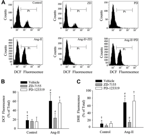
(2) 666. C. Cabello-Verrugio et al. / Biochemical and Biophysical Research Communications 410 (2011) 665–670. Fig. 1. Angiotensin-II induces fibrosis via an AT-1-dependent mechanism in skeletal muscle cells. (A) C2C12 myoblasts were incubated with Ang-II (500 nM), TGF-b1 (10 ng/ ml) or CTGF (80 ng/ml) for 48 h. The extracts obtained were separated by SDS–PAGE. The presence of collagen-III (Col-III) was evaluated by Western blot using anti-Col-III antibody. Levels of GAPDH are shown as a loading control. (B) C2C12 cells were pre-incubated with ARB ZD-7155 (10 lM) and then incubated with Ang-II (500 nM) as in A. Levels of collagen-III (Col-III) and fibronectin (FN) were determined by Western blot. Levels of GAPDH are shown as a loading control. (C) Myoblasts were incubated with AngII (500 nM) or TGF-b1 (5 ng/ml) for 1 and 6 h respectively. Total RNA was isolated, blotted and probed with cDNA for mouse CTGF and tubulin as a loading control. The sizes of CTGF and tubulin mRNAs are shown in kilobases (kb). (D) C2C12 cells were pre-treated with ZD-7155 (10 lM) and incubated with Ang-II as in (A). Levels of CTGF protein were determined by Western blot. Levels of GAPDH are shown as a loading control. (E) Quantitative analysis of experiments shown in (B and D). The graph shows the fold of induction relative to control treatment. Values correspond to the mean ± standard deviation from three independent experiments (⁄, #, P < 0.05). In A, B and D the molecular weight standards are indicated in kilodaltons (kDa).. In the present study, we therefore investigated the role of Ang-II in skeletal muscle fibrosis using C2C12 myoblasts and the participation of ROS as a key mediator. We demonstrated that Ang-II induced an increase in ECM and CTGF protein levels through its AT-1 receptor. These pro-fibrotic effects of Ang-II were dependent on NAD(P)H oxidase-induced ROS generation. Indeed, the activation of NAD(P)H oxidase was a Ca2+-dependent PKC-induced event. Thus, our results provide an insight into the fundamental mechanisms of Ang-II-induced fibrosis in skeletal muscle cells.. USA) or Ang-II (500 nM) (Sigma, USA). The following inhibitors were pre-incubated for 1 h prior to exposure to Ang-II: AT-1 and AT-2 receptor blockers ZD-7155 (5 lM) and PD-123319 (10 lM) (both from Tocris, USA) respectively, dithiothreitol (DTT) (0.5 mM, Invitrogen, USA), NAD(P)H oxidase inhibitors diphenylene iodonium (DPI) (0.1, 1 and 5 lM, Sigma, USA) or apocynin (1 mM, Sigma, USA), or PKC inhibitor chelerythrine (10 lV, Sigma, USA). 2.2. Measurement of intracellular ROS production and [Ca2+] by FACS. 2. Materials and methods 2.1. Cell cultures The skeletal muscle cell line C2C12, obtained from adult mouse leg (American Type Culture Collection), was grown as described previously [20]. Cells were serum-starved for 18 h and then subjected to different treatments. For pro-fibrotic cytokine treatment myoblasts were incubated for 48 h with TGF-b1 (10 ng/ml) (R&D System, USA), recombinant CTGF (80 ng/ml) [16], H2O2 (Merck,. Treated C2C12 cells were harvested, resuspended, and loaded with the following cell permeant dyes: dichlorodihydrofluorescein (DCF, 5 lM) or dihydroethdium (DHE, 10 lM) for ROS determination, and Fura-3 (5 lM) for Ca2+ measurements (all from Invitrogen, USA) for 15–30 min at room temperature in the absence of light. They were then analyzed immediately by Fluorescence Activated Cell Sorting (FACS) using a flow cytometry system (FACSCanto, BD Biosciences, USA). A minimum of 10,000 cells were analyzed per sample. The cellular intensity of the dyes was analyzed using FACSDiva software v4.1.1 (BD Biosciences, USA)..
(3) C. Cabello-Verrugio et al. / Biochemical and Biophysical Research Communications 410 (2011) 665–670. 2.3. RNA isolation and Northern blot analysis Myoblasts were serum-starved for 18 h and then incubated for 1 h with Ang-II (500 nM) or for 6 h with TGF-b1 (10 ng/ml). Total RNA was isolated from cell cultures at the indicated times using Trizol (Invitrogen, USA). RNA samples (20 lg/lane) were electrophoresed, transferred and hybridized with probes for mouse tubulin and CTGF as described previously [21]. 2.4. Immunoblot analysis For cell immunoblot analyses, protein extracts from myoblasts were prepared as previously described [16,17] and subjected to SDS–PAGE, electrophoretically transferred onto PDVF membranes (Schleicher & Schuell, USA) and probed with goat anti-CTGF (Santa Cruz Biotechnology, USA), rabbit anti-fibronectin and mouse antitubulin (Sigma–Aldrich, USA), rabbit anti-collagen-III (Rockland, USA), and mouse anti-GAPDH (Chemicon, USA). All immunoreactions were visualized by enhanced chemiluminescence (Pierce, USA). 2.5. Protein determination Proteins were determined in aliquots of cell extracts using the bicinchoninic acid protein assay kit (Pierce, USA) with BSA as the standard.. 667. analysis of variance (ANOVA) with a post hoc Bonferroni multiple-comparison test (Sigma Stat 3.5 Software). A difference was considered statistically significant at a P value <0.05. 3. Results 3.1. Angiotensin-II-induced fibrotic response is AT-1 receptordependent in skeletal muscle cells We studied the effect of Ang-II as a possible inducer of fibrosis in skeletal muscle cells. Fig. 1A and B show an increase in the amount of collagen-III and fibronectin of C2C12 cells in response to Ang-II. TGFb1 and CTGF are showed as fibrotic factors increasing the levels of collagen-III and fibronectin [16,17]. Fig. 1B also shows that the increase in collagen-III and fibronectin induced by Ang-II in C2C12 myoblasts was prevented in presence of the Ang-II receptor type 1 blocker (ARB) ZD-7155 suggesting that is mediated by AT-1 receptor. Fig. 1C and D show that Ang-II induces increased levels of CTGF mRNA and protein respectively. Fig. 1D also shows that Ang-II-induced CTGF levels are dependent on the AT-1 receptor. Fig. 1E shows the quantitative analysis of the ARB ZD-7155 effect on Ang-II-induced collagen-III, fibronectin and CTGF levels. Together, these results suggest that Ang-II induces pro-fibrotic effects in skeletal muscle cells.. 2.6. Statistics. 3.2. Angiotensin-II-induced intracellular production of reactive oxygen species (ROS) is AT-1 receptor-dependent and participates in the fibrosis of skeletal muscle cells. The statistical significance of the differences between the means of the experimental groups was evaluated using one-way. We tested whether skeletal muscle cells increase their intracellular ROS production in response to Ang-II. By means of flow. Fig. 2. Reactive oxygen species (ROS) are induced by angiotensin-II via the AT-1 receptor in skeletal muscle cells. (A) Representative DCF fluorescence histograms from FACS experiments of C2C12 cells exposed to vehicle alone (left upper panel), ZD-7155 alone (middle upper panel), PD-123319 alone (right upper panel), Ang-II (left lower panel), Ang-II + ZD-7155 (middle lower panel), or Ang-II + PD-123319 (right lower panel). Data obtained from three to four independent experiments such as those depicted in the histograms above are summarized in (B and C) for DCF and DHE respectively. Bars represent the percentage (mean ± standard deviation) of cells within the P1 population of DCF (B) or DHE (C) fluorescence. (⁄, #, P < 0.05)..
(4) 668. C. Cabello-Verrugio et al. / Biochemical and Biophysical Research Communications 410 (2011) 665–670. Fig. 3. Angiotensin-II-induced ROS production is mediated by NAD(P)H oxidase and participates in the fibrosis of skeletal muscle cells. C2C12 cells were exposed to vehicle alone, DPI (5 lM) or apocynin (1 mM) in the absence (control) or presence of Ang-II (500 nM), and DCF (A) or DHF (B) fluorescence from three to four independent experiments was measured. Bars represent the percentage (mean ± standard deviation) (⁄, #, &P < 0.05). Myoblasts were pre-incubated with DPI (5 lM) (C) or apocynin (1 mM) (D) and then incubated with Ang-II (500 nM). After 48 h extracts were obtained, separated by SDS–PAGE, blotted and probed with anti-fibronectin and anti-CTGF antibodies. Tubulin levels are shown as a loading control. The molecular weight standards are indicated in kilodaltons (kDa). (E) Quantitative analysis of experiments shown in (C and D). The graph represents the fold of induction relative to control treatment. Values correspond to the mean ± standard deviation of three independent experiments (⁄, #, , &P < 0.05).. cytometry, we determined that intracellular ROS levels were increased by Ang-II evaluated using a ROS-sensitive fluorescent probe DCF in C2C12 cells (Fig. 2A). This increase in Ang-II-induced ROS was completely decreased in presence of ARB ZD-7155 (Fig. 2A), whereas AT-2 blocker PD-123319 had no significant inhibitory effect (Fig. 2A). The quantitative analysis of these experiments is shown in Fig. 2B. Similar results were obtained with ROSsensitive fluorescent probe DHE (Fig. 2C). These results suggest that Ang-II induces an increase in ROS production through an AT1-dependent mechanism. The Ang-II-induced ROS production participates in the generation and development of fibrosis in skeletal muscle cells since pre-incubation of cells with DTT prevented the Ang-II-dependent increase in ECM protein (collagen-III and fibronectin) and CTGF protein levels (Supplementary 1A). An exogenous source of ROS (H2O2) induced similar increase in levels of fibronectin and CTGF (Supplementary 1B). The results suggest that Ang-II induces skeletal muscle fibrosis via a mechanism involving ROS generation through its AT-1 receptor.. 3.3. Angiotensin-II-induced ROS production is mediated by the NAD(P)H oxidase and participates in the fibrosis of skeletal muscle cells We evaluated the participation of NAD(P)H oxidase in the production of ROS induced by Ang-II in skeletal muscle cells. As depicted in Fig. 3A, two NAD(P)H oxidase inhibitors such as DPI or apocynin completely prevented the increase in Ang-II-induced ROS production measured as DCF fluorescence. Similar results were obtained using the DHE probe (Fig. 3B). When DPI was used in a lower concentration, the decrease in the ROS production also was observed (Supplementary 2A and B). Then we evaluated the participation of ROS in Ang-II-induced pro-fibrotic effects. DPI (Fig. 3C) or apocynin (Fig. 3D) prevented the increase in Ang-II-induced fibronectin and CTGF protein levels in C2C12 myoblasts. Fig. 3E shows the quantitative analysis of the experiments depicted in Fig. 3C and Fig. 3D. A low DPI concentration produced the same effect on fibronectin and CTGF levels induced by Ang-II (Supplementary 2C)..
(5) C. Cabello-Verrugio et al. / Biochemical and Biophysical Research Communications 410 (2011) 665–670. 669. affected by DPI or apocynin, suggesting that this calcium increase occurs upstream of the NAD(P)H oxidase activity and ROS generation induced by Ang-II (Supplementary 3B). Together, these results suggest that PKC activity is required for induction of fibrosis by Ang-II and required to activate NAD(P)H oxidase.. 4. Discussion. Fig. 4. Angiotensin-II-induced ROS production and fibrosis require PKC activity in skeletal muscle cells. C2C12 cells were exposed to vehicle alone or the generic PKC inhibitor chelerythrine (10 lM) in the absence (control) or presence of Ang-II, and DCF (A) or DHE (B) fluorescence from three to four independent experiments was measured. Bars represent the percentage (mean ± standard deviation) (⁄, #P < 0.05). (C) Myoblasts were pre-incubated with chelerythrine (10 lM) and then incubated with Ang-II (500 nM). After 48 h extracts were obtained and fibronectin (FN) and CTGF protein levels were determined by Western blot analysis. Tubulin levels are shown as a loading control. The molecular weight standards are indicated in kilodaltons (kDa). The images are representative of two independent experiments.. These results strongly suggest that NAD(P)H oxidase-induced ROS is involved in the pro-fibrotic effects induced by Ang-II in skeletal muscle cells. 3.4. Protein kinase C is required for angiotensin-II-induced intracellular ROS production and fibrosis of skeletal muscle cells We determined that inhibition of PKC activity by chelerythrine prevented Ang-II-induced ROS production in C2C12 cells, as evaluated using DCF (Fig. 4A) and DHE (Fig. 4B) probes. Also, we show that inhibition of PKC reduced Ang-II-induced fibronectin and CTGF protein levels to basal levels in C2C12 myoblast (Fig. 4C). These results suggest that PKC activity is essential for mediating Ang-II-induced ROS production and fibrosis in skeletal muscle cells. Ang-II also increased levels of intracellular Ca2+ mediated by AT-1 receptor in C2C12 cells (Supplementary 3A) which was not. In this paper we show that Ang-II produces pro-fibrotic effects in skeletal muscle cells through an AT-1-dependent mechanism involving the participation of ROS generated by NAD(P)H oxidase, which is activated by Ca2+-dependent PKC. Skeletal muscles from DMD or its murine model mdx mice develop fibrosis [22,23]. These dystrophic skeletal muscles have increased levels of angiotensin-II converting enzyme (ACE) and AT1 receptor compared to normal muscle [24]. According our results in vitro, the RAS axis would be more active in DMD, producing more intramuscular Ang-II which could augment signaling through its fibrotic receptor AT-1, thus contributing to the fibrosis found in dystrophic skeletal muscle. Experimental evidence from Cohn et al. and our laboratory (data not published) suggests that AT-1 receptor blockade decreases skeletal muscle fibrosis and improves tissue regeneration [25]. However, the direct involvement of Ang-II in skeletal muscle fibrosis in vivo must be analyzed further. Our study is the first evidence of the participation of Ang-II-induced ROS in the skeletal muscle fibrosis. Inhibition of the Ang-IIinduced fibrotic effect using DTT demonstrates a redox effect of ROS. Moreover the participation of ROS in fibrotic skeletal muscle associated with DMD or mdx mice is strongly supported by the use of antioxidants as a therapeutic tool to decrease fibrosis [26,27]. However, the precise mechanism by which ROS produces its fibrotic effect is unknown. One possibility is that gene expression of the pro-fibrotic factor CTGF and ECM proteins can be induced in a ROS-dependent manner by Ang-II such as have been described in other models [28,29]. Our results show that Ang-II-induced ROS production appeared to be generated by NAD(P)H oxidase activation. The expression and activity of NAD(P)H oxidase in skeletal muscle have been reported previously [30–32]. The reduction in Ang-II-induced ROS generation produced by DPI and apocynin NAD(P)H oxidase inhibitors indicates that activated NAD(P)H oxidase is the source of intracellular ROS. A similar pharmacological strategy to demonstrate that NAD(P)H oxidase is responsible for ROS generation in myotubes and T-tubules have been previously described [32,33]. Moreover, the inhibition of NAD(P)H oxidase by apocynin suggests that a phagocytic-like oxidase (NAD(P)H oxidase type 2 is involved in the Ang-II-induced ROS generation in skeletal muscle cells. Inhibition of PKC activity significantly diminished ROS production, fibronectin and CTGF levels induced by Ang-II. These effects were probably produced by inhibition of NAD(P)H oxidase since calcium-dependent PKC-mediated serine phosphorylation is the main mechanism for activating NAD(P)H oxidase [34,35]. The PI3-K and PKB/Akt pathways have also been reported to activate NAD(P)H oxidase in the presence or absence of PKC activity [36]. However, further experiments are needed to determine whether these pathways are involved. Our results indicate that Ang-II induced a significant increase in intracellular calcium levels which was not inhibited by NAD(P)H oxidase blockers, suggesting that Ang-II-induced ROS are not involved in the increase in calcium. Thus, Ang-II-induced ROS signals act differently to previously observed in myotubes and isolated triads (containing T-tubules), in which NAD(P)H oxidase-generated ROS induced an increase in calcium levels [32,33]. Further experiments must be performed to determine the mechanism by which.
(6) 670. C. Cabello-Verrugio et al. / Biochemical and Biophysical Research Communications 410 (2011) 665–670. Ang-II increases calcium levels. In our model, it is possible to hypothesize that the Ang-II-induced calcium increment was required to activate PKC and thus to promote NAD(P)H oxidase activation, ROS generation, and the subsequent Ang-II-induced fibrotic effect. However, the direct participation of PKC and which PKC isoform could be involved in the NAD(P)H oxidase-induced ROS and Ang-II-induced fibrosis has not yet been demonstrated. To summarize, in this report we show that Ang-II induces fibrosis in skeletal muscle through its AT-1 receptor, inducing ROS production via a PKC-dependent NAD(P)H oxidase mechanism. These results strongly show the importance of Ang-II in the fibrotic phenotype in skeletal muscle and support the use of ARB or ACE inhibitors as possible therapeutic drugs to decrease skeletal muscle fibrosis associated with dystrophies such as DMD.. [12]. [13]. [14]. [15]. [16]. [17]. Conflict of interest [18]. The authors confirm that there are no conflicts of interest. [19]. Acknowledgments This study was supported by research grants from FONDECYT 11080212, CARE PFB12/2007, Conicyt AT-24100061, Conicyt AT24100047, MDA 89419, FONDECYT 11080119 and UNAB DI 3311/R. Appendix A. Supplementary data. [20]. [21]. [22] [23]. Supplementary data associated with this article can be found, in the online version, at doi:10.1016/j.bbrc.2011.06.051.. [24]. References. [25]. [1] R. Bataller, E. Gabele, C.J. Parsons, et al., Systemic infusion of angiotensin II exacerbates liver fibrosis in bile duct-ligated rats, Hepatology 41 (2005) 1046– 1055. [2] P. Brecher, Angiotensin II and cardiac fibrosis, Trends Cardiovasc. Med. 6 (1996) 193–198. [3] G. Guo, J. Morrissey, R. McCracken, et al., Contributions of angiotensin II and tumor necrosis factor-alpha to the development of renal fibrosis, Am. J. Physiol. Renal Physiol. 280 (2001) F777–F785. [4] T.M. Seccia, A.S. Belloni, R. Kreutz, et al., Cardiac fibrosis occurs early and involves endothelin and AT-1 receptors in hypertension due to endogenous angiotensin II, J. Am. Coll. Cardiol. 41 (2003) 666–673. [5] N. Tarif, G.L. Bakris, Angiotensin II receptor blockade and progression of nondiabetic-mediated renal disease, Kidney Int. Suppl. 63 (1997) S67–S70. [6] A.P. Lakshmanan, K. Watanabe, R.A. Thandavarayan, et al., Telmisartan attenuates oxidative stress and renal fibrosis in streptozotocin induced diabetic mice with the alteration of angiotensin-(1-7) mas receptor expression associated with its PPAR-gamma agonist action, Free Radic. Res. 45 (2011) 575–584. [7] A.M. Briones, N. Rodriguez-Criado, R. Hernanz, et al., Atorvastatin prevents angiotensin II-induced vascular remodeling and oxidative stress, Hypertension 54 (2009) 142–149. [8] B. Lassegue, R.E. Clempus, Vascular NAD(P)H oxidases: specific features, expression, and regulation, Am. J. Physiol. Regul. Integr. Comp. Physiol. 285 (2003) R277–R297. [9] B.M. Babior, The activity of leukocyte NADPH oxidase: regulation by p47PHOX cysteine and serine residues, Antioxid. Redox Signal. 4 (2002) 35–38. [10] C. Doerries, K. Grote, D. Hilfiker-Kleiner, et al., Critical role of the NAD(P)H oxidase subunit p47phox for left ventricular remodeling/dysfunction and survival after myocardial infarction, Circ. Res. 100 (2007) 894–903. [11] K. Block, A. Eid, K.K. Griendling, et al., Nox4 NAD(P)H oxidase mediates Srcdependent tyrosine phosphorylation of PDK-1 in response to angiotensin II:. [26]. [27]. [28]. [29] [30]. [31] [32]. [33]. [34] [35]. [36]. role in mesangial cell hypertrophy and fibronectin expression, J. Biol. Chem. 283 (2008) 24061–24076. I.R. Hanna, Y. Taniyama, K. Szocs, et al., NAD(P)H oxidase-derived reactive oxygen species as mediators of angiotensin II signaling, Antioxid. Redox Signal. 4 (2002) 899–914. T. Hannken, R. Schroeder, R.A. Stahl, et al., Angiotensin II-mediated expression of p27Kip1 and induction of cellular hypertrophy in renal tubular cells depend on the generation of oxygen radicals, Kidney Int. 54 (1998) 1923–1933. V. Mezzano, D. Cabrera, C. Vial, et al., Constitutively activated dystrophic muscle fibroblasts show a paradoxical response to TGF-beta and CTGF/CCN2, J. Cell. Commun. Signal. 1 (2007) 205–217. R. Fadic, V. Mezzano, K. Alvarez, et al., Increase in decorin and biglycan in Duchenne Muscular Dystrophy: role of fibroblasts as cell source of these proteoglycans in the disease, J. Cell. Mol. Med. 10 (2006) 758–769. C. Vial, L.M. Zuniga, C. Cabello-Verrugio, et al., Skeletal muscle cells express the profibrotic cytokine connective tissue growth factor (CTGF/CCN2) which induces their dedifferentiation, J. Cell. Physiol. 215 (2008) 410–421. C. Cabello-Verrugio, G. Cordova, C. Vial, et al., Connective tissue growth factor induction by lysophosphatidic acid requires transactivation of transforming growth factor type beta receptors and the JNK pathway, Cell. Signal. 23 (2011) 449–457. V.M. Shkryl, A.S. Martins, N.D. Ullrich, et al., Reciprocal amplification of ROS and Ca(2+) signals in stressed mdx dystrophic skeletal muscle fibers, Pflugers Arch. 458 (2009) 915–928. N.P. Whitehead, E.W. Yeung, S.C. Froehner, et al., Skeletal muscle NADPH oxidase is increased and triggers stretch-induced damage in the mdx mouse, PLoS One 5 (2010) e15354. C. Cabello-Verrugio, E. Brandan, A novel modulatory mechanism of transforming growth factor-beta signaling through decorin and LRP-1, J. Biol. Chem. 282 (2007) 18842–18850. C. Riquelme, J. Larrain, E. Schonherr, et al., Antisense inhibition of decorin expression in myoblasts decreases cell responsiveness to transforming growth factor beta and accelerates skeletal muscle differentiation, J. Biol. Chem. 276 (2001) 3589–3596. S. Caceres, C. Cuellar, J.C. Casar, et al., Synthesis of proteoglycans is augmented in dystrophic mdx mouse skeletal muscle, Eur. J. Cell Biol. 79 (2000) 173–181. K. Alvarez, R. Fadic, E. Brandan, Augmented synthesis and differential localization of heparan sulfate proteoglycans in Duchenne muscular dystrophy, J. Cell. Biochem. 85 (2002) 703–713. G. Sun, K. Haginoya, H. Dai, et al., Intramuscular renin-angiotensin system is activated in human muscular dystrophy, J. Neurol. Sci. 280 (2009) 40–48. R.D. Cohn, C. van Erp, J.P. Habashi, et al., Angiotensin II type 1 receptor blockade attenuates TGF-beta-induced failure of muscle regeneration in multiple myopathic states, Nat. Med. 13 (2007) 204–210. N.P. Whitehead, C. Pham, O.L. Gervasio, et al., N-Acetylcysteine ameliorates skeletal muscle pathophysiology in mdx mice, J. Physiol. 586 (2008) 2003– 2014. L. Austin, M. de Niese, A. McGregor, et al., Potential oxyradical damage and energy status in individual muscle fibres from degenerating muscle diseases, Neuromuscul. Disord. 2 (1992) 27–33. H.D. Yan, X.Z. Li, J.M. Xie, et al., Effects of advanced glycation end products on renal fibrosis, oxidative stress in cultured NRK-49F cells, Chin. Med. J. (Engl.) 120 (2007) 787–793. M. Wang, J. Zhang, S.J. Walker, et al., Involvement of NADPH oxidase in ageassociated cardiacremodeling, J. Mol. Cell. Cardiol. 48 (2010) 765–772. D. Javesghani, S.A. Magder, E. Barreiro, et al., Molecular characterization of a superoxide-generating NAD(P)H oxidase in the ventilatory muscles, Am. J. Respir. Crit. Care Med. 165 (2002) 412–418. G. Cheng, Z. Cao, X. Xu, et al., Homologs of gp91phox: cloning and tissue expression of Nox3, Nox4, and Nox5, Gene 269 (2001) 131–140. C. Hidalgo, G. Sanchez, G. Barrientos, et al., A transverse tubule NADPH oxidase activity stimulates calcium release from isolated triads via ryanodine receptor type 1 S-glutathionylation, J. Biol. Chem. 281 (2006) 26473–26482. A. Espinosa, A. Leiva, M. Pena, et al., Myotube depolarization generates reactive oxygen species through NAD(P)H oxidase; ROS-elicited Ca2+ stimulates ERK, CREB, early genes, J. Cell. Physiol. 209 (2006) 379–388. B.M. Babior, NADPH oxidase, Curr. Opin. Immunol. 16 (2004) 42–47. L.R. Lopes, C.R. Hoyal, U.G. Knaus, et al., Activation of the leukocyte NADPH oxidase by protein kinase C in a partially recombinant cell-free system, J. Biol. Chem. 274 (1999) 15533–15537. C.R. Hoyal, A. Gutierrez, B.M. Young, et al., Modulation of p47PHOX activity by site-specific phosphorylation: Akt-dependent activation of the NADPH oxidase, Proc. Natl. Acad. Sci. USA 100 (2003) 5130–5135..
(7)
Figure


Documento similar