Facultad de Odontología
Vol. 15, No. 2 April-June 2011 pp 124-129
Revista Odontológica Mexicana
CASE REPORT
www.medigraphic.org.mx
Unusual mandibular lesion of central odontogenic fibroma
combined with mandibular giant cell central granuloma
Lesión mandibular inusual de fibroma odontogénico central
combinado con granuloma central de células gigantes mandibular
Gabriel Cortés Castillo,* Rodrigo Liceaga Reyes,§ Adalberto Mosqueda TaylorII
* Oral and Maxillofacial Surgery Department, ISSEMYM Ecate-pec.
§ Oral and Maxillofacial Surgery Department, Hospital Juárez de
México.
II Professor, Health Care Department, UAM Xochimilco.
Este artículo puede ser consultado en versión completa en http://www.medigraphic.com/facultadodontologiaunam
ABSTRACT
It is very hard to find two different lesions in the jaws, but to find a central odontogenic fibroma coexisting with another lesion is even harder. This article presents a case of a 14 years old patient with a central odontogenic fibroma. and a central giant cell granuloma. A literature review is provided.
RESUMEN
En pocas ocasiones encontramos dos lesiones distintas en los maxilares, pero es todavía menos común ubicar entre ellas a
le-siones tan poco frecuentes como el fibroma odontogénico central.
Presentamos el caso de un paciente de 14 años de edad con una lesión osteolítica mandibular que el examen histopatológico reportó
como fibroma odontogénico central combinado con un granuloma
central de células gigantes y se presenta una revisión de la literatu-ra sobre el tema.
Key words: Central odontogenic fibroma, central giant cell granuloma, hybrid lesion.
Palabras clave: Fibroma odontogénico central, granuloma central de células gigantes, tumor híbrido.
IntroductIon
Odontogenic Fibromas are strange tumors.1 The
World Health Organization (WHO) defines them as an
ectomesenchymal benign proliferation characterized
by fibroblastic tissue with a variable amount of odonto -genic epithelium inactive apparently.2 WHO classified
in two, depending on their location: intraosseous or
central (COF) and extraosseous or peripheral (POF), but it does not subdivide COF.3 Gardner in 1980 tried to establish criteria to diagnose POF, separates them
into two variables of central lesions, type 1 Simple COF and type 2 WHO COF. The latter with greater cellular content, than the simple one with epithelial
cells, that conforms a substantial part of the lesion,
and show many collagen fibers and calcifications.4 Dif-ferent histological varieties of this lesion have been reported although they have not been included in the
classification of maxillary neoplasms, as the granular cells type, the OF with a central giant cell lesion and OF with pleomorphic fibroblasts.3
A mandibular central odontogenic fibroma
com-bined with a giant cell central granuloma in a 14 years
old patient is presented. The clinical, radiographical, surgical and histopathological features are discussed
comparing it with the few cases reported previously.
case report
A 14 year old boy was admitted to the dental clinic at the Hospital Juárez de México in July 2004, with
a right mandibular tumor like lesion, of apparently 7 months of evolution. The patient reported tenderness
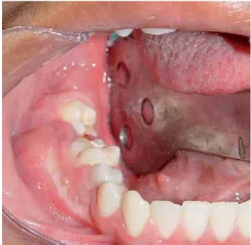
and there was no other relevant history for this lesion. The swelling was located from the right second pre
-molar to the third -molar zone with vestibular expansion with displacement and mobility of the first and second
molar ipsilateral (Figure 1). The panoramic radiograph
showed a unilocular radiolucid lesion that extended from the first and second right molars with a very close relation to the third molar, well defined borders and
measuring approximately 30 x 20 mm, producing dis-placement of the teeth involved (Figure 2). The simple
www.medigraphic.org.mx
www.medigraphic.org.mx
CT scan showed a lesion that produced vestibular andlingual cortical slimming and expansion with preserva -tion of the mandibular border (Figure 3).
An incisional biopsy was taken and the histopatho
-logical result was as follows: Central Giant Cell Granu
-loma (GCCG) combined with Central Odontogenic Fibroma. Surgery was programmed for September of
the same year.
Under general anesthesia and nasotracheal
intuba-tion, an outlining incision was designed with distal ret -romolar extension and anterior vestibular,
mucoperi-osteal flap elevation, complete curettage of the lesion,
extractions of the involved teeth and surgical drilling of the borders, protecting the displaced alveolar nerve (Figure 4). First an absorbable haemostatic sponge
was placed in the gap then the mucoperiosteal flap was sutured.
Microscopically the lesion had well defined borders, a rough surface, firm consistency and pale yellow col -or, producing lingual and vestibular expansion of 40 x 30 x 25 mm (Figure 5).
Histologically the tumor was conformed by fibrocel -lular vascularized tissue forming cross linked bundles
with a scarcity of collagen production.
Histological features: the tumor was conformed by fibro-cellular tissue vascularized forming cross linked bundles of fusiform cells with scarce production of col
-lagen, between them islands and strands of non active odontogenic epithelium were found. In some zones specially in the outer area, there was a highly vascu
-larized fibro-cellular tissue where could be found many
osteoclastic giant multinucleated cells type, distributed around vessels and some intralesional hemorrhages (Figures 6 and 7).
On a follow up 2 years after the treatment the pa
-tient was found without any clinical or radiographic evi -dence of recurrence (Figure 8).
dIscussIon
Among the reported cases of this rare intraosseous
lesion with histological features of COF and GCCG Al -len et al. reported a three cases series.5 Odell et al
referred ten lesions with the same features and sug
-gested that those were hybrid lesions.6 Mosqueda et
al reported another case with the same histological
pattern.7 It should be noticed that there are not many cases published of this unusual lesion.
The Central Odontogenic Fibroma (COF) is a rare benign neoplasm that only appears in the upper and
lower jaws, the reason for this is that the lesion de -rives of mesenchimal tissue of dental origin: periodon-tal ligament, denperiodon-tal papilla or denperiodon-tal follicle. The WHO
defines it as a fibroblastic neoplasia with a variable
amount of seemingly inactive odontogenic epithelium. Some lesions have a certain amount of hard tissue that resembles dysplastic cement or bone.8
COF can appear at any age, but it is more frequent
between the second and fourth decade of life, twice more common in women than in men. It can affect ei
-Figure 1. Clinical appearance.
www.medigraphic.org.mx
ther the maxilla or the mandible, posterior or anteriorzone of the mouth.8
Svirsky (1986), analyzed 15 cases of COF, he
re-ported that 80% of the cases were in the mandible, 60% of them affected women, ranging from 11–80 years, with a 29 years old.9
Handlers et al, showed among 39 cases, 22 cas
-es maxillar and 17 mandibular, with a range of 3:1
female-male incidence, in patients from 11 to 80 years old.9
Ramer et al revision in 2002, showed a 1:1 man -dibular and maxillar incidence, predominating female
in 69%. 25 of the 34 mandibular cases were posterior while in the maxilla 25 of 34 were found in the anterior zone; the range of age of the patients was varied from
4 to 80 years and a media of 35 years.9
Figure 3. Computed tomography scan.
Figure 4. Surgical procedure.
Figure 5. Surgical specimen.
Figure 6. Histological image of the giant cell central lesion
within Central Odontogenic Fibroma with epithelial islands in
www.medigraphic.org.mx
Este documento es elaborado por Medigraphic
Radiographically it has been described as a
radio-lucent area well defined that resembles a unilocular
ameloblastoma or an odontogenic cyst and as a
ra-diolucent lesion with well defined borders with a ten -dency to be smaller than other radiolucent unilocular
lesions, while greater lesions frequently are mul
-tilocular. In most cases the lesion has well defined
borders but also can be seen as a lesion of mixed
appearance with difuse borders.8 Sometimes these
lesions can be associated with root resorption of the
involved teeth.9 There are not radiographic pathogno-monic features of COF.
Microscopically the differentiation spectrum is di-verse. The simple type COF is mode of star-shaped fibroblasts, fine collagen fibers and an important amount of fundamental substance, scarce amount of
odontogenic epithelium; dystrophic calcifications may
be found. The WHO type COF along the same fea-tures of the simple type can also have odontogenic
epithelium and calcifications similar to cement and to
dentine. Other histological variants include granular cell type and hybrid tumor of COF and giant cells. The COFs have been related to intracranial aneurysms and tuberous sclerosis.10
It is thought to derive from mesenchymal elements of the dental germ, as the dental follicle, the dental pa-pillae and periodontal ligament and it is possible that
in his final and mature spectrum it could be seen as
odontogenic myxoma, myxofibroma or odontogenic
fibroma.11
The usual treatment of COF is enucleation. Recur-rence it is not common. Dunlap and Barrer reported
2 cases of COF treated with curettage with 9 and 10
years follow up without recurrence. However, some
cases of recurrence have been reported.9
The Central Giant Cell Granuloma (GCCG) was
described by Jaffe in 1953.12 It is a common lesion that represents 7% of all benign tumors of the max-illa.14 The histological features of GCCG have been
widely discussed13 and have been defined by WHO
as an intraosseous lesion compound of fibrous cel
-lular tissue with multiple hemorrhagic foci and multi -nucleated giant cells and sometimes inmature bone trabecular tissue.2
The clinical feature of this lesion ranges from an
asymptomatic slow growth lesion to an aggressive de
-structive and painful one with root resorption or dental
displacement. The aggressive subtypes of this lesion have a tendency to recurrence after treatment.13 It
can be seen more frequently in women than in men
around, 30 years of age and it is more common in mandible than in maxilla.15 Commonly this lesion has
been found in areas with teeth14 in the anterior zone. The radiological features have not been clearly
de-fined and there are many contradictory descriptions
in the literature.14 The lesion can be found as a uni
or multilocular radiolucency with well or badly defined borders and with different degrees of cortical expan -sion. It is important to notice that this lesion has not a radiologic pathognomonic appearance therefore it can
be confused with other maxillary lesions.16
The usual treatment for GCCG is surgical removal.
However the surgical treatment varies from a simple
curettage to a complete resection of the tumor. The
cu-rettage most be follow up by cryosurgery 17 or
peripher-Figure 7. Epithelial odontogenic islands distributed within
the area of giant cell central lesion (H&E 400X).
Figure 8.
De-tail of control panoramic.
www.medigraphic.org.mx
al ostectomy.18 The GCCG has been recently treatedby non surgical methods utilizing systemic calcito-nin19,20 or an intralesional injection of corticosteroids.21 The presentation of this unusual lesion associated
with COF, shows prevalence in women (12 cases in women to 1 case in men) with a range of age between
5 and 66 years old (average 35.5) more common in mandible (11 mandibular cases compared to 2 maxil-lary cases); in the mandibular cases the posterior zone
was the affected one.7
There are not many publications where the clinical
features of this combined lesion can be found. Most
of the cases reported were asymptomatic with a slow growth and cortical expansion with dental mobility and
in some cases displacement of the teeth. In this case it
was observed the relation of the lesion with an impact -ed third molar that seem-ed a dentigerous cyst. There
were no trauma antecedents or systemic diseases and the bibliography only one case reported showed re
-lation with orthodontic treatment, root canal treatment
and antecedent of dental extraction in the zone of the
lesion, not knowing if the lesion was pre-existent. Only one case mentioned a fast growth with expan -sion and cortical perforation.5-7 Because the scarce
number of cases reported and the resemblance with
the clinical features of other kinds of lesions, it is
dif-ficult to establish the pattern for this combined lesion. From a radiographical point of view the scarce
number of cases reported makes it difficult to
com-pare with previous descriptions of COF and GCCG. Of six cases reported with radiographical description there were 3 unilocular and 3 multilocular lesions.6,7
The case here described showed an unilocular le
-sion associated with an impacted third molar, and it
had a different radiographical pattern.6,7 Kaffe and Buchner reported that 55% of the lesions described
as COF had a radiolucent unilocular lesion with well defined borders, and 29.4% were multilocular. The
GCCG can have either a multilocular or unilocular
presentation and the borders can be well or poorly
defined, it can present different degrees of cortical
expansion and can be confused with other maxil -lary lesions.14 It has not been possible to establish a specific radiographic pattern of the combined lesion (COF and GCCG).
The case here reported histologically is formed by
fibro-cellular tissue well vascularized forming cross
linked bundles of fusiform cells and scarce
produc-tion of collagen fibers, abundant strings and islands
of inactive odontogenic epithelium. In some areas of
the fibro-cellular tissue many giant multinucleated os
-teoclast type cells may be identified specially around
the blood vessels and intralesional hemorrhage zones.
These findings are similar to those described in the
previously reported cases.
Allen et al5 refer to an unusual association of COF
and GCCG reaction, thus confirming the presence of
a histopathological component and considers the pos-sibility that his cases represent a «collision» of tumors in the same area. This appears to be a very rare
prob-ability in which a rare lesion like FOC WHO type de
-velops along with another uncommon lesion like the
GCCG.5
An alternative explanation could be that type WHO COF, could by some means evoke a formation of GCCG in these patients. It is important to mention that the aneurismal bone cyst that histopathologically
re-sembles GCCG has been reported in association with
a certain number of intraosseous lesions. Recently there have been reports of giant cells reactions in
as-sociation with ameloblastoma. Some researchers be
-lieve that the giant cells identified surrounding the am
-eloblastoma were more a result of a reactive process
than a feature of the lesion itself.5
The paper of Mosqueda et al. mentioned that 3 cases among those published until 1999 (25%) have
had recurrence 3 years after the treatment with similar histological findings than the first lesions.7 Allen et al. mentioned that in one of the recurrence cases there
were histological components of COF WHO type and
giant cells.5
These findings, according to Odell et al, suggest a combination of two entities as a main feature of the le -sion therefore they consider it an hybrid tumor.6
So far it has not been possible to determine if the development of COF in a central position produced a surrounding giant cells reaction or if both tissues de-veloped at random in the same place.5
The antecedents of recurrence in the published cases of this type of lesion suggest that it should be treated surgically as GCCG because of its possibility of recurrence, they also suggest to monitor constantly after the surgical excision.
conclusIon
Because of the scarce number of reported cases in the literature, it is not possible to establish clinical and radiographical features to diagnose this neoplasia among the group of odontogenic tumors but like most of the lesions only histological findings can confirm this particular entity.
It has not been possible to determine the origin of
this lesion or to find out if one of the lesions derives
from the other or if they developed simultaneous in the same zone but the recommended treatment should
www.medigraphic.org.mx
take into consideration the most aggressivecompo-nent of the hybrid tumor.
references
1. Handlers JP, Abrams AM, Melrose RJ et al. Central odontogenic
fibroma: Clinicopathologic features of 19 cases and review of the
literature. J Oral Maxillofac Surg 1991; 49: 46.
2. Kramer IRH, Pindborg JJ, Shear M. Histological Typing of Odon-togenic Tumor. Berlin Germany, Springer-Verlag, 1992. 3. Nicolás C, Damián A, Mercedes P, Luis J. Central odontogenic
fibroma granular cell variant: A case report and review of the
literature. J Oral Maxillofac Surg 2002; 60: 1192-1194.
4. Gardner DG. The central odontogenic fibroma: An attempt at clarification. Oral Surg Oral Med Oral Pathol 1980; 50: 425.
5. Allen CM, Hammond HL, Stimson PG. Central odontogenic fi
-broma, WHO type: A report of three cases with an unusual asso -ciated giant cell reaction. Oral Surg Oral Med Oral Pathol 1992; 73: 62.
6. Odell EW, Lombardi T, Barrett AW et al. Hybrid central giant cell granuloma and central odontogenic fibroma-lie lesions of the
jaws. Histopathology 1997; 30: 165.
7. Mosqueda TA, Bermudez FV, Diaz FMA. Combined central
odontogenic fibroma and giant cell granuloma-like lesion of the mandible: report of a case and review of the literature. J Oral Maxillofac Surg 1999; 57: 1258-1262.
8. Kaffe I, Buchner, Buchner A. Radiologic features of central
odon-togenic fibroma. Oral Surg Oral Med Oral Pathol 1994; 78: 811.
9. Spencer MJD. Central odontogenic fibroma of mandible: a case report and review of the literature. Oral Surg Oral Med Oral
Pathol Oral Radiol Endod 2004; 98: 295-300.
10. Erich JR, Claudia EN. Central odontogenic fibroma-like tumors, hypodontia, and enamel dysplasia: review of the literature and
report of a case. Oral Surg Oral Med Oral Pathol Oral Radiol
Endod 2002; 94: 74-7.
11. Zachariades N. Odontogenic fibroma. Int J Oral Maxillofac Surg
1986; 15: 102.
12. Jan de L, Hans P, van den Akker. Clinical and radiological
fea-tures of central giant-cell lesions of the jaw. Oral Surg Oral Med
Oral Pathol Oral Radiol Endod 2005; 99: 464-70.
13. Chuong R, Kaban LB, Kozakewich H et al. Central giant cell le
-sions of the jaws: A clinicopathologic study. J Oral Maxillofac Surg 1986; 44: 708.
14. Bataineh AB, Al-Khateeb T, Rawashdeh MA. The surgical treat -ment of central giant cell granuloma of the mandible. J Oral
Max-illofac Surg 2002; 60: 756-761.
15. Whitaker SB, Waldron CA. Central giant cell lesions of the jaws.
Oral Surg Oral Med Oral Pathol 1993; 75: 199.
16. Cohen MA, Hertzanu Y. Radiologic features, including those
seen with computed tomography, of central giant cell granuloma of the jaws. Oral Surg Oral Med Oral Pathol 1988; 65: 255. 17. Webb DJ, Brockbank J. Combined curettage and cryosurgical
treatment for the aggressive «giant cell lesion» of the mandible.
Int J Oral Maxillofac Surg 1986; 5: 780.
18. Eisenbud L, Stern M, Rothberg M et al. Central giant cell
granu-loma of the jaws: Experiences in the management of thirty-seven
cases. J Oral Maxillofac Surg 1988; 46: 376-84.
19. Harris M. Central giant cell granulomas of the jaws regress with
calcitonin therapy. Br J Oral Maxillofac Surg 1993; 31: 89. 20. de Lange J, Rosenberg AJ, van den Akker HP et al. Treatment of
central giant cell granuloma of the jaw with calcitonin. Int J Oral Maxillofac Surg 1999; 28: 372.
21. Kermer C, Millesi W, Watzke IM. Local injection of corticoste -roids for central giant cell granuloma: A case report. Int J Oral
Maxillofac Surg 1994; 23: 366.
Address correspondence:
Adalberto Mosqueda MD