R E S E A R C H A R T I C L E
Open Access
Vitamin D deficiency in chronic inflammatory
rheumatic diseases: results of the cardiovascular
in rheumatology [CARMA] study
Ana Urruticoechea-Arana
1†, María A. Martín-Martínez
2†, Santos Castañeda
3†, Carlos A. Sanchez Piedra
2,
Carlos González-Juanatey
4, Javier Llorca
5, Federico Díaz-Gonzalez
2,6,7, Miguel A. González-Gay
8,9*and on behalf of the CARMA Project Collaborative Group
Abstract
Introduction: The aim was to study the association between 25-hydroxyvitamin D (25(OH)D) levels and the clinical
characteristics of patients with chronic inflammatory rheumatic diseases (CIRD).
Methods: We studied a cross-section from the baseline visit of the CARMA project (CARdiovascular in
rheuMAtology), a 10-year prospective study evaluating the risk of cardiovascular events in rheumatoid arthritis (RA),
ankylosing spondylitis (AS) and psoriatic arthritis (PsA) patients, and non-CIRD patients who attended rheumatology
outpatient clinics from 67 hospitals in Spain. Non-CIRD group was frequency matched by age with the joint
distribution of the three CIRD groups included in the study. 25(OH)D deficiency was defined if 25(OH)D vitamin
levels were < 20 ng/ml.
Results: 2.234 patients (775 RA, 738 AS and 721 PsA) and 677 non-CIRD subjects were assessed. The median
(p25-p75) 25(OH)D levels were: 20.4 (14.4-29.2) ng/ml in RA, 20.9 (13.1-29.0) in AS, 20.0 (14.0-28.8) in PsA, and 24.8
(18.4-32.6) ng/ml in non-CIRD patients. We detected 25(OH)D deficiency in 40.5 % RA, 39.7 % AS, 40.9 % PsA and
26.7 % non-CIRD controls (p < 0.001). A statistically significant positive association between RA and 25(OH)D
deficiency was found (adjusted (adj.) OR = 1.46; 95 % CI = 1.09-1.96); p = 0.012. This positive association did not reach
statistical significance for AS (adj. OR 1.23; 95 % CI = 0.85-1.80) and PsA (adj. OR 1.32; 95 % CI = 0.94-1.84). When the
parameters of disease activity, severity or functional impairment were assessed, a marginally significant association
between 25(OH)D deficiency and ACPA positivity in RA patients (adj. OR = 1.45; 95 % CI = 0.99-2.12; p = 0.056), and
between 25(OH)D deficiency and BASFI in AS patients (adj. OR = 1.08; 95 % CI = 0.99-1.17); p = 0.07) was also found.
Conclusions: Patients with RA show an increased risk of having 25(OH)D deficiency compared to non-CIRD controls.
Introduction
Vitamin D has raised great interest in recent decades
due to its multiple physiological functions, a including
significant role in the regulation of the immune system
[1–7]. Vitamin D deficiency is an extremely common
health problem that affects up to 50 % of the general
population during winter months in the Northern
hemi-sphere [8]. Several studies have pointed out a potential
as-sociation between vitamin D deficiency and cancer, some
chronic infections, cardiovascular mortality and increased
risk of some autoimmune diseases [8–13], such as type I
diabetes mellitus [10], multiple sclerosis [10], systemic
lupus erythematosus (SLE) [9, 11, 12] and rheumatoid
arthritis (RA) [9, 13]. In this regard, some authors have
reported an inverse relationship between serum levels of
25-hydroxyvitamin D (25(OH)D) and disease activity or
functional impairment in patients with RA or early arthritis
[14–21]. In a recent study, vitamin D deficiency was found
in 30 % of RA patients [8]. Most studies assessing serum
* Correspondence:[email protected]†Equal contributors 8
Division of Rheumatology, Hospital Universitario de Canarias, La Laguna, Tenerife, Spain
9
Division of Rheumatology, Hospital Universitario Marqués de Valdecilla, Santander, and Epidemiology, Genetics and Atherosclerosis Research Group on Systemic Inflammatory Diseases, Rheumatology Division, IDIVAL, Avenida de Valdecilla, s/n, 39008 Santander, Spain
Full list of author information is available at the end of the article
© 2015 Urruticoechea-Arana et al.Open AccessThis article is distributed under the terms of the Creative Commons Attribution 4.0 International License (http://creativecommons.org/licenses/by/4.0/), which permits unrestricted use, distribution, and reproduction in any medium, provided you give appropriate credit to the original author(s) and the source, provide a link to the Creative Commons license, and indicate if changes were made. The Creative Commons Public Domain Dedication waiver (http://creativecommons.org/publicdomain/zero/1.0/) applies to the data made available in this article, unless otherwise stated.
25(OH)D levels in patients with chronic inflammatory
rheumatic diseases (CIRD) were focused on patients with
RA [9, 13–15, 17–20]. However, fewer studies have
ana-lyzed the presence of 25(OH)D deficiency in other CIRD
such as ankylosing spondylitis (AS) or psoriatic arthritis
(PsA) [21–24].
The aim of this study was to assess 25(OH)D levels in
a cohort of Spanish patients with CIRD that included
patients with RA, AS, PsA and non-CIRD, who were
at-tending rheumatology outpatient clinics, and to
deter-mine the potential relationship between 25(OH)D levels
and clinical characteristics of every disease included in
the group of patients with CIRD.
Methods
Study design
Cross-sectional analysis from the baseline visit of the
project, CARdiovascular in rheuMAtology (CARMA), a
10-year prospective cohort study designed to determine
the cardiovascular mortality risk in patients with CIRD
compared to a cohort of patients without inflammatory
pathological disease [25]. Information on this cohort has
recently been reported. The institutional review board of
each center approved the study and all patients signed
an informed consent agreement.
Patients and controls
Based on the information from the Spanish Society of
Rheumatology (SER), all the Spanish public hospitals
(university and general community hospitals) that have
rheumatology units were invited to take part in the present
study. Finally, 67 (63.2 %) of the 106 centers agreed to
par-ticipate in the study. They recruited 2,986 patients who
attended rheumatology outpatient clinics from July 2010 to
January 2012. Seventy-five patients declined the invitation.
Therefore, 2,911 patients over 18 years old were included
in the study. They were split into two different cohorts
ac-cording to CIRD exposition. The CIRD patients consisted
of 775 patients diagnosed with RA (1987 American College
of Rheumatology (ACR) classification criteria) [26], 738
diagnosed with AS (modified New York criteria) [27]
and 721 patients with PsA (Moll and Wright criteria)
[28]. The control (non-CIRD) group included 677 patients
without inflammatory rheumatic disease. The non-CIRD
group was frequency-matched by age with the joint
distri-bution of the three CIRD groups. Therefore, its
distribu-tion approximates the joint age distribudistribu-tion of the three
CIRD. To see the list of participating centers, we advise
readers to see the Acknowledgements section.
Variables and operative definitions
Both cohorts were evaluated following international
proto-cols including standardized definitions and validated
ques-tionnaires. All patients included were continuously and
systematically evaluated online and, to verify the quality of
the information, an in situ monitoring data assessment
was performed in 15 % of patients randomly selected.
The primary endpoint was the presence of 25(OH)D
deficiency defined as 25(OH)D levels below 20 ng/ml.
The variable sunshine hours per month and province
(geographical area in which the hospital is located) was
established considering the hours of sunlight in the period
of time between 60 and 90 days prior to the visit of
inclu-sion of each patient. For this purpose we used the
infor-mation published by the Spanish Meteorological Agency
(SMA) [29]. When information on sunshine hours of the
month and year was not available, we used the mean value
of sunshine hours of the last 5 years in which information
on sunshine hours in the same period of time and site was
available. The 25(OH)D analysis was locally performed
according to the methodology and reproducibility level
of each institution.
Other variables analyzed were: (1) obesity (BMI
≥30,
kg/m
2) and main physical activity during working hours
(low activity: sitting most of the time; moderate activity:
standing most of the time and with little movement or
effort; intense activity: walking most of the time or
perform-ing tasks that require high physical activity); (2) disease
characteristics and parameters of disease activity:
rheuma-toid factor (RF), anti-cyclic citrullinated peptide antibodies
(ACPA), HLA-B27 positivity, erythrocyte sedimentation
rate (ESR) (mm/1
sth), ultra-sensitive C-reactive protein
(CRP) (mg/l), disease activity score including 28
joints-erythrocyte sedimentation rate (DAS28-ESR), health
as-sessment questionnaire (HAQ (0–3)), Bath ankylosing
spondylitis disease activity index (BASDAI (0–10)) and
Bath ankylosing spondylitis functional index (BASFI
(0–10)); (3) sociodemographic variables, and (4) other
factors: disease severity, duration of the disease and
therapies administered including calcium and vitamin D
supplementation.
Statistical analysis
Numerical variables with a normal distribution were
expressed as mean and standard deviation. Variables not
normally distributed were shown as median and
interquar-tile range (IQR, perceninterquar-tile (p)25
− p75). Absolute and
rela-tive frequencies of qualitarela-tive variables were calculated.
We performed analysis of the main demographic and
clin-ical variables stratified by type of disease. Stratified analysis
of 25(OH)D deficiency (<20 ng/ml) was performed for
each group of patients according to sociodemographic
characteristics and clinical factors, using the Student
t test
or the Mann-Whitney
U test. Qualitative variables were
assessed using the Chi-square test, Yates correction, or
Fisher test using 2 × 2 tables.
To study the association between 25(OH)D deficiency
and CIRD, logistic regression models were constructed
by calculating the odds ratios (OR) with 95 % CI and
adjusting for potential confounding factors. In this regard,
an adjusted model for sunshine hours was performed. It
was carried out considering for this purpose the period of
time between 60 and 90 days prior to the baseline visit
blood test, which included assessment of 25(OH)D levels.
To reduce variability in the methods of measurement of
25(OH)D from each participating hospital, the mixed
logistic regression models were constructed with robust
variance estimators using the hospital as cluster variable
es-timation. The same procedure was carried out to identify
specific features of each disease. The selection of
independ-ent variables in the multivariate models was based on
clin-ical judgment and those with a
p-value <0.20 in the bivariate
analysis. In all models of logistic regression, the independent
variables were adjusted for the other variables in the model.
Data management and statistical analysis were centralized
at the Research Unit of the SER following a pre-established
analysis plan. All the analyses were performed using the
SPSS 21.0 statistical program. Statistical significance
was assumed at
p <0.05.
Results
Sociodemographic and clinical characteristics
Demographic and clinical characteristics of patients
in-cluded in this study are summarized in Table 1. There was
a predominance of women in the group with RA, whereas
most AS patients were men. Sex distribution was similar
in the group with PsA. The mean age in patients with RA
was higher than in patients with other CIRD. Although
patients with AS were younger, the duration of disease
was longer than in the other groups. The frequency of
obesity was higher among PsA patients, whereas it was
lower among controls, despite being the group that
in-cluded more sedentary individuals (p <0.001). Smoking
history was more commonly observed in those with AS.
It is worth noting that a great majority of patients with
CIRD included in the study had low disease activity at
time of recruitment. In this regard, CRP and ESR levels
in RA, AS and PsA patients were remarkably low at the
time of inclusion in the study, as well as the
functional-ity scores (HAQ and BASFI, respectively).
Individuals without CIRD (controls) had the following
rheumatic diseases: osteoarthritis (30 %), osteoporosis
(15.2 %), soft tissue disorders (18.8 %) and other
non-inflammatory diseases (36 %). The distribution at
recruit-ment of individuals with CIRD per hospital was uniform
throughout the year 2011, whereas the recruitment of
pa-tients without CIRD occurred mainly between October
and November 2011 (Additional file 1: Table S1).
25(OH)D levels and CIRD
Patients with CIRD had lower 25(OH)D levels than those
from the non-CIRD controls (Table 1). The median
(p25
− p75) 25(OH)D levels were: 20.4 (14.4 − 29.2)] ng/ml
in RA, 20.9 (13.1
− 29.00 ng/ml in AS, 20.0 (14.0 − 28.8)
ng/ml in PsA and 24.8 (18.4
− 32.6) ng/ml in non-CIRD
individuals. Globally, 25(OH)D deficiency was detected in
40.5 % of patients with RA, 39.7 % of patients with AS,
40.9 % of patients with PsA and 26.7 % of individuals with
non-CIRD (p <0.001). The median of sunshine hours in
the group of non-CIRD controls was higher than in the
three groups of patients with CIRD (Table 2).
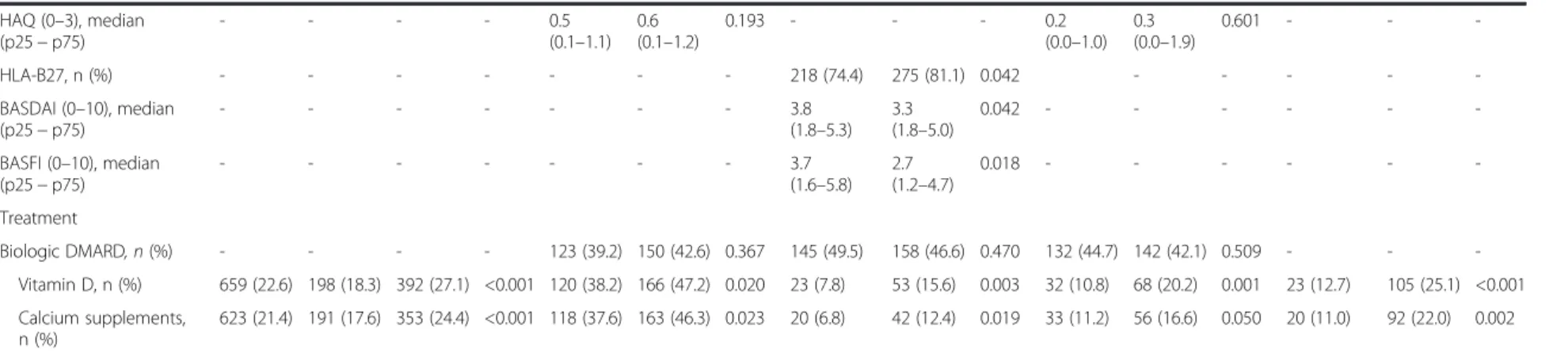
Among the variables related to activity and severity of
inflammatory diseases, ACPA-positive RA patients had a
higher frequency of 25(OH)D deficiency (66.9 %). It was
also the case for AS, with higher values of BASDAI and
BASFI in 25(OH)D-deficient patients (p <0.05 in both
cases) (Table 2).
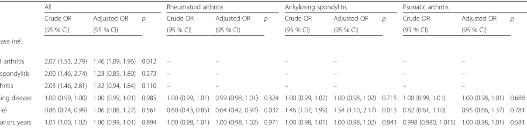
Multivariate analysis
Multivariate analysis (Table 3) disclosed a positive
asso-ciation with 25(OH)D deficiency in the patients with
CIRD when compared with the non-CIRD subjects. This
association with 25(OH)D deficiency was statistically
sig-nificant in the group of patients with RA (adjusted (adj.)
OR = 1.46; 95 % CI = 1.09, 1.96);
p = 0.012. However, the
positive association was not statistically significant for
AS (adj. OR = 1.23; 95 % CI = 0.85, 1.80) and PsA (adj.
OR = 1.32; 95 % CI = 0.94, 1.84). Women with RA had
significantly higher risk of 25(OH)D deficiency than men
(p <0.01). Likewise, obese RA and PsA patients (BMI ≥30)
had higher risk of 25(OH)D deficiency
When the parameters of disease activity, severity or
functional impairment were assessed, a marginally
signifi-cant association between 25(OH)D deficiency and
ACPA-positivity in RA patients (adj. OR = 1.45; 95 % CI = 0.99,
2.12;
p = 0.056), and between 25(OH)D deficiency and
BASFI in AS patients (adj. OR = 1.08; 95 % CI = 0.99, 1.17;
p = 0.07) was also found (Table 3).
Discussion
Our results show that Spanish patients with RA
attend-ing rheumatology outpatient clinics have 25(OH)D
defi-ciency. This baseline result is from a cohort of patients
that has been followed prospectively to determine the
car-diovascular outcome. To establish comparisons, we also
assessed baseline 25(OH)D levels in non-CIRD controls
attending the same rheumatology outpatient clinics [25].
Vitamin D plays an important role in the immune
regu-lation [2]. Vitamin D deficiency has been observed in some
autoimmune diseases, in particular in SLE [11, 12, 30] and
RA [8, 20]. However, information related to
undifferenti-ated spondyloarthropathies and AS is limited [21, 22, 31].
It is also the case for PsA [24].
In our series, the frequency of 25(OH)D deficiency
(level <20 ng/ml) was higher in patients with RA than in
the individuals from the non-CIRD control group. The
present study raises several points of potential interest.
First, the CARMA cohort constitutes the largest series of
comparisons of 25(OH)D levels in three well-established
CIRD. In addition, a cohort of individuals without CIRD
was used for comparison. Second, we assessed patients
who were periodically followed at rheumatology
out-patient clinics. Nevertheless, it is important to emphasize
that baseline levels of 25(OH)D in the control population
Table 1 Sociodemographic characteristics, 25(OH)D levels and clinical characteristics of the population included in the study
Rheumatoid arthritis Ankylosing spondylitis Psoriatic arthritis Controls
(n = 775) (n = 738) (n = 721) (n = 677)
Age at inclusion, years, mean (SD) 57.1 (12.3) 48.1 (11.7) 51.8 (12.0) 54.0 (12.4)
Age at the beginning of disease, years, mean (SD) 45.8 (13.4) 29.7 (11.8) 39.5 (13.3) 48.5 (12.4)
Female sex, n (%) 581 (75.0) 200 (27.1) 327 (45.4) 437 (64.5) Educational level, n (%) Elementary 467 (60.9) 318 (43.3) 331 (46.3) 229 (34.1) University /secondary 300 (39.1) 416 (56.7) 383 (53.7) 443 (65.9) Caucasian Race, n (%) 747 (96.6) 723 (98.0) 712 (98.9) 668 (98.7) Others 26 (3.4) 15 (2.0) 8 (1.1) 9 (1.3) Main activity, n (%) Sedentary 236 (35.0) 263 (39.3) 253 (38.9) 291 (46.3) Moderate 290 (43.0) 238 (35.5) 241 (37.1) 207 (32.9)
Active with displacement 148 (22.0) 169 (25.2) 156 (24.0) 131 (20.8)
BMI, kg/m2, mean (SD) 26.9 (4.8) 27.4 (4.4) 28.2 (4.7) 26.7 (4.4) Obesity (BMI≥30), n (%) 180 (23.2) 186 (25.2) 209 (29.1) 147 (21.8) Smoking status, n (%) Current smokers 189 (24.4) 254 (34.4) 157 (21.8) 143 (21.2) Past smokers 202 (26.1) 240 (32.5) 227 (31.5) 176 (26.0) Never smokers 384 (49.5) 244 (33.1) 337 (46.7) 357 (52.8) 25 (OH) D 25(OH)D (ng/ml), median (p25− p75) 20.4 (14.4–29.2) 20.9 (13.1–29.0) 20.0 (14.0–28.8) 24.8 (18.4–32.6) 25(OH)D deficiency, n (%) 314 (40.5) 293 (39.7) 295 (40.9) 181 (26.7)
Sunshine hours/month*, median (p25− p75) 162 (122–219) 165 (136–233) 178 (128–235) 301 (202–345)
Disease duration, years, median (p25− p75) 8.0 (3.0–14.0) 15.0 (8.0–26.0) 9.0 (4.0–16.0) 2.0 (0.0–6.0)
RF-positive, n (%) 528 (68.1) – – – ACPA-positive, n (%) 482 (62.2) – – – DAS28-ESR, mean (SD) 3.2 (1.2) – 3.0 (1.3) – HAQ (0–3), median (p25 − p75) 0.5 (0.1–1.1) – 0.4 (0.0–0.9) – ESR (mm/h), median (p25− p75) 17.0 (9.0–29.0) 10.0 (6.0–21.0) 12.0 (6.0–21.0) 10.0 (5.0–18) CRP (mg/l), median (p25− p75) 3.1 (1.2–8.0) 3.6 (1.6–8.9) 2.9 (1.4–6.1) 1.9 (1.3–3.3) BASDAI (0–10), median (p25 − p75) – 3.5 (1.7–5.3) – – BASFI (0–10), median (p25 − p75) – 3.1 (1.3–5.2) – – HLA-B27, n (%) – 561 (76) – – Erosions (RA), n (%) 352 (45.4) – – –
Biologic therapy (% ever treated), n (%) 313 (40.4) 349 (47.4) 300 (41.7) –
Vitamin D, n (%) 325 (41.9) 82 (11.1) 114 (15.8) 138 (20.4)
Calcium supplements, n (%) 328 (42.3) 68 (9.2) 105 (14.6) 122 (18.0)
25(OH)D deficiency is defined as 25(OH)D <20 ng/ml. *Hours of sunshine per month considering the period of time between 60 and 90 days prior to the baseline visit (blood test to determine the levels of 25(OH)D was performed at the baseline visit).BMI body mass index, 25(OH)D 25-hydroxyvitamin D, p25 − p75 25th
to 75th
percentile,RF rheumatoid factor, ACPA anti-cyclic citrullinated peptide antibodies, DAS28-ESR, disease activity score using 28 joints-erythrocyte sedimentation rate,HAQ (0–3): health assessment questionnaire, ESR erythrocyte sedimentation rate, CRP C-reactive protein, BASDAI (0–10) Bath ankylosing spondylitis disease activity index,BASFI (0–10) Bath ankylosing spondylitis functional index, HLA-B27 histocompatibility antigen HLA-B27, RA rheumatoid arthritis
Female, n (%) 1545 (53.1)
554 (51.2) 796 (55.0) 0.054 220 (70.1) 280 (79.5) 0.005 92 (31.4) 81 (23.9) 0.035 127 (43.1) 162 (48.1) 0.206 115 (63.5) 273 (65.2) 0.703 Age beginning disease,
years, mean (SD) 40.5 (14.5) 40.4 (14.5) 41.4 (14.7) 0.079 45.8 (13) 46.1 (13.6) 0.743 30.1 (11.7) 29.6 (11.8) 0.620 39.2 (13.1) 39.4 (13.3) 0.846 49.4 (12.6) 48.5 (12.2) 0.406 Disease duration, years,
mean (SD) 11.03 (10.4) 11.5 (10.2) 10.7 (10.5) 0.005 10.1 (8.5) 10.4 (9.1) 0.782 17.6 (11.9) 17.72 (12.1) 0.950 11.2 (8.4) 11.4 (9.3) 0.983 4.4 (6.9) 4.5 (6.2) 0.321 Educational level, n (%) Elementary 1693 (58.6) 646 (60.4) 819 (56.8) 0.071 212 (68.6) 242 (68.9) 0.926 176 (60.7) 182 (53.8) 0.84 173 (59.5) 200 (59.5) 0.985 85 (47.5) 195 (46.9) 0.891 University/secondary 1194 (41.4) 423 (39.6) 622 (43.2) 97 (31.4) 109 (31.1) 114 (39.3) 156 (46.2) 118 (40.5) 136 (40.5) 94 (52.5) 221 (53.1) Smoking status, n (%) Current smokers 743 (25.5) 303 (28.0) 335 (23.2) 83 (24.4) 76 (21.6) 107 (36.5) 113 (33.3) 59 (20.0) 73 (21.6) 54 (30.0) 73 (17.4) Past smokers 845 (29.0) 332 (30.7) 407 (28.1) 0.001 86 (27.4) 92 (26.1) 0.226 95 (32.4) 107 (31.6) 0.530 101 (34.2) 104 (30.9) 0.650 50 (27.8) 104 (24.8) <0.001 Never smokers 1322 (45.5) 447 (41.3) 705 (48.7) 145 (46.2) 184 (52.3) 91 (31.1) 119 (35.1) 135 (45.8) 160 (47.5) 76 (42.2) 242 (57.8) Obesity (BMI≥30), n (%) 2184 (75.0) 327 (30.2) 308 (21.3) 0.001 88 (28.0) 71 (20.2) 0.018 83 (28.3) 75 (22.1) 0.072 102 (34.7) 89 (26.5) 0.025 54 (30.0) 73 (17.5) 0.001 Main physical activity, n (%)
Sedentary 1043 (39.8) 380 (39.3) 553 (40.8) 92 (34.3) 113 (36.1) 103 (39.6) 120 (38.7) 110 (41.5) 119 (39.1) 75 (42.9) 181 (47.6) Moderate 976 (37.2) 363 (37.5) 472 (36.1) 0.734 113 (42.2) 136 (43.5) 0.669 90 (34.6) 109 (35.2) 0.976 100 (37.7) 107 (35.2) 0.386 60 (34.3) 120 (31.6) 0.576 Active with displacement 604 (23.0) 225 (23.2) 302 (23.1) 63 (23.5) 64 (20.4) 67 (25.8) 81 (26.1) 55 (20.8) 78 (25.7) 40 (22.9) 79 (20.8) Sunshine hours /month*,
median (p25− p75) 189 (138–269) 161 (122–225) 210 (159–320) <0.001 156 (101–193) 184 (141–234) <0.001 161 (122–202) 200 (157–251) <0.001 159 (103–200) 201 (150–263) <0.001 252 (162–345) 303 (202–345) 0.031 Clinical characteristics ESR (mm/h), median (p25− p75) - - - - 18.0 (9.0–28.0) 16.0 (9.0–30.0) 0.631 10.0 (6.0–23.2) 11.0 (6.0–19.0) 0.691 12.0 (5.0–21.0) 12.0 (6.0–21.0) 0.471 11.0 (5.0–18.0) 10.0 (5.0–18.0) 0.189 CRP (mg/l), median (p25− p75) - - - - 3.1 (1.4–8.0) 3.0 (1.0–7.3) 0.543 4.1 (1.5–10.6) 3.2 (1.6–7.6) 0.333 3.0 (1.4– 6.1) 2.9 (1.3–6.2) 0.449 2.0 (0.9– 4.1) 1.9 (1.0–3.2) 0.358 RF positive, n (%) - - - - 252 (80.3) 264 (75.0) 0.105 - - - -ACPA positive, n (%) - - - - 210 (66.9) 205 (58.2) 0.022 - - - -Erosions, n (%) - - - - 143 (45.5) 169 (48.0) 0.524 - - - 79 (26.8) 92 (27.3) 0.883 - - -DAS28-ESR, median (p25− p75) - - - - 3.0 (2.2–3.8) 3.0 (2.3–4.0) 0.937 - - - 2.8 (1.9–3.9) 2.9 (2.0–3.9) 0.807 - - -a et al. Arthritis Research & Therapy (2015) 17:211 Page 5 of 10
Table 2 Bivariate analysis according to each specific entity and the occurrence of 25(OH)D deficiency (25(OH)D <20 ng/ml) (Continued)
HAQ (0–3), median (p25− p75) - - - - 0.5 (0.1–1.1) 0.6 (0.1–1.2) 0.193 - - - 0.2 (0.0–1.0) 0.3 (0.0–1.9) 0.601 - - -HLA-B27, n (%) - - - 218 (74.4) 275 (81.1) 0.042 - - - - -BASDAI (0–10), median (p25− p75) - - - 3.8 (1.8–5.3) 3.3 (1.8–5.0) 0.042 - - - -BASFI (0–10), median (p25− p75) - - - 3.7 (1.6–5.8) 2.7 (1.2–4.7) 0.018 - - - -Treatment Biologic DMARD, n (%) - - - - 123 (39.2) 150 (42.6) 0.367 145 (49.5) 158 (46.6) 0.470 132 (44.7) 142 (42.1) 0.509 - - -Vitamin D, n (%) 659 (22.6) 198 (18.3) 392 (27.1) <0.001 120 (38.2) 166 (47.2) 0.020 23 (7.8) 53 (15.6) 0.003 32 (10.8) 68 (20.2) 0.001 23 (12.7) 105 (25.1) <0.001 Calcium supplements, n (%) 623 (21.4) 191 (17.6) 353 (24.4) <0.001 118 (37.6) 163 (46.3) 0.023 20 (6.8) 42 (12.4) 0.019 33 (11.2) 56 (16.6) 0.050 20 (11.0) 92 (22.0) 0.002*Hours of sunshine per month considering the period of time between 60 and 90 days prior to the baseline visit (blood test to determine the levels of 25(OH)D was performed at the baseline visit).25(OH)D 25-hydroxyvitamin D,BMI body mass index, p25 − p75 25th
to 75th
percentile,ESR erythrocyte sedimentation rate, CRP C-reactive protein, RF rheumatoid factor, ACPA anti-cyclic citrullinated peptide antibodies, DAS28-ESR, disease activity score using 28 joints-erythrocyte sedimentation rate,HAQ (0–3): health assessment questionnaire, HLA-B27 histocompatibility antigen HLA-B27, BASDAI (0–10) Bath ankylosing spondylitis disease activity index,BASFI (0–10) Bath ankylosing spondylitis functional index, DMARD disease-modifying anti-rheumatic drugs
Urruticoec hea-Aran a et al. Arthritis Research & Therapy (2015) 17:211 Page 6 of 10
Table 3 Multivariate analysis of 25(OH)D deficiency (25(OH)D levels <20 ng/ml) in patients with chronic inflammatory rheumatic diseases
All Rheumatoid arthritis Ankylosing spondylitis Psoriatic arthritis
Variables Crude OR Adjusted OR p Crude OR Adjusted OR p Crude OR Adjusted OR p Crude OR Adjusted OR p (95 % CI) (95 % CI) (95 % CI) (95 % CI) (95 % CI) (95 % CI) (95 % CI) (95 % CI) Kind of disease (ref.
controls)
Rheumatoid arthritis 2.07 (1.53, 2.79) 1.46 (1.09, 1.96) 0.012 – – – – – –
Ankylosing spondylitis 2.00 (1.46, 2.74) 1.23 (0.85, 1.80) 0.273 – – – – – –
Psoriatic arthritis 2.03 (1.46, 2.81) 1.32 (0.94, 1.84) 0.110 – – – – – –
Age beginning disease 1.00 (0.99, 1.00) 1.00 (0.99, 1.01) 0.985 1.00 (0.99, 1.01) 0.99 (0.98, 1.01) 0.324 1.00 (0.99, 1.02) 1.00 (0.98, 1.02) 0.715 1.00 (0.99, 1.01) 1.00 (0.98, 1.01) 0.688 Sex (ref. male) 0.86 (0.74, 0.99) 1.06 (0.88, 1.27) 0.561 0.60 (0.43, 0.85) 0.64 (0.42, 0.97) 0.037 1.46 (1.07, 1.99) 1.54 (1.10, 2.17) 0.013 0.82 (0.61, 1.10) 0.95 (0.66, 1.37) 0.781 Disease duration, years 1.01 (1.00, 1.02) 1.00 (0.99, 1.01) 0.894 1.00 (0.98, 1.01) 1.00 (0.98, 1.02) 0.971 1.00 (0.98, 1.01) 1.00 (0.98, 1.02) 0.841 0.998 (0.980, 1.015) 1.00 (0.98, 1.01) 0.581 Educational level (ref.
elementary)
University/secondary 0.86 (0.67, 1.11) 0.99 (0.77, 1.29) 0.963 1.02 (0.71, 1.45) 1.17 (0.69, 1.72) 0.430 0.76 (0.52, 1.10) 0.81 (0.55, 1.19) 0.280 1.00 (0.67, 1.50) 0.94 (0.62, 1.41) 0.765 Smoking status (ref.
current smokers) Past smokers 0.90 (0.74, 1.10) 0.91 (0.73, 1.13) 0.387 0.86 (0.58, 1.26) 0.80 (0.49, 1.29) 0.354 0.94 (0.62, 1.41) 1.11 (0.72, 1.70) 0.639 1.20 (0.77, 1.88) 1.20 (0.72, 1.99) 0.480 Never smokers 0.70 (0.57, 0.86) 0.73 (0.58, 0.91) 0.005 0.72 (0.51, 1.02) 0.74 (0.50, 1.11) 0.144 0.81 (0.53, 1.22) 0.84 (0.56, 1.26) 0.404 1.04 (0.66, 1.64) 1.12 (0.67, 1.87) 0.662 Obesity (BMI≥30. kg/m2) 1.60 (1.32, 1.94) 1.96 (1.28, 1.90) <0.001 1.54 (1.09, 2.17) 1.76 (1.18, 2.62) 0.006 1.39 (0.93, 2.09) 1.20 (0.78, 1.86) 0.408 1.47 (1.04, 2.09) 1.41 (0.98, 2.05) 0.067 Sunshine hours/month* 0.99 (0.99, 0.99) 0.99 (0.99, 0.99) <0.001 0.99 (0.99, 0.99) 0.99 (0.99, 0.99) 0.001 0.99 (0.99, 0.99) 0.99 (0.99, 0.99) 0.001 0.99 (0.99, 0.99) 0.99 (0.99, 0.99) <0.001 HAQ (0–3) 0.86 (0.69, 1.07) 0.90 (0.71, 1.14) 0.384 – – – – ACPA-positive – – 1.45 (1.00, 2.09) 1.45 (0.99, 2.12) 0.056 – – – – HLA-B27 – – – – 0.68 (0.50, 0.92) 0.70 (0.48, 1.02) 0.062 – – BASFI (0–10) – – – – 1.10 (1.02, 1.18) 1.08 (0.99, 1.17) 0.070 – – Vitamin D therapy 0.60 (0.47, 0.77) 0.57 (0.43, 0.76) <0.001 0.69 (0.48, 0.99) 0.78 (0.52, 1.18) 0.245 0.46 (0.26, 0.81) 0.43 (0.26, 0.81) 0.008 0.48 (0.28, 0.82) 0.54 (0.29, 1.01) 0.053
*Hours of sunshine per month considering the period of time between 60 and 90 days prior to the baseline visit (blood test to determine the levels of 25(OH)D was performed at the baseline visit).25(OH)D 25-hydroxyvitamin D,OR odds ratio, BMI body mass index, HAQ (0–3): health assessment questionnaire, ACPA anti-cyclic citrullinated peptide antibodies, HLA-B27 histocompatibility antigen HLA-B27, BASFI (0–10) Bath ankylosing spondylitis functional index
a et al. Arthritis Research & Therapy (2015) 17:211 Page 7 of 10
were also low, due to the inclusion in this population of a
high percentage of subjects with osteoarthritis and/or
osteoporosis, who are also likely to have low baseline 25
(OH)D levels.
Nowadays it is not clear whether vitamin D deficiency
is the cause or effect of the inflammatory process. In this
regard, in a model of acute phase response after surgery,
plasma concentrations of 25(OH)D were found to
de-crease after elective knee arthroplasty [32]. Furthermore,
several studies have found an inverse association between
25(OH)D levels and activity parameters of some CIRD,
such as DAS28, swollen joints and HAQ in RA and BASFI
and BASDAI in AS [17, 19, 21]. Although the results from
our study do not fully support all these findings, in the
multivariate analysis a marginally statistically significant
association between 25(OH)D deficiency and ACPA in
RA and BASFI in AS was found. It is worth noting that
our patients with CIRD had decreased 25(OH)D levels
despite the fact that a great majority had low activity at
the time of inclusion. Patients with CIRD have less
mobil-ity and life outdoors, which would also contribute
nega-tively to maintain adequate levels of vitamin D. Therefore,
25(OH)D deficiency in these patients may be explained by
a dual mechanism. On the one hand, chronic diseases can
predispose to 25(OH)D deficiency directly by decreasing
synthesis or increasing vitamin D catabolism, and on
the other hand, indirectly lowering sunlight exposure in
phases of reduced mobility and ability to spend time
outdoors in patients with worse functional status.
We feel that our results may be considered of potential
interest in daily clinical practice, as our population
encom-passed individuals periodically followed at rheumatology
outpatient clinics, many of whom are controlled under
biological treatment.
Although a recent umbrella review of systematic
re-views and meta-analyses of observational studies and
randomized trials did not demonstrate that
supplemen-tation of vitamin D improves the health of the general
population [33], we believe it is important to monitor
and supplement vitamin D to patients with CIRD and
vitamin D deficiency, regardless of whether the deficiency
of vitamin D may or may not have a pathogenic role, or
whether it is merely an epiphenomenon associated with
inflammatory disease.
Our study has some limitations. First, a potential
limi-tation of the study was that the non-CIRD subjects were
not completely healthy, as a high percentage of individuals
included had osteoarthritis, osteoporosis and/or other
musculoskeletal diseases, which by themselves are
associ-ated with some risk of 25(OH)D deficiency. Another
limitation of this study may be that the control group
had more sunshine hours because many controls were
recruited in the months of October and November, and
several studies indicate that the level of 25(OH)D is the
result of sun exposure in a period of time between 60
to 90 days prior to the 25(OH)D assessment [34], which
in our study corresponded with the months of July and
August. With respect to the variability of the vitamin D
measurement among all participating centers, we
per-formed a mixed model of logistic regression to reduce
the variability in the method of assessment of 25(OH)D
levels.
Finally, another limitation is the possible ecological
fallacy that we may be committing to impute the average
hours of sunshine from one province to every individual.
As the CARMA study was designed to determine the
causality of cardiovascular mortality in patients with
CIRD, information on the length of time during which
individual patients were exposed to sunshine was not
collected. Therefore, and because sun exposure is a key
factor in the blood levels of vitamin D, and patients were
not recruited in the same period of the year and in the
same geographical area of the country, we decided to
collect aggregate information on sunshine hours provided
by the SMA as an adjustment variable in the multivariate
model.
Conclusions
In summary, patients with RA followed at rheumatology
outpatient clinics have high risk of 25(OH)D deficiency,
in spite of presenting low-to-moderate disease activity due
to tight control of the disease. In consequence, we believe
that we must monitor the levels of vitamin D at baseline
and during follow up, and supplement vitamin D if any
deficiency is detected.
Additional file
Additional file 1: Table S1. Distribution of the patients and controls according to the geographic area (region) and the month of the year of inclusion in the study. (DOC 70 kb)
Abbreviations
25(OH) D:25-hydroxyvitamin D; ACPA: anti-cyclic citrullinated peptide antibodies; ACR: American College of Rheumatology; AS: ankylosing spondylitis; BASDAI (0–10): Bath ankylosing spondylitis disease activity index; BASFI (0–10): Bath ankylosing spondylitis functional index; BMI: body mass index; CARMA: Cardiovascular in rheumatology project; CIRD: chronic inflammatory rheumatic diseases; CRP: C-reactive protein; DAS28-ESR: Disease activity score including 28 joints-erythrocyte sedimentation rate; HAQ (0–3): Health assessment questionnaire; HLA-B27: histocompatibility leucocyte antigen B27; IQR: interquartile range; OR: odds ratio; p25− p75: 25thto 75thpercentile; PsA: psoriatic arthritis; RA: rheumatoid arthritis; RF: rheumatoid factor SLE, systemic lupus erythematosus; SMA: Spanish Meteorological Agency.
Competing interests
The authors declare that they have no competing interests. Authors’ contributions
AUA, MAMM and SC carried out the data analysis and drafted the manuscript. CSP helped interpret the data and improve the manuscript. CGJ helped develop the study protocol and the manuscript, and also assisted in data interpretation. JL helped design the study protocol, interpret the data,
strengthen the manuscript and also performed the statistical analysis. FDG helped interpret the data and strengthen the manuscript. MAGG helped design and developed the CARMA project, assisted in data interpretation, and was responsible for the final draft of the manuscript. All authors read and approved the final manuscript.
Acknowledgements
This publication was aided by members of the Research Unit of the SER. Dedicated to Dr José L Fernández Sueiro who took part in the initial design of this project and passed away in 2012. The authors thank all of the health professionals and patients who generously participated in this study. Furthermore, the authors thank the approval of the study from all participating centers: Complejo Hospitalario A Coruña, A Coruña; Instituto Dexeus, Barcelona; Hospital Universitari Vall d’Hebron, Barcelona; Hospital Infanta Sofía, Madrid; Hospital S. Pedro de Alcántara, Cáceres; Hospital Son Llatzer, Palma de Mallorca; Hospital Univ. de Guadalajara; Hospital Clinic i Provincial, Barcelona; Hospital Clínico Univ. San Carlos, Madrid; Hospital de Barbastro, Huesca; Hospital Univ. de Bellvitge, Barcelona; Hospital Univ. de La Princesa, Madrid; Hospital de Mérida, Badajoz; Hospital General Carlos Haya, Málaga; Hospital General Virgen de la Concha, Zamora; Hospital Virgen de la Salud, Toledo; Hospital del Sureste, Madrid; Hospital Ramón y Cajal, Madrid; Hospital Univ. Miguel Servet, Zaragoza; Hospital Dr. Negrín, Las Palmas de Gran Canaria; Hospital de Cabueñes, Gijón; Hospital Gregorio Marañón, Madrid; Hospital Univ. de Salamanca; Hospital Univ. Marqués de Valdecilla, Santander; Hospital de la Marina Baixa, Alicante; Hospital de San Rafael, Barcelona; Hospital General. Universitario, Valencia; Instituto Poal, Barcelona; Hospital Universitario Puerta de Hierro, Madrid; Hospital Clínico Univ. San Cecilio, Granada; Hospital Santiago Apóstol, Vitoria-Gasteiz; Consorci Sanitari de Terrassa, Terrassa; Hospital de Viladecans, Barcelona; Hospital General de Albacete; Hospital Mutua Terrassa, Terrassa; Hospital Ntra. Sra. de Candelaria, Santa Cruz de Tenerife; Hospital Univ. de Canarias, La Laguna, Tenerife; Hospital Univ. de Valme, Sevilla; Instituto Provincial de Rehabilitación, Madrid; Hospital de Cantoblanco, Madrid; Hospital de Jerez de la Frontera, Cádiz; Hospital Obispo Polanco, Teruel; Hospital Infanta Leonor, Madrid; Hospital General de Elda, Alicante; Hospital Los Arcos, Murcia; Hospital Severo Ochoa, Madrid; Hospital Príncipe de Asturias, Madrid; Hospital Univ. 12 de Octubre, Madrid; Hospital Univ. Reina Sofía, Córdoba; Hospital Univ. La Paz, Madrid; Hospital Gutiérrez Ortega, Valdepeñas, Ciudad Real; Hospital Virgen de la Arrixaca, Murcia; Hospital de El Escorial, Madrid; Hospital de Basurto, Bilbao; Hospital Dos de Maig, Barcelona; Hospital del Mar, Barcelona; Hospital Universitario Son Espases, Palma de Mallorca; Hospital de Donostia, Donostia; Hospital de la Santa Creu i Sant Pau, Barcelona; Hospital de Palamós, Gerona; Hospital Comarcal de L’Alt Penedés, Vilafranca del Penedès, Barcelona; Hospital Sierrallana, Torrelavega; Complejo Asistencial de León; Hospital General de Ciudad Real; Hospital General de Móstoles, Madrid; Hospital General Universitario de Elche, Alicante; Hospital Xeral Calde, Lugo. This project has been supported by an unrestricted grant from Abbvie, Spain. The design, analysis, interpretation of results and preparation of the manuscript has been done independently of Abbvie. Dr González-Gay’s studies have been supported by grants from Fondo de Investigaciones Sanitarias, PI06/0024, PS09/00748 and PI12/00060, and RD12/0009/0013 (RIER) from Instituto de Salud Carlos III (ISCIII) (Spain).
The members of the CARMA Project Collaborative Group include: José L Fernández Sueiro and Eugenia Gonzalez de Rábago (Complejo Hospitalario A Coruña, Xubias de Arriba, A Coruña); María J. González Fernández, Ramón Huguet Codina, Beatriz Yoldi and Mercedes Ramentol (Instituto Dexeus, Barcelona); Sara Marsal, Gabriela Ávila and Cayetano Alegre (Hospital Universitari Vall d’Hebron, Barcelona); Martina Steiner, Tatiana Cobo and Santiago Muñoz (Hospital Infanta Sofía, Madrid); Fernando Gamero, José García Torón and María P. Moreno Gil (Hospital S. Pedro de Alcántara, Cáceres); Antonio J. Mas, Pilar Espiño, Inmaculada Ros and Mónica Ibañez (Hospital Son Llatzer, Palma de Mallorca); Jesús Tornero and José A. Piqueras (Hospital Univ. de Guadalajara); Raimon Sanmartí and Horacio Berman (Hospital Clinic i Provincial, Barcelona); Oscar Fontseré Patón, Benjamín Fernández Gutiérrez and Lydia Abasolo (Hospital Clínico Univ. San Carlos, Madrid); José M. Pina Salvador and María D. Fábregas (Hospital de Barbastro, Huesca); Montserrat Romera and Joan M. Nolla (Hospital Univ. de Bellvitge, Barcelona); Miriam García Arias, Jesús A. García Vadillo and Rosario García de Vicuña (Hospital Univ. de La Princesa, Madrid); Eugenio Chamizo Carmona (Hospital de Mérida, Badajoz); Antonio Fernández. Nebro, Inmaculada Ureña, María A. Belmonte and María V. Irigoyen (Hospital General Carlos Haya, Málaga); Olga Martínez
González (Hospital General Virgen de la Concha); Rebeca Belmonte Gómez, Pastora Granados Bautista, Azucena Hernández Sanz and José Santos Rey (Hospital Virgen de la Salud, Toledo); Carmen O. Sánchez González (Hospital del Sureste, Madrid); Javier Bachiller and Antonio Zea (Hospital Ramón y Cajal, Madrid); Francisco J. Manero, Chesús Beltrán Audera, Marta Medrano and Ángela Pecondón (Hospital Univ. Miguel Servet, Zaragoza); Celia Erausquin, Soledad Ojeda and Carlos Rodríguez. Lozano (Hospital Dr. Negrín, Las Palmas de Gran Canaria); Jesús Babío Herráez (Hospital de Cabueñes, Gijón); Francisco J. López Longo, Luis Carreño and Indalecio Monteagudo (Hospital Gregorio Marañón, Madrid); Javier del Pino and Ruth López González (Hospital Univ. de Salamanca); Miguel A. González-Gay, Alfonso Corrales and María Enriqueta Peiró (Hospital Univ. Marqués de Valdecilla, Santander); José M. Senabre and José C. Rosas (Hospital de la Marina Baixa, Alicante); Isabel Rotés, Estefanía Moreno and Alba Erra (Hospital de San Rafael, Barcelona); Javier Calvo and Amalia Rueda (Hospital General. Universitario, Valencia); Ingrid Möller and Isabel Rodríguez (Instituto Poal, Barcelona); Carmen Barbadillo (Hospital Universitario Puerta de Hierro, Madrid); Enrique Raya, Pilar Morales and Ana Nieto (Hospital Clínico Univ. San Cecilio, Granada); Ana Ruibal Escribano (Hospital Santiago Apóstol, Vitoria-Gasteiz); Carmen García Gómez (Consorci Sanitari de Terrassa, Terrassa); Sergio Ros Expósito (Hospital de Viladecans, Barcelona); Ginés Sánchez Nievas, Enrique Júdez Navarro and Manuela Sianes Fernández (Hospital General de Albacete); Silvia Martínez. Pardo and Manel Pujol (Hospital Mutua Terrassa, Terrassa); Beatriz Gónzález Alvarez and Alberto Cantabrana (Hospital Ntra. Sra. de Candelaria, Santa Cruz de Tenerife); Sagrario Bustabad and Esmeralda Delgada (Hospital Univ. de Canarias, La Laguna, Tenerife); Alejandro Muñoz and Sergio Rodríguez Montero (Hospital Univ. de Valme, Sevilla); Javier Rivera Redondo and Teresa González Hernández (Instituto Provincial de Rehabilitación, Madrid); Francisco J. González. Polo (Hospital de Cantoblanco, Madrid); Raúl Menor Almagro (Hospital de Jerez de la Frontera, Jerez de la Frontera, Cádiz); José M. Moreno and Emilio Giner Serret (Hospital Obispo Polanco, Teruel); Laura Cebrián Méndez and María Teresa Navío (Hospital Infanta Leonor, Madrid); Cristina Fernández Carballido (Hospital General de Elda, Alicante); Encarnación Pagán and Pablo Mesa del Castillo (Hospital Los Arcos, Murcia); Esperanza Naredo and Ana Cruz (Hospital Severo Ochoa, Madrid); Ana Turrión (Hospital Príncipe de Asturias, Madrid); Isabel Mateo, Julio Sánchez and María Galindo (Hospital Univ. 12 de Octubre, Madrid); Eduardo Collantes, Desireé Ruíz and Pilar Font (Hospital Univ. Reina Sofía, Córdoba); Gema Bonilla (Hospital Univ. La Paz, Madrid); Antonio López Meseguer (Hospital Gutiérrez Ortega, Valdepeñas, Ciudad Real); Manuel J. Moreno and Luis F. Linares (Hospital Virgen de la Arrixaca, Murcia); Mercedes Morcillo and María L. González Gómez (Hospital del Escorial, Madrid); María L. García Vivar, Natalia A. Rivera and Olaia Fernández. Berrizbeitia (Hospital de Basurto, Bilbao); Manel Riera and Yolanda María León (Hospital Dos de Maig, Barcelona); Joan Maymó and Miriam Amirall (Hospital del Mar, Barcelona); Jordi Fiter, Julia Fernández Melón and Luis Espadaler (Hospital Universitario Son Espases, Palma de Mallorca); Joaquín Belzunegui and Inmaculada Bañegil (Hospital de Donostia, Donostia); César Díaz (Hospital de la Santa Creu i Sant Pau, Barcelona); Ramón Valls (Hospital de Palamós, Gerona); Iván Castellví and María Bonet (Hospital Comarcal de L’Alt Penedés, Vilafranca del Penedès, Barcelona); Jaime Calvo Alen (Hospital Sierrallana, Torrelavega); Trinidad Pérez Sandoval (Complejo Asistencial de León); Eva Revuelta Evrard (Hospital General de Ciudad Real); Javier R. Godo (Hospital General de Móstoles, Madrid); Francisco Navarro Blasco (Hospital General Universitario de Elche, Alicante); José A. Miranda-Filloy (Hospital Xeral Calde, Lugo).
Author details
1Division of Rheumatology, Hospital Can Misses, Calle Corona s/n, 07800
Ibiza, Spain.2Research Unit of Spanish Society of Rheumatology, Calle
Marqués del Duero, 5 1°A, 28001 Madrid, Spain.3Division of Rheumatology,
Hospital U de la Princesa, IIS-IPrincesa, Calle Diego de León 62, 28006 Madrid, Spain.4Division of Cardiology, Hospital Lucus Augusti, Lugo 28001,
Spain.5Division of Epidemiology and Computational Biology, School of
Medicine, University of Cantabria, CIBER Epidemiología y Salud Pública (CIBERESP), Santander, Spain.6Research Unit of Spanish Society of
Rheumatology, Madrid, Spain.7School of Medicine, Universidad de La
Laguna, Tenerife, Spain.8Division of Rheumatology, Hospital Universitario de
Canarias, La Laguna, Tenerife, Spain.9Division of Rheumatology, Hospital
Universitario Marqués de Valdecilla, Santander, and Epidemiology, Genetics and Atherosclerosis Research Group on Systemic Inflammatory Diseases, Rheumatology Division, IDIVAL, Avenida de Valdecilla, s/n, 39008 Santander, Spain.
Received: 11 January 2015 Accepted: 2 July 2015
References
1. Bikle DD. What is new in vitamin D: 2006–2007. Curr Opin Rheumatol. 2007;19:383–8.
2. Hewison M. Vitamin D, and the immune system: new perspectives on an old theme. Endocrinol Metab Clin North Am. 2010;39:365–79.
3. Verstuyf A, Carmeliet G, Bouillon R, Mathieu C. Vitamin D: a pleiotropic hormone. Kidney Int. 2010;78:140–5.
4. Arnson Y, Amital H, Shoenfeld Y. Vitamin D and autoimmunity: new aetiological and therapeutic considerations. Ann Rheum Dis. 2007;66:1137–42.
5. Gatenby P, Lucas R, Swaminathan A. Vitamin D deficiency and risk for rheumatic diseases: an update. Curr Opin Rheumatol. 2013;25:184–91. 6. Wobke TK, Sorg BL, Steinhilber D. Vitamin D in inflammatory diseases. Front
Physiol. 2014;5:244.
7. Brance ML, Brun LR, Lioi S, Sanchez A, Abdala M, Oliveri B. Vitamin D levels and bone mass in rheumatoid arthritis. Rheumatol Int. 2015;35:499–505. 8. Ranganathan P, Khalatbari S, Yalavarthi S, Marder W, Brook R, Kaplan MJ. Vitamin D deficiency, interleukin 17, and vascular function in rheumatoid arthritis. J Rheumatol. 2013;40:1529–34.
9. Souberbielle JC, Body JJ, Lappe JM, Plebani M, Shoenfeld Y, Wang TJ, et al. Vitamin D and musculoskeletal health, cardiovascular disease, autoimmunity and cancer: Recommendations for clinical practice. Autoimmun Rev. 2010;9:709–15.
10. Holick MF. Vitamin D, deficiency. N Engl J Med. 2007;357:266–81. 11. Cutolo M. Vitamin D, or hormone D deficiency in autoimmune rheumatic
diseases, including undifferentiated connective tissue disease. Arthritis Res Ther. 2008;10:123.
12. Borba VZ, Vieira JG, Kasamatsu T, Radominski SC, Sato EI, Lazaretti-Castro M. Vitamin D deficiency in patients with active systemic lupus erythematosus. Osteoporos Int. 2009;20:427–33.
13. Avina-Zubieta JA, Thomas J, Sadatsafavi M, Lehman AJ, Lacaille D. Risk of incident cardiovascular events in patients with rheumatoid arthritis: a meta-analysis of observational studies. Ann Rheum Dis. 2012;71:1524–9. 14. Oelzner P, Muller A, Deschner F, Huller M, Abendroth K, Hein G, et al.
Relationship between disease activity and serum levels of vitamin D metabolites and PTH in rheumatoid arthritis. Calcif Tissue Int. 1998;62:193–8. 15. Craig SM, Yu F, Curtis JR, Alarcon GS, Conn DL, Jonas B, et al. Vitamin D
status and its associations with disease activity and severity in African Americans with recent-onset rheumatoid arthritis. J Rheumatol. 2010;37:275–81. 16. Cooles FA, Pratt AG, Wilson G, Isaacs JD, Ng WF. Prevalence and diagnostic outcome relating to vitamin D deficiency in new patients presenting to an early arthritis clinic over 12 months. Clin Rheumatol. 2011;30:1137–8. 17. Cutolo M, Otsa K, Laas K, Yprus M, Lehtme R, Secchi ME, et al. Circannual
vitamin d serum levels and disease activity in rheumatoid arthritis: Northern versus Southern Europe. Clin Exp Rheumatol. 2006;24:702–4.
18. Haque UJ, Bartlett SJ. Relationships among vitamin D, disease activity, pain and disability in rheumatoid arthritis. Clin Exp Rheumatol. 2010;28:745–7. 19. Patel S, Farragher T, Berry J, Bunn D, Silman A, Symmons D. Association
between serum vitamin D metabolite levels and disease activity in patients with early inflammatory polyarthritis. Arthritis Rheum. 2007;56:2143–9. 20. Rossini M, Maddali Bongi S, La Montagna G, Minisola G, Malavolta N, Bernini L,
et al. Vitamin D deficiency in rheumatoid arthritis: prevalence, determinants and associations with disease activity and disability. Arthritis Res Ther. 2010;12:R216.
21. Zhao S, Duffield SJ, Moots RJ, Goodson NJ. Systematic review of association between vitamin D levels and susceptibility and disease activity of ankylosing spondylitis. Rheumatology (Oxford). 2014;53:1595–603. 22. Erten S, Kucuksahin O, Sahin A, Altunoglu A, Akyol M, Koca C. Decreased
plasma vitamin D levels in patients with undifferentiated spondyloarthritis and ankylosing spondylitis. Intern Med. 2013;52:339–44.
23. Franck H, Keck E. Serum osteocalcin and vitamin D metabolites in patients with ankylosing spondylitis. Ann Rheum Dis. 1993;52:343–6.
24. Touma Z, Eder L, Zisman D, Feld J, Chandran V, Rosen CF, et al. Seasonal variation in vitamin D levels in psoriatic arthritis patients from different latitudes and its association with clinical outcomes. Arthritis Care Res. 2011;63:1440–7.
25. Castañeda S, Martín-Martínez MA, González-Juanatey C, Llorca J, García-Yébenes MJ, Pérez-Vicente S, et al. Cardiovascular morbidity and associated risk factors in Spanish patients with chronic inflammatory
rheumatic diseases attending rheumatology clinics: Baseline data of the CARMA Project. Semin Arthritis Rheum. 2015;44:618–26. doi:10.1016/j. semarthrit.2014.12.002.
26. Arnett FC, Edworthy SM, Bloch DA, McShane DJ, Fries JF, Cooper NS, et al. The American Rheumatism Association 1987 revised criteria for the classification of rheumatoid arthritis. Arthritis Rheum. 1988;31:315–24. 27. Van der Linden S, Valkenburg HA, Cats A. Evaluation of diagnostic criteria
for ankylosing spondylitis. A proposal for modification of the New York criteria. Arthritis Rheum. 1984;27:361–8.
28. Moll JM, Wright V. Psoriatic arthritis. Semin Arthritis Rheum. 1973;3:55–78. 29. Instituto Nacional de Estadística. IneBase. Número de horas de sol por región,
estación, años y meses. Madrid: http://www.ine.es/jaxi/tabla.do?path=/t43/ a012/a1998/l0/&file=t200111b.px&type=pcaxis. Accessed 13 March 2015. 30. Ruiz-Irastorza G, Egurbide MV, Olivares N, Martinez-Berriotxoa A, Aguirre C.
Vitamin D deficiency in systemic lupus erythematosus: prevalence, predictors and clinical consequences. Rheumatology (Oxford). 2008;47:920–3.
31. Obermayer-Pietsch BM, Lange U, Tauber G, Fruhauf G, Fahrleitner A, Dobnig H, et al. Vitamin D receptor initiation codon polymorphism, bone density and inflammatory activity of patients with ankylosing spondylitis. Osteoporos Int. 2003;14:995–1000.
32. Reid D, Toole BJ, Knox S, Talwar D, Harten J, O'Reilly DS, et al. The relation between acute changes in the systemic inflammatory response and plasma 25-hydroxyvitamin D concentrations after elective knee arthroplasty. Am J Clin Nutr. 2011;93:1006–11.
33. Theodoratou E, Tzoulaki I, Zgaga L, Ioannidis JP. Vitamin D and multiple health outcomes: umbrella review of systematic reviews and meta-analyses of observational studies and randomised trials. BMJ. 2014;348:g2035. 34. Holick MF. McCollum Award Lecture, 1994: Vitamin D− new horizons for
the 21st century. Am J Clin Nutr. 1994;60:619–30.
Submit your next manuscript to BioMed Central
and take full advantage of:
• Convenient online submission • Thorough peer review
• No space constraints or color figure charges • Immediate publication on acceptance
• Inclusion in PubMed, CAS, Scopus and Google Scholar • Research which is freely available for redistribution
Submit your manuscript at www.biomedcentral.com/submit