Neuroanatomic Correlates of Anomia: A Scoping Review
9
0
0
Texto completo
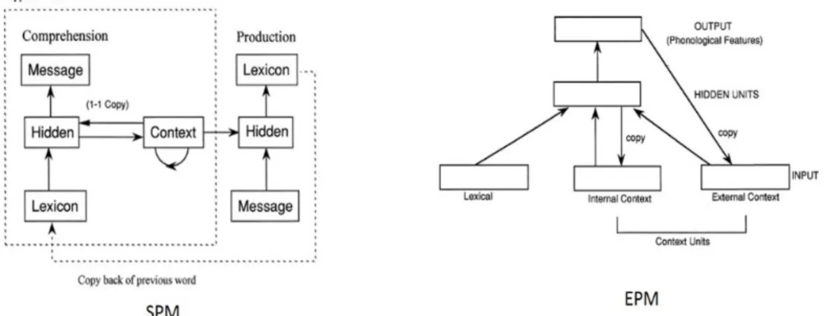
(2) Zambrano-Cruz, R., & Méndez-Ramírez, L.F.. PSICOLOGÍA. Pág 89. 2. Estudiante de Doctorado en Ciencias Cognitivas. Magíster en Neuropsicología Clínica. Psicóloga. Miembro de grupo de investigación “Neuroaprendizaje” de la Universidad Autónoma de Manizales.. Introduction. Anomia, nominal aphasia, or amnesic aphasia, has been described in the classic aphasia syndromes as an alteration in the production of words, this deficit is more prominent to nouns than for verbs, characterized by a preservation of understanding and repetition (Webb, 2017; Caplan, 2003; Fernández-Blázquez et. al., 2012). During naming process difficulties are in lexical access and lexical retrieval of word (Fernández-Turrado et al., 2006). The neurologic conditions causing aphasia and anomia are: stroke, dementia, epilepsy and traumatic brain injury (TBI) (Fridriksson, 2011; Laganaro, Di Pietro, & Schnider, 2006; Hoffmann & Chen, 2013; Lorenz and Ziegler, 2009). Additionally, anomia is one of the most common symptoms of patients with post-stroke aphasia (Van Hees, McMahon, Angwin, de Zubicaray, & Copland, 2014; Fridriksson et al., 2007). The neuroanatomical correlates are inferior parietal lobe, plus connections between parietal and temporal lobes (Caplan, 2003). It is estimated that one million people in the USA has an aphasia (The National Institute on Deafness and Other Communication Disorders –NIDCD-, 2015), and the prevalence of these is between 20% and 40% after a cerebrovascular disease around the world (Martínez, Reyes-Saborit, Turtós-Carbonell and Dusu -Contreras, 2014). One of the most common characteristics is the difficulty to find words, this is present in approximately 70% of aphasias, it even manages to configure an independent disorder (anomic aphasia). Dell, Chang and Griffin (1999) propose that the Parallel Distributed Processing models (PDP) are the best models to explain production of words and they integrate linguistic and no-linguistic features. In this models, structure and order are entwined in the sequential schemata that develop from the superimposed weight changes associated with the training set. Structural Priming Model and Phonological Error Model are both PDP Models. The first is about the grammatical encoding feature while the second is about the phonological component (See Figure 1).. Figure 1. Structural Priming Model (SPM) and Phonological Error Model (PEM) (each rectangle indicates a group of units). Source: Dell et al. 1999.. In its treatment, there have been evidence of changes in the neural activity in both hemispheres (Fridriksson et al., 2007). Fridriksson (2011) suggests that rehabilitation depends of the anomia subtype and the best results are seen in disorders adjusted to the specific language of each individual therapy. http://dx.doi.org/10.21615/cesp.11.2.8.
(3) Neuroanatomic Correlates of Anomia: A Scoping Review. PSICOLOGÍA. Pág 90. Treatment for anomia is an objective of the therapies of language and cognitive disorders. The more common difficulties in this kind of patients have trouble retrieving names of places, people, monuments, this impairment has an impact in everyday life such as in social, working and other contexts. Knowing more precisely the neurological components associated with anomia has two important implications, the first is to establish if the brain areas are more related to the phonological component (PEM) or the grammatical component (SPM); the second one would be to know areas or related brain networks that would allow to give indications for possible intervention strategies, both pharmacological and cognitive. This led to ask, what are the neuroanatomical correlates of anomia?. Metodology Design This study is developed through the methodology of scoping review (Levac, Colquhoun, & O’Brien, 2010; Pham et al., 2014). This review synthetized the literature on a particular topic and provide evidence on that field. Selection criteria The papers should be in English or Spanish, should be published between the years 2009-2016; and as the main criterion, the paper should establish neurological findings associated with anomia. Those items showing evaluation results of other language disorders, such as dyslexia or other aphasia were excluded. The data bases used were: EBSCOhost and Science Direct, using the keyword for the search terms “anomia” and “neurological”. The procedure was to search items in the database containing the previously named certain keywords, for which the criteria of inclusion and exclusion are considered. Was important that the articles were about research, not reviews or considerations about the topic. Analysis plan We considered the next components for the article analysis: - Author(s), year of publication and study location - Sample size - The analysis focus on the anatomic structure and cognitive functionality in accordance with the results of the studies reviewed. Ethical Standards The authors assert that all procedures contributing to this work comply with the ethical standards of the relevant national and institutional committees on human experimentation and with the Helsinki Declaration of 1975, as revised in 2008.. Results. After the bibliographic search, only 10 articles remained (See Figure 2).. http://dx.doi.org/10.21615/cesp.11.2.8.
(4) PSICOLOGÍA. Zambrano-Cruz, R., & Méndez-Ramírez, L.F. Pág 91. Figure 2. Diagram about the articles search strategy.. They highlight the importance of additional neuropsychological deficits to visual present in lesions of the aforementioned areas, also showing that anomia and some memory impairments are related to lesions in the lower occipital rotation in the lingual gyrus in hippocampus and injuries of some major white matter tracts.. When we reviewed all the articles, we found that many of them appeared based on the fact that the topic is common in journals and texts of neurology and neuropsychology, without an analysis of brain activity, focusing on the psychological characteristic and on the results of applications of neuropsychological tests and their relationship with other psychological or linguistic features. Of these few found texts, almost all are methodologically focused on magnetic resonance studies (Magnetic resonance imaging -MRI- and functional MRI –fMRI-) and only one of Evoked Related Potential (ERP) (See Table 1). About the last one, Neumann, Obler, Gomez and Schafer (2009) studied the brain activity to a phonological response. Thirty-two people divided into two age groups (young and old) were evaluated with a naming exercise and response times to different stimuli were obtained. They found that there are no substantial differences between the two groups, people got less time between 300ms and 450ms, seniors got time between 351ms and 500ms. The role of the phonological component is discussed in the response naming. The second group of articles found were about magnetic resonance imaging (MRI). In 2012, Kraft et. al. defined the anatomic correlates of neuropsychological dysfunctions using mapping symptoms and voxel-based lesion in 61 patients with cerebral stroke; patients underwent a neuro-ophthalmological, neurological and neuropsychological evaluation test and a brain MRI. They highlight the importance of additional neuropsychological deficits to visual present in lesions of the aforementioned areas, also showing that anomia and some memory impairments are related to lesions in the lower occipital rotation in the lingual gyrus in hippocampus and injuries of some major white matter tracts.. http://dx.doi.org/10.21615/cesp.11.2.8. In 2014 were published two papers, in the first one, Van Hees et al. (2014) established the relationship of anomia with neural activation in the context of a series of interventions to improve such neurolinguistic condition. Evaluated eight people diagnosed with speech pathology after stroke (Cerebrovascular Accident –CVA-); assessed language with Boston-Naming-Test (BNT) and battery name based on international project database naming photographs and for neuronal activation images were used a MRI. For the study, had fourteen healthy control patients with similar demographic characteristics. Before the intervention found that areas related to anomia were the superior frontal gyrus, the angular gyrus and the superior temporal gyrus and a half I left mainly.
(5) Neuroanatomic Correlates of Anomia: A Scoping Review. PSICOLOGÍA. Pág 92. hemisphere. While intervention shows changes in brain activation for the eight different patients, it was found that an improvement in the treatment is generally associated with an activation on the left supramarginal gyrus and the right precuneus. In the second one, Toumiranta et al. (2014) conducted a study with patients with extensive brain damage in the left temporal lobe and phonological impairment, in order to test the claim that the ability to learn and use new words depends on the left temporal lobe. Patients and controls results were compared. In MRI was observed in the left hemisphere a complete shutdown of the white matter of the arcuate fasciculus, which was confirmed by tractography. The remaining articles were about functional magnetic resonance imaging (fMRI), that uses a technology based on oxygen rich-blood. This articles were clasified in two, treatment and no treatment articles.. They found activation in the left superior temporal gyrus before the intervention; after the intervention activity was seen in the upper, middle and inferior temporal gyrus, plus activations were found in the fronto-medial turn and right turn supramarginal.. In the first group found studies about brain activity after therapy. Dressel et al. (2010) examined changes in brain activity associated with a treatment for anomia. With a unique case of a patient with semantic dementia they used different neuropsychological and neurolinguistic evidence, including the Naming Test and the BNT, in addition to conducting fMRI. They found activation in the left superior temporal gyrus before the intervention; after the intervention activity was seen in the upper, middle and inferior temporal gyrus, plus activations were found in the fronto-medial turn and right turn supramarginal. In the left hemisphery the middle frontal and cingulate gyrus were activated, also the putamen, caudate and thalamus. However, the authors emphasize that right temporal region, the superior temporal and inferior gyrus were more representative. In a similar study, Rochon et al. (2010) studied the characteristics of neural processing skills associated with access to the words after treatment for anomia. The research was made with two patients and ten controls were applied to different neuropsychological instruments, including the Boston-Naming-Test and the Philadelphia-Naming-Test. After the speech, the inferior frontal and middle temporal gyrus in the left hemisphere and the left cuneus the right precuneus and supramarginal turn in both hemispheres were activated. The authors discuss about the importance of phonological route in the recovery of anomia. Fridriksson, Richardson, Fillmore and Cai (2012) studied the treatment of anomia and the left hemisphere in 2010. In 2012 added more patients to the sample and improved methods of study. The study aimed to characterize precisely the changes in the left hemisphere taking into account a larger patient sample; plus: 1. Compare the role of perilesional cortex, and 2. Understand of how brain circulation and brain activation evaluated before starting treatment is associated with improvement in the naming after treatment. Thirty patients were administered the Western Aphasia Battery (WAB). Patients received treatment of anomia for three hours a day, half received phonological treatment using keys in the first week and then treated using semantic keys. Twenty-nine patients underwent fMRI and participated in naming tasks. Subsequently they conducted a multivariate statistical analysis and concluded that treatment increases brain activation in areas surrounding the injured cortical area while improving the naming, through modulation of the left frontal lobe, independent of the type of aphasia.. http://dx.doi.org/10.21615/cesp.11.2.8.
(6) PSICOLOGÍA. Zambrano-Cruz, R., & Méndez-Ramírez, L.F. Pág 93. In the second group we found two studies, Bonelli et al (2011) researched the correlation of hippocampal activation with visual confrontation naming in patients with temporal lobe epilepsy; made a retrospective investigation of the relationship between the name and integrity of the networks of language using fMRI with simple tasks of verbal fluency. Included 22 healthy subjects and 66 patients with temporal lobe epilepsy candidates for surgery, all with preoperative evaluation and pharmacological treatment; the test were McKenna naming, verbal fluency test, and underwent structural and fMRI. They demonstrated the importance of the hippocampus in the denomination key to patients and controls. In 2009, Mesulam et al. studied the brain areas related to anomia. They studied seven patients (four women) with a part of the Peabody Picture Vocabulary Test (PPVT-IV) and functional MRI. Founded an atrophy in the left inferior temporal orders, upper half, the fusiform gyrus and the anterior parahippocampal gyrus. Finally, Hoffmann and Chen (2013) studied the spectrum of aphasia, subtypes and etiology after subacute stroke. Of the 625 patients who met the selection criteria, 165 patients had anomic aphasia, this corresponds to 26.40%, making it the second most common after Broca’s aphasia (27.20%). They found that anomia occurred most commonly in small vessel disease (subcortical level), specifically in the thalamus, corona radiata and the globus pallidus. They concluded that aphasia subtypes are heterogeneous in etiology and could be useful to adopt a new taxonomy of aphasia based on independent component analysis, which may be based on studies with fMRI. Table 1. Findings of studies about neurological correlates of anomia Authors. Country. Sample size. Relevant findings. Dressel et al (2010). Germany. 1. The left superior temporal gyrus activated without intervention. Right superior and inferior temporal gyrus activated after intervention.. Hees et al (2014). Australia. 8. Superior frontal gyrus, angular gyrus and superior left temporal gyrus activated without intervention. Left supramarginal gyrus and right precuneus activated after intervention.. Rochon et al (2010). Canada. 2. Activation of the left inferior frontal gyrus, left middle temporal gyrus, left cuneus, turn right precuneus and supramarginal in both hemispheres.. Neumann, Obler, Gomes & Shafer (2009. United States. 32. No significant differences in evoked potentials in naming among young people and seniors.. Hoffmann and Chen (2013). United States. 625. Some stroke patients presented anomia. MRI showed lesions in subcortical thalamus, white matter, corona radiata and globus pallidus.. Toumiranta et al (2014) Multicentric. 1. Left temporal lobe and the arcuate fasciculus.. Fridriksson, Richardson, Fillmore & Cai (2012). United States. 30. Brain changes after treatment of anomia are associated with increased in left frontal activity and perilesional activity.. Bonelli et al (2011). Germany and United Kingdom. 22controls 66patients. Controls and patients of left temporal lobe epilepsy showed activity in the hippocampal fMRI when performing verbal fluency and naming and left medial frontal activation and lower in verbal fluency tasks.. Mesulam et al (2009). United States. 7. Changes in temporal gyrus, fusiform gyrus and parahippocampal gyrus. Everything in the left hemisphere.. Kraft et al (2014). Germany and United Kingdom. 128. Anomia and memory impairments were related to injuries in the lower occipital and lingual gyrus and hippocampus.. http://dx.doi.org/10.21615/cesp.11.2.8.
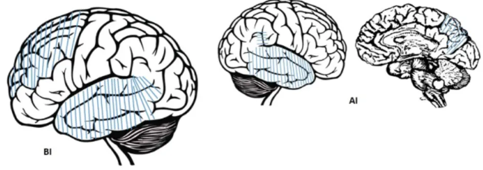
(7) PSICOLOGÍA. Neuroanatomic Correlates of Anomia: A Scoping Review Pág 94. The main findings were that prior language intervention the brain areas implicated are the temporal and frontal gyrus of the left hemisphere. After intervention, the activation is in the temporal gyrus, precuneus and the supramarginal gyrus of the right hemisphere (See Figure 3).. Figure 3. Cortex related to lexical access before (BI) and after an intervention (AI).. Discussion. According to the findings published in recent years, there is evidence for differential naming tasks: before and after non-pharmacological intervention.. Related to the time when the damage that generates anomia occurred, an activation of temporary gyrus and frontal gyrus in the left hemisphere was found. This evidence suggests that anomia is not dependent on specific structures, since these areas are also related to memory tasks.. Related to the time when the damage that generates anomia occurred, an activation of temporary gyrus and frontal gyrus in the left hemisphere was found. This evidence suggests that anomia is not dependent on specific structures, since these areas are also related to memory tasks. These findings are like those reported in the literature, but expand understanding of related areas since reportedly are the areas 39 and 40 of Brodmann those involved in the process of anomia (Montañés & de Brigard, 2001). These findings show the importance of the connections in the left hemisphere in temporal and frontal areas in anomia. It is also evidence related to the neurological processes in naming tasks after a non-pharmacological intervention. Then it found that after surgery the temporal gyrus, that the right precuneus, cuneus and supramarginal gyrus are active. This would show that connections between areas that link the sensory, memory and executive functions of the right hemisphere occur in the naming recovery. This evidence implies that naming is a complex process that involves primary, secondary structures and its connections in a multimodal manner. This categorization shows the similarity in recognition and motor interaction. Probably in language development in childhood same areas are activated due to sensory multimodality, then other areas leading to the consolidation and specialization of the areas in the left hemisphere. This evidence supports the PDP models (Dell et al., 1999). This would be an indication of processes compensation, restitution and replacement of the right hemisphere when intervention.. http://dx.doi.org/10.21615/cesp.11.2.8. Such evidence would give us two lines: 1. First, the naming and its alteration could be a linguistic marker for the presence of Alzheimer’s Disease. In turn, detection of abnormalities in these brain areas could help the remediation of functional alterations in Alzheimer’s Disease (Cuetos-Vega, Arango-Lasprilla, Uribe-Pérez, Valencia-Marín, & Lopera-Restrepo, 2007)..
(8) Zambrano-Cruz, R., & Méndez-Ramírez, L.F.. PSICOLOGÍA. Pág 95. 2. Second, pharmacological and non-pharmacological stimulation centered interventions in temporal gyrus, precuneus, cuneus and supramarginal gyrus. This could end up slowing the progression of impairment associated with Alzheimer’s disease. Other dementias such as fronto-temporal could be affected positively to the patient, interventions in this regard. Limitations The information about the anomia is subject to the presence of aphasia, and in some studies it is not specified about the characteristics of the aphasias, much less the anomia. Another problem lies in the methods used, since in some studies the MRI is used and in others the fMRI, we suggest that in future studies more robust methods such as fMRI should be considered. Finally, there are several strategies for measuring the denomination and the lexicon, such as the BNT or the PPVT, this could cause problems in the conclusions of the different anomia studies. The number of articles included in the study draws attention, this shows a low production in these issues in recent years, possibly because the rise of research in this subject in previous years.. References. http://dx.doi.org/10.21615/cesp.11.2.8. Bonelli, S. B., Powell, R., Thompson, P. J., Yogarajah, M., Focke, N. K., Stretton, J., ... Koepp, M. J. (2011). Hippocampal activation correlates with visual confrontation naming: fMRI findings in controls and patients with temporal lobe epilepsy. Epilepsy research, 95(3), 246-254. doi: https://doi.org/10.1016/j.eplepsyres.2011.04.007 Caplan D., (2003). Aphasic Syndromes. In Heilman, M. K. M., & Valenstein, E. (Eds.). (2011). Clinical neuropsychology. Oxford University Press. Cuetos-Vega, F., Arango-Lasprilla, J., Uribe-Pérez, C., Valencia-Marín, C., & Lopera-Restrepo, F. (2007). Linguistic Changes in verbal expression: a preclinical marker of Alzheimer’s Disease. Journal of the International Neuropsychological Society, 13, 433-439. doi: https://doi.org/10.1017/S1355617707070609 Dell, G., Chang, F., & Griffin, Z. (1999). Connectionist Models of Language Production: Lexical Access and Grammatical Encoding. Cognitive Science, 23(4), 517-542. doi: https://doi.org/10.1016/S0364-0213(99)00014-2 Dressel, K., Huber, W., Frings, L., Kümerer, D., Saur, D., Mader, I., … Abel, S. (2010). Modeloriented naming therapy in semantic dementia: A single-case fMRI study. Aphasiology, 24(12), 1537-1558. doi: https://doi.org/10.1080/02687038.2010.500567 Fernández-Blázquez, M. A., Ruiz-Sánchez de León, J. M., López-Pina, J. A., Llanero-Luque, M., Montenegro-Peña, M., & Montejo-Carrasco, P. (2012). Nueva versión reducida del test de denominación de Boston para mayores de 65años: aproximación desde la teoría de respuesta al ítem. Rev Neurol, 55, 399-407. Fernández-Turrado, T., Tejero-Juste, C., Santos-Lasaosa, S., Pérez-Lázaro, C., Piñol-Ripoll, G., Mostacero-Miguel, E., & Pascual Millán, L. F. (2006). Lenguaje y deterioro cognitivo: un estudio semiológico en denominación visual. Rev Neurol, 42, 578-83. Fridriksson, J. (2011). Measuring and inducing brain plasticity in chronic aphasia. Journal of communication disorders, 44(5), 557-563. doi: https://doi.org/10.1016/j. jcomdis.2011.04.009 Fridriksson, J., Morrow-Odom, L., Moser, D., Fridriksson, A., & Baylis, G. (2006). Neural recruitment associated with anomia treatment in aphasia. Neuroimage, 32(3), 1403-1412. doi: https://doi.org/10.1016/j.neuroimage.2006.04.194 Fridriksson, J., Moser, D., Bonilha, L., Morrow-Odom, K. L., Shaw, H., Fridriksson, A., ... Rorden, C. (2007). Neural correlates of phonological and semantic-based ano-.
(9) Neuroanatomic Correlates of Anomia: A Scoping Review. PSICOLOGÍA. Pág 96. mia treatment in aphasia. Neuropsychologia, 45(8), 1812-1822. doi: https://doi. org/10.1016/j.neuropsychologia.2006.12.017 Fridriksson, J., Richardson, J. D., Fillmore, P., & Cai, B. (2012). Left hemisphere plasticity and aphasia recovery. Neuroimage, 60(2), 854-863. doi: https://doi. org/10.1016/j.neuroimage.2011.12.057 Hoffmann, M., & Chen, R. (2013). The spectrum of aphasia subtypes and etiology in subacute stroke. Journal of Stroke and Cerebrovascular Diseases, 22(8), 13851392. doi: https://doi.org/10.1016/j.jstrokecerebrovasdis.2013.04.017 Kraft, A., Grimsen, C., Kehrer, S., Bahnemann, M., Spang, K., Prass, M., ... Brandt, S. A. (2012). Neurological and neuropsychological characteristics of occipital, occipito-temporal and occipito-parietal infarction. Cortex, 56, 38-50. doi: https://doi. org/10.1016/j.cortex.2012.10.004 Laganaro, M., Di Pietro, M., & Schnider, A. (2006). What does recovery from anomia tell us about the underlying impairment: The case of similar anomic patterns and different recovery. Neuropsychologia, 44(4), 534-545. doi: https:// doi.org/10.1016/j.neuropsychologia.2005.07.005 Levac, D., Colquhoun, H., & O’Brien, K. (2010). Scoping studies: advancing the methodology. Implementation Science, 5(69). https://doi.org/10.1186/1748-5908-5-69 Lorenz, A., & Ziegler, W. (2009). Semantic vs. word-form specific techniques in anomia treatment: A multiple single-case study. Journal of Neurolinguistics, 22(6), 515-537. doi: https://doi.org/10.1016/j.jneuroling.2009.05.003 Martínez, E., Reyes-Saborit, A., Turtós-Carbonell, L., & Dusu-Contreras, R. (2014). Epidemiología de la afasia en Santiago de Cuba. Neurología Argentina, 6(2), 7782. doi: https://doi.org/10.1016/j.neuarg.2013.12.002 Mesulam, M., Rogalski, E., Wieneke, C., Cobia, D., Rademaker, A., Thompson, C., & Weintraub, S. (2009). Neurology of anomia in the semantic variant of primary progressive aphasia. Brain, 132, 2553-2565. doi: https://doi.org/10.1093/brain/awp138 Montañés, P., & de Brigard, F. (2001). Neuropsicología clínica y cognoscitiva. Bogotá: Universidad Nacional de Colombia. National Institute on Deafness and Other Communication Disorders. (2015). NIDCD fact sheet: Aphasia [PDF] [NIH Pub. No. 97-4257]. Retrieved from https://www. nidcd.nih.gov/sites/default/files/Documents/health/voice/Aphasia6-1-16.pdf Neumann, Y., Obler, L., Gomes, H., & Shafer, V. (2009). Phonological vs sensory contributions to age effects in naming: An electophysiological study. Aphasiology, 23(7-8), 1028-1039. doi: https://doi.org/10.1080/02687030802661630 Pham, M. T., Rajic, A., Greig, J. D., Sargeant, J. M., Papadopoulos, A., & McEwen, S. A. (2014). A scoping review of scoping reviews: advancing the approach and enhancing the consistency. Research Synthesis Methods, 5, 371 – 385. doi: https:// doi.org/10.1002/jrsm.1123 Rochon, E., Leonard, C., Burianova, H., Laird, L., Soros, P., Graham, S., & Grady, C. (2010). Neural changes after phonological treatment for anomia: An fMRI study. Brain & Language, 114, 164-179. doi: https://doi.org/10.1016/j.bandl.2010.05.005 Tuomiranta, L. M., Càmara, E., Froudist Walsh, S., Ripolles, P., Saunavaara, J. P., Parkkola, R., ... Laine, M. (2014). Hidden word learning capacity through orthography in aphasia. Cortex, 50, 174-191. doi: https://doi.org/10.1016/j.cortex.2013.10.003 Van Hees, S., McMahon, K., Angwin, A., de Zubicaray, G., & Copland, D. (2014). Neural activity associated with semantic versus phonological anomia treatments in aphasia. Brain & Language, 129, 47-57. doi: https://doi.org/10.1016/j.bandl.2013.12.004 Webb, W. (2017). Neurology for the Speech-Language Pathologist (Sixth edition). Missouri: Elsevier. doi: https://doi.org/10.1016/B978-0-323-10027-4.12001-9. http://dx.doi.org/10.21615/cesp.11.2.8.
(10)
Figure

Documento similar
The expansionary monetary policy measures have had a negative impact on net interest margins both via the reduction in interest rates and –less powerfully- the flattening of the
Although some public journalism schools aim for greater social diversity in their student selection, as in the case of the Institute of Journalism of Bordeaux, it should be
In the “big picture” perspective of the recent years that we have described in Brazil, Spain, Portugal and Puerto Rico there are some similarities and important differences,
Table 2 shows the descriptive statistics (M ± SD) of the success rate and reaction time for each group in the laboratory anticipation test, before and after the
Keywords: Metal mining conflicts, political ecology, politics of scale, environmental justice movement, social multi-criteria evaluation, consultations, Latin
Table VII shows the results of the five tests assessing visual object and visuospatial skills. The cognitively preserved group did not show statistically significant differences
The results showed more pronounced activation of facial expressions of emotion than neutral faces in the left cuneus, right superior temporal gyrus, right cingulate gyrus,
Even though the 1920s offered new employment opportunities in industries previously closed to women, often the women who took these jobs found themselves exploited.. No matter
