Hepatitis E : latest developments in knowledge / M Teresa Pérez Gracia, Beatriz Suay García, Mario García & M Luisa Mateos Lindemann
20
0
0
Texto completo
(2) Review Pérez-Gracia, Suay-García, García & Mateos-Lindemann History of hepatitis E The first epidemic attributed to HEV occurred during 1955 [6] in New Delhi (India) when approximately 30,000 individuals fell ill due to a supposed hepatitis A infection after consuming contaminated water. However, it was not until 1980 when it was observed that they did not react against HAV antigens [7] , thus this epidemic was attributed to a new etiological agent. In 1983, a volunteer was experimentally infected by ingesting a mixture of extracts obtained from an individual who suffered the disease in the 1955 epidemic in Kyrgyzstzan [8] . Viral particles, 27–34 nm in size, were observed in the feces. This observation was confirmed in patients from distant geographical regions, all of them affected by hepatitis no-A no-B [9] . In 1989, the first cynomolgus macaque monkeys were effectively infected [10] . In 1990, HEV RNA was detected in clinical samples of affected individuals in Burma, Mexico, Somalia and Pakistan [11] . Experimental infection of pigs was also performed, achieving the excretion of the virus in feces [12] . This new agent was named hepatitis E virus. The letter ‘E’ makes reference to the enteric, endemic and epidemiologic characteristics of the disease. In 1996, a strain different from those known at the time was isolated in an American individual with no prior history of trips overseas [13] . This fact, along with the emergence of a great number of hepatitis E cases in developed countries considered nonendemic, such as Holland [14] , Japan [15] and Spain [16] , suggested the existence of animal reservoirs. Consequently, many seroprevalence studies were performed in different animal species, for example, pigs [17–19] , poultry [20] and rodents [21] . In 1997, the first HEV strain of porcine origin was isolated in the USA [22] , with only a 74% of resemblance in the nucleotide sequence when compared with the classic strains isolated in Burma and Mexico, but with a resemblance of 90% when compared with the human isolates of the same region. Since then, there have been many cases of porcine strains isolated in industrialized countries, such as Spain, The Netherlands and the UK [23] . Viral particle HEV is a nonenveloped virus with an icosahedral symmetry and size between 32 and 34 nm in diameter. The buoyant density of HEV is. 10.2217/fmb-2016-0012. Future Microbiol. (Epub ahead of print). between 1.39 and 1.40 g/cm3 in CsCl and its sedimentation coefficient is 183S [24] . HEV genome is between 6.6 and 7.3 kb long and comprises a single-stranded positive polarity RNA molecule showing three open reading frames (ORFs) [25] . The ORF1 is the longest reading frame at the 5′-end of the genome. The ORF1 encodes a nonstructural polyprotein including a methyltransferase, a Y-domain, a papain-like protease, a polyproline region (hypervariable region), a macro-domain, a helicase and an RNA-dependent RNA polymerase. ORF2 is located at the 3′-end of the genome and encodes the major capsid protein. ORF3 overlaps partially with ORF2. It encodes a phosphoprotein that modules cellular activities [26] . All ORFs are expressed during viral infection since antibodies against these regions have been detected in naturally infected humans and in experimentally infected monkeys [27] . Additionally, there are two untranslated regions in 3′- and 5′-terminal portions. Classification Although a single serotype has been identified, a great genetic diversity between the different HEV isolates has been widely reported [28] . Recent studies have proposed several classifications of HEV in different genotypes and subtypes [29,30] . The International Committee of Taxonomic Virology, in its last meeting that took place in July 2014 in Montreal, Canada [31] , classified HEV into the family Hepeviridae (Table 1) . This family is divided in two genera, Orthohepevirus (all mammalian and avian HEV isolates) and Piscihepevirus (cutthroat trout HEV). Species within the genus Orthohepevirus are designated Orthohepevirus A (isolates from human, pig, wild boar, deer, mongoose, rat, rabbit and camel), Orthohepevirus B (isolates from chicken), Orthohepevirus C (isolates from greater bandicoot, Asian musk shrew, ferret and mink) and Orthohepevirus D (bat isolates). Within species Orthohepevirus A, there are currently four genotypes described that infect humans (G1, G2, G3 and G4) and three genotypes (G5, G6 y G7) that affect wild boards and camels [32] . Two genotypes exist within the genus Orthohepevirus C, C1 has been detected in greater bandicoot and Asian musk shrew and genotype C2, detected in ferret and mink [33,34] . The cutthroat trout HEV isolate has been classified in the genus Piscihepevirus A. This isolate exhibits the characteristic three ORFs. future science group.
(3) Hepatitis E: latest developments in knowledge. Review. Table 1. Classification of the family Hepeviridae. Family. Genus. Species. Genotype. Source. Hepeviridae. Orthohepevirus. Orthohepevirus A. 1 2 3. Human Human Human, swine, mongoose, rabbit, deer, rat Human, swine Wild board Wild board Camel Avian Greater bandicoot, Asian musk shrew Ferret, mink Bat Cutthroat trout. Piscihepevirus. Orthohepevirus B Orthohepevirus C. 4 5 6 7 – C1. Orthohepevirus D Piscihepevirus A. C2 – –. genome arrangement seen among avian and mammalian members of the family Hepeviridae. Additionally, motifs characteristics for methyltransferase, papain-like protease, macro domain, helicase and replicase are found in the cutthroat trout virus ORF1. However, amino acid sequence similarities between cutthroat trout virus and others members of this family are only 26–27% (ORF1), 18–21% (ORF2) and 13–16% (ORF3). By contrast, these similarities are 42–49% (ORF1), 42–55% (ORF2) and 20–29% (ORF3) between avian, rat and mammalian Hepeviridae [31,35] . Cell culture models of HEV replication The molecular mechanisms of HEV replication are not entirely understood, mostly because there are no efficient cell culture systems. The first cell culture model for G3 and G4 of HEV that showed optimal results was developed using an inoculum with high titer of virus from serum and feces, diffused efficiently in PLC/PRF/5 cells derived from human hepatocarcinoma and A549 cells obtained from lung cancer (Table 2) [36–38] . Although A549 cells are not hepatocytes, it was found that the virus was able to replicate in other tissues of infected pigs, such as kidney, liver, spleen, tonsils and colon [39] . Subsequently, in 2009, Tanaka et al. [38] designed a cell culture with G4 strains HE/JF5/15F isolated from the feces of a patient with fulminant hepatitis E, which grew more efficiently in PLC/PRF/5 and A549 cells than the strain A549 JE03-1760F [44] . Thus, this model has been proposed to study the factors related to fulminant hepatitis.. future science group. Another study [40] demonstrated that it was possible to grow the G3 KernowC1 strain, isolated from the feces of an infected patient, in human hepatoma cells HepG2/C3A. This strain contains a recombinant insert in the ORF1 region of the viral genome of 174 nucleotides of a human gene for a ribosomal protein, which apparently facilitated the adaptation to cell culture [45] . Moreover, several studies have focused on getting spread of HEV strains isolated from pigs in human cells, and vice versa, to obtain information about the zoonotic potential of the virus, the mechanisms that facilitate cross-species infection and help to elucidate the replicative cycle of the virus. Thus, one study showed that HEV G3 could infect LLC-PK1 pig cells more efficiently than human cells HepG2/C3A [40] . In that sense, it also described that porcine hepatitis E G4 strains could replicate in human A549 cells [46] . Additionally, in 2012, Rogée et al. [41] developed two cell culture models HepaRG from human hepatoma cells and PICM-19 cell line from pig embryonic stem cells. Recently, a new in vitro cell culture model has been performed with HEV G3 and G4 strains (G3jp, G3US, G3sp and G4jp) isolated from the feces of naturally infected pigs [42] , in PHCs cells (primary human hepatocytes). ●●Life cycle of HEV. The mechanism by which the virus penetrates the cell depends largely on the type of viral structure; big differences exist between enveloped viruses and nonenveloped viruses, such. www.futuremedicine.com. 10.2217/fmb-2016-0012.
(4) Review Pérez-Gracia, Suay-García, García & Mateos-Lindemann Table 2. Cell lines used for propagation of different hepatitis E virus strains. Cell line. Origin. Strain (genotype, origin). PLC/PRF/5 A549 HepG2/C3A. Human liver hepatoma Human lung carcinoma Human hepatocellular carcinoma. JE03-1760F (G3, H) HE/JF5/15F (G4, H). LLC-PK1 Hepa-RG PICM-19 PHCs S10-3. Swine kidney epithelial Human hepatocytes Pig liver stem Primary human hepatocytes Human hepatocellular carcinoma. Ref. [36–38]. KernowC1 (G3, H) SAR55 (G1, H) KernowC1 (G3, H) GenBank no JN906976 (G3f, S). [40]. G3jp (G3, S), G3US (G3, S), G3sp (G3, S), G4jp (G4, S) SAR55 (G1, H). [42]. [40] [41]. [43]. H: Human; S: Swine.. as HEV. Furthermore, the site of replication of HEV RNA, is located in the cytoplasm, unlike DNA viruses that typically replicate in the cell nucleus (Figure 1) [47] . ●●HEV binding to cell process. The specific life cycle of HEV is largely unknown due to the lack of an efficient cell culture system. In-depth knowledge of the capsid protein encoded by ORF2 has allowed the study of HEV interactions with target cells. It is believed that the C-terminal region of the ORF2 plays an important role in this stage of HEV life cycle [48] . This protein region first interacts with specific receptors on the cell membrane, including: heparan sulfate proteoglycans [49] and heat-shock protein cognate 70, facilitating virus binding to the hepatocyte. A recent study [50] performed with HEV-like particles, showed how the entrance of HEV into Huh-7 hepatoma cells is associated with endocytosis-mediated GTPase Dynamin-2, playing an essential role in the cleavage of plasma membrane to form Clathrin-coated vesicles. Additionally, the process involves the cholesterol membrane, clathrin and actin (clathrinmediated endocytosis), suggesting that macropinocytosis mechanisms are not involved in this process. ●●Intracellular trafficking. HEV enters the cell through endosomes and replicates in the cytoplasm for subsequent release. This process is pH-independent, unlike other viruses that require acidic pH conditions [51,52] . The heat-shock 90 and Grp78 proteins bind to the cell membrane once the virus contacts the cell, participating in the intracellular plasma transportation of viral capsids in the early stages of infection with HEV [53,54] .. 10.2217/fmb-2016-0012. Future Microbiol. (Epub ahead of print). ●●Replication. Once the RNA is released from the virion to the cell cytoplasm, translation of ORF1 nonstructural polyprotein occurs [55] . It is not yet confirmed if proteolytic processes occur or not. An RNA-dependent RNA polymerase synthesizes intermediate negative polarity copies from positive polarity RNA [56,57] . These serve as a template to synthesize new copies of viral RNA. The RNA-dependent RNA polymerase interacts with cis-reactive elements (CRE) in the 3′NCR of the genome of the HEV, which could have an important role in HEV replication. Moreover, ORF2 and ORF3 proteins are translated in the cytoplasm. ORF2 proteins with new copies of viral RNA are assembled to form new virus particles. In this regard, it is thought that the C-terminal region of viral capsid is essential for encapsidation and particle stabilization [48] , as suggested by a study of mutant strains of HEV for the region C-terminal 52 amino acids, in which premature signal STOP was artificially generated. This prevents the correct synthesis of this domain, which apparently protects the particle of C-terminal degradation. A recent study [58] conducted a mutagenic analysis in the active site of ‘X domain’ suggests that X-domain residues intervene decisively in HEV replication. However, additional studies are required to confirm this relationship with the pathogenesis of the virus. ●●Egression stage. New viral particles assembled in the cytoplasm are transported to the cell membrane; the ORF3 protein facilitates the process [59] . In the release process of new viral particles through the membrane of the infected cell, the action of PSAP (Pro, Ser, Ala and Pro) motif in ORF3 protein [60] is important. This protein is highly. future science group.
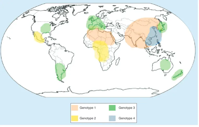
(5) Hepatitis E: latest developments in knowledge conserved in all the HEV strains isolated from mammals and poultry. Finally, the particles leave the cell and are released into the environment. In this last phase, it has been seen that the PSAP motif of the ORF3 protein interacts with the Tsg101, which is a cellular protein linked to the process of egress of other viruses, such as HIV [59,61] . The Tsg101 is a required component for the transportation related to the mechanism of egress associated with multivesicular bodies. Thus, it is suggested that HEV uses multivesicular bodies pathway to exit the cell. Epidemiology Hepatitis E infection is an increasingly common cause of acute hepatitis, especially in developing countries, such as India or Bangladesh, where robust sanitation systems and hygiene are more required. HEV infection is found worldwide, with East and South Asia having the highest prevalence rates [62] . Different studies have shown that HEV has spread globally [63] , but the genotypes found vary depending on the geographical region. G1 (Central and South Asia and North Africa) and G2 (West Africa. Review. and Mexico) are predominant in developing countries, whereas G3 is mostly found in the American continent and Europe, and G4 is most common in China (Figure 2) . Regardless of the global distribution, the majority of the disease burden is in less developed countries. According to WHO, there are approximately 20 million hepatitis E infections every year, causing over 3 million acute cases of hepatitis and 56,600 hepatitis E-related deaths [1] . Types of presentation (sporadic vs epidemic) Infection with HEV has two types of presentations, as endemic outbreaks or as sporadic events. Whereas sporadic hepatitis E occurs worldwide, outbreaks generally appear in developing countries and are mainly due to water sources that are contaminated with fecal matter [8] . These endemics become more frequent after natural disasters or in overcrowded refugee camps. Between one and 15% of the population is usually affected when an outbreak of hepatitis E occurs in a community [64] . HEV infection is considered endemic in India, Central Asia,. HEV particles Released virions. Cell membrane HSPGs HS70 Golgi Cytoplasm RNA Transcription. ER. Viral genomes. ORF1 proteins Assembly Translation. Nucleus. ORF2 proteins ORF3 proteins. Figure 1. Hepatitis E virus replicative cycle.. future science group. www.futuremedicine.com. 10.2217/fmb-2016-0012.
(6) Review Pérez-Gracia, Suay-García, García & Mateos-Lindemann. Genotype 1. Genotype 3. Genotype 2. Genotype 4. Figure 2. Geographic distribution of the hepatitis E virus genotypes affecting humans.. Africa and Mexico. India is one of the countries where HEV is responsible for a high percentage of acute viral hepatitis cases, accounting for approximately 40% according to CDC estimates. In fact, the first identified epidemic outbreak of HEV took place in New Delhi in 1955 [65] . The most recent epidemic took place in 2014, in Biratnagar, eastern Nepal, due to a contaminated water supply [66] . Epidemic outbreaks registered in each continent from 1955 to the present day are described in Table 3. Historically, HEV infections in industrialized countries were connected to patients that had traveled to endemic areas [78–80] , but recent studies found locally acquired HEV infections in these nonendemic countries that prove this theory wrong. G3 and G4 are responsible for autochthonous cases in nonendemic countries, such as the USA, Japan or European countries [81] . In these countries, the most probable transmission route for these sporadic cases is the contact with animals that act as reservoirs. This could include both, cattle handlers and veterinarians who work with pigs, and people. 10.2217/fmb-2016-0012. Future Microbiol. (Epub ahead of print). who ingest raw or undercooked meat from these infected animals [82] . Transmission routes As with genotype distribution, there is also a remarkable difference in transmission routes between geographical areas (Figure 3) . The most predominant transmission route in developing countries is the fecal–oral route; these countries have deficient hygienic conditions, which increase the contamination of food and, especially, drinking water [81] . Generally, contamination occurs after torrential rains, floods or natural disasters, when consumption water is mixed with animal and human sewage [83] . This is the main transmission route for G1 and G2, which can only be acquired person-to-person. Other potential human-to-human transmission routes include sexual transmission and in utero vertical transmission. In utero transmission has been described in several studies [84,85] . Mother-to-child transmission rates vary between 23.3 and 50% depending on the study [85] , and. future science group.
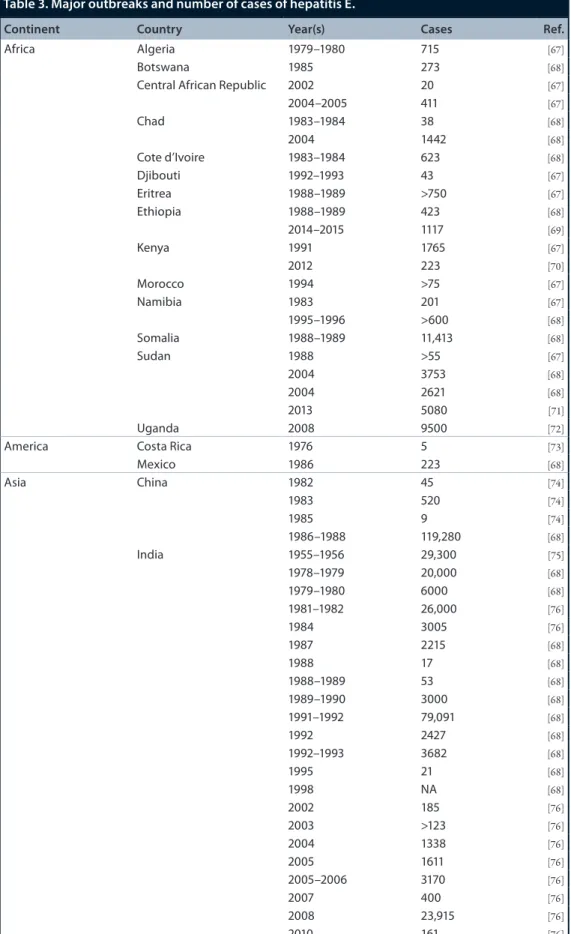
(7) Hepatitis E: latest developments in knowledge. Review. Table 3. Major outbreaks and number of cases of hepatitis E. Continent. Country. Year(s). Cases. Africa. Algeria Botswana Central African Republic. 1979–1980 1985 2002 2004–2005 1983–1984 2004 1983–1984 1992–1993 1988–1989 1988–1989 2014–2015 1991 2012 1994 1983 1995–1996 1988–1989 1988 2004 2004 2013 2008 1976 1986 1982 1983 1985 1986–1988 1955–1956 1978–1979 1979–1980 1981–1982 1984 1987 1988 1988–1989 1989–1990 1991–1992 1992 1992–1993 1995 1998 2002 2003 2004 2005 2005–2006 2007 2008 2010. 715 273 20 411 38 1442 623 43 >750 423 1117 1765 223 >75 201 >600 11,413 >55 3753 2621 5080 9500 5 223 45 520 9 119,280 29,300 20,000 6000 26,000 3005 2215 17 53 3000 79,091 2427 3682 21 NA 185 >123 1338 1611 3170 400 23,915 161. Chad Cote d’Ivoire Djibouti Eritrea Ethiopia Kenya Morocco Namibia Somalia Sudan. America Asia. Uganda Costa Rica Mexico China. India. future science group. Ref. [67] [68] [67] [67] [68] [68] [68] [67] [67] [68] [69] [67] [70] [67] [67] [68] [68] [67] [68] [68] [71] [72] [73] [68] [74] [74] [74] [68] [75] [68] [68] [76] [76] [68] [68] [68] [68] [68] [68] [68] [68] [68] [76] [76] [76] [76] [76] [76] [76] [76]. www.futuremedicine.com. 10.2217/fmb-2016-0012.
(8) Review Pérez-Gracia, Suay-García, García & Mateos-Lindemann Table 3. Major outbreaks and number of cases of hepatitis E (cont.). Continent. Country. Year(s). Cases. Ref.. Asia (cont.).. Indonesia Kyrgyzstan. 1998 1955 1987–1989 1982 1999 1981 1986 1987 1998 2000–2001 1985 1994. >600 NA >500 399 111 4337 85 133 109 18 16,175 150. [68]. Myanmar Nepal Pakistan. Turkmenistan Vietnam. are most frequent in the third trimester of pregnancy. In one of these studies, premature births and increased prenatal mortality were observed in a group of infected pregnant women [86] . Homosexual males show a higher prevalence of HEV antibodies (20%) than the general population, thus it has been suggested that sexual intercourse is a viable transmission route, however, further data are required [87] . In industrialized countries, hepatitis E is most commonly identified as a zoonotic disease, with swine being the main reservoir. In these countries, the infection can be acquired in one of two different ways: direct contact with infected animals (veterinarians and cattle handlers) or the ingestion of foods that have been in contact with infected animals or feces. Pig farmers, veterinarians, slaughterhouse workers and farmers have shown a high prevalence of HEV antibodies [88] . Infection through the consumption of undercooked or raw animal meat is also common in Europe and the USA, so much so that some epidemiological studies have identified the consumption of game and swine meat as a risk factor for the infection with HEV [89,90] . Bearing in mind that animal sewage can also contaminate water in developed countries, it is not surprising that the consumption of raw seafood could also lead to HEV infection. This was the case of a hepatitis E outbreak that took place in 2008 during a cruise [91] . The concept of hepatitis E as a food-borne disease is particularly strong in western Europe, where the food chain is the main source of infection [92] . Awareness of transfusion-transmitted HEV is increasing exponentially as more cases are retrospectively identified in both industrialized and developing countries. The first proven. 10.2217/fmb-2016-0012. Future Microbiol. (Epub ahead of print). [68] [76] [68] [68] [68] [68] [68] [68] [68] [68] [77]. hepatitis E transmission through transfusion therapy in a developed country took place in Hokkaido (Japan) in 2002, when a group of researchers showed an identical sequence of G4 HEV RNA genome in a blood donor and a patient who had received his plasma unit during open-heart surgery [93] . After several other cases with the same characteristics were identified, inhouse HEV RNA testing has been implemented in Japan in addition to blood donor screening for elevated ALT levels. Europe’s first cases of post-transfusion HEV infections were reported in the UK [94] and France [95] during 2006 and 2007, respectively. Many studies show that any blood product (red blood cells, platelets, fresh frozen plasma, etc.) can transmit HEV, however, the viral load required to induce symptoms is unclear [96] . Clinical manifestations HEV infection may cause a wide range of clinical presentations from subclinical or asymptomatic forms to fulminant liver failure [97,98] . However, acute hepatitis is the most common presentation in both industrialized and developing countries, although the mortality rate is much higher in the latter. Chronic hepatitis has also recently been described but only in infection with HEV G3. Common features are: ●● The incubation period is approximately. 40 days, ranging from 2 to 10 weeks; ●● Viremia, being transitory, occurs primarily. during the preicteric phase and disappears with the development of clinical symptoms, except in cases of chronic hepatitis E;. future science group.
(9) Hepatitis E: latest developments in knowledge ●● Fecal excretion of the virus begins around. Review. may play a role. On the other hand, in endemic areas, the large majority of patients are young adults from 10 to 40 years. The reason for that remains unclear, although it may be related to major exposure to contaminated water [100] . The clinical features in endemic areas are indistinguishable from those in industrialized countries. During pregnancy, the number of cases of severe disease and mortality caused by G1 or G2 is very high, approximately 15–20%. Complications in the mother, such as eclampsia, hemorrhage and fulminant hepatic failure, can occur at a high rate. Abortion, premature delivery, death of the mother and fetus or of a live-born baby after birth have also been reported [102] . In industrialized countries, sporadic acute hepatitis E is commonly misdiagnosed as autoimmune hepatitis or drug-induced liver injury, with the HEV infection being diagnosed later, during retrospective serological testing [103] . As liver histology is not required for most patients, there are few studies regarding to support this. Acute cholangitis and polymorph inflammation, severe intralobular necrosis and lobular disarray with reticulin framework distortion have been. 5 days prior to jaundice and diminishes 2 or 3 weeks later, at the onset of jaundice [83] . ●●Acute hepatitis E. The illness is usually self limiting, lasting <6–7 weeks and is almost identical to other acute viral hepatitis, such as hepatitis A or B. Jaundice is present in approximately 40% of patients [99] and is the most common clinical manifestation of the infection. It may last for 2–4 weeks in most cases, or longer if cholestasis is prolonged. Flu-like myalgia, asthenia, fever, nausea, vomiting, joint and abdominal pain are other common symptoms [100] . The prevalence of the disease in industrialized countries is much higher among the middle aged and elderly. Patients are mostly males, with the median age being 65 years [101] . The reason why symptomatic infection is more common in this population still remains unknown, but it may have a relationship with the transmission route. HEV G3 infections are considered a zoonotic disease, although most patients have no contact with pigs, other animals or inadequate handling of foods. Certain nonconfirmed environmental factors, such as recreational waters,. Sporadic. Endemic. Crops Vertical ‘ in utero’ transmission. Parenteral (blood transfusions) Water sources contaminated with fecal matter Pig farmers and slaughterhouse workers Ingestion of raw meat and molluscus. Figure 3. Transmission routes of hepatitis E virus.. future science group. www.futuremedicine.com. 10.2217/fmb-2016-0012.
(10) Review Pérez-Gracia, Suay-García, García & Mateos-Lindemann detected. Portal tracts are expanded by a severe mixed polymorph and lymphocytic inflammatory infiltrate but no characteristic lesions have been observed [103] . Polymorphs concentrated at the interface and periphery of the liver, with lymphocytes, including aggregates, concentrated centrally were detected in three patients with autochthonous hepatitis E [104] . These findings may be useful when differentiating autochthonous hepatitis E from other causes of hepatitis (autoimmune hepatitis). However, these data are based on a small number of cases and further research is required. ●●Chronic hepatitis E. The principal characteristic of chronic HEV infection is the presence of HEV RNA and/or IgM anti-HEV in blood and/or stools for >6 months in association with increased liver enzyme levels. However, if the virus has not been cleared in 3 months (HEV RNA persistence in blood by PCR) no spontaneous clearance is observed thereafter without treatment, suggesting that chronic hepatitis E can be diagnosed as soon as 3 months after infection [105] . HEV infection most frequently evolves into chronic liver disease in immunocompromised patients (patients with solid organ transplants, lymphoma, HIV, hematological patients receiving chemotherapy, primary immunodeficiencies and those under treatment with corticosteroids and immunosuppressive agents, such as rheumatology patients) [106] . Currently, only two cases of chronic hepatitis E in immunocompetent individuals have been reported [107,108] . Chronic hepatitis E has also been reported in children after liver or bone marrow transplantation [109] . Usually, there are no characteristic clinical features related to chronic hepatitis E as most patients only present a slight increase in liver enzyme levels and generally remain asymptomatic. Moreover, IgG and IgM anti-HEV in blood might not be detected due to the immunosuppression, leaving HEV RNA detection the only technique available to confirm the diagnosis of chronic hepatitis E. To date, chronic hepatitis E has been observed almost exclusively in locally acquired cases of patients infected with G3. No chronic disease has ever been related to G1 and G2 [110] and concerning to G4 there is only one report in China in a patient with acute lymphoblastic leukemia [111] .. 10.2217/fmb-2016-0012. Future Microbiol. (Epub ahead of print). ●●Chronic hepatitis E in transplant patients. Kamar et al. in France [112] first described chronic courses of hepatitis E in solid organ transplant patients and later on in different populations of immunosuppressed patients. With 60% of transplant patients being unable to eliminate HEV, and its corresponding evolution to liver fibrosis, cirrhosis and death, chronicity rates are elevated [113] . Estimates show that 10% of transplant patients suffering hepatitis E develop cirrhosis several years after primary infection [114] and, therefore, antiviral treatment must be started as soon as possible. This progressive evolution to cirrhosis has also been confirmed in liver-transplanted children infected with HEV [115] . The incidence of new HEV infection in liver transplant patients is 4.8 cases per year and HEV RNA is present in 0.9–3.2% of transplanted patients [106] . Immunosuppression degree, leukocyte count, T-cell composition and tacrolimus use are strongly related to the inability to clear HEV after acute infections. HEV reactivation, however rare, may occur, as it has been communicated in two allogenic stem cell transplant patients [116,117] . However scarce, the data available on liver pathology show progressive fibrosis and portal hepatitis with lymphocytic infiltration and piecemeal necrosis with progression to cirrhosis in a short period of time [111,118] . Extrahepatic manifestations of acute and chronic HEV infections are also described in transplant patients (see the ‘Extrahepatic manifestations’ section). In addition to the well-known modes of transmission of HEV, other routes, such as transfusion of blood products, nosocomial infection or most rarely the graft must be considered in the transplant patients. In one case, an HEV infected liver has been transplanted to an IgG anti-HEV-negative patient. The transmission was confirmed by phylogenetic analysis [119] . Screening of blood or organ donors have not yet been recommended but this risk for patients in the transplant setting may be underestimated as supported by a recent study [120] . ●●Chronic hepatitis E in HIV patients. Chronic hepatitis E has been described in HIVinfected patients mainly in those treated with antiretroviral drugs [121,122] . The seroprevalence of IgG anti-HEV can be as high as 10.4% in some European countries [123] , but according. future science group.
(11) Hepatitis E: latest developments in knowledge to some authors it may not be so common and only observed in individuals with strong immune impairments. Low CD4 counts may be considered a risk factor for progression to chronicity since many authors have confirmed that all HIV-infected patients who developed chronic infection had low CD4 counts [124,125] . Since HEV infection may be fulminant in the presence of underlying liver disease or may lead to chronic infection [126] , testing for detection of HEV RNA in blood should be considered essential for diagnosis and treatment. ●●HEV infection in pre-existing chronic liver. disease. Acute HEV infection coexisting with previous chronic liver disease with diverse origins has been widely reported. Initial symptoms are similar to those of acute hepatitis but soon develop complications due to the decompensation of chronic liver disease, appearance of ascites and hepatic encephalopathy. Alcohol, being a relevant risk factor, favors the onset of clinical symptoms and determines severity of the pathology. Patients with hepatic steatosis or hepatic fibrosis caused by alcohol consumption have been found to have a more severe host response to HEV infection [127] . In the absence of liver transplant, a negative outcome is highly probable, approaching a mortality rate of 70% in individuals infected with HEV G1 [128] . The liver histology of hepatitis E is nonconclusive for patients with underlying cirrhosis seeing as it can be mistaken for alcoholic hepatitis [129] . Reports show that, in some cases, a short treatment with ribavirin has avoided the need for liver transplants [129] . ●●Extrahepatic manifestations. Several nonhepatic diseases have been described in relation to HEV based on laboratory diagnosis of hepatitis E in addition to presence of symptoms in other organ systems apart from the liver. Neurological manifestations are the most frequent complications that have been described (5.5% of patients with acute and chronic HEV infection). Said manifestations include Guillain–Barré syndrome, Bell’s palsy, meningoencefalitis and inflammatory polyradiculopathy, among others [130] . On the other hand, a large case–control study confirmed that 5% of patients with Guillain–Barré syndrome had acute hepatitis previously [131] . The cerebrospinal fluid of patients with neurological disorders has. future science group. Review. been analyzed for HEV RNA. HEV sequences in these individuals presented quasispecies, which suggests that neurotropic variants and extrahepatic replication can exist [113] . Several cases of pancreatitis have been reported. The symptom usually developed in the second or third week after the onset of jaundice and disappeared spontaneously in some cases [132] . Interestingly, acute pancreatitis has only been reported from endemic countries related to G1. On the other hand, hematological manifestations, such as thrombocytopenia and hemolytic anemia, have been communicated in endemic and nonendemic countries and it is associated to immune-mediated injury [133] . Henoch–Schonlein purpura in a child [134] has also been reported. Kidney dysfunction has been communicated less frequently. In solid organ transplant patients, a decrease in glomerular filtration rate is observed in infections with G3, and some cases of membranous glomerulonephritis and cryoglobulinemia have also been reported [135] . Diagnosis The tests carried out in the laboratory in order to detect HEV infection include molecular techniques and/or serological tests to detect specific humoral response in the host and IgM and IgG anti-HEV. HEV RNA levels in both serum and feces are transient. The virus can be detected in feces 1 week before the onset of the clinical signs and persists for 2 weeks, although, in some cases, the virus has been detected in feces up to 52 days after the appearance of the first symptoms. In blood, viremia is present during the incubation period and in the early symptomatic phase and became undetectable within 21 days of symptom onset. ●●Detection of HEV RNA. There are some in-house real-time PCR assays to quantify HEV RNA in fecal and serum samples. The advantages of these techniques include a high sensitivity (ten molecules DNAc/PCR) and a high specificity. On the downside, their performance may vary between tests and laboratories. Due to this, and the short time of viremia, undetectable HEV RNA does not exclude HEV infection. However, detection of HEV RNA by PCR plays a critical role in the diagnosing of HEV infection, as well as the monitoring of antiviral therapy [136] in immunosuppressed patients in which diagnosis of acute or chronic. www.futuremedicine.com. 10.2217/fmb-2016-0012.
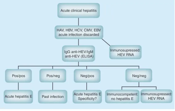
(12) Review Pérez-Gracia, Suay-García, García & Mateos-Lindemann hepatitis E is of some concern since seroconversion to detectable levels of anti-HEV antibodies is delayed or not present at all in these patients. Recently, commercially available tests for quantification of HEV RNA have appeared but these have not yet been approved by the US FDA. ●●Serologic assays. Most laboratories are choosing the method of serologic diagnosis for HEV infection, using ELISA (Figure 4) . The diagnosis of acute infection is based on the presence of IgM anti-HEV antibodies that can be detected during the acute phase of the illness and are present for up to 4–5 months. IgG anti-HEV are detected just after the raising of IgM anti-HEV, increasing from the acute phase until the convalescent phase. IgG anti-HEV have been detected up to 14 years after the acute phase [137] . Therefore, increased IgM anti-HEV levels represent acute infection, whereas IgG anti-HEV levels indicate previous contact with HEV. Recombinant proteins or synthetic peptides corresponding to immunodominant epitopes from ORF2 and/or ORF3 belonging to Burma and/or Mexico strains are being used as antigens in these assays. In vitro studies showed that all four HEV genotypes can be included in a single serotype. The performance characteristics of the commercially. available ELISA tests are considered suboptimal since several studies have demonstrated that the results are frequently discordant. The sensitivity ranged from 17 to 100% and some of them did not detect IgM anti-HEV antibodies in patients infected with G2–G4 [100] . By contrast, specificity needs to be increased since high false-positive rates with IgM anti-EBV and anti-CMV have been recorded [138] . In addition, IgG immunoblot assays frequently used as a confirming test have been shown to be unreliable [139] . More research must be done for improving the laboratory diagnosis of HEV to provide more reliable and reproducible tests either molecular or serological assays especially in low prevalence areas. Treatment Currently, there is no specific antiviral drug for HEV infection since it was not considered necessary until recently when chronic cases were reported. This fact promoted the treatment studies for isolated cases in which drugs, such as ribavirin and/or pegylated IFN-α (peginterferon), were used empirically [140] . Peginterferon is an immunostimulator that is being used in hepatitis B and C treatments. In transplant patients, its use can cause allogeneic immunity, which could lead to rejection of the transplanted organ,. Acute clinical hepatitis. HAV, HBV, HCV, CMV, EBV acute infection discarded. IgG anti-HEV/IgM anti-HEV (ELISA). Inmunosupressed: HEV RNA. Pos/pos. Pos/neg. Neg/pos. Neg/neg. Acute hepatitis E. Past infection. Acute hepatitis E Specificity?. Immunocompetent: Immunosupressed: HEV RNA no hepatitis E. Figure 4. Proposed algorithm for the diagnosis of acute hepatitis E in low prevalence countries. CMV: Cytomegalovirus; EBV: Epstein–Barr virus; HAV: Hepatitis A virus; HCV: Hepatitis C virus; HEV: Hepatitis E virus; Neg: Negative; Pos: Positive.. 10.2217/fmb-2016-0012. Future Microbiol. (Epub ahead of print). future science group.
(13) Hepatitis E: latest developments in knowledge. Review. Table 4. Ribavirin therapy in organ transplant patients. Transplanted organ. Patients (n). Dose. Duration median RVR, month (%) (months). SVR (%). Year. Ref.. KT/SPK. 8. 400–800 mg/day. 3. 5 (63). 2010. [144]. SPK HT HT KT LT LT. 2 1 4 1 1 11. 12 mg/kg/day 17 mg/kg/day 200–800 mg/day 800 mg/day 200 mg/day 600–1000 mg/day. 3 3 5 3 3 5. 2 (100) 1 (100) 3 (75) 1 (100) 1 (100) 9 (82). 2010 2011 2012 2012 2012 2013. KT/LT/HT/SPK/LungT. 59. 600 mg/day. 3. 46 (78). 2014. [106]. LungT. 4. 600–800 mg/day. 4.5. 2 (50). 2014. [151]. LT KT KT. 4 2 1. 400–800 mg/day 600–800 mg/day 600 mg/day. 3 3 4. 4 (100) 2 (100) 1 (100). 2015 2015 2015. [152]. 2 weeks (50) 3 weeks (12) 2 (38) 1 (50) 6 (100) 1 (100) 1 (50) 2 (100) 1 (100) 1 (36) 2 (46) 1 (64) 3 (31) 1 (25) 2 (25) 3 (25) 3 (100) 2 weeks (100) 2 (100). [145] [146] [147] [148] [149] [150]. [153] [154]. HT: Heart transplant; KT: Kidney transplant; LT: Liver transplant; LungT: Lung transplant; RVR: Rapid viral response; SPK: Simultaneous pancreas kidney; SVR: Sustained virological response.. thus its use could be limited in the treatment of chronic hepatitis E. Ribavirin is an antiviral agent that blocks nucleic acid synthesis and is used against DNA and RNA viruses. Although several studies suggest the beneficial effect of both antivirals for chronic hepatitis E, these therapies are in their experimental phases. The time to start the treatment, the optimal dose, the duration of the treatment and its safety, are currently unknown as there are no guidelines and neither of these drugs have been approved for this use. Tables 4 & 5 show the studies performed using ribavirin or pegylated interferon in transplant recipients. Haagsma et al. [141] have published a study in which two liver transplant patients with chronic hepatitis E were treated with peginterferon during 52 and 20 weeks, respectively, observing a significant decrease in viremia,. transaminase normalization and no HEV RNA detection in serum between weeks 4 and 12 of the treatment. However, one of the patients only showed positive results after lowering the immunosupressor dose. Similarly, other studies [118,142] refer to an improvement in HEV clearance when the patients’ immunosupressor dose was lowered, seeing as this improves the patient’s immunity [114,143] . In another study, by Kamar et al. [156] , a patient suffering from chronic hepatitis E was treated with peginterferon, presenting a satisfactory evolution up to 3 months after the treatment. In the other investigation [144] , they concluded that monotherapy with ribavirin inhibits HEV replication and may induce a sustained virologic response in patients with chronic HEV infection. Therefore, ribavirin monotherapy could be an effective and safe treatment in. Table 5. Pegylated interferon therapy in organ transplant patients. Transplanted organ. Patients (n). Dose (μg/kg/week). Duration median (months). RVR, month (%) SVR (%). Year. Ref.. LT LT. 2 3. α-2b: 1.5 α-2a: 135. 5 (50) 2 (67). 1 (50) 2 (67). 2010 2010. [141]. KT. 1. α-2a: 135. 8 1.5 (67) 3 (33) 3. 1 (100). 1 (100). 2010. [156]. [155]. KT: Kidney transplant; LT: Liver transplant; RVR: Rapid viral response; SVR: Sustained virological response.. future science group. www.futuremedicine.com. 10.2217/fmb-2016-0012.
(14) Review Pérez-Gracia, Suay-García, García & Mateos-Lindemann immunocompromised patients with chronic hepatitis E. The use of pegylated interferon in transplant patients may lead to transplant rejection and is not recommended. Ribavirin should be the antiviral treatment of choice in chronic hepatitis E [140] . There is scarce information available regarding the administration of immunoglobulins, but the few studies existing suggest that they do not grant protection against HEV infection [157] . Vaccines As HEV infection is significantly prevalent in developing countries, as well as among those working close to infection sources, many efforts have been placed in developing a vaccine. HEV is considered a good candidate for the development of a vaccine because it only presents one serotype and natural infection leads to protective antibodies [3] . Vaccination in developing countries could remarkably decrease the number of people infected by large waterborne HEV epidemics. In the early stages of vaccine development, it was observed that several recombinant proteins corresponding to the HEV capsid protein induced specific antibodies in animals and protected against liver injury following subsequent challenge with the virus [158] . Additionally, an HEV DNA vaccine was tested in cynomolgus macaques, having shown induction of anti-HEV serum production and protection against rechallenging with a heterologous HEV strain [159] . Bearing this in mind, two separate subunit vaccines were developed and tested. The first vaccine was a 56-kDa truncated HEV ORF2 protein produced from a baculovirus forming virus-like particles. In Phase I trials, an alum adjuvant was added to the formulation, which was then administered in three doses of 1, 5, 20 or 40 μg [158] . The results showed a dosedependent production of anti-HEV antibodies. This vaccine went into Phase II and III trials, where nearly 2000 volunteers from the Nepalese Army, lacking detectable anti-HEV antibodies, received either a 20-μg vaccine or a matching placebo in three doses (at 0, 1 and 6 months) [160] . After a follow-up of >2 years, it was observed that clinically overt acute hepatitis E occurred less frequently among vaccine recipients who completed the three-dose schedule than among placebo recipients, showing a vaccine efficacy of 95.5%. A lower efficacy rate of 87% was found in individuals who only received two doses.. 10.2217/fmb-2016-0012. Future Microbiol. (Epub ahead of print). Meanwhile, a Chinese group prepared another vaccine named the HEV 239 vaccine. It contains a more truncated HEV capsid protein (amino acids 368–606) expressed in Escherichia coli, purified and absorbed on aluminum hydroxide [161] . In Phase II trials, all the volunteers lacking anti-HEV antibodies seroconverted 1 month after having received a treatment consisting of three doses of a 20-μg vaccine at 0, 1 and 6 months [162] . Following this, a randomized, double-blind, placebocontrolled, community-based, Phase III trial has been successfully completed in China [163] . For 4.5 years, 112,604 participants with ages between 16 and 65, regardless of their anti-HEV antibody status, were divided in two groups: one group received the Hecolin® vaccine while the control group received the HBV vaccine. As a result, the vaccine showed an efficacy of 86.8%, with only seven of the 60 identified cases of hepatitis E belonging to the 239 vaccine group. This vaccine has been registered in China under the name Hecolin and is already available for use. Despite being based on the G1 virus, the Chinese vaccine has proven to provide protection against G4 HEV infections. Further studies are required to determine whether these vaccines provide protection against G3 virus strains, prevalent in developed countries. How this vaccine should be used is still unclear. In nonendemic regions, a vaccine could be used in cases of residents who are planning to travel to an endemic area. On the other hand, endemic areas could use the vaccine for pregnant women and patients with pre-existing chronic liver disease. However, cost considerations and the duration of protection afforded will need to be taken into account when deciding whether HEV vaccines should be used for the general population in endemic regions. The WHO has developed a three-stage approach to the threat of hepatitis E infections in which steps 2 and 3 refer to Hecolin as the only vaccine available in the market [164] . After evaluating the safety, immunogenicity, efficacy, cost–effectiveness and programmatic considerations of said vaccine for its use in prevention, control and treatment HEV, the WHO did not recommend the use of this vaccine routinely in countries where HEV is endemic. However, WHO also specifies that there might be special situations (outbreaks, travelers, pregnant women) in which individual countries can. future science group.
(15) Hepatitis E: latest developments in knowledge decide to use the vaccine as part of a prevention program [165] . Moreover, the use of these vaccines in animals should be considered seeing as they act as a reservoir and are becoming one of the main transmission routes, especially in developed countries. The efficacy of the HEV 239 vaccine was tested in 12 specific pathogen-free rabbits divided randomly into two groups and inoculated with HEV 239 and placebo (phosphatebuffered saline), respectively [166] . All animals were exposed to swine G4 HEV or rabbit HEV 7 weeks after the initial dose. Infection was monitored for 10 weeks measuring parameters, such as, duration of viremia, viral presence in stool, HEV antibody response and serum ALT levels. The group immunized with the HEV 239 vaccine showed no signs of HEV infection for the duration of the experiment, while those inoculated with placebo developed viral hepatitis. The results of this study show that the HEV 239 vaccine is also effective in rabbits and could possibly be extended to other animals, such as pigs. The positive results obtained with the HEV 239 vaccine have encouraged researchers to develop vaccines that will provide combined protection against viruses sharing the same transmission route. This is the case of Wang et al. [167] who are developing a vaccine to protect simultaneously against Norovirus (NoV) and HEV. This bivalent vaccine is still in the early stages of development.. Review. Conclusion & future perspective For the time being, HEV infection can be considered an emerging disease for chronic and progressive severe hepatitis in immunosupressed patients in developed countries. Several studies report that the different available immunosuppressants may play a role on the severity of the infection, mainly in the transplant setting. Regarding these controversial issues, more data are needed to control the high HEV replication in these patients [168,169] . The newly marketed vaccine (HEV 239 or Hecolin) is a giant leap in the prevention of hepatitis E in developing countries. However, WHO does not recommend its use routinely in countries where HEV is endemic. Moreover, its activity is yet to be proven in the target population of this vaccine in developed countries: immunosuppressed patients and pregnant women. Therefore, further efficacy studies are necessary. Financial & competing interests disclosure Work cited in this review from the author’s laboratory was supported in part by grants from the Universidad CEU Cardenal Herrera (PRUCH 25/10, PRUCH 39/11 and Santander-PRUCH 19/12; INDI1420). The authors have no other relevant affiliations or financial involvement with any organization or entity with a financial interest in or financial conflict with the subject matter or materials discussed in the manuscript apart from those disclosed. No writing assistance was utilized in the production of this manuscript.. EXECUTIVE SUMMARY ●●. Hepatitis E is a highly prevalent disease in developing countries.. ●●. According to the WHO, 20 million cases of Hepatitis E virus (HEV) infections are registered annually, of which >3 million are acute cases and approximately 56,600 result in death.. ●●. HEV classification is being constantly reviewed; the last classification was developed in 2014 by the International Committee of Taxonomic Virology.. ●●. More data about replication and cell culture systems are constantly being developed, which will result in a better understanding of the damages this virus may cause.. ●●. The main transmission route for HEV is the fecal–oral route, but the parenteral route has also been described.. ●●. HEV can cause chronic hepatitis in organ transplant recipients and immunocompetent patients.. ●●. The diagnosis is based in serological studies and detection of HEV-RNA in blood and stool samples.. ●●. There is no specific treatment against HEV, however, ribavirin monotherapy may be an effective and safe treatment in immunocompromised patients with chronic hepatitis E.. ●●. The development and commercialization of the first vaccine in China may help reduce future devastating epidemics caused by this virus. Vaccination is a good option to prevent infection, especially in countries where HEV is endemic.. future science group. www.futuremedicine.com. 10.2217/fmb-2016-0012.
(16) Review Pérez-Gracia, Suay-García, García & Mateos-Lindemann References. 13. Papers of special note have been highlighted as: • of interest; •• of considerable interest 1. WHO (2015). www.who.int. 2. Arankalle VA, Tsarev SA, Chadha MS et al. Age-specific prevalence of antibodies to hepatitis A and E viruses in Pune, India, 1982 and 1992. J. Infect. Dis. 2(171), 447–450 (1995).. 3. Purcell RH, Emerson SU. Hepatitis E: an emerging awareness of an old disease. J. Hepatol. 48, 494–503 (2008).. ••. Provides excellent review of Hepatitis E virus (HEV) clinical presentation, pathogenesis and diagnosis.. 4. Pérez-Gracia MT, Mateos ML, Galiana C et al. Autochthonous hepatitis E infection in a slaughterhouse worker. Am. J. Trop. Med. Hyg. 5(77), 893–896 (2007).. 5. Riveiro-Barciela M, Mínguez B, Gironés R, Rodriguez-Frías F, Quer J, Buti M. Phylogenetic demonstration of hepatitis E infection transmitted by pork meat ingestion. J. Clin. Gastroenterol. 49(2), 165–168 (2015).. 6. Viswanathan R, Sidhu AS. Infectious hepatitis; clinical findings. Indian J. Med. Res. (Suppl. 45), 49–58 (1957).. 7. Khuroo MS. Study of an epidemic of non-A, non-B hepatitis. Possibility of another human hepatitis virus distinct from post-transfusion non-A, non-B type. Am. J. Med. 6(68), 818–824 (1980).. 8. 9. 10. 14. Zaaijer HL, Yin MF, Lelie PN. Seroprevalence of hepatitis E in The Netherlands. Lancet 8820(340), 681 (1992).. 15. Paul DA, Knigge MF, Ritter A et al. Determination of hepatitis E virus seroprevalence by using recombinant fusion proteins and synthetic peptides. J. Infect. Dis. 4(169), 801–806 (1994).. 16. Buti M, Jardi R, Cotrina M et al. Hepatitis E virus infection in acute hepatitis in Spain. J. Virol. Methods. 1(55), 49–54 (1995).. 17. Meng XJ, Dea S, Engle RE et al. Prevalence of antibodies to the hepatitis E virus in pigs from countries where hepatitis E is common or is rare in the human population. J. Med. Virol. 3(59), 297–302 (1999).. 18. Fernández-Barredo S, Galiana C, García A, Vega S, Gómez MT, Pérez-Gracia MT. Detection of hepatitis E virus shedding in feces of pigs at different stages of production using reverse transcription-polymerase chain reaction. J. Vet. Diagn. Invest. 5(18), 462–465 (2006).. 19. 20. Balayan MS, Andjapardize AG, Savinskaya SS et al. Evidence for a virus in non-A, non-B hepatitis transmitted via faecal–oral route. Intervirology 20, 23–31 (1983). Bradley D, Andjaparidze A, Cook EH et al. Aetiological agent of enterically transmitted non-A, non-B hepatitis. J. Gen. Virol. 3, 731–738 (1988). Krawczynski K, Bradley DW. Enterically transmitted non-A, non-B hepatitis: identification of virus-associated antigen in experimentally infected cynomolgus macaques. J. Infect. Dis. 6(159), 1042–1049 (1989).. 11. Reyes GR, Purdy MA, Kim JP et al. Isolation of a cDNA from the virus responsible for enterically transmitted non-A, non-B hepatitis. Science 4948(247), 1335–1339 (1990).. 12. Balayan MS, Usmanov RK, Zamyatina NA, Djumalieva DI, Karas FR. Brief report: experimental hepatitis E infection in domestic pigs. J. Med. Virol. 1(32), 58–59 (1990).. 10.2217/fmb-2016-0012. Schlauder GG, Dawson GJ, Erker JC et al. The sequence and phylogenetic analysis of a novel hepatitis E virus isolated from a patient with acute hepatitis reported in the United States. J. Gen. Virol. 79(Pt 3), 447–456 (1998).. 21. 22. and sequencing of the full-length viral genome. Virology 185, 120–131 (1991). 26. Parvez MK, Al-Dosari MS. Evidence of MAPK-JNK1/2 activation by hepatitis E virus ORF3 protein in cultured hepatoma cells. Cytotechnology 67(3), 545–550 (2015).. 27. Khudyakov YE, Favorov MO, Khudyakova NS et al. Artificial mosaic protein containing antigenic epitopes of hepatitis E virus. J. Virol. 68, 7067–7074 (1994).. 28. Schlauder GG, Mushahwar IK. Genetic heterogeneity of hepatitis E virus. J. Med. Virol. 2(65), 282–292 (2001).. 29. Lu L, Li C, Hagedorn CH. Phylogenetic analysis of global hepatitis E virus sequences: genetic diversity, subtypes and zoonosis. Rev. Med. Virol. 16, 5–36 (2006).. 30. Smith DB, Simmonds P, International Committee on Taxonomy of Viruses Hepeviridae Study Group. Consensus proposals for classification of the family Hepeviridae. J. Gen. Virol. 95(Pt 10), 2223–2232 (2014).. ••. Provides information on HEV classification.. 31. Smith DB, Purdy MA, Simmonds P. Genetic variability and the classification of hepatitis E virus. J. Virol. 87(8), 4161–4169 (2013).. 32. Woo PC, Lau SK, Teng JL et al. New hepatitis E virus genotype in camels, the Middle East. Emerg. Infect. Dis. 20(6), 1044–1048 (2014).. 33. Pérez-Gracia MT, Suay B, MateosLindemann ML. Hepatitis E: an emerging disease. Infect. Genet. Evol. 22, 40–59 (2014).. ••. Provides a detailed review on HEV virology, epidemiology, clinical presentation, epidemiology, pathogenesis and diagnosis.. 34. He J, Innis BL, Shrestha MP et al. Evidence that rodents are a reservoir of hepatitis E virus for humans in Nepal. J. Clin. Microbiol. 12(40), 4493–4498 (2002).. Guan D, Li W, Su J. Asian musk shrew as a reservoir of rat hepatitis E virus, China. Emerg. Infect. Dis. 19(8), 1341–1343 (2013).. 35. Meng XJ, Purcell RH, Halbur PG et al. A novel virus in swine is closely related to the human hepatitis E virus. Proc. Natl Acad. Sci. USA 18(94), 9860–9865 (1997).. Batts W, Yun S, Hedrick R, Winton J. A novel member of the family Hepeviridae from cutthroat trout (Oncorhynchus clarkii). Virus Res. 158, 116–123 (2011).. 36. Takahashi M, Tanaka T, Takahashi H et al. Hepatitis E virus (HEV) strains in serum samples can replicate efficiently in cultured cells despite the coexistence of HEV antibodies: characterization of HEV virions in blood circulation. J. Clin. Microbiol. 48, 1112–1125 (2010).. Banks M, Bendall R, Grierson S, Heath G, Mitchell J, Dalton H. Human and porcine hepatitis E virus strains, United Kingdom. Emerg. Infect. Dis. 5(10), 953–955 (2004). Huang FF, Haqshenas G, Guenette DK et al. Detection by reverse transcription-PCR and genetic characterization of field isolates of swine hepatitis E virus from pigs in different geographic regions of the United States. J. Clin. Microbiol. 4(40), 1326–1332 (2002).. 23. Pérez-Gracia MT, García M, Suay B, Mateos-Lindemann ML. Current knowledge on Hepatitis E. J. Clin. Transl Hepatol. 3(2), 117–126 (2015).. 24. Li TC, Yamakawa Y, Suzuki K et al. Expression and self-assembly of empty virus-like particles of hepatitis E virus. J. Virol. 71, 7207–7213 (1997).. 37. Tanaka T, Takahashi M, Kusano E, Okamoto H. Development and evaluation of an efficient cell-culture system for Hepatitis E virus. J. Gen. Virol. 88, 903–911 (2007).. 25. Tam AW, Smith MM, Guerra ME et al. Hepatitis E virus (HEV): molecular cloning. 38. Tanaka T, Takahashi M, Takahashi H et al. Development and characterization of a. Future Microbiol. (Epub ahead of print). future science group.
(17) Hepatitis E: latest developments in knowledge genotype 4 hepatitis E virus cell culture system using a HE-JF5/15F strain recovered from a fulminant hepatitis patient. J. Clin. Microbiol. 47, 1906–1910 (2009). 39. 40. 41. 42. 43. 44. 45. 46. 47. Williams TP, Kasorndorkbua C, Halbur PG et al. Evidence of extrahepatic sites of replication of the hepatitis E virus in a swine model. J. Clin. Microbiol. 39, 3040–3046 (2001). Shukla P, H.T Nguyen, K Faulk et al. Adaptation of a genotype 3 hepatitis E virus to efficient growth in cell culture depends on an inserted human gene segment acquired by recombination. J. Virol. 86, 5697–5707 (2012). Rogee S, Talbot N, Caperna T et al. New models of hepatitis E virus replication in human and porcine hepatocyte cell lines. J. Gen. Virol. 94, 549–558 (2013). Oshiro Y, Yasue H, Takahashi K et al. Mode of swine hepatitis E virus infection and replication in primary human hepatocytes. J. Gen. Virol. 95, 2677–2682 (2014). Emerson SU, Nguyen HT, Torian U, Burke D, Engle R, Purcell RH. Release of Genotype 1 hepatitis E Virus from cultured hepatoma and polarized intestinal cells depends on open reading frame 3 protein and requires an intact PXXP motif. J. Virol. 84(18), 9059–9069 (2010). Okamoto H. Efficient cell culture systems for hepatitis E virus strains in feces and circulating blood. Rev. Med. Virol. 21, 18–31 (2011). Shukla P, Nguyen HT, Torian U et al. Cross-species infections of cultured cells by hepatitis E virus and discovery of an infectious virus-host recombinant. Proc. Natl Acad. Sci. USA 108, 2438–2443 (2011). Zhang HY, Chen DS, Wu YQ et al. Both swine and human cells are capable to support the replication of swine hepatitis E virus type 4 in vitro. Virus Res. 158, 289–293 (2011).. 50. 51. Helenius A. Virus entry: what has pH got to do with it? Nat. Cell Biol. 15, 125 (2013).. 52. Yamauchi Y, Helenius A. Virus entry at a glance. J. Cell Sci. 126, 1289–1295 (2013).. 53. Yu H, Li S, Yang C, Wei M et al. Homology model and potential virus-capsid binding site of a putative HEV receptor Grp78. J. Mol. Model. 17, 987–995 (2011).. 54. Zheng ZZ, Miao J, Zhao M et al. Role of heat-shock protein 90 in hepatitis E virus capsid trafficking. J.Gen.Virol. 91, 1728–1736 (2010).. 55. 56. 57. 58. 59. •. Provides review of HEV molecular virology. Shiota T, Li TC, Yoshizaki S et al. The hepatitis E virus capsid C-terminal region is essential for the viral life cycle: implication for viral genome encapsidation and particle stabilization. J. Virol. 87, 6031–6036 (2013).. 49. Kalia M, Chandra V, Rahman SA, Sehgal D, Jameel S. Heparan sulfate proteoglycans are required for cellular binding of the hepatitis E virus ORF2 capsid protein and for viral infection. J. Virol. 83, 12714–12724 (2009).. future science group. Perttila J, Spuul P, Ahola T. Early secretory pathway localization and lack of processing for hepatitis E virus replication protein pORF1. J. Gen.Virol. 94, 807–816 (2013). Agrawal S, Gupta D, Panda SK. The 3′ end of hepatitis E virus (HEV) genome binds specifically to the viral RNA-dependent RNA polymerase (RdRp). Virology 282, 87–101 (2001). Graff J, Nguyen H, Kasorndorkbua C et al. In vitro and in vivo mutational analysis of the 3′-terminal regions of hepatitis E virus genomes and replicons. J. Virol. 79, 1017–1026 (2005). Parvez MK. The hepatitis E virus ORF1 ‘X-domain’ residues form a putative macrodomain protein/Appr-1′′-pase catalytic-site, critical for viral RNA replication. Gene 566(1), 47–53 (2015). Emerson SU, Nguyen HT, Torian U et al. Release of genotype 1 hepatitis E virus from cultured hepatoma and polarized intestinal cells depends on open reading frame 3 protein and requires an intact PXXP motif. J. Virol. 84, 9059–9069 (2010).. 60. Nagashima S, Takahashi M, Jirintai, et al. A PSAP motif in the ORF3 protein of hepatitis E virus is necessary for virion release from infected cells. J. Gen. Virol. 92, 269–278 (2011).. 61. Surjit M, Oberoi R, Kumar R, Lal SK. Enhanced alpha1 microglobulin secretion from hepatitis E virus ORF3-expressing human hepatoma cells is mediated by the tumor susceptibility gene 101. J. Biol. Chem. 281, 8135–8142 (2006).. Ahmad I, Holla RP, Jameel S. Molecular virology of hepatitis E virus. Virus Res. 161, 47–58 (2011).. 48. Holla P, Ahmad I, Ahmed Z, Jameel S. Hepatitis e virus enters liver cells through a dynamin-2, clathrin and membrane cholesterol-dependent pathway. Traffic 16, 398–416 (2015).. 62. Jia Z, Yi Y, Liu J et al. Epidemiology of hepatitis E virus in China: results from the Third National Viral Hepatitis Prevalence Survey, 2005–2006. PLoS ONE 9(10), e110837 (2014).. Review. 63. Kamar N, Bendall R, Legrand-Abravanel F et al. Hepatitis E. Lancet 379, 2477–2488 (2012).. ••. Provides review of HEV history, epidemiology and diagnosis.. 64. Kumar S, Subhadra S, Singh B, Panda BK. Hepatitis E: the current scenario. Int. J. Infect. Dis. 17(4), e228–e233 (2013).. 65. Kmush B, Wierzba T, Krain L, Nelson K, Labrique AB. Epidemiology of hepatitis E in low- and middle-income countries of Asia and Africa. Semin. Liver Dis. 33, 15–29 (2013).. 66. CIWEC Clinic Travel Medicine Center. www.ciwec-clinic.com. 67. Kim JH, Nelson KE, Panzner U et al. A systematic review of the epidemiology of hepatitis E virus in Africa. BMC Infect Dis. 14, 308 (2014).. 68. Aggarwal R. The global prevalence of hepatitis E virus infection and susceptibility: a systematic review. WHO, Geneva, Switzerland (2010).. 69. Browne LB, Menkir Z, Kahi V et al. Notes from the field: hepatitis E outbreak among refugees from South Sudan – Gambella, Ethiopia, April 2014–January 2015. MMRW Morb Mortal Wkly Rep. 64(19), 537 (2015).. 70. Ahmed JA, Moturi E, Spiegel P et al. Hepatitis E outbreak, Dadaab refugee camp, Kenya, 2012. Emerg. Infect. Dis. 19(6), 1010–1012 (2013).. 71. Centers for Disease Control and Prevention (CDC). Investigation of hepatitis E outbreak among refugees – Upper Nile, South Sudan, 2012–2013. MMRW Morb Mortal Wkly Rep. 62(29), 581–586 (2013).. 72. Teshale EH, Grytdal SP, Howard C et al. Evidence of person-to-person transmission of hepatitis E virus during a large outbreak in Northern Uganda. Clin. Infect. Dis. 50(7), 1006–1010 (2010).. 73. Villarejos VM, Provost PJ, Ittensohn OL, McLean AA, Hilleman MR. Seroepidemiologic investigation of human hepatitis caused by A, B and a possible third virus. Proc. Soc. Exp. Biol. Med. 152(4), 524–528 (1976).. 74. Zhuang H, Cao XY, Liu CB, Wang GM. Epidemiology of hepatitis E in China. Gastroenterol Jpn 26(Suppl. 3), 135–138 (1991).. 75. Arankalle VA, Chadha MS, Tsarev SA et al. Seroepidemiology of water-borne hepatitis in India and evidence of third entericallytransmitted hepatitis agent. Proc. Natl Acad. Sci. USA 91(8), 3428–3432 (1994).. www.futuremedicine.com. 10.2217/fmb-2016-0012.
(18) Review Pérez-Gracia, Suay-García, García & Mateos-Lindemann 76. 77. Hepatitis D, E and GB-C: global status. Gideon e-books. (2016). www.gideononline.com Corwin AL, Khiem HB, Clayson ET et al. A waterborne outbreak of hepatitis E virus transmission in southwestern Vietnam. Am. J. Trop. Med. Hyg. 54(6), 559–562 (1996).. Wichmann O, Schimanski S, Koch J et al. Phylogenetic and case–control study on hepatitis E virus infection in Germany. J. Infect. Dis. 198, 1732–1741 (2008).. 102 Navaneethan U, Al Mohajer M, Shata MT.. 91. Said B, Ijaz S, Kafatos G et al. Hepatitis E outbreak on cruise ship. Emerg. Infect. Dis. 15, 1738–1744 (2009).. •. 90. 78. Dawson GJ, Mushahwar IK, Chau KH, Gitnick GL. Detection of long-lasting antibody to hepatitis E virus in a US traveller to Pakistan. Lancet 240, 426–427 (1992).. 92. Echevarría JM. Light and darkness: prevalence of hepatitis E virus infection among the general population. Scientifica (Cairo) 2014, 481016 (2014).. 79. Chapman BA, Burt MJ, Wilkinson ID, Schousboe MI. Community acquired viral hepatitis in New Zealand: a case of sporadic hepatitis E virus infection. Aust. NZ J. Med. 23, 722–723 (1993).. 93. Matsubayashi K, Nagaoka Y, Sakata H et al. Transfusion-transmitted hepatitis E caused by apparently indigenous hepatitis E virus strain in Hokkaido, Japan. Transfusion 44(6), 934–940 (2004).. 80. Fletcher J. A traveller returning from Nepal with hepatitis E. Med. J. Aust. 159, 563 (1993).. 94. 81. Acharya SK, Panda SK. Hepatitis E: water, water everywhere – now a global disease. J. Hepatol. 54, 9–11 (2011).. Boxall E, Herborn A, Kochethu G et al. Transfusion-transmitted hepatitis E in a ‘nonhyperendemic’ country. Transfus. Med. 16(2), 79–83 (2006).. 95. 82. García M, Fernández-Barredo S, Pérez-Gracia MT. Detection of hepatitis E virus (HEV) through the different stages of pig manure composting plants. Microb. Biotechnol. 7(1), 26–31 (2013).. Colson P, Coze C, Gallian P, Henry M, De Micco P, Tamalet C. Transfusion-associated hepatitis E, France. Emerg. Infect. Dis. 13(4), 648–649 (2007).. 83. 84. 85. 86. 87. 88. 89. Aggarwal R, Kini D, Sofat S, Naik SR, Krawczynski K. Duration of viraemia and faecal viral excretion in acute hepatitis E. Lancet 356, 1081–1082 (2000). Khuroo MS, Kamili S. Clinical course and duration of viremia in vertically transmitted hepatitis E virus (HEV) infection in babies born to HEV-infected mothers. J. Viral Hepat. 16, 519–523 (2009). Kumar RM, Uduman S, Rana S, Kochiyil JK, Usmani A, Tomas L. Sero-prevalence and mother-to-infant transmission of hepatitis E virus among pregnant women in the United Arab Emirates. Eur. J. Obstet. Gynecol. Reprod. Biol. 100, 9–15 (2001). Khuroo MS, Kamili S, Jameel S. Vertical transmission of hepatitis E virus. Lancet 345, 1025–1026 (1995). Psichogiou M, Tzala E, Boletis J et al. Hepatitis E virus infection in individuals at high risk of transmission of non-A, non-B hepatitis and sexually transmitted diseases. Scand. J. Infect. Dis. 28, 443–445 (1996). Galiana C, Fernández-Barredo S, Pérez-Gracia MT. Prevalence of hepatitis E virus (HEV) and risk factors in pig workers and blood donors. Enferm. Infecc. Microbiol. Clin. 28, 602–607 (2010). Mansuy JM, Bendall R, Legrand-Abravanel F et al. Hepatitis E virus antibodies in blood donors, France. Emerg. Infect. Dis. 17, 2309–2323 (2011).. 10.2217/fmb-2016-0012. 96. Mirazo S, Ramos N, Mainardi V, Gerona S, Arbiza J. Transmission, diagnosis and management of hepatitis E: an update. Hepat. Med. 6, 45–59 (2014).. ••. Provides review of HEV transmission, diagnosis and management.. 97. Mateos-Lindemann ML, Diez-Aguilar M, González-Galdamez A, Graus-Morales J, Moreno-Zamora A, Pérez-Gracia MT. Acute, chronic and fulminant hepatitis E: seven years of experience (2004–2011). Enferm. Infecc. Microbiol. Clin. 31, 595–598 (2013).. 98. 99. Mateos-Lindemann ML, Diez-Aguilar M, González-Galdámez A et al. Acute, chronic and fulminant hepatitis E: ten years of experience (2004–2013). Int. J. Gastroenterol. Dis. Ther. 1, 102–105 (2014). Labrique AB, Kuniholm MH, Nelson K. The global impact of hepatitis E: new horizons for an emerging virus. In: Emerging Infections (9th Edition). Scheld WM, Grayson ML, Hughes JM (Eds). ASM Press, VA, USA, 53–92 (2010).. 100 Aggarwal R. Hepatitis E: clinical. presentation in disease-endemic areas and diagnosis. Semin. Liver Dis. 33, 30–40 (2013). •. Provides review of HEV clinical presentation and diagnosis.. 101 Dalton HR, Bendall RP, Rashid M et al.. Host risk factors and autochthonous hepatitis E infection. Eur. J. Gastroenterol. Hepatol. 23, 1200–1205 (2011).. Future Microbiol. (Epub ahead of print). Hepatitis E and pregnancy: understanding the pathogenesis. Liver Int. 28, 1190–1199 (2008). Provides review of HEV pathogenesis in pregnancy.. 103 Davern TJ, Chalasami N, Fontana RJ et al.. Acute hepatitis E infection accounts for some cases of suspected drug-induced liver injury. Gastroenterology 41, 1665–1672 (2011). 104 Peron JM, Dalton H, Izopet J, Kamar N.. Acute autochthonous hepatitis E in western patients with underlying chronic liver disease: a role for ribavirin? J. Hepatol. 54, 1323–1324, author reply 1324–1325 (2011). 105 Kamar N, Rostaing L, Legrand-Abravanet F,. Izopet J. How should hepatitis E virus infection be defined in organ-transplant recipients? Am. J. Transplant. 13, 1935–1936 (2013). 106 Kamar N, Abravanel F, Lhomme S, Rostaing. L, Izopet J. Hepatitis E virus: chronic infection, extra-hepatic manifestations, and treatment. Clin. Res. Hepatol. Gastroenterol. 39, 20–27 (2015). ••. Provides review of HEV chronic infection, extrahepatic manifestations and treatment.. 107 González-Tallón AI, Moreira Vicente V,. Mateos Lindemann ML. Hepatitis crónica E en paciente immunocompetente. Gastroenterol. Hepatol. 34, 398–400 (2011). 108 Grewal P, Kamili S, Motamed D. Chronic. hepatitis E in an immunocompetent patient: a case report. Hepatology 59, 347–348 (2013). 109 Junge N, Pischke S, Baumann U et al. Results. of single-center liver transplantation and report on successful treatment with ribavirin. Pediatr. Transplant. 17, 343–347 (2013). 110 Kamar N, Rostaing L, Izopet J. Hepatitis E. virus infection in immunosuppressed patients: natural history and therapy. Semin. Liver Dis. 33, 62–70 (2013). 111 Geng Y, Zhang H, Huang W, Geng K, Li Z.. Persistent hepatitis E virus genotype 4 infection in a child with acute lymphoblastic leukaemia. Hepat. Mon. 14, e15618 (2014). 112 Kamar N, Selves J, Mansuy JM et al.. Hepatitis E virus and chronic hepatitis in organ-transplant recipients. N. Engl. J. Med. 358, 811–817 (2008). 113 Kamar N, Izopet J, Cintas P et al. Hepatitis E. virus induced-neurological symptoms in a kidney-transplant patient with chronic hepatitis. Am. J. Transplant. 10, 1321–1324 (2010). 114 Kamar N, Abravanel F, Selves J et al.. Influence of immunosuppressive therapy on. future science group.
Figure


+4


Documento similar
Several systems, molecules and responses involved in the pathogenesis of the pathological fibrosis of chronic kidney disease (CKD) will be discussed in this review, putting
– Large Multiplexing: > 100 stars in one single configuration (potentially!) – We aim at ~100 stars with V= 17-18 in two fiber configurations. • Although WYFOSS
Kopple, “Association of dietary phosphorus intake and phosphorus to protein ratio with mortality in hemodialysis patients,” Clinical Journal of the American Society of Nephrology,
The PICS technique: A novel approach for residual curvature correction during penile prosthesis im- plantation in patients with severe Peyronie’s disease using the collagen
The expansionary monetary policy measures have had a negative impact on net interest margins both via the reduction in interest rates and –less powerfully- the flattening of the
Jointly estimate this entry game with several outcome equations (fees/rates, credit limits) for bank accounts, credit cards and lines of credit. Use simulation methods to
In our sample, 2890 deals were issued by less reputable underwriters (i.e. a weighted syndication underwriting reputation share below the share of the 7 th largest underwriter
The assessment of perceived health status in patients with chronic diseases is essential for measuring the impact and burden of disease, as in the case of patients with
