Role of Sirt1 During the Ageing Process: Relevance to Protection of Synapses in the Brain
13
0
0
Texto completo
(2) Mol Neurobiol (2014) 50:744–756. of oxidative phosphorylation and ATP production. Therefore, ATP synthesis and redox potential are directly proportional to intracellular NAD+ concentrations. More recently, NAD+ has been identified as a ubiquitous molecule which plays a critical role in several biological processes, including cellular respiration, DNA repair, transcriptional regulation, posttranslational protein modifications, signal transduction and apoptosis [5]. Disruption of fusion results in mitochondrial heterogeneity and dysfunction. Indeed, NAD+ is quite important for a number of ADP-ribosylation reactions associated with cell regulation and repair mechanisms, which are mediated by CD38 [6]. Importantly, changes in NAD+ metabolism have been associated with several pathologies, including neurodegenerative diseases, cancer, cardiovascular disease and normal ageing [7]. Moreover, we have previously shown that NAD+ levels decline with age in ‘physiologically’ aged Wistar rats [8] and in aged human pelvic skin tissue [7]. Therefore, promotion of cellular NAD+ anabolism is crucial for maintaining longevity and neuroprotection against chronic oxidative stress insult. It is well established that damaged organelles and misfolded or malfunctioning cellular proteins increase in differentiated cells, such as neurons, where the functional characteristics decline with age. The clearance of aberrant malfunctioning cellular components by autophagy, or cellular self-digestion, is a highly conserved intracellular process in eukaryotes and is characterized by protein and organelle degradation into double membrane vesicles which are delivered to the lysosome/vacuole for breakdown and recycling of the resulting macromolecules [9]. The gradual age-related decline in autophagic activity could play a further role in the functional deterioration of ageing organisms [10]. Moreover, recent studies have evidenced that autophagy might be at the basis of the anti-ageing mechanism observed under calorie restriction, which mediates life extension in an NAD+-dependent manner [11]. Calorie restriction (10–30 % less calorie intake than the average diet) is the only intervention known to be able to slow down several aspects of the ageing process in mammals, including age-related mortality [12]. Nutritional modulation through calorie restriction has been shown to be effective as an anti-ageing intervention [13] and paves the way for the development of new therapeutic strategies to treat age-dependent neurological disorders that cannot, as yet, be cured once initiated [14]. Sirtuins or silent mating-type information regulator 2 (Sir2) proteins catalyze numerous activities including NAD+-dependent protein deacetylation reaction, which couples lysine deacetylation to NAD+ hydrolysis [15, 16] (Fig. 1). This new class of enzymes is thought to function as nutrient sensors, which mediate the beneficial effects of calorie restriction and can extend lifespan in several organisms. Moreover, activators of sirtuins such as resveratrol, a naturally occurring. 745. phytochemical present in grapes and red wine, and its endogenous substrate, NAD+, have been shown to promote dendritic branching and neuronal survival in aged neuronal populations (Fig. 2). Indeed, an extra copy of Sir2 gene is able to extend replicative lifespan in yeasts and nematodes [17]. Seven Sir2 homologs (Sirt1 to 7) have been identified in mammals, and several lifespan key components, such as the peroxisome proliferator-activated receptor gamma coactivator 1-alpha (PGC1-α), PPARγ, Ku70, histones, p300, p53, FoxO, TAF 168, PCAF/MyoD, HIV tat, and NF-κB [15, 18, 19], are considered among its targets (Fig. 3). Although sirtuins are widely expressed through the mammalian organism, they are also selectively localized at the subcellular level: Sirt3, 4 and 5 are mitochondrial; Sirt1, 6 and 7 are mainly nuclear, while Sirt2 is primarily cytosolic [20]. Among these, Sirt3 has been proposed to regulate mitochondrial metabolism and might be critical in sensing NAD+ levels in the mitochondria, since increased NAD+ triggers a regulatory pathway that activates Sirt3, leading to the deacetylation of specific targets [21] (Fig. 3). Moreover, it has been demonstrated that Sirt3deficient mice exhibit protein hyper-acetylation [22] of the metabolic enzyme glutamate dehydrogenase (GDH), suggesting that Sirt3 has a critical role in metabolic control [23]. On the other hand, Sirt1, which is commonly expressed in the developing and in the adult mammalian brain [24], has been implicated in various biological processes such as stress resistance, reduced apoptosis and prevention of axonal degeneration, among others [25, 26]. Sirt1 expression is decreased in the brain in some senescence and neurodegenerative disease models [27]. Moreover, Sirt1 activity is closely related to the ratio NAD+/NADH, and as mentioned previously, NAD+ depletion is a major cause of genotoxic cellular damage [28–33]. Taken together, selectively targeting Sirt1 might represent a potential therapeutic target for the prevention and neuroprotection against age-related neurodegenerative disorders (Fig. 5).. Ageing and Oxidative Stress The major cause of cellular damage and tissue decline during ageing is due to the cytotoxic effects of ROS and free radicals, formed because of external inducers of damage and as a consequence of an impaired cellular metabolism involving oxygen, redox-active metals and other metabolites, nutritional glucose and spontaneous errors in biochemical processes and reduced endogenous antioxidant defenses [4, 34, 35]. Oxidative stress refers to the imbalance between the cellular productions of ROS and the antioxidant mechanisms that remove them. The ROS, which are formed mainly in the mitochondria by the membrane-bound enzyme complex NADPH oxidase, xanthine oxidase and endoplasmic reticulum P450 oxidase, during normal cellular metabolism, are.
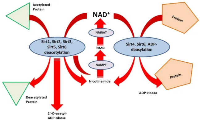
(3) 746. Mol Neurobiol (2014) 50:744–756. Fig. 1 Sirtuin enzymatic activities. Sirtuins are NAD+-dependent histone deacetylases which carry out several deacetylation and ADPribosylation reactions. They have been related with critical lifespan regulatory pathways, and recent studies suggest the modulation of sirtuin activity as a promising target against several cellular lifespan-. related pathologies, such as cancer and neurodegenerative disorders. Sirtuin activities are inhibited by nicotinamide. NAD+ nicotinamide adenine dinucleotide, NMN nicotinamide mononucleotide, NMNAT nicotinamide mononucleotide adenyl transferase, NAMPT nicotinamide phosphoribosyl transferase. Fig. 2 Sirt1 regulates neuronal lifespan. As critical lifespan regulator, sirtuins could be modulated by several external stimuli, affecting the cell behavior. Agonists, such as resveratrol, NAD+, and calorie restriction, or inhibitors, such as nicotinamide, or fasting, signal through Sirt1 inducing the activation of key metabolic/cell cycle effectors, such as PGC-1α deacetylation, allowing direct regulation of the cell lifespan. In neurons, the. activation of Sirt1 is related with increased dendritic expansion and improved neuronal survival. The precise mechanism underlying how Sirt1 deacetylates PGC-1α in the brain remains unclear. Sirt1 activity can be inhibited by the endogenous NAD+ precursor, nicotinamide. Other Sirt1 inhibitors have been synthetically developed with greater Sirt1 affinity than nicotinamide. PGC1α peroxisome proliferator-activated receptor co-activator 1α.
(4) Mol Neurobiol (2014) 50:744–756. 747. Fig. 3 Regulation of ROS by sirtuins during AD and ageing. ROS have been linked with critical changes that verify in cells after highly demanding physiological/pathological challenges. Organelle damage as well as modification of critical macromolecules, such as DNA, is often described as the common result of the ROS accumulation within the cells. Moreover, increased ROS levels have been also described as a common feature of the ageing process and in several neurodegenerative diseases, such as AD, PD and HD. There is a close link between Sirt1 and ROS, and the figure illustrates how oxidative DNA damage. regulates the activity of the nuclear sirtuin, Sirt1, and the mitochondrial sirtuin, Sirt3. When Sirt1 and Sirt3 are activated, these sirtuins deacetylate several proteins that regulate protection against oxidative stress. ROS reactive oxygen species; Sirt1 sirtuin 1; PGC-1α peroxisome proliferator-activated receptor gamma co-activator 1-alpha; NF-κB nuclear factor kappa-light-chain-enhancer of activated B cells; FOXO forkhead box proteins; Sirt3 sirtuin 3; HIF1-α hypoxia-inducible factor 1-alpha; SOD2 superoxide dismutase 2, mitochondrial; IDH2 isocitrate dehydrogenase 2. considered the major contributors to the ageing-damaging process [36]. The brain is the organ with the highest oxygen consumption rate, and oxidative damage has long been considered to play a primarily deleterious effect on neurons both during normal ageing and in neurodegenerative disorders [37]. Transient production of ROS plays a role in synaptic signaling, with ROS acting as secondary messenger molecules. In particular, superoxide can serve as a cellular messenger in the process of long-term potentiation (LTP) [38], a well-known model for synaptic plasticity and learning. However, when. superoxide levels are increased, it provides a substrate for the Haber Weiss reaction leading to the production of the highly reactive hydroxyl radical which inflicts profound cellular degeneration and cell death. In the hippocampus, N-methyl-D-aspartate (NMDA) receptor activation increases oxidative and nitrosative stress by inducing free radical production and lipid peroxidation [39]. Consequently, the NMDA receptor-mediated components of synaptic activity enhance the antioxidant defenses of neurons, triggering several transcriptional events and stimulating enzymatic activity to detoxify peroxides and ROS [40]. Soluble.
(5) 748. Aβ oligomers, which are well-known neurotoxins [41, 42], induce a prominent increase in ROS formation because of abnormal overactivation of the NMDA receptor [43]. Moreover, in aged brains, there is a decrease in the expression of the NR-1 subunit of the NMDA receptor subtype in the hippocampus [44]. This downregulation, which leads to an altered configuration of the NMDA receptor and enhances glutamate excitotoxicity, might represent a potential mechanism of the increased oxidative stress in neurons and might contribute to the development and progression of neurodegenerative disease, such as AD. Furthermore, the accumulation of macromolecular damage has long been thought to underlie the ageing process at a fundamental level. Several studies have suggested that DNA damage is an important triggering factor for nuclear ageing, thus providing additional support to the free radical theory of ageing [45]. Although it could also be argued that chromatin structure is directly affected during the ageing process through an as-yetnot fully understood mechanism that leads to increased DNA damage, and to a permanent modification that alters gene expression patterns, several research groups have documented the accumulation of oxidative DNA damage during the mammalian brain ageing [46, 47]. Once DNA damage has occurred, it is the role of specific cellular repair systems to prevent its accumulation and initiate the repair process. However, different studies have shown that ageing is also associated with a general reduction of DNA repair capacity [48], preventing the efficient repair of single- or double-stranded DNA breaks. Interestingly, a differential accumulation of oxidativerelated DNA lesions in different brain regions has also been reported, suggesting that some areas of the brain are more vulnerable to oxidative DNA modifications than others [49]. Moreover, Lu et al. [50] found that at the molecular level, oxidative DNA damage accumulates preferentially in the promoter regions of several genes involved in synaptic plasticity (i.e., GluN1, NMDA receptor 2A, GABA receptor β3, serotonin receptor, voltage-gated Na channel II β, voltagedependent calcium channel β2, neurexin 1, synaptobrevin, synapsin IIb, γSNAP, αSNAP, RAB3A), vesicular transport (RAB1A, RAB3A, RAB5A, RAB6A, kinesin 1B, sortilin 1, dynein, dynamin 1-like, trans Golgi network protein 2, phosphotidylinositol transfer protein β, clathrin light polypeptide) and mitochondrial proteins (ATP synthase, mitochondrial ribosomal protein L28 and S12, cytochrome c synthase and translocase of inner mitochondrial membrane 17A), suggesting a close relation between oxidative damage and memory and cognitive decline because of an impaired synaptic functionality. As well, certain types of neurons, as demonstrated in the mouse brain, such as hippocampal, pyramidal and granule cells, usually do not decrease in number during ageing, but can accumulate nuclear DNA damage [51]. Several phytochemicals, such as resveratrol and quercetin, have shown to be able to prevent the accumulation of ROS and. Mol Neurobiol (2014) 50:744–756. attenuate the depletion of cellular glutathione and to reduce the oxidative DNA damage [52] and the apoptosis [53] of dopaminergic neurons exposed to oxidative stress. Apart from its strong antioxidant potential, resveratrol has been previously shown to rescue neurons from polyglutamine neurotoxicity in Caenorhabditis elegans through a Sir2-dependent mechanism [29]. These data raise the possibility that resveratrol might be useful in brain protection and constitutes a novel candidate drug for the development of neuroprotective therapies aimed for the treatment and prevention of neurodegenerative disorders. In fact, the identification and validation of a molecule able to act against multiple age-related diseases would have a major impact on global health and economics. On this regard, the Sirt1 deacetylase has drawn considerable attention as a target for drug design. Yet, controversy remains behind the mechanism of sirtuin-activating compounds (STACs), and further studies should be conducted in order to assess and validate the potentialities of such drugs. Interestingly, specific hydrophobic motifs found in Sirt1 substrates, such as peroxisome proliferator-activated receptor co-activator 1α (PGC1α) and FOXO3a, facilitate Sirt1 activation by the STACs [54]. Moreover, a single Sirt1 amino acid, the Glu230, located in a structured N-terminal domain, has been reported as critical for the activation by currently known STACs. In fact, in primary Sirt1 defective cells, the metabolic effects of STACs were blocked, suggesting that Sirt1 can be directly activated through an allosteric mechanism common to chemically diverse STACs [54].. Ageing and Autophagy The term autophagy is used to illustrate the observations from electron microscope studies, which showed novel single or double membrane vesicles that contain parts of the cytoplasm, including organelles, in different degrees of disintegration [55]. The process of autophagy exists in yeast and can be stimulated by different nutrients. Moreover, at least 16 separate autophagy genes (Atg) have been identified and are necessary for the formation of the autophagosome and the subsequent induction of autophagy [56, 57]. There are several types of autophagy, namely micro- and macroautophagy, as well as chaperone-mediated autophagy (CMA). Both microand macroautophagy have the capacity to engulf large structures through selective and non-selective mechanisms, whereas CMA degrades only soluble proteins [10]. The autophagy regulation pathway in mammals is stimulated by deprivation of insulin and other growth factors. When present, these factors signal through tyrosine kinasesreceptors to activate the autophagy-inhibitory class I phosphatidyl-inositol-3P kinase (PI3K) pathway and its downstream kinases, including Akt and mTOR. In response to low energy, reduced ATP levels and increased AMP levels,.
(6) Mol Neurobiol (2014) 50:744–756. the activation of AMPK inhibits mTOR, thus activating autophagy [10, 19, 58]. mTOR is an evolutionary conserved serine/threonine protein kinase, associated with normal cellular physiology, such as cell growth, metabolism and autophagy. mTOR exerts its functions mainly through two wellrecognized molecules: the p70 s6 kinase (p70S6K) and the repressor protein of the eukaryotic initiation factor 4E (eIF4E) known as e4EBP1 [59]. Upstream, mTOR integrates signals from several signaling molecules, such as Akt and GSK-3 [60]. Among its main functions, protein translation and cell sensor are often described as the key roles played by mTOR [59, 61]. Indeed, mTOR deregulation has been described in chronic-degenenerative disorders, such as cancer and diabetes [33]. Moreover, increased mTOR activity is related to the inhibition of autophagy, leading to impaired elimination of dysfunctional organelles, adaptation to starvation and accelerated ageing [62]. Recently, it has also been suggested that mTOR plays a critical role in neurons, affecting synaptic health, synaptic plasticity and dendritic arborization shape [59, 63]. Additionally, Cunningham et al. showed that the mitochondrial oxidative metabolism could be downregulated through mTOR-mediated inhibition of the ying-yang 1 (YY1)-peroxisome proliferator-activated receptor coactivator 1α (PGC-1α) complex, leading to reduced ROS production [64]. Autophagy occurs at basal, constitutive levels, but experiments conducted on hepatocytes from the liver of aged animals have shown that there is a decrease in autophagic vesicle formation rate, as well as a decrease in autophagic vesicle elimination in advanced ageing [65]. Studies have highlighted the importance of basal autophagy in the intracellular quality control processes. In normal cells with damaged mitochondria, these are rapidly autophagocytosed and degraded, but in ageing cells, this process occurs more slowly, increasing intracellular ROS levels derived from damaged mitochondria and decreasing the clearance of misfolded proteins originating from the intracellular interaction of toxic compounds and proteins [66]. The basal autophagy is particularly important in tissues where the cells do not further divide after differentiation, such as neurons. Indeed, the close relation between autophagy and neurological diseases has emerged from several studies of conditional knockout models for the atg7 or atg5 genes that exhibit significantly accelerated development of neuropathologies, usually restricted to older animals [67]. It is important to notice that degenerative changes are most prominent in cerebellar Purkinje cells, while inclusion bodies of misfolded protein, that accumulate in the cytoplasm, are most prominent in neurons of the thalamus, pons, medulla, dorsal root ganglion, midbrain, cerebral cortex, hippocampus (especially in the CA3 and CA4 regions), striatum and olfactory bulb [68, 69]. It has been demonstrated that Sirt1 often plays a dual role in regulating autophagy: by (1) directly controlling autophagy. 749. [70] and (2) mediating cellular organelle biogenesis due to crosstalk with complementary signaling pathways, such as the mitochondria through the PGC1-α regulation axis [15, 71]. Particularly for the regulatory role of autophagy, the absence of Sirt1 inhibits autophagy in vivo; however, a transient enhancement of Sirt1 activity is sufficient to activate autophagy in cells even in the presence of nutrients [70]. Moreover, sirtuin expression is downregulated by the insulin signaling cascade suggesting an important link between calorie restriction/ lifespan and adaptation to starvation/autophagy [72]. In summary, autophagy-deficient cells have been shown to accumulate insoluble misfolding proteins and malfunctioning organelles more rapidly than autophagy-proficient cells, and the activation of autophagy allows to alleviate the toxicity of different aggregate-prone proteins [73]. However, how autophagy can prevent neurodegeneration or the relationship between autophagy and ageing is not completely understood; nevertheless, the potential of autophagy as a therapeutic target against neurodegenerative diseases is very promising.. Molecular Mechanism of Ageing and Neurodegeneration Although it is has been demonstrated that the human brain shrinks during ageing, as evidenced by both total and specific region volume reduction [74–76], selective neuronal loss is a well-recognized feature of neurodegenerative disorders, such as AD. Healthy ‘physiological’ ageing does not exhibit a critical decrease in neuronal numbers, at least not enough to be related with the commonly reported age-related memory and cognitive decline [75–78]. Moreover, recent studies have suggested that these age-related impairments are closely related to microstructural changes to neurons, which induce synaptic modifications and the loss of dendritic arborization, leading to poor neuronal circuitry performance, thus making neurons more sensitive to neurotoxic insult [75, 76, 78]. Together with the age-related decrease in cellular antioxidant capacity and the subsequent increase in oxidative stress status, this might account for several physiological and molecular changes registered during ageing and the modification of several neurochemical markers, in particular those linked to the synaptic function, which have been considered obvious candidates for modulation of the age-related behavioral and cognitive decline. Cholinergic and GABAergic systems appear moderately compromised in several brain regions and in the spinal cord, and specific markers of these neurotransmitters showed significant decreases ranging from 15 to 30 % in aged rats. Moreover, the expression of the GluN1, GluN2A and GluN2B subunits of the NMDA receptor also decreases in the hippocampus of aged rats [44, 79, 80]. These changes, in the composition of the NMDA receptor and in neurotransmitters levels, are more profound in neurodegenerative diseases. The precise mechanism of the age-related loss of the GluN1.
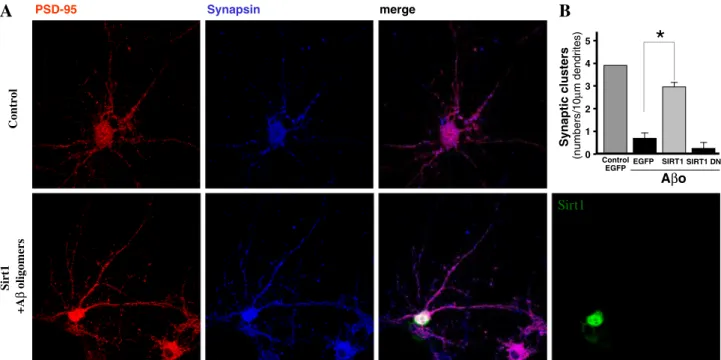
(7) 750. Mol Neurobiol (2014) 50:744–756. Neurodegenerative disorders are characterized by the progressive loss of specific neuronal populations and impaired cognitive and locomotor functions. The accumulation of mutant proteins such as α-synuclein and mutant huntingtin (Htt) are major pathological hallmark in PD and HD, respectively. A large body of evidence suggests a central role of mitochondrial dysfunction in the pathophysiology of these chronic neurodegenerative disorders. Here, we discuss the role of mitochondrial dysfunction, mitochondrial bioenergetics, mitophagy,. PSD-95. Synapsin. Control. A. merge. B Synaptic clusters. Mitochondrial Metabolism in Ageing and Neurodegeneration. mitochondrial fusion/fission and transcriptional dysregulation in the pathogenesis of these neurodegenerative diseases. The mitochondria are the principal energy source of the cell that converts nutrients into energy through cellular respiration. Compromised mitochondrial function has been described as a critical factor for enhanced ROS production and could be linked to numerous diseases, including those of the metabolic, cardiovascular systems and neurodegenerative disorders. Evidence suggests that mitochondrial biogenesis is regulated at least in part by PGC-1α, a transcriptional co-activator of peroxisome proliferator- activated receptor-γ (PPARγ), as well as other transcription factors [82]. It was therefore of considerable interest when it was shown that PGC-1α was in fact a deacetylation target of Sirt1 and that acetylation regulates PGC-1α activity [19, 83]. Recently, Sirt1 has been shown to function together with PGC-1α to promote adaptation to calorie restriction by regulating the genetic programs for gluconeogenesis and glycolysis in the liver. Sirt1 physically interacts with and deacetylates PGC-1α at multiple lysine sites, consequently increasing PGC-1α activity and leading to the induction of liver gluconeogenesis-related gene. (numbers/10μm dendrites). subunit remains unclear. However, these synaptic age-related changes may be a consequence of increased oxidative stress, decreased autophagy and/or accumulation of damaged cellular structures. The molecular and cellular basis of how calorie restriction might ameliorate some of the cognitive deficits associated with the ageing process warrants further investigation [81].. *. 5 4 3 2 1 0. Control EGFP EGFP. SIRT1 SIRT1 DN. Aβo. Sirt1 +Aβ oligomers. Sirt1. Fig. 4 Sirt1 protects rat hippocampal neurons against Aβ challenge. Aβ-derived neuronal damage is a well-recognized mechanism that occurs during AD. Moreover, increased oxidative stress caused by increased ROS levels is at the basis of Aβ neurotoxicity. Based on this knowledge, several research groups have focused to assess whether Sirt1 expression could lead to improved neuronal survival in the presence of Aβ. As indicated by different studies, the selected microphotographs show how Sirt1 overexpression not only leads to increased neuronal survival, but also to the protection of several synaptic markers related to the neuronal network functionality. Importantly, because neuronal loss is not a common condition during normal ageing, the protection of neuronal functionality demonstrated by Sirt1 suggests that its role goes further than the disease, being possible to consider it as a. support agent during normal ageing and during the treatment of some neurodegenerative disorders. a Primary rat E18 hippocampal neurons were transfected with Sirt1 (DIV 3); at day 14 post-transfection, neurons were treated; representative images of the control neurons and transfected with Sirt1 (green) challenged with Aβ oligomers, stained against PSD-95 (red) and synapsin (blue) stain, are shown (upper row). Neurons transfected with Sirt1 (green) and treated with Aβo are shown (bottom row). b Graph shows the different experimental groups analysed, control EGFP without treatment; neurons transfected with EGFP treated with Aβo; neurons transfected with Sirt1 and challenged with Aβo and neurons transfected with a dominant negative of Sirt1 challenged with Aβo, the pair of PSD-95/synapsin under control and treated with Aβo for 12 h.
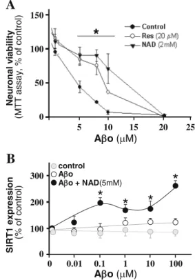
(8) Mol Neurobiol (2014) 50:744–756. the Aβ peptide derived from the proteolytic cleavage of the amyloid precursor protein (APP) and the intra-neuronal neurofibrillary tangles formed by aggregation of hyperphosphorylated tau [90]. Although the etiology of AD remains unclear [91], recently, increased importance has been granted to studies that attempt to identify the responsible toxin(s) for synaptic dysfunction and neuronal loss in AD. Several lines of evidence point to the involvement of mitochondrial dysfunction and oxidative stress as major factors in the progression of the disease [92]. Soluble Aβ oligomers have shown able to inhibit LTP, a paradigm in memory and synaptic plasticity [93]. In fact, oligomers are able to bind to the dendritic tree and are usually found at synaptic terminals, leading to reduced PSD-95 protein level, a main component of the post-synaptic densities which has a critical function in forming synaptic plasticity [41, 93]. Additional observations also suggest that deregulation of NMDA receptor, induced by soluble Aβ peptide bound to neuronal synapses, may lead to synaptic mitochondrial dysfunction and excessive ROS formation [43], probably by enhancing the synaptic failure. Calorie restriction increases the lifespan and decreases Aβ deposition in transgenic animal for AD [94]. The induction and activation by calorie restriction in the brain has been linked to neuroprotection against AD neuropathology [95]. Neuronal viability (MTT assay, % of control). A 150. * 100. 50. 0 5. 10. 15. 20. 25. A o ( M). B SIRT1 expression (% of control). transcription [84]. Given the role of Sirt1 as a mediator of calorie restriction and longevity, and the central role of reactive oxygen species, which are mainly produced as a consequence of mitochondrial dysfunction during the ageing process, it is plausible that PGC-1α and Sirt1 functions converge in tissues beyond the liver and in other catabolic tissues which have a high level of mitochondrial activity, such as the muscle and brown adipose tissue (BAT). Moreover, several mitochondrial fission-fusion proteins have been described under the control of PGC-1α, such as DRP1, FIS1 and Mfn [85]. Interestingly, it has been suggested that consecutive mitochondrial fission-fusion cycles allow both the final degradation and/or rescue through genetic material exchange of the defective organelles [86]. Since such a convergence could potentially impact on metabolic diseases, we have addressed part of our work not only in the context of the calorie restriction, but under conditions of calorie excess using the specific Sirt1 activator, resveratrol [87]. Together, these studies suggest that mitochondrial biogenesis might be regulated by tissue energetic status and that sirtuins represent important energy biosensors. Indeed, the notion that PGC-1α acetylation and function and, by extension, the mitochondrial activity are regulated in a nutrientdependent fashion by Sirt1 seems quite possible [19]. Nonetheless, the concept that Sirt1 is in turn able to respond to nutrient-sensitive changes in basal NAD+ levels has had until recently received little experimental support [88]. Indeed, despite resveratrol shown to induce mitochondrial biogenesis and to protect against metabolic decline, whether Sirt1 mediates these benefits is still the subject of continuous debate. In order to elucidate this relation, an inducible system which permits whole-body deletion of Sirt1 in adult mice, to circumvent the developmental defects of Sirt1 germline knockout mice, was used by Price et al. (2012). In this study, mice treated with a moderate dose of resveratrol showed increased mitochondrial biogenesis and function, AMPK activation and increased NAD+ levels in the skeletal muscle, whereas adult-induced Sirt1 knockout mice displayed none of these benefits. On the contrary, mice overexpressing Sirt1 mimicked these effects. Importantly, independent of resveratrol dosage, no improvements in mitochondrial function were observed in animals lacking Sirt1. Together, these data indicate that Sirt1 plays an essential role in resveratrol-induced stimulation of AMPK and improvement of mitochondrial function both in vitro and in vivo [89].. 751. control A o A o + NAD(5mM). 300. *. 200. * *. *. 1. 10. 100 0 0. 0.01. 0.1. 100. A o ( M). Alzheimer’s Disease (AD) AD is a progressive age-related neurodegenerative disorder characterized by the progressive loss of cholinergic neurons. The two main pathological hallmarks of AD include the extracellular amyloid plaques, formed by the aggregation of. Fig. 5 a Effect of resveratrol or NAD+ on cell viability measured by MTT assays. a Hippocampal neurons were treated with different Aβo concentrations for 24 h (black circles) or preincubated with 20 μM resveratrol for 2 h (white circle) or 2 mM of NAD+ (black triangle) before Aβo treatment. b ELISA was used to detect Sirt1 on hippocampal neurons. Cells were treated by 24 h with Aβo (white circle) or treated with Aβo plus 5 mM of NAD.
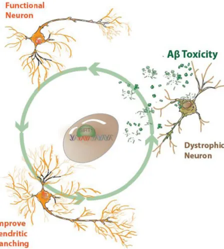
(9) 752. and is involved in the axonal protection against degeneration [26] and protection in models of neuronal damage based on hyperphosphorylation of the tau protein [30]. In a mice model of neurodegeneration overexpressing SOD1G37R and CDK5, Sirt1 appears upregulated, and this may be in response to neurodegenerative conditions [30]. On the other hand, a dramatic reduction of Sirt1 has been observed in cell culture experiments under acute stress by neurotoxins, in the same way to senescence mice models showing a downregulation of Sirt1 protein expression [27]. This differential regulation of Sirt1 protein levels in response to neurotoxic conditions may represent a neuroprotective adaptative response. Sirt1 Regulates Dendritic Spines and Dendritic Branching and Protects Hippocampal Neurons from Aβ Oligomers Damage Synapses have long been at the centre of neurobiology research. Synapses are the main communication gateway between neurons and the primary connections underlying neural circuits. Molecular interactions between the presynaptic and postsynaptic components induce a chemical matching which modulates the size and strength of synaptic signaling. Moreover, dendritic spines play a major role in memory Fig. 6 Neuronal effects derived from Sirt1 activation. The in vitro activation of Sirt1 by either NAD+ or resveratrol, or overexpression of Sirt1, induces several cellular mechanisms which lead to increased neuronal survival and protection of the neuronal network, at least through improved dendritic branching and preventing neuron dystrophy. It has been demonstrated that Sirt1 can significantly lower the levels of oligomerized Aβ; however, the precise molecular mechanisms related to this and other effects are still unknown. Mol Neurobiol (2014) 50:744–756. acquisition and LTP [96]. The number, size and shape of dendritic spines are critical parameters to determine neuronal function and to provide the structural basis for synaptic plasticity. Dendritic spines can be classified into three classes which are as follows: mushroom spines—the diameter of the head is much higher than the diameter of the neck; stubby spines—the diameter of the neck is similar to that of the head; and thin spines—the length is greater than the diameter and the length is less than 3 μm [97]. Overexpression of sirtuins has been shown to be sufficient to change dendritic morphogenesis and enhance dendritic arborization at early stages of development, while the interference of the catalytic deacetylase activity of Sirt1 leads to reduced number of dendritic branches. Indeed, it has been reported that Sirt1 regulates dendritic development and increases dendritic arborization in hippocampal neurons [28]. Brains of Sirt1 knockout mice exhibit normal morphology and dendritic spine structure, but display a decrease in dendritic branching, branch length and complexity of neuronal dendritic arborization [98]. These results suggest that the catalytic activity of Sirt1 is able to modulate spine architecture. Furthermore, resveratrol, a modulator of the Sirt1 activity, mimics the effect on dendritic arborization at early stages of development [98]. Similarly, NAD+, the Sirt1 co-substrate, has a protective effect against damage from Aβ oligomers (see Fig. 4), an effect that.
(10) Mol Neurobiol (2014) 50:744–756. has not been observed in neurons overexpressing a dominant negative form of Sirt1 (Sirt1 DN). On the other hand, hippocampal neurons overexpressing Sirt1 are more resistant against cytotoxic damage induced by Aβ oligomers [98]. The underlying pathways downstream of Sirt1 have been assessed by several authors which have verified the contribution of the Jun-terminal kinase (JNK), a downstream target of Rac [99]. Therefore, the Sirt1-mediated activity of resveratrol in dendritic development involves the contribution of Rho GTPases, specifically the ROCK pathway [100, 101]. In addition to the effect of Sirt1 in normal dendrite development and maintenance of synapsis (Fig. 5), it has been proposed that Sirt1 activity might play an important role also in the protection of several synaptic markers in different neurodegenerative disorders (Fig. 4) [30, 94, 102]. Regarding neuronal survival, one proposed mechanism by which Sirt1 might induce neuronal survival in AD is related to the beneficial effects observed for resveratrol, which exerts increased Sirt1 levels and promotes the proteasomal degradation of the Aβ peptide [103]. In hippocampal neuronal cultures treated with Aβ oligomers at different concentrations, the addition of either 20 μM of resveratrol or 2 mM of NAD+ induces an increase in neuronal viability (Fig. 5a) compared to the treatment with cytotoxic concentrations of Aβ oligomers alone. Additionally, neurons exposed to Aβ oligomers in the presence of NAD+ showed a significant increase in Sirt1 protein expression. These findings further highlight another NAD+-dependent mechanism that promotes resistance against Aβ neurotoxicity (Fig. 5b). Another possible mechanism by which Sirt1 induces neuronal survival in AD considers an increased α-secretase activity and the insulin degradation enzyme-IDE and warrants further investigation [95].. Conclusions As the world’s ageing population continues to rise and age appears to be a prominent risk factor in most neurodegenerative diseases, novel therapeutic strategies which delay the onset of age-related diseases are highly desirable. The agerelated molecular changes that occur in the brain are increased in age-related neurological diseases. There are multiple connections between neurodegenerative diseases, increased oxidative stress, decreased autophagy, mitochondrial dysfunction and the formation of misfolded proteins. The function of sirtuins in neuronal protection has been investigated in several models. Strong evidence suggests that sirtuins might be key master controllers of important metabolic pathways. In particular, if Sirt1 activity is increased, neurons are likely to be less vulnerable to neurotoxic damage, but if neurons are exposed to neurotoxic stimuli, Sirt1 expression level increases as a neuroprotective adaptative response. These two different mechanisms highlight how Sirt1 has a neuroprotective. 753. function in human neurodegenerative diseases (Fig. 6). The sirtuin co-substrate NAD+ demonstrates neuroprotective properties similar to Sirt1-dependent pathway activation involving calorie restriction. Mitochondrial dysfunction and changes to the mitochondria represent additional pathological characteristics of AD. Specifically, hippocampal neurons show a reduction in the number of mitochondrial enzyme activities including dehydrogenase enzymes such pyruvate dehydrogenase and cyclooxygenase enzymes. Increased activity of sirtuins can affect ROS production and increase resistance to these damaging effects. Oxidative stress has been shown to decrease Sirt1 expression in the rat hippocampus and cortex [104]. Sirt1 overexpression prevents oxidative stress-induced apoptosis and increases resistance to oxidative stress through deacetylation of a number of different transcription factors. The sirtuin family of enzymes, most importantly Sirt1, is strongly influenced by calorie restriction, which refers to a reduction in calorie intake without malnutrition, and is likely to have a wide range of benefits in humans. Acknowledgements This work was supported by grants PFB 12/2007 from the Basal Centre for Excellence in Science and Technology, FONDECYT 1120156 and MIFAB Institute and Fundación Ciencia y Vida to NCI and FONDECYT No. 11130033 to JMZ. NB is a recipient of the Alzheimer’s Australia and NHMRC Early Career Postdoctoral Research Fellowship at the University of New South Wales, Sydney, Australia. Conflict of Interest The authors declare that they have no competing interests.. References 1. Lutz W, Sanderson W, Scherbov S (2008) The coming acceleration of global population ageing. Nature 451:716–719 2. Joseph JA, Shukitt-Hale B, Casadesus G (2005) Reversing the deleterious effects of aging on neuronal communication and behavior: beneficial properties of fruit polyphenolic compounds. Am J Clin Nutr 81(1 Suppl):313S–316S 3. Rattan S (2006) Theories of biological aging: genes, proteins, and free radicals. Free Radic Res 40:1230–1238 4. Harman D (2006) Free radical theory of aging: an update. Ann NY Acad Sci 1067:10–21 5. Chen H, Chomyn A, Chan DC (2005) Disruption of fusion results in mitochondrial heterogeneity and dysfunction. J Biol Chem 280: 26185–92 6. Massudi H, Grant R, Guillemin GJ, Braidy N (2012) NAD+ metabolism and oxidative stress: the golden nucleotide on a crown of thorns. Redox Rep 17:28–46 7. Massudi H, Grant R, Braidy N, Guest J, Farnsworth B, Guillemin GJ (2012) Age-associated changes in oxidative stress and NAD+ metabolism in human tissue. PLoS One 7:e42357 8. Braidy N, Guillemin G, Mansour H, Chan-Ling T, Poljak A, Grant R (2011) Age related changes in NAD+ metabolism, oxidative stress and Sirt1 activity in Wistar rats. PLoS One 6:e19194 9. Wong E, Cuervo AM (2010) Integration of clearance mechanism: the proteosome and autophagy. Cold Spring Harb Perspect Biol 2: a006734.
(11) 754 10. Mizushima N, Levine B, Cuervo AM, Klionsky DJ (2008) Autophagy fights disease through cellular self-digestion. Nature 451:1069–1075 11. Bergamini E, Cavallini G, Donati A, Gori Z (2007) The role of autophagy in aging: its essential part in the anti-aging mechanism of caloric restriction. Ann N Y Acad Sci 1114:69–78 12. Bordone L, Guarente L (2005) Calorie restriction, SIRT1 and metabolism: understanding longevity. Nat Rev Mol Cell Biol 6:298–305 13. Hekimi S (2006) How genetic analysis tests theories of animal aging. Nat Genet 38:985–991 14. Jayasena T, Poljak A, Smythe G, Braidy N, Munch G, Sachdev P (2013) The role of polyphenols in the modulation of sirtuins and pathways involved in Alzheimer’s disease. Ageing Res Rev 12: 867–883 15. Haigis MC, Guarente LP (2006) Mammalian sirtuins—emerging roles in physiology, aging, and calorie restriction. Genes and Dev 20:2913–2921 16. Tanner KG, Landry J, Sternglanz, Denu JM (2000) Silent information regulator 2 family of NAD-dependent histone/protein deacetylases generates a unique product, 1-O-acetyl-ADP-ribose. Proc Natl Acad Sci U S A 97:14178–82 17. Fu XH, Meng FL, Hu Y, Zhou JQ (2008) Candida albicans, a distinctive fungal model for cellular aging study. Aging Cell 7: 746–57 18. Zhang F, Wang S, Gan L, Vosler PS, Gao Y, Chen J (2011) Protective effects and mechanisms of sirtuins in the nervous system. Prog Neurobiol 95:373–395 19. Herskovits AZ, Guarente L (2013) Sirtuin deacetylases in neurodegenerative diseases of aging. Cell Res 23:746–758 20. Cooper HM, Spelbrink JN (2008) The human SIRT3 protein deacetylase is exclusively mitochondrial. Biochem J 411:279–85 21. Rose G, Dato S, Altomare K, Bellizzi D, Garasto S, Greco V, Passarino G, Feraco E, Mari V, Barbi C, BonaFe M, Franceschi C, Tan Q, Boiko S, Yashin AI, De Benedictis G (2003) Variability of the SIRT3 gene, human silent information regulator Sir2 homologue, and survivorship in the elderly. Exp Gerontol 38:1065–70 22. Lombard DB, Alt FW, Cheng HL, Bunkenborg J, Streeper RS, Mostoslavsky R, Kim J, Yancopoulos G, Valenzuela D, Murphy A, Yang Y, Chen Y, Hirschey MD, Bronson RT, Haigis M, Guarente LP, Farese RV Jr, Weissman VE, Schwer B (2007) Mammalian Sir2 homolog SIRT3 regulates global mitochondrial lysine acetylation. Mol Cell Biol 27:8807–8814 23. Schlicker C, Gertz M, Papatheodorou P, Kachholz B, Becker CF, Steegborn C (2008) Substrates and regulation mechanisms for the human mitochondrial sirtuins Sirt3 and Sirt5. J Mol Biol 382: 790–801 24. Hisahara S, Chiba S, Matsumoto H, Horio Y (2005) Transcriptional regulation of neuronal genes and its effect on neural functions: NAD-dependent histone deacetylase SIRT1 (Sir2alpha). J Pharmacol Sci 98:200–204 25. Blander G, Guarente L (2004) The Sir2 family of protein deacetylases. Annu Rev Biochem 73:417–435 26. Araki T, Sasaki Y, Milbrandt J (2004) Increased nuclear NAD biosynthesis and SIRT1 activation prevent axonal degeneration. Science 305:1010–1013 27. Pallàs M, Pizarro JG, Gutierrez-Cuesta J, Crespo-Biel N, Alvira D, Tajes M, Yeste-Velasco M, Folch J, Canudas AM, Sureda FX, Ferrer I, Camins A (2008) Modulation of SIRT1 expression in different neurodegenerative models and human pathologies. Neuroscience 154:1388–1397 28. Braidy N, Jayasena T, Poljak A, Sachdev P (2012) Sirtuins in cognitive ageing and Alzheimer’s disease. Curr Opin Psychiatry 25:226–30 29. Parker JA, Arango M, Abderrahmane S, Lambert E, Tourette C, Catoire H, Néri C (2005) Resveratrol rescues mutant polyglutamine. Mol Neurobiol (2014) 50:744–756. 30.. 31.. 32.. 33. 34.. 35. 36.. 37.. 38. 39.. 40.. 41.. 42.. 43.. 44.. 45.. 46.. cytotoxicity in nematode and mammalian neurons. Nat Genet 37: 349–50 Kim D, Nguyen MD, Dobbin M, Fischer A, Sananbenesi F, Rodgers J, Delalle I, Baur J, Sui G, Armour S, Puigserver P, Sinclair D, Tsai L (2007) SIRT1 deacetylase protects against neurodegeneration in models for Alzheimer’s disease and amyotrophic lateral sclerosis. The EMBO Journal 26:3169–3179 Chen D, Steele AD, Hutter G, Bruno J, Govindarajan A, Easlon E, Lin SJ, Aguzzi A, Lindquist S, Guarente L (2008) The role of calorie restriction and SIRT1 in prion-mediated neurodegeneration. Exp Gerontol 43:1086–93 Barger JL, Kayo T, Vann JM, Arias EB, Wang J, Hacker TA, Wang Y, Raederstorff D, Morrow JD, Leeuwenburgh C, Allison DB, Saupe KW, Cartee GD, Weindruch R, Prolla TA (2008) A low dose of dietary resveratrol partially mimics caloric restriction and retards aging parameters in mice. PLoS ONE 3:e2264 Yang Q, Guan KL (2007) Expanding mTOR signaling. Cell Res 17: 666–681 Loeb LL, Wallace DC, Martin GM (2005) The mitochondrial theory of aging and its relationship to reactive oxygen species damage and somatic mtDNA mutations. Proc Natl Acad Sci U S A 102:18769–18770 Rattan SI (2006) Theories of biological aging: genes, proteins, and free radicals. Free Radic Res 40:1230–1238 Keating DJ (2008) Mitochondrial dysfunction, oxidative stress, regulation of exocytosis and their relevance to neurodegenerative disease. J Neurochem 104:298–305 Miranda S, Opazo C, Larrondo LF, Muñoz FJ, Ruiz F, Leighton F, Inestrosa NC (2000) The role of oxidative stress in the toxicity induced by amyloid beta-peptide in Alzheimer’s disease. Prog Neurobiol 62:633–48 Serrano F, Klann E (2004) Reactive oxygen species and synaptic plasticity in the aging hippocampus. Ageing Res Rev 3:431–443 Ueda Y, Doi T, Nagatomo K, Nakajima A (2007) In vivo activation of N-methyl-D-aspartate receptors generates free radicals and reduces antioxidant ability in the rat hippocampus: experimental protocol of in vivo ESR spectroscopy and microdialysis for redox status evaluation. Brain Res 1178:20–27 Papadia S, Soriano FX, Léveillé F, Martel MA, Dakin KA, Hansen HH, Kaindl A, Sifringer M, Fowler J, Stefovska V, McKenzie G, Craigon M, Corriveau R, Ghazal P, Horsburgh K, Yankner BA, Wyllie DJ, Ikonomidou C, Hardingham GE (2008) Synaptic NMDA receptor activity boosts intrinsic antioxidant defenses. Nat Neurosci 11:476–487 Lacor PN, Buniel MC, Chang L, Fernandez SJ, Gong Y, Viola KL, Lambert MP, Velasco PT, Bigio EH, Finch CE, Krafft GA, Klein WL (2004) Synaptic targeting by Alzheimer’s-related amyloid βoligomers. J Neurosci 24:10191–10200 Dinamarca MC, Ríos JA, Inestrosa NC (2012) Postsynaptic receptors for amyloid-β oligomers as mediators of neuronal damage in Alzheimer’s disease. Front Physiol 3:464 De Felice FG, Velasco PT, Lambert MP, Viola K, Fernandez SJ, Ferreira ST, Klein WL (2007) Aβ oligomers induce neuronal oxidative stress through an N-methyl-D-aspartate receptor-dependent mechanism that is blocked by the Alzheimer drug memantine. J Biol Chem 282:11590–11601 Magnusson KR, Nelson SE, Young AB (2002) Age-related changes in the protein expression of subunits of the NMDA receptor. Mol Brain Res 99:40–45 Lombard DB, Chua KF, Mostoslavsky R, Franco S, Gostissa M, Alt FW (2005) DNA repair, genome stability, and aging. Cell 120: 497–512 Hirano T, Yamaguchi R, Asami S, Iwamoto N, Kasai H (1996) 8Hydroxyguanine levels in nuclear DNA and its repair activity in rat organs associated with age. J Gerontol A Biol Sci Med Sci 51: B303–B307.
(12) Mol Neurobiol (2014) 50:744–756 47. Shen S, Cooley DM, Glickman LT, Glickman N, Waters DJ (2001) Reduction in DNA damage in brain and peripheral blood lymphocytes of elderly dogs after treatment with dehydroepiandrosterone (DHEA). Mutat Res 480–481:153–162 48. Krishna TH, Mahipal S, Sudhakar A, Sugimoto H, Kalluri R, Rao KS (2005) Reduced DNA gap repair in aging rat neuronal extracts and its restoration by DNA polymerase beta and DNA-ligase. J Neurochem 92:818–823 49. Giovannelli L, Decorosi F, Dolara P, Pulvirenti L (2003) Vulnerability to DNA damage in the aging rat substantia nigra: a study with the comet assay. Brain Res 969:244–247 50. Lu T, Pan Y, Kao SY, Li C, Kohane I, Chan J, Yankner BA (2004) Gene regulation and DNA damage in the ageing human brain. Nature 429:883–891 51. Rutten BP, Schmitz C, Gerlach OH, Oyen HM, de Mesquita EB, Steinbusch HW, Korr H (2007) The aging brain: accumulation of DNA damage or neuron loss? Neurobiol Aging 28:91–98 52. Okawara M, Katsuki H, Kurimoto E, Shibata H, Kume T, Akaike A (2007) Resveratrol protects dopaminergic neurons in midbrain slice culture from multiple insults. Biochem Pharmacol 73:550–560 53. Bureau G, Longpré F, Martinoli MG (2008) Resveratrol and quercetin, two natural polyphenols, reduce apoptotic neuronal cell death induced by neuroinflammation. J Neurosci Res 86:403–10 54. Hubbard BP, Gomes AP, Dai H, Li J, Case AW, Considine T, Riera TV, Lee JE, SY E, Lamming DW, Pentelute BL, Schuman ER, Stevens LA, Ling AJ, Armour SM, Michan S, Zhao H, Jiang Y, Sweitzer SM, Blum CA, Disch JS, Ng PY, Howitz KT, Rolo AP, Hamuro Y, Moss J, Perni RB, Ellis JL, Vlasuk GP, Sinclair DA (2013) Evidence for a common mechanism of SIRT1 regulation by allosteric activators. Science 339:1216–9 55. Klionsky DJ (2007) Autophagy: from phenomenology to molecular understanding in less than a decade. Nat Rev Mol Cell Biol 8: 931–937 56. Shintani T, Klionsky DJ (2004) Autophagy in health and disease: a double-edged sword. Science 306:990–995 57. Lum JJ, DeBerardinis RJ, Thompson CB (2005) Autophagy in metazoans: cell survival in the land of plenty. Nat Rev Mol Cell Biol 6:439–448 58. Blommaart EF, Krause U, Schellens JP, Vreeling-Sindelarova H, Meijer AJ (1997) The phosphatidylinositol 3-kinase inhibitors wortmannin and LY294002 inhibit autophagy in isolated rat hepatocytes. Eur J Biochem 243:240–246 59. Ma T, Hoeffer CA, Capetillo-Zarate E, Yu F, Wong H, Lin MT, Tampellini D, Klann E, Blitzer RD, Gouras GK (2010) Dysregulation of the mTOR pathway mediates impairment of synaptic plasticity in a mouse model of Alzheimer’s disease. PLoS One 5:e12845 60. Yang H, Yang T, Baur JA, Perez, Matsui T, Carmona JJ, Lamming DW, Souza-Pinto NC, Bohr VA, Rosenzweig A, de Cabo R, Sauve AA, Sinclair DA (2007) Nutrient-sensitive mitochondrial NAD+ levels dictate cell survival. Cell 130:1095–1107 61. Gouras GK (2013) mTOR: at the crossroads of aging, chaperones, and Alzheimer’s disease. J Neurochem 124(6):747–748. doi:10. 1111/jnc.12098 62. Cai Z, Yan LJ (2013) Rapamycin, autophagy, and Alzheimer’s disease. J Biochem Pharmacol Res 1:84–90 63. Urbanska M, Gozdz A, Swiech LJ, Jaworski J (2012) Mammalian target of rapamycin complex 1 (MTORC1) and 2 (MTORC2) control the dendritic arbor morphology of hippocampal neurons. J Biol Chem 287:30240–56 64. Cunningham JT, Rodgers JT, Arlow DH, Vazquez F, Mootha VK, Puigserver P (2007) mTOR controls mitochondrial oxidative function through a YY1-PGC-1α transcriptional complex. Nature 450: 736–740 65. Terman A (1995) The effect of age on formation and elimination of autophagic vacuoles in mouse hepatocytes. Gerontology 41:319–26. 755 66. Kurz T, Terman A, Gustafsson B, Brunk UT (2008) Lysosomes and oxidative stress in aging and apoptosis. Biochim Biophys Acta 1780:1291–303 67. Ahmed I, Liang Y, Schools S, Dawson VL, Dawson TM, Savitt JM (2012) Development and characterization of a new Parkinson’s disease model resulting from impaired autophagy. J Neurosci 14: 16503–9 68. Hara T, Nakamura K, Matsui M, Yamamoto A, Nakahara Y, SuzukiMigishima R, Yokoyama M, Mishima K, Saito I, Okano H, Mizushima N (2006) Suppression of basal autophagy in neural cells causes neurodegenerative disease in mice. Nature 441:885–889 69. Komatsu M, Wang QJ, Holstein GR, Friedrich VL Jr, Iwata J, Kominami E, Chait BT, Tanaka K, Yue Z (2007) Essential role for autophagy protein Atg7 in the maintenance of axonal homeostasis and the prevention of axonal degeneration. Proc Natl Acad Sci U S A 104:14489–14494 70. Lee IH, Cao L, Mostoslavsky R, Lombard DB, Liu J, Bruns NE, Tsokos M, Alt FW, Finkel T (2008) A role for the NAD-dependent deacetylase Sirt1 in the regulation of autophagy. Proc Natl Acad Sci U S A 105:3374–3379 71. Raghavan A, Shah ZA (2012) Sirtuins in neurodegenerative diseases: a biological-chemical perspective. Neurodegenerative Dis 9: 1–10 72. McBurney MW, Clark-Knowles KV, Caron AZ, Gray DA (2013) SIRT1 is a highly networked protein that mediates the adaptation to chronic physiological stress. Genes Cancer 4:125–34 73. King MA, Hands S, Hafiz F, Mizushima N, Tolkovsky AM, Wyttenbach A (2008) Rapamycin inhibits polyglutamine aggregation independently of autophagy by reducing protein synthesis. Mol Pharmacol 73:1052–1063 74. Drachman DA (2006) Aging of the brain, entropy, and Alzheimer disease. Neurology 67:1340–1352 75. Stark AK, Toft MH, Pakkenberg H, Fabricius K, Eriksen N, Pelvig DP, Moller M, Pakkenberg B (2007) The effect of age and gender on the volume and size distribution of neocortical neurons. Neuroscience 150:121–130 76. Sherwood CC, Gordon AD, Allen JS, Phillips KA, Hof PR, Hopkins WD (2011) Aging of the cerebral cortex differs between humans and chimpanzees. Proc Natl Acad Sci U S A 108: 13029–12034 77. Rapp PR, Deroche PS, Mao Y, Burwell RD (2002) Neuron number in the parahippocampal region is preserved in aged rats with spatial learning deficits. Cereb Cortex 12:1171–1179 78. Burke SN, Ryan L, Barnes CA (2012) Characterizing cognitive aging of recognition memory and related processes in animal models and in humans. Front Aging Neurosci 4:15 79. Virgili M, Monti B, Polazzi E, Angiolini G, Contestabile A (2001) Topography of neurochemical alterations in the CNS of aged rats. Int J Dev Neurosci 19:109–116 80. Monti B, Virgili M, Contestabile A (2004) Alterations of markers related to synaptic function in aging rat brain, in normal conditions or under conditions of long-term dietary manipulation. Neurochem Int 44:579–84 81. Eckles-Smith K, Clayton D, Bickford P, Browning MD (2000) Caloric restriction prevents age-related deficits in LTP and in NMDA receptor expression. Brain Res Mol Brain Res 78:154–162 82. Fernandez-Marcos PJ, Auwerx J (2011) Regulation of PGC-1α, a nodal regulator of mitochondrial biogenesis. Am J Clin Nutr 93: 884S–890S 83. Nemoto S, Fergusson MM, Finkel T (2005) SIRT1 functionally interacts with the metabolic regulator and transcriptional coactivator PGC-1α. J Biol Chem 280:16456–16460 84. Yamamoto T, Shimano H, Nakagawa Y, Ide T, Yahagi N, Matsuzaka T, Nakakuki M, Takahashi A, Suzuki H, Sone H, Toyoshima H, Sato R, Yamada N (2004) SREBP-1 interacts with hepatocyte nuclear factor-4 alpha and interferes with PGC-1.
(13) 756. 85.. 86. 87. 88.. 89.. 90.. 91. 92.. 93.. 94.. Mol Neurobiol (2014) 50:744–756 recruitment to suppress hepatic gluconeogenic genes. J Bol Chem 279:12027–35 Zolezzi JM, Silva-Alvarez C, Ordenes D, Godoy JA, Carvajal FJ, Santos MJ, Inestrosa NC (2013) Peroxisome proliferator-activated receptor (PPAR) γ and PPARα agonists modulate mitochondrial fusion-fission dynamics: relevance to reactive oxygen species (ROS)-related neurodegenerative disorders? PLoS One 8:e64019 Itoh K, Nakamura K, Iijima M, Sesaki H (2012) Mitochondrial dynamics in neurodegeneration. Trends Cell Biol 23:64–71 Finkel T, Deng CX, Mostoslavsky R (2009) Recent progress in the biology and physiology of sirtuins. Nature 460:587–591 Sack MN, Finkel T (2012) Mitochondrial metabolism, sirtuins, and aging. Cold Spring Harb Perspect Biol 4:1–10. doi:10.1101/ cshperspect.a013102 Price NL, Gomes AP, Ling AJ, Duarte FV, Martin-Montalvo A, North BJ, Agarwal B, Ye L, Ramadori G, Teodoro JS, Hubbard BP, Varela AT, Davis JG, Varamini B, Hafner A, Moaddel R, Rolo AP, Coppari R, Palmeira CM, de Cabo R, Baur JA, Sinclair DA (2012) SIRT1 is required for AMPK activation and the beneficial effects of resveratrol on mitochondrial function. Cell Metab 15:675–90 Selkoe DJ (2004) Cell biology of protein misfolding: the examples of Alzheimer’s and Parkinson’s diseases. Nat Cell Biol 6: 1054–1061 Mattson MP (2004) Pathways towards and away from Alzheimer’s disease. Nature 430:631–639 Reddy PH, Beal MF (2008) Amyloid beta, mitochondrial dysfunction and synaptic damage: implications for cognitive decline in aging and Alzheimer’s disease. Trends Mol Med 14:45–53 Haass C, Selkoe D (2007) Soluble protein oligomers in neurodegeneration: lessons from the Alzheimer’s amyloid β-peptide. Nat Rev Mol Cell Biol 8:101–112 Patel NV, Gordon MN, Connor KE, Good RA, Engelman RW, Mason J, Morgan DG, Morgan TE, Finch CE (2005) Caloric restriction attenuates Aβ deposition in Alzheimer transgenic models. Neurobiol Aging 26:995–1000. 95. Qin W, Yang T, Ho L, Zhao Z, Wang J, Chen L, Zhao W, Thiyagarajan M, MacGrogan D, Rodgers JT, Puigserver P, Sadoshima J, Deng H, Pedrini S, Gandy S, Sauve AA, Pasinetti GM (2006) Neuronal SIRT1 activation as a novel mechanism underlying the prevention of Alzheimer disease amyloid neuropathology by calorie restriction. J Biol Chem 281:21745–21754 96. Alvarez VA, Sabatini BL (2007) Anatomical and physiological plasticity of dendritic spines. Annu Rev Neurosci 30:79–97 97. Harris KM, Jensen FE, Tsao B (1992) Three-dimensional structure of dendritic spines and synapses in rat hippocampus (CA1) at postnatal day 15 and adult ages: implications for the maturation of synaptic physiology and long-term potentiation. J Neurosci 12: 2685–705 98. Michán S, Li Y, Chou MM, Parrella E, Ge H, Long JM, Allard JS, Lewis K, Miller M, Xu W, Mervis RF, Chen J, Guerin KI, Smith LE, McBurney MW, Sinclair DA, Baudry M, de Cabo R, Longo VD (2010) SIRT1 is essential for normal cognitive function and synaptic plasticity. J Neurosci 30:9695–707 99. Weston CR, Davis RJ (2002) The JNK signal transduction pathway. Curr Opin Genet Dev 12:14–21 100. Codocedo JF, Allard C, Godoy JA, Varela-Nallar L, Inestrosa NC (2012) SIRT1 regulates dendritic development in hippocampal neurons. PLoS One 7:e47073. doi:10.1371/journal.pone.0047073 101. Negishi M, Katoh H (2002) Rho family GTPases as key regulators for neuronal network formation. J Biochem 132:157–66 102. Duan W, Mattson MP (1999) Dietary restriction and 2deoxyglucose administration improve behavioral outcome and reduce degeneration of dopaminergic neurons in models of Parkinson’s disease. J Neurosci Res 57:195–206 103. Marambaud P, Zhao H, Davies P (2005) Resveratrol promotes clearance of Alzheimer’s disease amyloid-β peptides. J Biol Chem 280:37377–37382 104. Wu A, Ying Z, Gomez-Pinilla F (2006) Oxidative stress modulates Sir2alpha in rat hippocampus and cerebral cortex. Eur J Neurosci 23:2573–2580.
(14)
Figure
Documento similar
Astrometric and photometric star cata- logues derived from the ESA HIPPARCOS Space Astrometry Mission.
Ribeiro, M.O., et al., Expression of uncoupling protein 1 in mouse brown adipose tissue is thyroid hormone receptor-beta isoform specific and required for adaptive
Given the much higher efficiencies for solar H 2 -generation from water achieved at tandem PEC/PV devices ( > 10% solar-to-H 2 energy efficiency under simulated sunlight) compared
TipC and the chorea-acanthocytosis protein VPS13A regulate autophagy in Dictyostelium and human HeLa cells.. Sandra Muñoz-Braceras a , Rosa Calvo a & Ricardo
teriza por dos factores, que vienen a determinar la especial responsabilidad que incumbe al Tribunal de Justicia en esta materia: de un lado, la inexistencia, en el
Even though the 1920s offered new employment opportunities in industries previously closed to women, often the women who took these jobs found themselves exploited.. No matter
In the “big picture” perspective of the recent years that we have described in Brazil, Spain, Portugal and Puerto Rico there are some similarities and important differences,
SIRT1 Tg/Tg mice displayed a moderate SIRT1 overexpression in skeletal muscle, liver, white adipose tissue (WAT) and BAT, both at the mRNA and protein levels (Figure 1A,B).. In
