Peroxisome Proliferator activated Receptors and Alzheimer's Disease: Hitting the Blood Brain Barrier
14
0
0
Texto completo
(2) Mol Neurobiol (2013) 48:438–451. 439. (1) the BBB, (2) the blood–CSF barrier, and (3) the arachnoid epithelium [9, 10]. The BBB formation begins during embryonic development as a consequence of the close relationship established between blood vessels and neuroectodermal cells [11]. From the subarachnoid space, the pial arteries, the main blood vessels of the brain, project the intracerebral arteries to the brain, which in turn, ramify to ultimately form the brain capillaries [12]. The neurovascular unit is considered to be the minimal functional unit of the BBB, and three cellular subtypes can be identified: (1) vascular, composed of endothelial cells, pericytes, and vascular smooth muscular cells; (2) glial,. composed of astrocytes, microglia, and oligodendroglia; and (3) neurons [11, 12] (Fig. 1). When mature, the BBB is a highly specialized structure where brain capillaries comprise a single endothelial cell connected with itself and with neighbor cells through occluding TJ and through nonoccluding adherens junctions (AJ) [13, 14], each of these comprised of a set of specific proteins. This primary structure is surrounded by pericytes, which are in direct contact with astrocyte endfeet [11, 14, 15]. The BBB functions not only as a barrier limiting the entrance of several substances into the brain [14] but also as a permeable structure that is able to ensure oxygen. Fig. 1 Main components of the BBB. The BBB is a complex structure composed of several cell types and characterized by distinct expression patterns of different transport and adhesion proteins. The combination of these components allows the BBB to exhibit low paracellular permeability and critically determinant cellular transport. The diagram represents a brief description of the structure of the BBB. At the brain capillaries, a monocellular layer of endothelial cells constitutes the basis of the barrier. Each endothelial cell encircles the lumen of the capillary and seals it, through establishment of TJ and AJ at the ends of. the cell as well as with the adjacent cells. The endothelial wall is closely surrounded by pericytes, constituting an additional cellular layer of the BBB. Finally, astrocyte end-feet processes attach to the barrier forming an additional cellular envelope. Despite some of the most important BBB transport systems included in the diagram (ABC ATP-binding cassette, LRP low-density lipoprotein receptor-related protein, RAGE receptor for advanced glycation end products) shown only to be expressed by endothelial cells, several of these transporters are also expressed by the other cellular components of the BBB.
(3) 440. Mol Neurobiol (2013) 48:438–451. and glucose delivery to brain tissue as well as removal of different metabolic end products, which helps to maintain brain homeostasis [16]. Additionally, correct functionality of the BBB is critical for neurons mainly because of the precise balance of ion gradients required to allow for electrical communication between them [11].. that link membrane proteins to the actin cytoskeleton [16, 17], we found the following: zonula occludens (ZO) protein-1, protein-2, and protein-3 [23–25], containing a PDZ domain; and non-PDZ proteins, such as cingulin [16] and the junctionassociated coiled-coil protein (JACOP)/paracingulin [16, 26]. Adherens Junctions. BBB traffic can be separated into a low permeable paracellular component, including the TJ and AJ, and a cellular component, composed mainly of endothelial cells expressing several specific carriers [17]. Only small lipidsoluble molecules are allowed to cross the BBB freely [15].. AJ are also responsible for cell–cell adhesion and critical functions have been described for it, such as contact inhibition during vascular growth and remodeling, cell polarity initiation, and being fundamental for TJ formation [16, 17]. The main proteins of AJ are VE-cadherin [27], an armadillo protein, and platelet endothelial cell adhesion molecule 1 (PECAM-1) and they are usually linked to the catenin protein family [17] (Table 1).. Low Paracellular Permeability. Blood–Brain Barrier Transport. The seal between endothelial cells depends on TJ and AJ, and forces molecules to undergo transcellular transport [16]. Physically, from the lumen of brain capillaries, TJ are the first intercellular seal followed by AJ, as a combined unit.. The transport across the BBB is highly specialized and depends on the expression of several transporters by the endothelial cells, which allow bidirectional traffic in order to maintain brain homeostasis (Table 2). Different authors have used diverse criteria to classify these transporters [16, 17, 28]. Glucose transporter 1 (GLUT1), monocarboxylate transporter 1 (MCT1), L1, and y+ amino acid transporters, excitatory acidic amino acid transporters (EAAT-1, EAAT-2, and EAAT-3), constitute the first group of transporters in charge of nutrients, several energy source molecules, lactate, and amino acids into and out of the brain [17, 28]. It is important to mention that EAATs determine the removal of glutamate from the brain, playing a critical role in the prevention of glutamate-induced excitotoxicity [29]. Another important group of BBB transporters is composed of the adenosine triphosphate (ATP)-binding cassette (ABC) efflux transporters, particularly ABCB1 (P-glycoprotein) [30]; the multidrug resistance proteins (MRP or ABCC), such as MRP-1, MRP-4, MRP-5, and MRP-6 [16]; and the breast cancer resistance protein (BCRP or ABCG2) [31]. Moreover, endothelial protein C receptor (EPCR), insulin receptor (IR), transferrin receptor (TFR), low-density lipoprotein receptor-related protein 1 (LRP1), peptide transport system (PTS-1, PTS-2, and PTS-3), and PTS4-vasopressin V1a receptor (V1AR) are specialized transporters for peptides [17, 28, 32, 33]. As mentioned, the electrical properties of brain networks are provided by a precise ion balance achieved and maintained through a whole range of ion transporters. The sodium pump (Na + /K + -ATPase), sodium–potassium–2 chloride (Na + /K + /2Cl − ), sodium–hydrogen exchanger (Na+/H+), sodium–calcium (Na+/Ca++), and chloride–bicarbonate exchanger (Cl−/HCO3−) are the representative members of ion transporters present in the BBB [34–36]. Additionally, the BBB possesses enriched lipid microdomains (lipid rafts), composed of caveolae, which exert further. Blood–Brain Barrier Carrier System. Tight Junctions Molecular components of TJ (Table 1) can be divided into membrane and cytoplasmic proteins [16, 17]. Occludin [17, 18], claudins [17–19], the endothelial cell-selective adhesion molecules [20], and the junctional adhesion molecule-A [21, 22] constitutes the first group and are responsible for cell–cell anchorage. In the second group, acting as the scaffold proteins. Table 1 Main molecular components of tight and adherens junctions Tight junction Claudins Occludins Junctional adhesion molecule A (JAM-A) Endothelial cell-selective adhesion molecule (ESMA) Zonula occludens Calcium-dependent serine protein kinase (CASK) Cingulin Multi-PDZ protein 1 (MUPP1) Membrane-associated guanylate kinase (MAGI) Adherens junction Vascular-endothelial cadherin (VE-cadherin) Platelet endothelial cell adhesion molecule (PECAM-1) Catenin (α, β, χ).
(4) Mol Neurobiol (2013) 48:438–451 Table 2 BBB transporter system. 441. GLUT1 MCT1 L1 y+ XG− N ASC LNAA EAAT N. Glucose Lactate Essential amino acids Cationic amino acids Elimination of acidic amino acids Elimination of nitrogen rich amino acids Elimination of non-essential amio acids Elimination of essential amino acids Elimination of excitatory amino acids Nitrogen-rich amino acids. Na+/K+/ATPase Cl−/HCO3− Na+, K+/2Cl− H+/Na+. Ions. ATP-binding cassette (ABCB1, ABCC, ABCG2) Endothelial protein C receptor (EPCR) Insulin receptor (IR) Low-density lipoprotein receptor-related protein (LRP) Peptide transport system (PTS). Peptides. regulation of BBB traffic [37]. Furthermore, several receptors have been described to be associated with caveolar membranes, such as insulin and the receptor for advanced glycation end products (RAGE) [30]. Moreover, even when the BBB constitutes a physical barrier that allows for the exchange of a wide range of substances in and out of the brain through specific transporters, enzymatic activity has also been described for each cellular component of the BBB, offering additional metabolic protection against potentially neurotoxic compounds that could cross the BBB [32]. Available Cellular Blood–Brain Barrier Models Even when the main objective of the present review is not directly related with this particular issue, some words should be mentioned about this important matter. The current available BBB models can be divided into two groups: nonhuman and human-derived, and both can be further divided, according to the origin of the blood vessels, into noncerebral or cerebral endothelial models [16]. Despite several available nonhuman (noncerebral and cerebral) as well as human (noncerebral) cell models, such as MDCK, HUVEC, RBE4, GP8, GPNT, or primary cultures, it is important to mention that only a few of these retain the main characteristics of the BBB [16]. RBE4, GP8, GPNT, b.End3, and primary cultures of brain endothelial cells express most of the efflux/influx carriers as well as junctional proteins, but we must also remember in this case that these are murine models of the BBB and that, even by giving us an initial understanding of the complexity of the BBB, are quite far from indicating how the human BBB. functions [16, 38–41]. Considering the critical role that the BBB carrier system plays in Alzheimer's disease (AD) pathogenesis, it is of most importance to have a more reliable model of the human BBB that expresses as many components as possible, to understand its main characteristics. The hCMEC/D3 cells, described by Weksler et al. [42], are considered to be one of the most significant models for BBB studies, mainly because they correspond to a cerebral vessel human-derived BBB model that expresses the main characteristics of the human BBB [10, 42]. Additionally, considering the high complexity of the BBB, due to the interaction of several cell types, cocultures with glial cells and pericytes have emerged as more complete and complex models to study BBB properties [10, 16]. The selection of the appropriate model is critical when structural and physiological properties of the BBB are assessed, particularly in AD, not only because the integrity of the “barrier” is highly important but also because of the expression patterns of the several carriers involved in amyloid-β (Aβ) clearance.. Blood–Brain Barrier and Neurodegenerative Disorders Despite the known existence of the BBB for more than a century, only in the last few decades have significant efforts been made in order to understand the real impact of the role of the BBB on several neurodegenerative disorders. Of course, each of these disorders has its own etiology and particular hallmarks; however, the health or integrity of the BBB has often been shown to be compromised, and the.
(5) 442. severity of changes observed usually relates to the progression of the disease [17, 30, 43]. Several authors have analyzed the particularities of the BBB in different disorders, such as epilepsy [44, 45], multiple sclerosis [46], AD, Parkinson's disease, and Huntington's disease [17, 30, 31], among others, and have found an altered function of TJs, AJs, or in the carrier transport system that controls the BBB traffic, such as occludins, claudins, cadherins, EAAT, MCT1, and GLUT1.. Blood–Brain Barrier and Alzheimer's Disease AD is an age-associated neurodegenerative disorder characterized by progressive memory and cognitive impairment that eventually leads to death [47, 48]. Clinically, AD progression reflects gradual neurodegeneration with a compromise of short-term memory at the beginning of the disease followed by long-term memory loss [49]. Brain atrophy and gradual loss of neurons, mainly in the hippocampus (HC), frontal cortex (FC) and limbic areas, together with extracellular accumulation of Aβ plaques and intraneuronal formation of neurofibrillary tangles (NFT), composed of hyperphosphorylated aggregates of microtubule-associated protein tau, are pathologic hallmarks of AD [48–50]. In AD patients, whether in the familial or in the sporadic form, increased levels of Aβ are usual and considered to be the basis of the pathologic changes observed during AD progression [51]. When Aβ accumulates around blood vessels, it leads to neurovascular dysfunction and cerebral amyloid angiopathy [17]. Indeed, several changes take place in the cerebral blood vessels of AD patients, including loss of vascular density, decreased luminal diameter of vessels and capillaries, and thickness of vessels walls [52]. However, even when the relationship between Aβ accumulation and BBB damage seems evident, it is important to consider that increased levels of Aβ in the brain interstitial fluid depends not only on the production rate but also on the clearance rate from the brain. In fact, the recently published work of Cramer et al. [53] suggested the critical role of Aβ clearance in AD and the importance of considering Aβrelated transporters as targets in future AD therapies. Compromised Transporters in Alzheimer's Disease As a key hallmark of AD, the proper excretion of Aβ from the brain, preventing its neurotoxic accumulation, depends on an appropriate transport through the BBB (Fig. 1). LRP1 and LRP2 LRP are widely expressed by several cell types, including neurons, and constitute the main Aβ clearance system of the. Mol Neurobiol (2013) 48:438–451. brain [54, 55]. LRP1-associated Aβ clearance requires Aβ binding to specific proteins, such as apolipoprotein E (ApoE), apolipoprotein J (ApoJ), and α2-macroglobulin. ApoE, the main apolipoprotein of the brain, binds to Aβ forming a complex, which is the substrate of LRP1 [56–60]. In the same way, LRP2 needs the clusterin (or ApoJ)–Aβ complex in order to remove Aβ [56, 61, 62]. In fact, several studies have evaluated how decreased gene expression of LRP1 and/or LRP2 leads to an increased risk of AD [59, 61, 63, 64]. Moreover, it has been demonstrated that LRP as well as neprilysin, the main brain Aβ-degrading enzyme, are target genes of the Aβ precursor protein (APP) intracellular domain (AICD), a small peptide derived from APP γsecretase processing [65, 66]. ApoE Several authors have reported the critical role of isoform variations of ApoE or ApoJ on Aβ clearance and BBB integrity [58, 61, 62, 67]. Furthermore, specific ApoE isoform 4 (ApoE4) is related with decreased Aβ clearance from the brain and constitutes a recognized genetic risk factor for AD development. On the other hand, ApoE isoform 2 (ApoE2) has shown to act as a protective factor, reducing the risk of developing AD [54, 68–70]. This point suggests that further therapies based on increased expression of chaperone proteins, such as ApoE, should be carefully studied and the genetic pull of each single patient must be considered in order to properly offer low-risk therapies. ABC ABCB1 (P-gp) is one of the most important members of the ABC transporters and its expression is often altered in AD [60, 71]. Mainly related with drug transport across the BBB [16], it is also related to Aβ clearance. Indeed, it has been observed that ABCB1 polymorphisms are associated with increased Aβ levels [71]. Additionally, it has been demonstrated that neuroinflammation, often present in several neurodegenerative disorders, also interferes with Aβ traffic through mechanisms that involve main carrier systems found in the BBB, such as ABCB1 [72, 73]. Despite some doubts regarding the real impact of altered ABCB1 function in AD pathogenesis [74], different studies have focused on the identification of different compounds that are able to rescue or enhance BBB traffic through ABCB1 modulation [60, 75]. Additional members of the ABC family have also been described as being related to Aβ efflux across the BBB, such as ABCC1 [76], ABCG2 (BCRP), and ABCG4 [31]. However, considering that the above-mentioned transporters work as required to remove Aβ from the brain once it reaches the luminal space of the brain microvessels, the.
(6) Mol Neurobiol (2013) 48:438–451. 443. Aβ must be eliminated in order to prevent influx to the brain. In fact, it has been demonstrated that peripheral injections of Aβ leads to increased Aβ brain levels and to the amyloid-associated pathology. Moreover, the link between hepatic failure and increased Aβ brain levels has also been established suggesting that a poor systemic excretion of Aβ contributes to brain amyloidosis [77–80]. In fact, it has recently been demonstrated that increasing liver LRP receptor expression is a valid strategy in order to favor circulating Aβ elimination and that it is possible to target distant organs in order to promote brain and systemic Aβ clearance [80]. RAGE has been described as the main carrier related to Aβ brain influx and this association, RAGE–Aβ, leads to several pathologic changes not only in the brain but also at the BBB affecting its permeability through several mechanisms that also include TJ alterations [81, 82] (Table 3).. pathways, inducing posttranslational events [89]. However, the mechanisms of action as well as the interactions with different cell signaling pathways remain to be fully elucidated [92]. Three different mammalian PPARs have been identified: PPARα, PPARβ/δ, and PPARγ with dissimilar distribution among different tissues [84, 91]. “PPARα is highly expressed in several tissues. PPARβ/δ is an APC-regulated target of nonsteroidal antiinflammatory drugs, and PPARγ participates in biological pathways of intense basic and clinical interest” [84]. Although all PPARs have been described in the adult and developing brain [93], PPARγ is the most studied isoform and has showed the most promising neuroprotective effects in various models of neurodegenerative disorders [84, 91, 92].. Peroxisome Proliferator-activated Receptors and Alzheimer's Disease Peroxisome Proliferator-activated Receptors Despite the fact that peroxisome proliferator-activated receptors (PPARs) have been known for a long time [83], the recent work of Cramer et al. [53] has redirected the attention to this nuclear receptor subgroup as a key target in AD therapy. Indeed, PPARs have already been suggested as potential targets for AD therapies [84–88]. Nuclear receptors are a class of transcription factors that sense both the extra- and the intracellular environment [89, 90]. PPARs correspond to a type 2 nuclear receptor characterized by the formation of heterodimers with the retinoid X receptor (RXR) [89]. The PPAR-RXR receptor, when inactivated, forms complexes with corepressor proteins and its activation induces transcriptional regulation of target genes through direct binding to the DNA peroxisome proliferator response elements (PPREs) [90, 91]. Additionally, it has been described that PPAR-RXR activation leads to interactions with different cell signaling transduction pathways, such as the MAPK, PI3K/Akt, and Wnt. Table 3 Compromised BBB transporters in AD LRP 1 ApoE allele ε4 LRP2 ABC transporters ABCB1 ABCC1 ABCG2 ABCG4 Receptor for advanced glycation end products (RAGE). Reduced expression Alters Aβ clearance by LRP1 Reduced expression Polymorphisms and reduced expression. Increased influx of Aβ into the brain. Experimental data have pointed out that insulin-sensitizing thiazolidinedione (TZD) drugs, such as troglitazone (TGZ) and rosiglitazone (RGZ), which are known PPARγ agonists and primarily used to treat type II diabetes, are able to delay Alzheimer's development and promote cell survival through PPARγ activation [84, 94]. PPARγ activity related to oxidative stress response is well documented and direct prooxidant as well as antioxidant activity have been described [50, 95]. However, interaction with several antioxidant and antiinflammatory regulatory pathways, such as nuclear factor kappa-light-chain-enhancer of activated B cells (NF-κB), nuclear factor erythroid 2-related factor (NRF2), or the Wnt/β-catenin pathway, have also been noted [96]. In the same way, Fuenzalida et al. [86] have proposed that PPARγ also upregulates Bcl-2, an antiapoptotic protein and a Wnt target gene [97], in addition to traditional survival pathways, such as MAPK or Akt, preventing neural degeneration and increasing mitochondrial stability. Recently, Cramer et al. [53] demonstrated that Aβ clearance can be enhanced through ApoE increased expression as well as its transporter proteins, ABCA1 and ABCG1, by the activation with the RXR agonist, bexarotene. Moreover, the bexarotene treatment was able to reverse the Aβ-induced neurotoxicity, improving mice behavior. Despite the impact derived from Cramer's work and the expectation of a successful therapy against AD, there are some questions that need to be answered. Although this finding offers a reliable mechanism of increased Aβ clearance from the brain mediated by ApoE expression modulation [98], it poorly explains all the benefits observed in the treated mice. Furthermore, the behavioral improvement suggests that additional underlying mechanisms, including the potential interactions of.
(7) 444. PPARγ:RXR and LXR:RXR heterodimers with cell signaling pathways related to neuron and synaptic recovery, such as Wnt, might play a critical role in the effects observed with the RXR agonist. Even when some authors have tried to explain the full range of effects observed by a stimulation of nuclear receptor agonist treatments, they are focused on the reduction in Aβ levels due to increased glial activity or increased chaperone protein expression [99, 100] and as of yet nothing has been described to explain the potential effects of PPARs on the Aβ toxicity-derived effects or on the BBB. Indeed, as mentioned previously and as has been pointed out by other authors, the effects observed in Cramer's research suggest the involvement of the BBB Aβ–efflux system [76].. Peroxisome Proliferator-activated Receptors and the Blood–Brain Barrier Our laboratory has been working with PPAR agonists for several years [84, 86, 97] and more recently, we have published a study using two different PPAR agonists: 4phenylbutyric acid (4-PB), a PPARγ agonist, and WY 14,643 (WY), a PPARα agonist, assessing the effects of these drugs in a double transgenic mouse model of AD [88]. We have found that both drugs are able to improve the cognitive impairment and alleviate the main pathological changes observed in this murine model, even when it has been suggested that WY cannot cross the BBB [101]. This result has prompted us to consider the possibility that part of the effects observed with the PPAR agonists are due to a direct effect of the drug on one or more components of the neurovascular unit, which leads to an increased Aβ clearance, and that its activation serves to alleviate or prevent the cerebral amyloid angiopathy. To our knowledge, all the information available regarding AD and PPAR effects are centered on neurons and astrocytes and not even one article was found directed at assessing the implications of PPAR activation on the BBB. The following lines constitute an attempt to relate what is known about the PPARs mechanisms of action and how their activation could induce a wide range of effects on the BBB, acting through the different cellular components of the neurovascular unit. Peroxisome Proliferator-activated Receptors and Blood–Brain Barrier Amyloid-β Clearance We have previously mentioned that the level of amyloidosis depends on the balance between production and excretion of Aβ from the brain and how the excretion also depends on the binding of Aβ with additional proteins, such as ApoE, which will serve as a substrate of BBB transporters for the final elimination of Aβ from the brain. The studies of. Mol Neurobiol (2013) 48:438–451. Cramer et al. [53] and Mandrekar-Colucci et al. [100] have highlighted the importance of the Aβ clearance in AD, including binding proteins and the contribution of the glial components to this process. However, this explains only one-half of the problem and does not consider the role of the neurovascular unit in the Aβ clearance process. Even more, it does not take into account the rescue function that must take place in order to induce the cognitive and behavioral improvements observed. Several authors have indicated that PPAR activation (α, β/δ, γ) is able to induce changes in the BBB, protecting the brain as well as the BBB itself under different negative stimuli. PPARα activation has been related to an increased expression of ABCG2 [102] and with protection against deprivation stimuli in BBB models [103]. On the other hand, the PPARβ/δ effects on the BBB are poorly studied but its overexpression has been related to increased protection during cerebral ischemia [104] and also with Aβ burden decrease in AD murine models [105]. PPARγ are the most studied subgroup of PPARs but only a few studies have focused on BBB changes due to PPARγ activation [106–110]. However, even when it is possible to infer that PPARγ activation leads to increased ABCA1 and ABCG1 levels [53], no studies have examined the changes in the expression of BBB transporters after PPARγ activation. From our point of view, the increased Aβ clearance must also be due to an increased expression of specialized transporters at the endothelial level. However, the absence of information regarding this issue induces the underestimation of PPAR agonist effects on the BBB and AD. Peroxisome Proliferator-activated Receptors and Blood–Brain Barrier Protection and Stabilization It has been well noted that one of the mechanisms involved in Aβ neurotoxicity is mediated by oxidative stress [50, 111–113] and through induction of mitochondrial dysfunction that further leads to oxidative damage by an increased production of reactive oxygen species (ROS) [114–116]. Indeed, from these observations, it has been proposed that enhancing the cellular antioxidant mechanism could prevent neurodegeneration [50, 86, 117, 118]. Considering the structure of the neurovascular unit, the increased Aβ-induced oxidative stress, and the concomitant mitochondrial dysfunction that enhance the production of ROS, it might be that the basis of the cerebral amyloid angiopathy and capillary disruption is due to the inability of endothelial cells, pericytes, and/or astrocytes to properly respond to the increasing levels of ROS, leading to oxidative damage of the BBB (Fig. 2). Additionally, the tasks carried out at the BBB are highenergy demanding [11] and the Aβ-induced mitochondrial dysfunction might also have an impact on the energy.
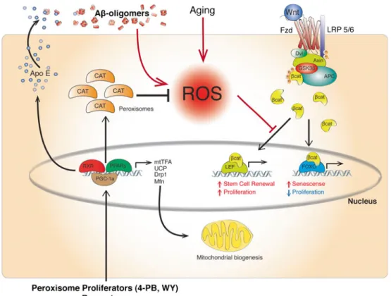
(8) Mol Neurobiol (2013) 48:438–451. 445. Fig. 2 Aβ/ROS-based BBB failure. It is well known that one of the main mechanisms involved in the Aβ-induced neurotoxicity is through increased oxidative stress derived by an increased production of reactive oxygen species (ROS). The present model suggests that an increased level of ROS might exert the same toxic effects at the BBB, disrupting the normal structure of the barrier, and altering the functionality of this critical structure. Moreover, it is well recognized that this event happens early in the development of AD and suggests that the altered function of the BBB could accelerate the progress of the pathology and might contribute to Aβ decreased clearance. balance of the different components of the neurovascular unit, altering the traffic across the BBB and perhaps also affecting the ability to maintain the brain environment, necessary to ensure electrical communication between neurons. PPAR activation by several PPAR agonists has proven to reduce oxidative damage through the reduction of ROS production in several tissues, including the brain, liver, and blood vessels, among others, due to the interaction of PPARs with antiinflammatory pathways such as NF-κB and NRF2, the reduction of COX-2 expression or the interaction with antiapoptotic pathways such as Bcl-2 [85, 86, 96, 119–122]. Additionally, we have recently found that PPAR agonist treatment also increases catalase activity in the brain of an AD mouse model [88], suggesting an enhancement of the antioxidant capacity mediated by PPAR activation. It is possible to hypothesize that astrocytes, pericytes, and endothelial cells are able to respond in a similar way as has been observed for other cell types, including highly specialized cells. such as neurons, controlling the surrounding environment and limiting the oxidative damage. Additionally, if the oxidative insult is controlled, functional rescue occurs as observed in Cramer's work [53] as well as in our research [88]. Also, in the latter case, the available information seems to suggest a ROS-mediated crosstalk between PPARs and proliferative/prosurvival pathways, such as the Wnt signaling pathway [123]. In fact, several authors have proposed that β-catenin-Tcf/Lef binding, a key step of the Wnt signaling pathway, can be modulated under oxidative stress and redirected to FoxO, inducing cell senescence and the release of proapoptotic signals [124, 125]. On the other hand, it has been described that targeting PPARs leads to the activation of the PPARγ coactivator 1-α (PGC1α) transcription factor, which has been related with several proteins linked to mitochondrial biogenesis and respiration, leading to higher mitochondrial density in neurons as well as increased activity of several.
(9) 446. Mol Neurobiol (2013) 48:438–451. antioxidant enzymes, such as SOD-1, SOD-2, CAT, and GPx [126, 127]. Moreover, PGC1α has become an interesting target in Huntington's disease due to the impact that this cofactor has on mitochondrial stabilization [128, 129]. Furthermore, our recent studies [88, 130] offer further support to the importance of Aβ peripheral clearance, as proposed by Nishitsuji et al. [58] and Sutcliffe et al. [79], due to the increased peroxisomal activity reported in the liver of 4-PB- and WY-treated mice and also allow us to hypothesize that PPAR activation, through different agonists, may lead to PGC1α-increased expression at the BBB. In fact, according to our results [88], part of the benefits observed after PPAR agonist (4-PB and WY) treatment should be related to a direct effect on the BBB components. Moreover, WY is a PPARα agonist that seems unable to cross the BBB. If we consider that PGC1α can also be activated as a consequence of the PPARα activation [131], this hypothesis could explain an additional mechanism that accounts for the wide range of effects observed after PPAR agonist treatment (Fig. 3).. Even when the importance of the BBB is out of discussion, there is an absence of information on the impact that several potential drugs may exert on the BBB. Recently, the work of Cramer et al. [53] has astonished the scientific community because of the possibility of having a novel and effective therapy against AD. However, some voices have called for calm and have reminded us that in the past, several therapies have promised a lot but when transferred to real patients, have failed [98, 132]. Indeed, potent PPARγ agonists, such as RGZ and pioglitazone [122], have shown impressive results in different AD models, but when transferred to patients have proven to be little effective. Well, maybe part of the problem involves the BBB and perhaps the lack of knowledge about what roles are played by the BBB in neurodegenerative disorder therapies have lead us once again to failure. Nuclear receptors, and particularly the PPARs family, are a quite complex group of receptors involved in several cellular physiological mechanisms and. Fig. 3 RXR/PPAR-based therapy model. According to current information as well as our recent results, it is possible to hypothesize a more complex mechanism of action of an RXR/PPAR-based therapy. Beyond the ApoE increased expression due to pharmacologic RXR: PPARγ dimer stimulation, additional underlying effects might be occurring. It is well noticed that PPAR agonist enhances antioxidant defenses through increased expression of several antioxidant enzymes, such as catalase (CAT), superoxide dysmutase (SOD), and glutation peroxidase (GPx). Despite the contribution to attenuate the oxidative. status of the cell, it is important to highlight the effects on cell signaling derived from ROS levels. The Wnt signaling pathway, a proliferative prosurvival pathway, is inhibited in the presence of high levels of ROS by diverting β-catenin from TCF/Lef to the FoxO pathway. Moreover, PPARs have been described to be able to activate PPARγ coactivator 1α (PGC-1α), a transcription coactivator related to mitochondrial dynamics and biogenesis, which could further account for mitochondrial stabilization enhancing efficient ROS management as well as reducing mitochondrial ROS production. Final Considerations.
(10) Mol Neurobiol (2013) 48:438–451. which effects are quite far to be fully addressed [122]. Perhaps the systemic administration of PPARs agonists leads to the BBB partial recovery and to an improved BBB trafficking, which in turn, might reduce the ability of the agonists to enter into the brain. Unfortunately, there is a lack of studies focused on BBB recovery related with PPARs agonists administration. We believe that the knowledge derived from this kind of studies will help to understand why several drugs fail when transferred from in vitro or in vivo neurodegenerative disease models to real patients. Scientific progress has allowed us to develop more complex BBB models than before [133], offering the possibility to answer some of the questions regarding the structure and function of the BBB. Moreover, new technologies, such as nanotechnology, have already shown promising results regarding drug delivery to the brain, suggesting that in the future, critical advances will be made in this field [134]. Nuclear receptors and particularly PPARs have become promising targets in several neurodegenerative disorders. The expectation for a novel AD therapy is higher than ever, but several questions about the mechanism of action and the potentialities of such a treatment must be answered in order to be certain of risks and benefits derived from PPAR activation. Some of these questions certainly involve the BBB structure and function. We have tried to briefly introduce, considering the available information about the mechanisms of action of PPARs, how its activation could also induce beneficial effects in the BBB and explain the improvements observed after PPAR agonist treatments in AD mouse models. Acknowledgments This work was supported by grants from the Basal Center of Excellence in Aging and Regeneration (CONICYT-PFB 12/ 2007) and FONDECYT (No. 1120156) to N.C.I. Graphic work was carried out by Graphique-Science (http://graphique-science.blogspot.com). Conflicts of interest None. References 1. Lewandowsky M (1900) ZurLehre der Cerebrospinal flussigkeit. Z Klin Med 40:480–494 2. Biedl A, Kraus R (1898) ÜbereinerbisherunbekanntetoxischeWikung der Gallensauren auf das Zentral-nervensystem. ZentralblattInnere Medizin 19:1185–1200 3. Goldmann EE (1909) Die aussere und innereSekretion des gesunden und krankenOrganismusimLichte der ‘vitalenFarbung’. BeitraegeKlinischenChirurgie 64:192–265 4. Goldmann EE (1913) Vitalfarbung am Zentral-nervensystem. AbhandlungenPreussischenAkademie der WissenschaftenPhysikalisch Mathematischklasse I 1–60 5. Krogh A (1946) The active and passive exchanges of inorganic ions through the surfaces of living cells and through living membranes generally. Proc Biol Sci 133:140–200. 447 6. Schultz RL, Maynard EA, Pease DC (1957) Electron microscopy of neurons and neuroglia of cerebral cortex and corpus callosum. Am J Anat 100:369–407 7. Reese TS, Karnovsky MJ (1967) Structural localization of a blood–brain barrier to exogenous peroxidase. J Cell Biol 34:207–217 8. Brightman MW, Reese TS (1969) Junctions between intimately opposed cell membranes in the vertebrate brain. J Cell Biol 40:648–677 9. Abbott NJ (2004) Prediction of blood–brain barrier permeation in drug discovery from in vivo, in vitro and in silico models. Drug Discov Today Technol 1:407–416 10. Abbott NJ, Patabendige AAK, Dolman DEM, Yusof SR, Begley DJ (2010) Structure and function of the blood–brain barrier. Neurobiol Dis 37:13–25 11. Liebner S, Plate KH (2010) Differentiation of the brain vasculature: the answer came blowing by the Wnt. J Angiogenes Res 2:1 12. Girouard H, Iadecola C (2006) Neurovascular coupling in the normal brain and in hypertension, stroke, and Alzheimer disease. J Appl Physiol 100:328–335 13. Schulze C, Firth JA (1993) Immunohistochemical localization of adherens junction components in blood–brain barrier microvessels of the rat. J Cell Sci 104:773–782 14. Roberts J, Kahle MP, Bix GJ (2012) Perlecan and the blood–brain barrier: beneficial proteolysis. Front Pharmacol 3:155 15. Pardridge WM (2007) Blood–brain barrier delivery. Drug Discov Today 12:54–61 16. Wilhelm I, Fazakas C, Krizbai IA (2011) In vitro models of the blood–brain barrier. Acta NeurobiolExp 71:113–128 17. Zlokovic BV (2008) The blood–brain barrier in health and chronic neurodegenerative disorders. Neuron 57:178–201 18. Furuse M, Hirase T, Itoh M, Nagafuchi A, Yonemura S, Tsukita S, Tsukita S (1993) Occludin: a novel integral membrane protein localizing at tight junctions. J Cell Biol 123:1777–1788 19. Gonzáles-Mariscal L, Betanzos A, Nava P, Jaramillo BE (2003) Tight junction proteins. Prog Biophys Mol Biol 81:1–44 20. Nasdala I, Wolburg-Buchholz K, Wolburg H, Kuhn A, Ebnet K, Brachtendorf G, Samulowitz U, Kuster B, Engelhardt B, Vestweber D, Butz S (2002) A transmembrane tight junction protein selectively expressed on endotelial cells and platelets. J Biol Chem 277:16294–16303 21. Martin-Padura I, Lostaglio S, Schneemann M, Williams L, Romano M, Fruscella P, Panzeri C, Stoppacciaro A, Ruco L, Villa A, Simmons D, Dejana E (1998) Junctional adhesion molecule, a novel member of the immunoglobulin superfamily that distributes at intercelular junctions and modulates monocyte transmigration. J Cell Biol 142:117–127 22. Bazzoni G, Tonetti P, Manzi L, Cera MR, Balconi G, Dejana E (2005) Expression of junctional adhesion molecule-A prevents spontaneous and random motility. J Cell Sci 118:623–632 23. Stevenson BR, Siliciano JD, Mooseker MS, Goodenough DA (1986) Identification of ZO-1: a high molecular weight polypeptide associated with the tight junction (zonula occludens) in a variety of epithelia. J Cell Biol 103:755–766 24. Gumbiner B, Lowenkopf T, Apatira D (1991) Identification of a 160-kDa polypeptide that binds to the tight junction protein ZO1. Proc Natl Acad Sci USA 88:3460–3464 25. Hawkins BT, Davis TP (2005) The blood–brain barrier neurovascular unit in health and disease. Pharmacol Rev 57:173–185 26. Ohnishi H, Nakahara T, Furuse K, Sasaki H, Tsukita S, Furuse M (2004) JACOP, a novel plaque protein localizing at the apical junctional complex with sequence similarity to cingulin. J Biol Chem 279:46014–46022 27. Breier G, Breviario F, Caveda L, Berthier R, Schnürch H, Gotsch U, Vestweber D, Risau W, Dejana E (1996) Molecular cloning.
(11) 448. 28.. 29.. 30.. 31.. 32. 33. 34.. 35.. 36.. 37.. 38.. 39.. 40.. 41. 42.. 43.. 44.. 45.. 46.. Mol Neurobiol (2013) 48:438–451 and expression of murine vascular endotelial-cadherin in early stage development of cardiovascular system. Blood 87:630–641 Redzic Z (2011) Molecular biology of the blood–brain and the blood–cerebrospinal fluid barrier: similarities and differences. Fluid Barriers CNS 8:3 O'Kane RL, Martinez-Lopez I, DeJoseph MR, Vina JR, Hawkins RA (1999) Na+ −dependent glutamate transporters (EAAT1, EAAT2, and EAAT3) of the blood–barrier. A mechanism for glutamate removal. J Biol Chem 274:31891–31895 Zlokovic BV (2011) Neurovascular pathways to neurodegeneration in Alzheimer's disease and other disorders. Nat Rev Neurosci 12:723–738 Do TM, Noel-Hudson MS, Ribes S, Besengez C, Smirnova M, Cisternino S, Buyse M, Calon F, Chimini G, Chacun H, Scherrmann JM, Farinotti R, Bourasset F (2012) ABCG2- and ABCG4-mediated efflux of amyloid-β peptide 1–40 at the mouse blood–brain barrier. J Alzheimers Dis 30:155–166 Pardridge WM (2005) Molecular biology of the blood–brain barrier. Mol Biotechnol 30:57–70 Banks WA (2006) The CNS as a target for peptides and peptidebased drugs. Expert Opin Drug Deliv 3:707–712 O'Donnell ME, Lam TI, Tran LQ (2006) Estradiol reduces activity of the blood–brain barrier Na–K–Cl cotransporter and decreases edema formation in permanent middle cerebral artery occlusion. J Cereb Blood Flow Metab 26:1234–1249 Taylor CJ, Nicola PA, Wang S, Barrand MA, Hladky SB (2006) Transporters involved in regulation of intracellular pH in primary cultured rat brain endothelial cells. J Physiol 576:769–785 Simard JM, Kent TA, Chen M, Tarasov KV, Gerzanich V (2007) Brain oedema in focal ischaemia: molecular pathophysiology and theoretical implications. Lancet Neurol 6:258–268 Parton RG, Richards AA (2003) Lipid rafts and caveolae as portals for endocytosis: new insights and common mechanisms. Traffic 4:724–738 Roux F, Durieu-Trautmann O, Chaverot N, Claire M, Mailly P, Bourre JM, Strosberg AD, Couraud PO (1994) Regulation of gamma-glutamyl transpeptidase and alkaline phosphatase activities in immortalized rat brain microvessel endothelial cells. J Cell Physiol 159:101–113 Régina A, Koman A, Piciotti M, El Hafny B, Center MS, Bergmann R, Couraud PO, Roux F (1998) Mrp1 multidrug resistance-associated protein and P-glycoprotein expression in rat brain microvessel endothelial cells. J Neurochem 71:705–715 Deli MA, Abrahám CS, Takahata H, Niwa M (2001) Tissue plasminogen activator inhibits P-glycoprotein activity in brain endothelial cells. Eur J Pharmacol 411:R3–R5.46 Ballard C, Gauthier S, Corbett A, Brayne C, Aarsland D, Jones E (2011) Alzheimer's disease. Lancet 377:1019–1031 Omidi Y, Campbell L, Barar J, Connell D, Akhtar S, Gumbleton M (2003) Evaluation of the immortalized mouse brain capillary endothelial cell line, b.End3, as an in vitro blood–brain barrier model for drug uptake and transport studies. Brain Res 990:95–112 Weksler BB, Subileau EA, Perrière N, Charneau P, Holloway K, Levenque M, Tricoire-Leignel H, Nicotra A, Bourdoulous S, Turowski P, Male DK, Roux F, Greenwood J, Romero IA, Couraud PO (2005) Blood–brain barrier-specific properties of a human adult brain endothelial cell line. FASEB J 19:1872–1874 Sagare AP, Bell RD, Zlokovic BV (2012) Neurovascular dysfunction and faulty amyloid β-peptide clearance in Alzheimer disease. Cold Spring Harb Perspect Med. doi:10.1101/ cshperspect.a011452 Heinemann U, Kaufer D, Friedman A (2012) Blood–brain barrier dysfunction, TGFβ signaling, and astrocyte dysfunction in epilepsy. Glia 60:1251–1257 Marchi N, Granata T, Ghosh C, Janigro D (2012) Blood–brain barrier dysfunction and epilepsy: pathophysiologic role and. 47.. 48. 49. 50.. 51.. 52.. 53.. 54.. 55.. 56.. 57.. 58.. 59.. 60.. 61.. 62.. 63.. therapeutic approaches. Epilepsia. doi:10.1111/j.1528-1167. 2012.03637.x Morgan L, Shah B, Rivers LE, Barden L, Groom AJ, Chung R, Higazi D, Desmond H, Smith T, Staddon JM (2007) Inflammation and dephosphorylation of the tight junction protein occluding in an experimental model of multiple sclerosis. Neuroscience 147:664–673 Salmon DP, Bondi MW (2009) Neuropsychological assessment of dementia. Annu Rev Psychol 60:257–282 Perl DP (2010) Neuropathology of Alzheimer's disease. Mt Sinai J Med 77:32–42 Manji H, Kato T, Di Prospero NA, Ness S, Beal MF, Krams M, Chen G (2012) Impaired mitochondrial function in psychiatric disorders. Nature Rev Neurosci 13:293–307 Selkoe DJ (2001) Alzheimer's disease results from the cerebral accumulation and cytotoxicity of amyloid beta-protein. J Alzheimers Dis 3:75–80 Bailey TL, Rivara CB, Rocher AB, Hof PR (2004) The nature and effects of cortical microvascular pathology in aging and Alzheimer's disease. Neurol Res 26:573–578 Cramer PE, Cirrito JR, Wesson DW, Daniel Lee CY, Colleen KJ, Zinn AE, Casali BT, Restivo JL, Goebel WD, James MJ, Brunden KR, Wilson DA, Landreth GE (2012) ApoE-directed therapeutics rapidly clear β-amyloid and reverse deficits in AD mouse models. Science 335:1503–1506 Bell RD, Zlokovic BV (2009) Neurovascular mechanisms and blood–brain barrier disorder in Alzheimer's disease. Acta Neuropathol 118:103–113 Kanekiyo T, Liu C, Shinohara M, Li J, Bu G (2012) LRP1 in brain vascular smooth muscle cells mediates local clearance of Alzheimer's amyloid-β. J Neurosci 32:16458–16465 Bell RD, Sagare AP, Friedman AE, Bedi GS, Holtzman DM, Deane R, Zlokovic BV (2007) Transport pathways for clearance of human Alzheimer's amyloid β-peptide and apolipoprotein E and J in the mouse central nervous system. J Cereb Blood Flow Metab 27:909–918 Jaeger LB, Dohgu S, Hwang MC, Farr SA, Murphy MP, FleegalDeMotta MA, Lynch JL, Robinson SM, Niehoff ML, Johnson SN, Kumar VB, Banks WA (2009) Testing the neurovascular hypothesis of Alzheimer's disease: LRP-1 antisense reduces blood–brain barrier clearance, increases brain levels of amyloidβ protein, and impairs cognition. J Biol Chem 258:22091–22102 Nishitsuji K, Hosono T, Nakamura T, Bu G, Michikawa M (2011) Apolipoprotein E regulates the integrity of tight junctions in an isoform-dependent manner in an in vitro blood–brain barrier model. J Biol Chem 286:17536–17542 Akram A, Schmeidler J, Katsel P, Hof PR, Haroutunian V (2012) Association of ApoE and LRP mRNA levels with dementia and AD neuropathology. Neurobiol Aging 33:628, e1-628.e14 Qosa H, Abuznait AH, Hill RA, Kaddoumi A (2012) Enhanced brain amyloid-β clearance by rifampicin and caffeine as possible protective mechanism against Alzheimer's disease. J Alzheimers Dis 31:151–165 Nielsen HM, Mulder SD, Beliën JA, Musters RJ, Eikelenboom P, Veerhuis R (2010) Astrocytic A beta 1–42 uptake is determined by A beta-aggregation state and the presence of amyloidassociated proteins. Glia 58:1235–1246 Kamboh MI, Minster RL, Demirci FY, Ganguli M, Dekosky ST, Lopez OL, Barmada MM (2012) Association of CLU and PICALM variants with Alzheimer's disease. Neurobiol Aging 33:518–521 Erickson MA, Niehoff ML, Farr SA, Morley JE, Dillman LA, Banks WA (2012) Peripheral administration of antisense oligonucleotides targeting the amyloid-β protein precursor reverses AβPP and LRP-1 overexpression in the aged SAMP8 mouse brain. J Alzheimers Dis 28:951–960.
(12) Mol Neurobiol (2013) 48:438–451 64. Natunen T, Helisalmi S, Vepsäläinen S, Sarajärvi T, Antikainen L, Mäkinen P, Herukka SK, Koivisto AM, Haapasalo A, Soininen H, Hiltunen M (2012) Genetic analysis of genes involved in amyloid-β degradation and clearance in Alzheimer's disease. J AlzheimersDis 28:553–559 65. Liu Q, Zerbinatti CV, Zhang J, Hoe HS, Wang B, Cole SL, Herz J, Muglia L, Bu G (2007) Amyloid precursor protein regulates brain apolipoprotein E and cholesterol metabolism through lipoprotein receptor LRP1. Neuron 56:66–78 66. Belyaev ND, Nalivaeva NN, Makova NZ, Turner AJ (2009) Neprilysin gene expression requires binding of the amyloid precursor protein intracellular domain to its promoter: implications for Alzheimer disease. EMBO Rep 10:94–100 67. Sen A, Alkon DL, Nelson TJ (2012) Apolipoprotein E3 (ApoE3) but no ApoE4 protects against synaptic loss through increased expression of protein kinase C epsilon. J Biol Chem 287:15947– 15958 68. Schmechel DE, Saunders AM, Strittmatter WJ, Crain BJ, Hulette CM, Joo SH, Pericak-Vance MA, Goldgaber D, Roses AD (1993) Increased amyloid β-peptide deposition in cerebral cortex as consequence of apolipoprotein E genotype in late-onset Alzheimer disease. Proc Natl Acad Sci USA 90:9649–9653 69. Bu G (2009) Apolipoprotein E and its receptors in Alzheimer's disease: pathways, pathogenesis and therapy. Nat Rev Neurosci 10:333–344 70. Holtzman DM, Herz J, Bu G (2012) Apolipoprotein E and Apolipoprotein E receptors: normal biology and roles in Alzheimer disease. Cold Spring Harb Perspect Med. doi:10.1101/ cshperspect.a006312 71. Assema DM, Lubberink M, Rizzu P, Swieten JC, Schuit RC, Eriksson J, Scheltens P, Koepp M, Lammertsma AA, Berckel BN (2012) Blood–brain barrier P-glycoprotein function in healthy subjects and Alzheimer's disease patients: effect of polymorphisms in the ABCB1 gene. EJNMMI Res 16:57 72. Erickson MA, Hansen K, Banks WA (2012) Inflammationinduced dysfunction of the low-density lipoprotein receptorrelated protein-1 at the blood–brain barrier: protection by the antioxidant N-acetylcysteine. Brain Behav Immun 26:1085–1094 73. Erickson MA, Hartvigson PE, Morofuji Y, Owen JB, Butterfield DA, Banks WA (2012) Lipopolysaccharide impairs amyloid beta efflux from brain: altered vascular sequestration, cerebrospinal fluid reabsorption, peripheral clearance and transporter function at the blood–brain barrier. J Neuroinflammation 9:150 74. Assema DM, Goos JD, van der Flier WM, Lubberink M, Boellaard R, Windhorst AD, Scheltens P, Lammertsma AA, van Berckel BN (2012) No evidence for additional blood–brain barrier P-glycoprotein dysfunction in Alzheimer's disease patients with microbleeds. J Cereb Blood Flow Metab 32:1468–1471 75. Durk MR, Chan GN, Campos CR, Peart JC, Chow EC, Lee E, Cannon RE, Bendayan R, Miller DS, Sandy PK (2012) 1α,25Dihydroxyvitamin D(3)-liganded Vitamin D receptor increases expression and transport activity of P-glycoprotein in isolated rat brain capillaries and human and rat brain microvessel endothelial cells. J Neurochem. doi:10.1111/jnc.12041 76. Krohn M, Lange C, Hofrichter J, Scheffler K, Stenzel J, Steffen J, Schumacher T, Brüning T, Plath AS, Alfen F, Schmidt A, Winter F, Rateitschak K, Wree A, Gsponer J, Walker LC, Pahnke J (2011) Cerebral amyloid-β proteostasis is regulated by the membrane transport protein ABCC1 in mice. J Clin Invest 121:3924– 3931 77. Eisele YS, Obermüller U, Heilbronner G, Baumann F, Kaeser SA, Wolburg H, Walker LC, Staufenbiel M, Heikenwalder M, Jucker M (2010) Peripherally applied Aβ-containing inoculates induce cerebral β-amyloidosis. Science 330:980–982 78. Sagare AP, Winkler EA, Bell RD, Deane R, Zlokovic BV (2011) From the liver to the blood–brain barrier: an interconnected. 449. 79.. 80.. 81.. 82.. 83.. 84.. 85.. 86.. 87.. 88.. 89.. 90. 91.. 92.. 93. 94.. system regulating brain amyloid-β levels. J Neurosci Res 89:967–968 Sutcliffe JG, Hedlund PB, Thomas EA, Bloom FE, Hilbush BS (2011) Peripheral reduction of β-amyloid is sufficient to reduce brain β-amyloid: implications for Alzheimer's disease. J Neurosci Res 89:808–814 Sehgal N, Gupta A, Valli RK, Joshi SD, Mills JT, Hamel E, Khanna P, Jain SC, Thakur SS, Ravindranath V (2012) Withania somnifera reverses Alzheimer's disease pathology by enhancing low-density lipoprotein receptor-related protein in liver. ProcNatl Acad Sci USA 109:3510–3515 Deane R, Singh I, Sagare AP, Bell RD, Ross NT, LaRue B, Love R, Perry S, Paquette N, Deane RJ, Thiyagarajan M, Zarcone T, Fritz G, Friedman AE, Miller BL, Zlokovic BV (2012) A multimodal RAGE-specific inhibitor reduces amyloid β-mediated brain disorder in a mouse model of Alzheimer disease. J Clin Invest 122:1377–1392 Kook SY, Hong HS, Moon M, Ha CM, Chang S, Mook-Jung I (2012) Aβ1-42–RAGE interaction disrupts tight junctions of the blood–brain barrier via Ca2+-calcineurin signaling. J Neurosci 32:8845–8854 Poellinger L, Göttlicher M, Gustafsson JA (1992) The dioxin and peroxisome proliferator-activated receptors: nuclear receptors in search of endogenous ligands. Trends Pharmacol Sci 13:241–245 Inestrosa NC, Godoy JA, Quintanilla RA, Koenig CS, Bronfman M (2005) Peroxisome proliferator-activated receptor gamma is expressed in hippocampal neurons and its activation prevents βamyloid neurodegeneration: role of Wnt signaling. Exp Cell Res 304:91–104 Santos MJ, Quintanilla RA, Toro A, Grandy R, Dinamarca MC, Godoy JA, Inestrosa NC (2005) Peroxisomal proliferation protects from beta-amyloid neurodegeneration. J Biol Chem 280:41057–41068 Fuenzalida K, Quintanilla R, Ramos P, Piderit D, Fuentealba RA, Martinez G, Inestrosa NC, Bronfman M (2007) Peroxisome proliferator-activated receptor γ up-regulates the Bcl-2 antiapoptotic protein in neurons and induces mitochondrial stabilization and protection against oxidative stress and apoptosis. J Biol Chem 282:37006–37015 Mandrekar-Colucci S, Landreth GE (2011) Nuclear receptors as therapeutic targets for Alzheimer's disease. Expert Opin Ther Targets 15:1085–1097 Inestrosa NC, Carvajal FJ, Zolezzi JM, Tapia-Rojas C, Serrano F, Karmelic D, Toledo EM, Toro A, Toro J, Santos MJ (2012) Peroxisome proliferators reduce spatial memory impairment, synaptic failure, and neurodegeneration in brains of a double transgenic mice model of Alzheimer's disease. J Alzheimers Dis. doi:10.3233/JAD-2012-120397 Mulholland DJ, Dedhar S, Coetzee GA, Nelson CC (2005) Interaction of nuclear receptors with the Wnt/β-catenin/Tcf signaling axis: Wnt you like to know? Endocr Rev 26:898–915 Hollenberg AN (2012) Metabolic health and nuclear-receptor sensitivity. N Engl J Med 366:1345–1347 Neher MD, Weckbach S, Huber-Lang MS, Stahel PF (2012) New insights into the role of peroxisome proliferator-activated receptors in regulating the inflammatory response after tissue injury. PPAR Res. doi:10.1155/2012/728461 Chen YC, Wu JS, Tsai HD, Huang CY, Chen JJ, Sun GY, Lin TN (2012) Peroxisome proliferator-activated receptor gamma (PPAR-γ) and neurodegenerative disorders. Mol Neurobiol. doi:10.1007/s12035-012-8259-8 Heneka MT, Landreth GE (2007) PPARs in the brain. Biochim Biophys Acta 177:1031–1045 Watson GS, Craft S (2003) The role of insulin resistance in the pathogenesis of Alzheimer's disease: implications for treatment. CNS Drugs 17:27–45.
(13) 450 95. Polvani S, Tarocchi M, Galli A (2012) PPARγ and oxidative stress: Con(β) catenating NRF2 and FOXO. PPAR Res. doi:10.1155/2012/641087 96. Martín A, Pérez-Girón JV, Hernanz R, Palacios R, Briones AM, Fortuño A, Zalba G, Salaices M, Alonso MJ (2012) Peroxisome p r o l i fe r at o r- a ct i v a t e d r e c ep t o r- γ ac t i v at i o n r e d u ce s cyclooxygenase-2 expression in vascular smooth muscle cells from hypertensive rats by interfering with oxidative stress. J Hypertens 30:315–326 97. Fuentealba RA, Farias G, Scheu J, Bronfman M, Marzolo MP, Inestrosa NC (2004) Signal transduction during amyloid-betapeptide neurotoxicity: role in Alzheimer disease. Brain Res Brain Res Rev 47:275–289 98. LaFerla FM (2012) Preclinical success against Alzheimer's disease with an old drug. N Engl J Med 367:570–572 99. Jiang Q, Lee CY, Mandrekar S, Wilkinson B, Cramer P, Zelcer N, Mann K, Lamb B, Willson TM, Collins JL, Richardson JC, Smith JD, Comery TA, Riddell D, Holtzman DM, Tontonoz P, Landreth GE (2008) ApoE promotes the proteolytic degradation of Aβ. Neuron 12:681–693 100. Mandrekar-Colucci S, Karlo JC, Landreth GE (2012) Mechanisms underlying the rapid peroxisome proliferator-activated receptor-γ-mediated amyloid clearance and reversal of cognitive deficits in a murine model of Alzheimer's disease. J Neurosci 32:10117–10128 101. Biserni A, Giannessi F, Sciarroni AF, Milazzo FM, Maggi A, Ciana P (2008) In vivo imaging reveals selective peroxisome proliferator activated receptor modulator activity of the synthetic ligand 3-(1-(4-chlorobenzyl)-3-t-butylthio-5-isopropylindol-2yl)-2,2-dimethylpropanoic acid (MK-886). Mol Pharmacol 73:1434–1443 102. Hoque MT, Robillard KR, Bendayan R (2012) Regulation of breast cancer resistant protein by peroxisome proliferatoractivated receptor α in human brain microvessel endothelial cells. Mol Pharmacol 81:598–609 103. Mysiorek C, Culot M, Dehouck L, Derudas B, Bordet R, Cecchelli R, Fenart L, Berezowski V (2009) Peroxisomeproliferator-activated receptor-alpha activation protects brain capillary endothelial cells from oxygen-glucose deprivationinduced hyperpermeability in the blood–brain barrier. Curr Neurovasc Res 6:181–193 104. Yin KJ, Deng Z, Hamblin M, Xiang Y, Huang H, Zhang J, Jiang X, Wang Y, Chen YE (2010) Peroxisome proliferator-activated receptor delta regulation of miR-15a in ischemia-induced cerebral vascular endothelial injury. J Neurosci 30:6398–6408 105. Kalinin S, Richardson JC, Feinstein DL (2009) A PPARdelta agonist reduces amyloid burden and brain inflammation in a transgenic mouse model of Alzheimer's disease. Curr Alzheimer Res 6:431–437 106. Huang W, Eum SY, András IE, Hennig B, Toborek M (2009) PPARalpha and PPARgamma attenuate HIV-induced dysregulation of tight junctions proteins by modulations of matrix metalloproteinase and proteasome activities. FASEB J 23:1596– 1606 107. Ramirez SH, Heilman D, Morsey B, Potula R, Haorah J, Persidsky Y (2008) Activation of peroxisome proliferator-activated receptor gamma (PPARgamma) suppresses Rho GTPases in human brain microvascular endothelial cells and inhibits adhesion and transendothelial migration of HIV-1 infected monocytes. J Immunol 180:1854–1865 108. Huang W, András IE, Rha GB, Hennig B, Toborek M (2011) PPARα and PPARγ protect against HIV-induced MMP-9 overexpression via caveolae-associated ERK and Akt signaling. FASEB J 25:3979–3988 109. Araújo CV, Estato V, Tibiriçá E, Bozza PT, Castro-Faria-Neto HC, Silva AR (2012) PPAR gamma activation protects the brain. Mol Neurobiol (2013) 48:438–451. 110.. 111.. 112.. 113.. 114.. 115.. 116.. 117.. 118.. 119.. 120.. 121.. 122.. 123.. 124.. 125.. 126.. against microvascular dysfunction in sepsis. Microvasc Res 84:218–221 Min LJ, Mogi M, Shudou M, Jing F, Tsukuda K, Ohshima K, Iwanami J, Horiuchi M (2012) Peroxisome proliferator-activated receptor-γ activation with angiotensin II type 1 receptor blockade is pivotal for the prevention of blood–brain barrier impairment and cognitive decline in type 2 diabetic mice. Hypertension 59:1079–1088 Smith MA, Perry G, Richey PL, Sayre LM, Anderson VE, Beal MF, Kowall N (1996) Oxidative damage in Alzheimer's disease. Nature 382:120–121 Abramov AY, Canevari L, Duchen MR (2004) Β-Amyloid peptides induce mitochondrial dysfunction and oxidative stress in astrocytes and death of neurons through activation of NADPH oxidase. J Neurosci 24:565–575 Small SA, Gandy S (2006) Sorting through the cell biology of Alzheimer's disease: intracellular pathways to pathogenesis. Neuron 60:534–542 Caspersen C, Wang N, Yao J, Sosunov A, Chen X, Lustbader JW, Xu HW, Stern D, McKhann G, Yan SD (2005) Mitochondrial Abeta: a potential focal point for neuronal metabolic dysfunction in Alzheimer's disease. FASEB J 19:2040–2041 Manczak M, Anekonda TS, Henson E, Park BS, Quinn J, Reddy PH (2006) Mitochondria are direct site of A beta accumulation in Alzheimer's disease neurons: implications for free radical generation and oxidative damage in disease progression. Hum Mol Genet 15:1437–1449 Reddy PH, Beal MF (2008) Amyloid beta, mitochondrial dysfunction and synaptic damage: implications for cognitive decline in aging and Alzheimer's disease. Trends Mol Med 14:45–53 Karran E, Mercken M, de Strooper B (2011) The amyloid cascade hypothesis for Alzheimer's disease: an appraisal for the development of therapeutics. Nat Rev Drug Discov 10:698–712 Miranda S, Opazo C, Larrondo LF, Muñoz FJ, Leighton F, Inestrosa NC (2000) The role of oxidative stress in the toxicity induced by amyloid β-peptide in Alzheimer's disease. Prog Neurobiol 62:633–648 Hernanz R, Martín A, Pérez-Girón JV, Palacios R, Briones AM, Miguel M, Salaices M, Alonso MJ (2012) Pioglitazone treatment increases COX-2-derived prostacyclin production and reduces oxidative stress. Br J Pharmacol 166:1303–1319 Iwaisako K, Haimerl M, Paik YH, Taura K, Kodama Y, Sirlin C, Yu E, Yu RT, Downes M, Evans RM, Brenner DA, Schnabl B (2012) Protection from liver fibrosis by a peroxisome proliferator-activated receptor δ agonist. Proc Natl Acad Sci USA 109:E1369–1376 Patterson AD, Shah YM, Matsubara T, Krausz KW, Gonzales FJ (2012) Peroxisome proliferator-activated receptor alpha induction of uncoupling protein 2 protects against acetaminophen-induced liver toxicity. Hepatology 56:281–290 Jiang Q, Heneka M, Landreth GE (2008) The role of peroxisome proliferator-activated receptor-γ (PPARγ) in Alzheimer's disease. CNS Drugs 22:1–14 Inestrosa NC, Toledo EM (2008) The role of Wnt signaling in neuronal dysfunction in Alzheimer's disease. Molec Neurodegener 3:9 Almeida M, Han L, Martín-Millan M, O'Brien CA, Manolagas SC (2007) Oxidative stress antagonizes Wnt signaling in osteoblast precursors by diverting β-catenin from T cell factor- to forkhead box O-mediated transcription. J Biol Chem 282:27298– 27305 Manolopoulos KN, Klotz L-O, Korsten P, Bornstein SR, Barthel A (2010) Linking Alzheimer's disease to insulin resistance: the FoxO response to oxidative stress. Mol Psychiatry 15:1046–1052 St-Pierre J, Drori S, Uldry M, Silvaggi JM, Rhee J, Jäger S, Handschin C, Zheng K, Lin J, Yang W, Simon DK, Bachoo R,.
(14) Mol Neurobiol (2013) 48:438–451. 127.. 128.. 129.. 130.. Spiegelman BM (2006) Suppression of reactive oxygen species and neurodegeneration by the PGC-1 transcriptional coactivators. Cell 127:397–408 Waresky P, Vaarmann A, Choubey V, Safiulina D, Liiv J, Kuum M, Kaasik A (2009) PGC-1α and PGC-1β regulates mitochondrial density in neurons. J Biol Chem 284:21379–21385 Cui L, Jeong H, Borovecki F, Parkhurst CN, Tanese N, Krainc D (2006) Transcriptional repression of PGC-1α by mutant Huntingtin leads to mitochondrial dysfunction and neurodegeneration. Cell 127:59–69 Martin E, Betuing S, Pagès C, Cambon K, Auregan G, Deglon N, Roze E, Caboche J (2011) Mitogen- and stress-activated protein kinase 1-induced neuroprotection in Huntington's disease: role on chromatine remodeling at the PGC-1-alpha promoter. Hum Mol Genet 20:2422–2434 Toledo EM, Inestrosa NC (2010) Activation of Wnt signaling by lithium and rosiglitazone reduced spatial memory impairment and neurodegeneration in brains of an APPswe/. 451. 131.. 132.. 133.. 134.. PSEN1DeltaE9 mouse model of Alzheimer's disease. Mol Psychiatry 15:272–285 Hondares E, Rosell M, Díaz-Delfin J, Olmos Y, Monsalve M, Iglesias R, Villarroya F, Giralt M (2011) Peroxisome proliferatoractivated receptor α (PPARα) induces PPARγcoactivator 1α (PGC-1α) gene expression and contributes to thermogenic activation of brown fat. J Biol Chem 286:43112–43122 Lowenthal J, Hull SC, Pearson SD (2012) The ethics of early evidence—preparing for a possible breakthrough in Alzheimer's disease. N Engl J Med 367:488–490 Mehdipour AR, Hamidi M (2009) Brain drug targeting: a computational approach for overcoming blood–brain barrier. Drug Discov Today 14:1030–1036 Raffa V, Gherardini L, Vittorio O, Bardi G, Ziaei A, Pizzorusso T, Riggio C, Nitodas S, Karachalios T, Al-Jamal KT, Kostarelos K, Costa M, Cuschieri A (2011) Carbon nanotube-mediated wireless cell permeabilization: drug and gene uptake. Nanomedicine (Lond) 6:1709–1718.
(15)
Figure

Documento similar
• How can massively parallel computing resources accelerate our understanding of brain function?. • How can our growing understanding of brain function point the way to more
Serotonin exerts its effects by signaling mechanisms through the 5-HT receptors located in postsynaptic and presynaptic neurons at CNS and intestinal serotonergic neurons, and
Objective: We examined physician perception of blood pressure control and treatment behavior in patients with previous cardiovascular disease and uncontrolled hypertension as defined
(B) A normal peritoneum shows baseline VEGF receptors and co-receptors expression, and normal VEGF and Sema-3A levels are secreted at the peritoneal cavity. During continuous
Background aims: After recent observations that intrathecal administration of autologous bone marrow mesenchymal stromal cells (MSCs) increases cerebral metabolism in patients
For real time access to the da- ta two options have been provided and rigorously tested during the last 10 years: METEOSAT and GOES Data Col- lection Systems, and recently, a
The following topics are reviewed: (a) intestinal barrier function and microbiota; (b) leaky-gut, gut microbiota relationship and liver disease; (c) current main strategies to
In the present study, we analysed the expression of TREM2 and its main sheddase ADAM10 in the brain of sporadic Creutzfeldt-Jakob disease (sCJD) patients and evaluated the role of
