Unrevaling the diversity of nitrogen fixers of the sediment of Posidonia Oceanica
48
0
0
Texto completo
(2)
(3) Abstract It has been proposed that biological nitrogen fixation represents an important process that can contribute to the nitrogen demand of seagrasses. Nitrogen-fixing organisms, known as diazotrophs, can inhabit both the phyllospere and rhizosphere of seagrasses. To date, the nitrogen-fixing community inhabiting the rhizosphere of Posidonia oceanica is still unknown. Here, we report the diversity of these diazotrophs by nested PCR (Polymerase Chain Reaction) amplifying nifH sequences (coding for the nitrogenase enzyme) from P. oceanica sediment samples taken at a Mallorcan site (Alcanada, Alcudia Bay) from 3 different sediment depths. Temporal variability in the diazotrophic community composition was also assessed by sampling at different seasons of the year. A clone library was constructed with a total of 104 new nifH clones and comparative diversity analyses were done. Results show that P. oceanica sediment contains a diverse community of heterotrophic diazotrophs. δ- and γ-Proteobacteria appeared to be the most representative taxa in our samples. Sulphate-reducing bacteria (SRB) was abundant suggesting a link between nitrogen fixation and sulphate reduction.. 2.
(4) Resumen Se ha propuesto que la fijación biológica de nitrógeno representa un proceso importante que contribuye en el demanda de nitrógeno de las praderías marinas. Los organismos fijadores de nitrógeno, conocidos como diazótrofos, pueden habitar tanto en la filosfera como en la rizosfera de la pradera. Hasta el momento, la comunidad que residente en la rizosfera de Posidonia oceanica es aún desconocida. En este trabajo se recoge la diversidad de estos diazótrofos a partir de una nested PCR (Polymerasa Chain Reaction) amplificando secuencias nifH (secuencias que codifican para la enzima nitrogenasa) de muestras de sedimento de P.oceanica tomadas en Mallorca (Alcanada, Bahía de Alcúdia), de tres profundidades de sedimento distintas. La variabilidad temporal de la composición de comunidad diazotrófica también fue evaluada tomando las muestras en las distintas estaciones del año. Se elaboró una librería de clones con un total de 104 clones nifH nuevos con los que se hizo un análisis comparativo de diversidad. Los resultados muestran que el sedimento de P.oceanica contiene una comunidad diversa de diazótrofos heterótrofos. δ- y γ-Proteobacteria resultaron ser el taxón más representativo en nuestras muestras. Bacterias reductoras de sulfato (SRB) fueron halladas en gran abundancia sugiriendo una conexión entre la fijación de nitrógeno y la reducción de sulfato.. 3.
(5) Index 1. Introduction ………………………………………………………… 5-12 p 2. Experimental procedures 2.1. Origin of the samples ………………………………….....…… 12-13 p 2.2. Acetylene reduction assay (ARA) and sediment analyses ……. 14-15 p 2.3. DNA extraction …………………………………………………… 15 p 2.4. DNA amplification ……………………………………………. 15-16 p 2.5. Cloning and target gene amplification ………………………... 16-18 p 2.6. Sequencing …………………………………………………..…… 18 p 2.7. Bioinformatics and phylogenetic analysis ……………………….. 19 p 3. Results ………………………………………………………………19-29 p 3.1. Ecological diversity ………………………………………...… 22-25 p 3.2. Diversity in Clusters and closest relatives ……………………. 25-28 p 3.3. Physicochemical variables ……………………………………. 28-29 p 4. Discussion …………………………………………………………. 29-36 p 4.1. Diversity and nitrogen fixing rates ………………………………. 33 p 4.2. Significant physicochemical variables ………………….……. 34-35 p 4.3. Phylogenetic distribution according to the habitat …………… 35-36 p 5. Conclusions …………………………………………………..………. 37 p 6. Acknowledgments …………………….…………...…………….... 37-38 p 7. References ………………………………………………………… 38-44 p ANNEX …………………………………………………………… 44-46 p. 4.
(6) 1. Introduction Posidonia oceanica ecology Posidonia oceanica is a common clonal angiosperm in Mediterranean coastal waters that supports highly complex productive ecological systems (Vizzini et al., 2002). It occupies 50.000km2, which represents an extension of 50% of the Mediterranean floor, meanwhile in the Balearic Islands, occupies approximately 1,200 km2. It dwells in the sublittoral zone, from mean sea level down to 30 to 40 m depth, depending upon water transparency (Personnic et al., 2014). The meadow can reach a length up to 120 cm and, together with the density of the leaves, makes decrease water movement and traps sediment on it, what prevents coastline erosion, sediment re-suspension, and protects beaches from storms. It regulates Earth climate as it can bury atmospheric CO2 and has the potential for buffering ocean acidification through its photosynthetic activity. For all these ecological services, Posidonia oceanica is considered a great ecosystem engineer (Jones et al., 1994). The structural unit of P. oceanica is called ramet, which is organized in roots, rhizomes and leaves. A set of ramets forms a matte (Figure 1).. Figure 1. Posidonia oceanica matte.. 5.
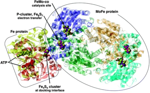
(7) In order to keep P. oceanica rending the key services, it has to satisfy its nutrient requirements. The main machinery that fuels this demand is the microbiological community. Among these microorganisms, nitrogen fixers are responsible for increase the availability of nitrogen to the plant growth and to associated microorganisms, particularly in nitrogen-limited conditions.. Nitrogenase complex Nitrogen fixing-activity comes from the presence of the nif genes that encode for the enzyme nitrogenase complex, which carries out the reduction of molecular dinitrogen (N2) to ammonim according to the reaction N2 + 8H+ + 8e´+16ATP à 2NH3 + H2 + 16ADP + 16Pi (Kneip et al., 2007; Ludden, 2001). This complex is composed of two multisubunit metallo-proteins: dinitrogenase (also known as component I or MoFe protein) and dinitrogenase reductase (component II or Fe protein) (Yun et al., 1984) (Figure 2). Dinitrogenase reductase is a α2 dimer of nifH gene product and it contains a Fe4S4 cluster that bridges the 2 subunits of the protein (Ludden, 2001). It serves as specific electron donor to dinitrogenase. Dinitrogenase is a α2β2 tetramer of the nifD and nifK genes (α- subunit and β-subunit, respectively) (Garcias et al., 2011), which has the substrate site reduction. This protein contains two each of the two unique metal clusters, P clusters and iron-molibdenum cofactor (FeMo-co) (Ludden, 2001). nifEN genes, that encode a protein resembling Component I, are related to nifDK genes. (Chien et al., 1996). In both Component I and Component II, Fe-S centres are present and coordinated between the subunits. Different types of nitrogenase have been described: (1) the “conventional” ones containing Mo in the Fe-S centre bridging the subunits, (2) the “alternative” nitrogenases replacing Mo with V (vnfH), and (3) “second alternative”. 6.
(8) nitrogenases replacing Mo with Fe (anfH) (Zehr et al., 2003). NifHDK operon is highly conserved so it shows a great degree of similarity among diazotroph organisms. In both Component I and Component II, Fe-S centres are present and co-ordinated between the subunits. Different types of nitrogenase have been described; the “conventional” ones contain Mo in the Fe-S centre bridging the subunits. “Alternative” nitrogenases replace Mo with V (vnfH), and “second alternative” nitrogenases replace Mo with Fe (anfH) (Zehr et al., 2003). NifHDK operon is highly conserved so it shows a great degree of similarity among diazotroph organisms.. Figure 2. Structure of the docked Fe protein and MoFe protein (Dance., 2013).. First diazotroph organisms The discovery of nitrogen fixation was attributed to Hellriegel and Wilfarth (1886), who reported that legumes carrying nodules could use molecular nitrogen (Franche et al., 2009), but was not till Martinus Beijerinck and Sergei Winogradsky, that was possible. 7.
(9) to isolate nitrogen-fixing organisms. M. Beijerinck developed the principles of enrichment culture, from which was possible to investigate about the role of the microorganisms in the natural processes and, hence, to reach the concept of microbial ecology (Chung et al., 1996; Franche et al., 2009). In 1901 he isolated Bacillus ridicicola proving the formation of nodules in roots of Leguminosae species. Later, also, he demonstrated nitrogen fixation of free-living organisms isolating Azotobacter chroococcum, an aerobic bacteria. S. Winogradsky took the success of the novel culturing technique to isolate an anaerobic diazotroph, Clostridium pasteuranum (Franche et al., 2009). Both works allowed knowing a bit more about the nitrogen fixation process. At the present time, we have a wider knowledge about this issue. Biological nitrogen fixation is well distributed among Bacteria and Archaea and is very diverse (Farnelid et al., 2013). NifH gene has evolved in a similar way to the 16S rRNA genes. This fact makes it suitable as molecular evolution marker (Young, 1992). Molecular methods have been used to examine diazotroph diversity in many environments. Comparison with available nifH sequences from databases provides taxonomical information about nitrogen fixing bacteria (Zehr et al., 2013). Nitrogen fixation was discovered in the Archaea in 1984, in both Methanosarcina barkery by Murray and Zinder, and Methanococcus thermolithotrophicus. Known nitrogen-fixing species among the Archaea are restricted to the methanogenic Euryarchaeota (Leigh, 2004). From Beijerinck and Winogradsky’s discoveries, is known that diazotrophs can be freeliving nitrogen-fixers, which don’t need any host to carry out the process, and symbiont nitrogen-fixing organisms, which only fix nitrogen in association with other organisms. The second ones have a suitable environment and take a benefit from the nutrition of their host (Mohamed et al., 2008), and in some cases, they are protected from the. 8.
(10) oxygen inhibitory action of nitrogenase enzyme (Fiore et al., 2010). Symbiont diazotrophs have been reported in association with different organisms such as termite gut (Ohkuma et al., 1999), marine sponges (Mohamed et al., 2008), coral reefs, urchins, shipworms; and protists, such as dinoflagelates, diatoms, radiolarians and tinnids (Fiore et al., 2010). Symbiontic relationship also exists between leguminous plants, in which groups Rhizobia (α-Proteobacteria) and Frankia (Actinobacteria) are forming nodules. In non-leguminous plants, diazotrophs have been reported as endophytes and root surface epiphytes (Franche et al., 2009). Among free-living diazotrophs, the cyanobacteria Trichodesmium is the most studied one, before known as blue-green algae. As seen above, diazotrophic organisms can be autotrophic and heterotrophic. The autotrophic are needed of light energy to fix CO2, therefore, in the aquatic environment inhabit up to 100 metres depth. By contrast, heterotrophic diazotrophs are free of such need, so they are able to life in dark areas. The dark ocean is the widest habitat of the Earth, which leads to think that the abundance of heterotrophic diazotrophs could be greater than the autotrophic ones.. State of the art The biological nitrogen fixation is well distributed among Bacteria and Archaea and is very diverse (Farnelid et al., 2013). NifH gene has evolved in a similar way to the 16S rRNA genes. This fact makes it suitable as molecular evolution marker (Young, 1992). Molecular methods have been used to examine diazotroph diversity in many environments. Comparison with available nifH sequences from databases provides taxonomical information about nitrogen fixing bacteria (Zehr et al., 2013). However, Zher et al (2003) also compared nifH gene and 16S rRNA phylogeny for cultivated organisms. The distribution of phylogenetic groups differed in some cases, suggesting. 9.
(11) that transfer process had occurred. In general, nifH gene shows a good correspondence with the taxonomic affiliation of diaotrophic bacteria (Garcias-Bonet et al., 2011), which suggests that these genes are suitable to be used to make a phylogenetic classification of diazotrophic organisms. Diazotrophs can be free-living organisms, symbiont and inhabitants in association with other organisms. Here, the diazotrophic community associated with P.oceanica, which inhabits its rhizophere, is our focus point. In this association, seagrasses provide DOC (Dissolved Organic Carbon) to bacteria and these, in turn, provide them DIN (Dissolved Inorganic Nitrogen). The study of diazotrophic communities associated to marine seagrasses is scarce in contrast with the wide knowledge of these organisms in the pelagic zone. In seagrasses, they inhabit inside the plant as endophytes (Garcias Bonet et al., 2012), on the surface as epiphytes (Hamisi et al., 2009) and in the sediment (Capone, 1982), providing up to 50% of the nitrogen demand of the seagrass (Hamisi et al., 2013). Seagrasses strongly influence the growth of the microbial community associated with them. Has been reported that between 6 and 28% of the carbon fixed by the leaves of the seagrass Halodule wrightii is translocated to the rhizomes and roots, and that about 10% of the total fixed carbon was released within 6h to the surrounding sediment where, in consequence, the bacterial production is stimulated (McGlathery et al., 1998). The bacterial abundance varies over the year and is often positively correlated with the seagrass production, being highest during summer, suggesting then that, the availability of organic matter and seagrass oxygen release are important factors in the control of the bacterial activity in seagrass rhizosphere. Bacterial activity generally follows the same pattern with high rates during summer (Duarte et al., 2005).. 10.
(12) Wide information is available about nitrogen-fixing communities associated with certain seagrasses such as Thalassia, Zostera (Capone, 1982) and Cyomodocea (Hamisi et al., 2013), but the knowledge about diazotrophic bacteria in association with Posidonia oceanica is still poor. Most of the reports available are focused on the endophytic community in their roots characterized as Proteobacteria, revealing that this community could play a major role in the nitrogen acquisition what would explain its success in a nutrient-poor environment (Garcias-Bonet, 2011). Very recently Agawin et al (2016) have reported for the first time nitrogen fixation rates and nifH sequences of P.oceanica epiphytes, but are overground estimates and is necessary to know more about belowground N2 fixation. Despite of these findings, even less is known about the diazotrophic community inhabiting the rhizosphere of Posidonia oceanica. Recently, Lehnen et al (2016) have conducted a study about the diazotrophs associated with the Posidonia oceanica´s roots. The rhizosphere is the sediment layer occupied by seagrass tissues, roots and rhizomes, and its extent is, therefore, dependent on the extent of the roots and rhizomes. Roots are representing only 1% of the rhizosphere, hence; most of the sediment is anoxic. This provides a large potential for nitrogen fixation (Duarte et al., 2005). Lots of works have been done in attempt to assess the diversity of nitrogenfixing organisms in the rhizosphere of many other seagrasses. For example, in sediments colonized by Zostera noltii, the most retrieved group was δ-Proteobacteria related to sulphate-reducing bacteria, and the second most abundant was γProteobacteria (Cifuentes et al., 2000). Similar findings resulted from the analysis of the Spartina alterniflora’s rhizosphere, in which γ-Proteobacteria was the most founded group (Lovell et al., 2000). Among the nitrogen fixers, has been reported that greater than 50% of the seagrass nitrogen requirement can be fulfilled with fixed nitrogen from sulphate-reducing bacteria (SRB). Has been estimated that SRB are responsible of. 11.
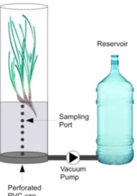
(13) mediating 60 to 95% of the nitrogen fixation in the seagrass rhizosohere (Smith et al., 2004).. The objective of this work is to fill the gap in the knowledge of the diazotrophic community inhabiting the rhizosphere of Posidonia oceanica. This is the first report about nitrogen fixing bacteria associated to its rhizosphere, what would prove that this community is also contributing to the nitrogen cycle as a new source of nitrogen and, hence, contributing to the maintenance of P.oceanica meadow, as they would supply the required nutrients.. 2. Experimental procedures 2.1. Origin of the samples Samples used to study the diversity of the community of nitrogen fixers came from a previous perfusion technique (figure 3). Intact plants were placed into incubation chambers composed of one-end capped PVC pipes (144 mm inner diameter) to hold intact rhizosphere, attached to 8 L metacrylate capped tube harbouring the phyllosphere. Chambers were filled with seawater containing acetylene (20% v/v). The PVC cap was removed from a hole in the base of the cores, and the acetylenated water perfused slowly through the sediment. Finally, more water with 20% acetylene was added in order to completely fill the chamber. Cores were incubated for 24 h periods with natural sunlight on outdoor thermostated aquaria mimicking in situ seawater temperatures.. 12.
(14) Figure 3. Diagram of perfusion technique apparatus.. The sampling location was a Posidonia oceanica meadow in the Alcudia Bay, Mallorca, Mediterranean Sea, Spain (39°50'12''N 3°10'15''E) (Figure 4). Sediment cores were collected in 4-time sampling, representing the four-time season: November 2012, February 2013, May 2013 and July 2013. 3 replicas were taken for each.. Figure 4. Alcudia Bay map with the red dot as study site.. 13.
(15) 2.2. Acetylene reduction assay (ARA) and sediment analyses Water column samples were taken in order to measure the oxygen concentration at the beginning and at the end of the experiment, so we could calculate respiration, net primary productivity and gross primary productivity. Every 2 hours for 6-8 h incubation time pore water samples were taken from the sediment at 3 depths: 2 cm, 6 cm and 10 cm of each core. Nitrogen fixation rates were calculated using Acetylene reduction Assay, which is a method that takes advantage of the inspecificity of the nitrogenase to reduce triple bond substrates (Staal et al., 2001). The acetylene (C2H2) is reduced to ethylene (C2H4) that is taken with a syringe and injected to a gas cromotograph equipped with a flame ionization detector (FID) to measure acetylene and ethylene concentrations. The conversion factor 4:1 is applied from the stoichiometry of the reactions (Staal et al., 2001): (1). N2 + 8[H] à 2NH3 + H2. (2). C2H2 + 2[H] à C2H4. At the end of the experiment, pore water was taken to measure nitrogen, carbon and phosphorous concentrations. Also, sediment and plant samples were taken to obtain the following data: grain size and organic matter corresponding to its sediment fraction size, biomass data of roots and rhizomes, density shoot, epiphytes in roots and rhizomes (in grams), and nitrogen, carbon and phosphorous concentrations in the sediment, roots, rhizomes, leaves and its epiphytes (%). Grain size analysis was done by air-drying for at least for 5 days and binned using series of sieves (pore sizes 1000 µm, 500 µm, 250 µm, 100 µm, 50 µm). Each retained fraction was weighted on an electronic balance. Ovendried method at 80ºC was used to determine the organic matter (OM) of sediment samples. Element Analyzer (EA1108, Carlo Erba Instruments) was used to analyse CN. 14.
(16) contents of bulk sediment and P content was measured following Aspila et al (1976) method. Density shoot was determined in situ using 6 randomly placed quadrats (0.4 x 0.4 m). All leaves of a shoot were measured for leaf morphometry and their adhering epiphytes were scarped-off at both sides using a clean scalpel and collected in dried and weighted eppendorf tubes. Dry-weight determination was done at 80ºC and after 24h. Also, dry-weight determination of the corresponding epiphyte-free leaves was done (60ºC). CNP analyses were done from both dried epiphyte-free leaves and dried epiphytes. C and N content were determined as described above and P content was measured using the method of Solórzano and Sharp (1980) modified. Net community production (NCP) was determined using the Winkler method from dissolved oxygen (DO) concentrations were estimated. DO was measured at time 0, during the day (after sunset) and during night (after sunrise), so community respiration (R) and NCP were calculated from the difference in DO concentrations in the chambers at night and during day, respectively. Gross primary production (GPP) was determined by the sum of R and NCP (Agawin et al., 2016). Significant variables are shown in table 4.. 2.3. DNA extraction Each core was cut in 3 sections what corresponded to depths between 0-12 cm: 0-4 cm, 4-8 cm and 8-12 cm. The DNA was extracted by a previous established procedure (Nogales et al., 2002).. 2.4. DNA amplification The worked sediment samples had a great amount of PCR inhibitors such as humid acids, fact that could be appreciated analysing absorbance values, specially 260/230. 15.
(17) ones, too low. Thus, before the amplification of the target gen, samples were purified with Wizard DNA Clean-up System of Promega and resuspended.. NifH gene amplification was performed by nested PCR. The final reaction mixture volume was 50 μl. The primers used were those from Zher et al. (1989): forward nifH3 primer. (5’-ATRTTRTTNGCNGCRTA-3’). and. reverse. nifH4. primer. (5’-. TTYTAYGGNAARGGNGG-3’) for the first round, and forward nifH1 primer (5’ADNGCCATCATYTCNCC-3’). and. reverse. nifH2. primer. (5’-. TGYGAYCCNAARGCNGA- 3’) for the second round. PCR mixture consisted in 1,5 mM MgCl2, 2 mM dNTP mixture, 10 μM of each primer and 5U DNA Platinum Polymerase (Invitrogen). The thermal cycling conditions for the first round were as follows: 35 cycles of 95 ºC for 1 min, 45 ºC for 1 min, and 72 ºC for 30 s, followed by extension at 72 ºC for 10 min. For the reactions with nifH1 and nifH2 were as follows: 30 cycles of 95 ºC for 1 min, 54 ºC for 1 min, and 72 ºC for 30 s, followed by extension at 72 ºC for 10 min. Both rounds needed hot start at 94 ºC just because Platinum polymerase requires this previous step to be activated. PCR products were run on a 2% agarose gel, and the 350 to 365-bp amplified nifH fragments were purified with Pure Link PCR Purification Kit (Invitrogen) in order to elute all primers, nucleotides, salts and polymerases from the previous process.. 2.5. Cloning and target gene amplification TOPO TA cloning kit (Invitrogen) with TOP10 chemically competent E.coli cells were used. To perform the cloning reaction (6 μl), a mix of 4 μl of fresh PCR product, 1 μl of salt solution and 1μl of TOPO vector was done. After the reaction, 50 μl of transformed. 16.
(18) cells were spread on LB plates containing 40 μl X-Gal. Plates were incubated overnight at 37 ºC. White colonies, corresponding to the positive clones, were picked for analysis. The vector contains lacZ gene (figure 5), which codifies for the galactosidase enzyme, responsible for the hydrolysis of X-Gal to galactose. The PCR product is inserted in the lacZ gen breaking it and making it no functional. E. coli not transformed cells are able to metabolize X-Gal, so these ones are expressed in blue, whilst white colonies have no functional gene that they can not use X-Gal.. M13 PCR was performed using M13 forward primer (5’-GTAAAACGACGGCCAG3’) and M13 reverse primer (5’-CAGGAAACAGCTATGAC-3’). The reaction mixture containing the following (50 μl reaction volume): 1.5 mM MgCl2, 2 mM dNTP mixture, 10 μM of each primer and 1U DNA Native Polymerase (Biotools). The thermal cycling conditions were 35 cycles of 94º C for 1 min, 55 ºC for 1 min, and 72 ºC for 2 min, followed by an extension step of 72 ºC for 10 min. PCR products were run on a 2% agarose gel, and 600-bp fragments were purified with Pure Link PCR Purification Kit (Invitrogen).. 17.
(19) Figure 5. PCR®2.1-TOPO® Map from Invtrogen user manual of TOPO TA Cloning Kit.. 2.6. Sequencing Positive PCR products were sequenced using Big Dye Terminator V3.1 Cycle Sequencing Kit (Applied Biosystems). The precipitation step was done: 10 μl sodium acetate 3 M and 250 μl ethanol 100% (-20 ºC) to each sample, mix by inversion and centrifuge 16.200 g for 30 min 4 ºC. Discard the supernatant and add 300 μl cold ethanol 70% and centrifuge again 16.200 g but for 15 min 4 ºC. The supernatant was discarded and pellet dried in air. Samples were kept at -20 ºC till sequencing. Before, the pellet was dissolved in 20 μl of sterile MilliQ water.. 18.
(20) 2.7. Bioinformatics and phylogenetic and physicochemical analysis Sequences were edited using Finch TV software and imported to CLC Sequence Viewer7 used to translate them to a protein sequence. NifH amino acid sequences were compared to the public database of NCBI by BLAST (Basic Local Alignment Search Tool) service (www.ncbi.nlm.nih.gov). The closest amino acid sequences were selected and used to create our database altogether with our sequences, which were treated with CLC sequence Viewer 7 at the beginning, just to have a first view of the alignment. Then, the sequences were exported to MEGA 6.06, with which the final alignment, the distance matrix and the phylogenetic tree were obtained. A Neighbour-joining tree was produced. Boostrap analysis was used to estimate the reliability of the phylogenetic reconstruction (1000 iterations). The model used was Maximum Composite Likelihood. Sequence from Methanosarcina lacustris (AAL02158) was used as outgrup. NifH sequences can fit in 4 Clusters previously used by Chien and Zinder (1996) to characterize nifH phylogeny. An OTU-based treatment was carried out in which all sequences were binned using a cutoff of 97% similarity at amino acid level. An identity >95% between two sequences they are within the same genus (Swee Hoe Omg et al., 2013). We checked this cutoff at nucleotide level and both of them matched, so we could consider this limit as suitable in our study. Rcmdr and Vegan R packages (http://www.r-project.org/) were used as statistic tool treat phylogenetic and physicochemical data.. 3. Results A total of 104 of new nifH clones were obtained from the four time sampling period and from the three different depths in Posidonia oceanica sediment.. 19.
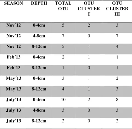
(21) We obtained 30 OTU and 48 different nifH- deduced amino acid sequences. No sequence was found to belong to Cluster II neither Cluster IV. 7 OTU were found to belong to Cluster I and 23 to Cluster III (Figure 6). Table 1 shows number of clones for each OTU binned to Cluster I and Cluster III according to the sampling conditions (season and depth). SEASON. DEPTH. TOTAL OTU. OTU CLUSTER I. OTU CLUSTER III. Nov´12. 0-4cm. 5. 2. 3. Nov´12. 4-8cm. 7. 0. 7. Nov´12. 8-12cm. 5. 1. 4. Feb´13. 0-4cm. 2. 1. 1. Feb´13. 8-12cm. 1. 0. 1. May´13. 0-4cm. 3. 1. 2. May´13. 8-12cm. 4. 1. 3. July´13. 0-4cm. 10. 2. 8. July´13. 4-8cm. 3. 0. 3. July´13. 8-12cm. 2. 0. 2. Table 1. Number of clones for OTU for each Cluster according to sampling conditions.. 20.
(22) Gr o u p 1. Uncultured bacterium clone I_3A.02 (ADV35102) Uncultured bacterium P.oceanica sediment Jul'13 0-4cm clone 34.18; 34.27. OTU 1. Uncultured microorganism H01_RNA_A3 (ABQ50691). Desulfobulbus mediterraneus (WP028585313). OTU 2. Uncultured bactrium P.oceanica sediment May'13 0-4cm clone 20.3; 20.4; Nov'12 4-8cm clone 24.8 90. Uncultured bacterium P.oceanica sediment Jul'13 0-4cm clone 34.7 64. Uncultured bacterium clone Xmnif13_LC_m (AFJ04673). Desulfovibrio zosterae (WP027723058) 81 73. -Proteobacteria. Uncultured bacterium P.oceanica sediment Jul'13 4-8cm clone 3.12; 3.13; May'13 0-4cm clone 20.1. Gr o u p 2. 52. Uncultured bacterium P.oceanica sediment Nov'12 4-8cm clone 24.5, Nov'12 8-12cm clone 25.1 OTU 3 Desulfobulbus japonicus (WP028579019) OTU 4. Uncultured bacterium P.oceanica sediment Nov'12 0-4cm clone 14.7. 61 74. Uncultured bacterium P.oceanica sediment Nov'12 0-4cm clone 14.9 Uncultured bacterium P.oceanica sediment Nov'12 0-4cm clone 14.3; 14.12; 14.14 Desulfovibrio desulfuricans (WP014321935) Desulfurivibrio alkaliphilus (WP013164631) Desulfobulbus sp. Tol-SR (WP035226442). Gr o u p 3. Uncultured nitrogen-fixing bacterium clone RcTDY73 (AFO55079) Uncultured bacterium clone MDE_elv_14d1 (AHN50870). 89. Gr o u p 4. OTU 5. Uncultured bacterium P.oceanica sediment Jul'13 0-4cm clone 34.10. OTU 6. Uncultured microorganism clone B10-8 (AJF13358). Chlorobi. Spirochaeta cellobiosiphila (WP028973004) Chloroherpeton thalassium (WP012499583). 75 70. Uncultured bacterium clone PJ1-28 (AHI71643) OTU 7. Uncultured bacterium P.oceanica sediment Jul'13 0-4cm clone 34.23; 34.26. Gr o u p 7 Gr o u p 6. Desulfurispora thermophila (WP018085363) Uncultured bacterium clone III-5.110 (ADD26590) 54. Uncultured bacterium P.oceanica sediment May'13 8-12cm clone 22.2; 22.3; 22.5; 22.6; 22.7. OTU 8. Uncultured bacterium clone PriLo-10343 (AAY85400). 71. Uucultured bacterium P.oceanica sediment Nov'12 0-4cm clone 14.10 OTU 9 Desulfobacca acetoxidans (WP013705354). 9 8 Uncultured bacterium P.oceanica sediment Nov'12 4-8cm clone 24.4; Nov'12 8-12cm clones 25.5; 25.7. OTU 10. Uncultured bacterium P.oceanica sediment Nov'12 8-12cm clone 25.6. 70. Gr o u p 8. Uncultured bacterium clone pCHL_W5 (AFK30433). 58. Uncultured bacterium P.oceanica sediment Jul'13 0-4cm clone 34.19. Gr o u p 9. Desulfovibrio fructosivorans (WP005994097) OTU 11. Uncultured bacterium clone 08-II.55 (ADD49205). Gr o u p 10. Uncultured bacterium P.oceanica sediment Jul'13 0-4cm clone 34.4 OTU 12 Desulfovibrio putealis (WP027192250). Uncultured bacterium P.oceanica sediment Jul'13 0-4cm clone 34.22; 34.25; 34.29 OTU 13 Uncultured bacterium clone Olson_Mlf22A1-F01 (ACD87623). Uncultured bacterium P.oceanica sediment Feb'13 0-4cm clone 8.3 OTU 14. Uncultured bacterium P.oceanica sediment May'13 0-4cm clone 20.5 79. Uncultured microorganism clone H01_DNA_E2 (ABQ50806). -Proteobacteria. Desulfovibrio magneticus (WP006922063) 99. CLUSTER III. Uncultured bacterium P.oceanica sediment Jul'13 4-8cm clone 3.1. Gr o u p 5. 62. Uncultured bacterium P.oceanica sediment Feb'13 0-4cm clone 8.2; 8.5; 8.7; 8.8; 8.11; 8.13; May'13 0-4cm clones 20.2; 20.6; 20.7; 20.8; 20.9 Uncultured bacterium P.oceanica sediment Jul'13 8-12cm clone 1.2; 1.4; 22.8; 22.10. Uncultured bacterium P.oceanica sediment Feb'13 8-12cm clone 10.4; 10.7; 10.8; 10.11; 10.16; 10.19; 10.20 95. Uncultured bacterium P.oceanica sediment Feb'13 8-12cm clone 10.14. OTU 15. Uncultured bacterium clone PJD2-17 (AHI71672). 50. Uncultured bacterium MDE_amb_32g12 (AHN50692). OTU 16. Uncultured bacterium clone 07-II.5 (ADD48905) Uncultured bacterium P.oceanica sediment Jul'13 8-12cm clone 1.8; 1.9; 1.11; 1.12; 1.13; 1.14; 1.15; 1.17; Jul'13 4-8cm clones 3.3; 3.5; 3.6; 3.7; 3.8; 3.9; 3.10, 3.11 Uncultured bacterium P.oceanica sediment Nov'12 8-12cm clone 25.27. Gr o u p 11. Uncultured bacterium P.oceanica sediment Nov'12 0-4cm clone 14.2. Uncultured bacterium P.oceanica sediment Nov'12 8-12cm clone 25.8 Uncultured bacterium clone MDE_amb_35f1 (AHN50740) OTU 17. Uncultured bacterium clone 07-II.1 (ADD48901) Uncultured bacterium P.oceanica sediment Nov'12 4-8cm clone 24.2. 87. Uncultured bacterium P.oceanica sediment Nov'12 4-8cm clone 24.13; 24.17 Uncultured bacterium clone OilLo-10301 (AAY85423) Uncultured bacterium P.oceanica sediment Nov'12 4-8cm clone 24.18. OTU 18. Uncultured bacterium clone MDE_amb_26a10 (AHN50513) Uncultured bacterium P.oceanica sediment Jul'13 0-4cm clone 34.30. OTU 19. Desulfatitalea sp. clone BRH c12 (WP045678441) Desulfatibacillum MULTISPECIES (WP012610655) Uncultured bacterium clone OilLo-10338 (AAY85434) Uncultured bacterium P.oceanica sediment Nov'12 8-12cm clone 25.3 Uncultured bacterium P.oceanica sediment May'13 8-12cm clone 22.1; 22.9. OTU 20 OTU 21. 97. Uncultured bacterium P.oceanica sediment Nov'12 4-8cm clone 24.15 OTU 22 Uncultured bacterium P.oceanica sediment Nov'12 4-8cm clone 24.20. 83. Uncultured bacterium P.oceanica sediment Nov'12 4-8cm clone 24.1. Gr o u p 12. 72. Firmicutes. Uncultured microorganism clone 507_161_B3 (AFH88470). OTU 23. 96. Uncultured bacterium clone MDE_amb_23f10 (AHN50488) Clostridium acetobutylicum (WP010963576) 67. Clostridium kluyveri (WP012101422). Uncultured bacterium P.oceanica sediment Nov'12 0-4cm clone 14.6 OTU 24. 95 64. Gr o u p 13. 9 8 Clostridium kluyveri NBRC 12016 (BAH06000). Uncultured bacterium clone Sipa-L23 (AGO36368). 80. Gr o u p 14. Candidatus Magnetoovum chiemensis strain CS-04 (KJR40986) Heliobacterium chlorum strain DSM 3682 (BAD80872) Uncultured bacterium P.oceanica sediment May'13 0-4cm clone 20.10 OTU 25 Thiobacillus prosperus (WP038093037). 99. Uncultured bacterium P.oceanica sediment Jul'13 0-4cm clone 34.3; 34.8. OTU 26. Uncultured bacterium P.oceanica sediment Nov'12 0-4cm clone 14.1; 14.13. Uncultured bacterium P.oceanica sediment Nov'12 0-4cm clone 14.4 Thiothrix nivea (WP002710379). - Proteobacteria. Uncultured bacterium clone Mf21-B01-A07 (ACD87576) Uncultured microorganism clone H05_DNA_E5 (ABQ50825) Halorhodospira halophila strain ABN10970 (ABN10970) Uncultured nitrogen-fixing bacterium clone 2#C2.36 (AHF50907) Uncultured bacterium clone MDE_amb_23h8 (AHN50510). CLUSTER I. Uncultured bacterium clone Mf20-B06-F02 (ACD87559). Gr o u p 15. Martelella endophytica (WP045682609) 6 7 Uncultured marine bacterium clone A11-0M-02 (ADT89921) 77. Uncultured bacterium P.oceanica sediment Jul'13 0-4cm clone 34.21 OTU 27 Ectothiorhodospira shaposhnikovii strain DSM 243 (ABN10972) Celerinatantimonas yamalensis strain DSM 21888 (AEC46862) Uncultured bacterium P.oceanica sediment Feb'13 0-4cm clone 8.1 Uncultured bacterium P.oceanica sediment Feb'13 0-4cm clone 8.9. Uncultured bacterium P.oceanica sediment Feb'13 0-4cm clone 8.6 Uncultured nitrogen-fixing bacterium clone B90 (AAZ06705) Vibrio diazotrophicus strain ATCC 33466 (AAF17302). 6 0 Uncultured bacterium P.oceania sediment May'13 8-12cm clone 22.4. OTU 29. Sedimenticola sp. SIP-G1 (WP046860621) Marinobacterium litorale (WP027854782) 6 2 Uncultured bacterium clone A5-H-48 (ACN77123). Uncultured bacterium clone III-5.1 (ADD26505) Pseudomonas stutzeri clone Gr16 (CBL85097) Uncultured bacterium P.oceanica sediment Nov'12 8-12cm clone 25.9 OTU 30 Pseudomonas stutzeri clone A15101 (CAC03734) Methanosarcina lacustris clone pms15 (AAL02158) 0.05. Gr o u p 16. 94. OTU 28. 55. 7 0 Uncultured bacterium P.oceanica sediment Feb'13 0-4cm clone 8.12. Figure 6. Neighbor-joining phylogenetic tree of nifH deduced amino acid sequences of nitrogenfixing bacteria associated with sediment of Posidonia oceanica. Sequences obtained in this study are marked in boldface letters. Relationships were bootstrapped 1000 times, and bootstrap values greater than 50% are indicated at respective nodes. Tree is rooted to nifH deduced amino acid sequence of Methanosarcina lacustris clone pms15 (AAL02158).. 21.
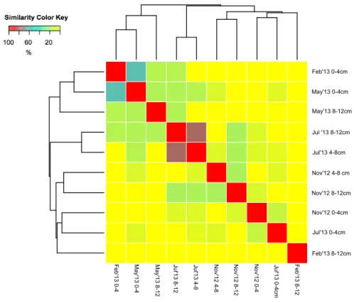
(23) 3.1. Ecological diversity A MANOVA and the results revealed that both of them were significant for the distribution of the communities in the sediment, as we obtained F1.9=0.1377 (p<0.05) and F3.9=0.4103 (p<0.05) for depth and season, respectively. We have used Shannon index (H) to assess the specific diversity in our samples, showed in table 2.. Section. Diversity (H). Nov 0-4cm. 1,37. N2 Fixation rate (nmol h-1)/mgDW 0,51. Nov 4-8cm. 1,83. 0,38. Nov 8-12cm. 1,49. 0,2. Feb 0-4cm. 0,66. 0,09. Feb 8-12cm. 0. 0,26. May 0-4cm. 0,9. 0,13. May 8-12cm. 1,22. 2,15. July 0-4cm. 2,21. 5,51. July 4-8cm. 0,76. 2,81. July 8-12cm. 0,5. 0,9. Table 2. Shannon values and fixation rates corresponding to their section.. Figure 7 revealed that there was a certain similarity in the community composition of the following samples, as they cluster together: shallow samples from February and May, shallow samples from November and July, deep samples from July (4-8cm and 812 cm) and deep samples from November (4-8 cm and 8-12 cm). Among these samples, July’13 4-8 cm and July’13 8-12 cm showed the highest similarity, with a similarity of 86%. A great similarity was showed too between Feb’13 0-4 cm and May’13 0-4 cm (67%).. 22.
(24) Sample from Feb’13 8-12 cm showed a unique community, as it did not share its composition with any other sample, this sample did not cluster with any other and appeared in soft colours, meaning a low similarity among the other communities. In the Bray Curtis dissimilarity index appeared with 0% similarity with the rest of samples. Nov’12 0-4 cm and July’13 0-4 cm (with similarity values of 0-15 % and 0-20 %, respectively) were, after Feb’13 8-12 cm, the samples with a most different composition with the rest. There was no sample from 0-4 cm clustering with any other deeper section, but we could find clusters with 4-8 cm and 8-12 cm depth. Sections from 0-4 cm depth of February and May clustered together and, July and November as well. Conditions from February and May were similar and they kept the composition of their communities during this period.. Feb'13 0-4cm May'13 0-4cm May'13 8-12cm Jul '13 8-12cm Jul'13 4-8cm Nov'12 4-8 cm Nov'12 8-12cm Nov'12 0-4cm Jul'13 0-4cm Feb'13 8-12cm Feb'13 8-12. Jul'13 0-4cm. Nov'12 0-4. Nov'12 8-12. Nov'12 4-8. Jul'13 4-8. Jul'13 8-12. May'13 8-12. May'13 0-4. Feb'13 0-4. Figure 7. Heat map based on a dendrogram of samples according to its time sampling and depth.. 23.
(25) To assess the distribution of our OTU regarding season and depth, we used Principal Components Analysis (PCA) with inertia by variance, which had a value of 39.09 (figure 8). This graphic let us know the reason of the clustering seen above, to know which OTU are affecting the evenness of communities. Can be appreciated that the section from February 8-12cm seemed to be the sample with the most different bacterial composition, as it appeared far away from the others in the graphic. Its position is determined by the composition of OTU 15, which was composed by 8 clones, only found in this section (see table 1 in annex). As seen above, the deepest sections of July were the ones with more similarity in their communities. They appeared together and far away from other samples. The composition of OTU 5 and 16 granted this position. OTU 16 had 8 sequences found in July 4-8cm section and another 8 in July 8-12cm, among others (1 sequence in November 0-4cm and 2 November 8-12cm). Hence, OTU 16, is the responsible that July 4-8 and July 8-12cm had a great similarity between them. Can also be remarkable the position of sections from February 0-4cm and May 0-4cm, which seemed to be more similar to each other than with section from May 8-12cm. OTU 14 was composed by 7 clones from Feb’12 0-4cm and by 6 from May’13 0-4cm, in addition to have 2 clones from May’13 8-12cm and 2 from July’13 8-12cm. Thus, the composition of OTU is responsible for the similarity between communities of upper sections. This OTU reflected as well similarity among this shallow sections and deep May and deep July, with 2 clones in each of these last two sections. In the central part of the graphic, the 3 sections of November and July 0-4cm were displayed. This suggested that there was no OTU which its composition made this samples differ from the rest and, therefore, bore some resemblance between them Dots. 24.
(26) in purple, orange, blue and yellow represent exclusive OTU for November 0-4 cm, November 4-8 cm, November 8-12 cm and July 0-4 cm, respectively.. Figure 8. PCA with inertia by variance of data from table 1 and table 1 in annex. Symbols represent a set of OTU in the same position: Green dot (CLONS OTU 8, 21, 29); blue dot (CLONS OTU 20, 30); yellow dot (CLONS OTU 1, 6, 7, 11, 12, 13, 19, 27) and purple dot (CLONS OTU 4, 9, 24).. 3.2. Diversity in Clusters and closest relatives The community composition was studied within Clusters. ANOVA results revealed that factor depth had a significant effect on the distribution in both Clusters: Cluster I (pvalue 0.024) and Cluster III (p-value 0.016).. 25.
(27) Two sequences belong to the same phylum with >80% identity (Swee Hoe Omg et al., 2013). 12 of the 104 nifH- deduced amino acid sequences were binned in Cluster I. Group 13 has just one clone (clone 14.6), which had 80% similarity with Heliobacterium chlorum (BAD80872), Firmicutes and Candidatus Magnetoovum chiemensis (KJR40986), Nitrospira (see table 3). It was not possible to determine in which phylogenetic group belonged this sequence. Group 14 included only one sequence, clone 20.10. This sequence had as closest relative Pseudomonas stutzeri (CBL85097), but their identity is not high enough to affirm that they were within the same genus, but within class γ-Proteobacteria. Group 15 had as closest relative Martelella endophytica, α- Proteobacteria (WP 045682609); Thiothrix nivea, γ- Proteobacteria (WP002710379), thus, was not possible to determine in which phylogenetic group they belonged to. In case of clone 34.21, we could say that it fitted within γ- Proteobacteria, as it had 93% identity with Halorhodospira halophila (ABN10970). We could say that all of 6 nifH-deduced amino acid sequences belonged to γ- Proteobacteria. Clone 25.9 had 100% identity with Pseudomonas stutzeri (CAC03734). Same situation happened with clone 22.4, with 100% identity with Sedimenticola sp.SIP-G1 (WP019604939). Nevertheless, as phylogenetic analyses were done with protein sequences, it cannot be concluded that those clones were Sedimenticola sp.SIP-G1 and Pseudomonas stutzeri, respectively. Can be appreciated that all clones within this group had an identity higher than 80% with their closest relative. Then, clones 8.1, 8.6, 8.9 were within class γ- Proteobacteria.. Regarding to Cluster III, most of nifH-deduced amino acid sequences belonged to δProteobacteria, except two sequences from group 5 (clones 34.23 and 34.26) that were. 26.
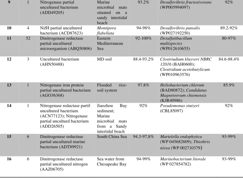
(28) found to the Chlorobi taxon with a great identity (95.5%). These green sulphur bacteria were found in 0-4c section. Also, three sequences from group 12 (clones 24.1, 24.15 and 24.20) belonged to Firmicutes. The great majority of closest cultured representatives were sulphate reducers, anaerobic microorganisms that use pyruvate, lactate, ethanol and certain fatty acids as electron donors during the sulphate reduction to sulphide. Desulfobacca acetoxidans, closest cultured representative for groups 7 and 8, specializes in sulphate reduction oxidizing acetate.. Group. No. of clones. Closest environmental nifH-deduced amino acid sequence. Source. 1. 2. Nitrogenase partial uncultured bacterium (ADV35102) Uncultured bacterium (ABQ50691). Florida Keys Admiarl´s Reef water column Eastern Mediterranean Sea.. 2. 14. 3. 1. Uncultured bacterium (AHN50870). 4. 1. Uncultured bacterium (AJF13358). 5. 2. Uncultured bacterium (AHI71643). 6. 5. Nitrogenase partial uncultured bacterium (AFK30433). 7. 1. Uncultured bacterium clone PriLo-10343 (AAY85400). 8. 4. Dinitrogenase reductase partial uncultured bacterium (AFK30433). % Identity. Closest environmental nifH-deduced amino acid sequence for a cultured bacterium Desulfovibrio putealis (WP027192250). % Identity. 92-100%. Desulfovibrio desulfuricans (WP014321935); Desulfobulbus mediterraneus (WP028585313). 93.2% 98.9%. MD soil. 90.8%. Desulfobulbus japonicus (WP 028579019); Desulfurivibrio alkaliphilus ( WP 013164631); Desulfovibrio zosterae (WP 027723058). 90.8%. Bohai Sea B10 site sediments (China) Healthy and diseased corals in Sanya Bay (Chian) Marine microbial mats situated on a Sandy intertidal beach Oilcontaminated marine sediment Rhizosphere of Colocasia esculenta. 97.8%. Desulfobulbus sp. Tol-SR (WP035226442). 93.2%. 95.5%. Chloroherpetum thalassium (WP012499583). 95.5%. 92%. Desulfurispora thermophila. 85.9%. 94.3%. Desulfobacca acetoxidans (WP013705354). 89.6%. 94.4-95.5%. Desulfobacca acetoxidans (WP013705354). 92%. 95.5%. 89.2-92%. 27.
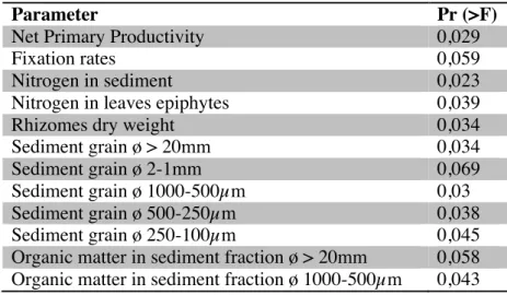
(29) 9. 1. Nitrogenase partial uncultured bacterium (ADD49205). Marine microbial mats situated on a sandy intertidal beach Montipora flabellata Eastern Mediterranean Sea. 10. 4. 11. 52. NifH partial uncultured bacterium (ACD87623) Dinitrogenase reductase partial uncultured microorganism (ABQ50806). 12. 3. Uncultured bacterium (AHN50488). MD soil. 13. 1. Nitrogenase iron protein partial uncultured bacterium (AGO36368). Flooded ricesoil system. 97.8%. 14. 1. Nitrogenase reductase partil uncultured bacterium (ACN77123); Nitrogenase partial unculturd bacterium (ADD26505). 92%. 15. 6. Dinitrogenase reductase partial uncultured marine bacterium (ADT89921). Jiaozhou Bay sediment; Marine microbial mats from a Sandy intertidal beach South China Sea. 16. 6. Dinitrogenase reductase partial uncultured nitrogen (AAZ06705). Sea water from Chesapeake Bay. 93.2%. Desulfovibrio fructosivorans (WP005994097). 94-98%. Desulfovibrio putealis (WP027192250) Desulfatibacillum multispecies (WP012610655). 92-100%. 88.4-93.2%. 94.3-97,8%. 94-99%. 92%. 89.2-92% 80-97%. Clostriudium kluyveri NBRC 12016 (BAH0600); Clostridium acetobutylicum (WP010963576). 84.6-88.4%. Heliobacterium chlorum (BAD80872); Candidatus Magnetoovum chiemensis (KJR40986) Pseudomonas stutzeri (CBL85097). 85.9%. Martelella endophytica (WP 045682609); Thiothrix nivea (WP 002710379). 93-99%. Marinobacterium litorale (WP 027854782). 93-99%. 92%. Table 3. Number of clones for each group and their closest nifH deduced amino acid sequence cultured and uncultured bacterium with their % identity.. 3.3. Physicochemical variables Sediment parameters were studied in order to find out which were relevant in the distribution of our community (see experimental procedures). Significant parameters are shown in table 4.. 28.
(30) Parameter Net Primary Productivity Fixation rates Nitrogen in sediment Nitrogen in leaves epiphytes Rhizomes dry weight Sediment grain ø > 20mm Sediment grain ø 2-1mm Sediment grain ø 1000-500µm Sediment grain ø 500-250µm Sediment grain ø 250-100µm Organic matter in sediment fraction ø > 20mm Organic matter in sediment fraction ø 1000-500µm. Pr (>F) 0,029 0,059 0,023 0,039 0,034 0,034 0,069 0,03 0,038 0,045 0,058 0,043. Table 4. Significant parametres in the distribution of nitrogen-fixing community of this study.. 4. Discussion Diazotrophs are distributed in habitats and ecosystems according to how genes are selected in those habitats (Zehr et al., 2003). NifH has been found previously in many environments, as can be seen in table 3: water column, sediments, soil, as coral symbionts, in termite guts, etc. In case of sediments, the diversity includes heterotrophs and phototrophs (Zher et al., 2003). In Cluster I, clones were mainly found in the shallowest part of sediment (0-4cm). They had great identity with purple sulphur bacteria (example Halorhodospira) and with sulphur reducing bacteria (Pseudomonas). Purple sulphur bacteria are phototrophic anoxigenic organisms, and sulphur reducing facultative aerobes, so in adequate nutritional conditions they can thrive in aerobiosis and anaerobiosis. These features for both bacterial groups may explain why Cluster I had had a great amount of clones sequenced from depth 0-4cm. They could have a great respiratory rhythm and a dense mucous layer to protect nitrogenase enzyme from oxygen inhibition (Sabra et al., 2000). The anaerobiosis of purple sulphur bacteria may suggest that the sampling area was poor of oxygen, as already in the surface of the sediment we could find bacterial. 29.
(31) communities that require absence or very short quantities of oxygen to thrive. The oxygen penetration to the sediment has to be considered, then. Has been reported in previous studies that oxygen penetrates approximately 4.5mm (Holmer et al., 2003) in vegetated marine sediment, fact that contributes to create anoxic conditions within the sediment. Both oxidizing and reducing sulphur bacteria live in the same habitat, and they have an association. Purple sulphur bacteria utilize hydrogen sulphide (H2S) as electron donor in the photosynthesis. H2S is oxidised to elemental sulphur that is kept and accumulated in globules inside the cell, which later disappeared when are oxidised to sulphate (SO4 2-). Has been reported that Halorhodospira keeps these globules outside the cell. The presence of Sulfur- Oxidizing bacteria has an important role in detoxifying sediments, what means that these bacteria are essential in maintenance of seagrasses. High inputs of organic matter enhance sulphate reduction and production of sulphide that is toxic to these plants (Garcias-Bonet et al., 2012). In iron-rich sediments, the effects of the sulphides are buffered by the precipitation and adsorption as sulphides combined with iron, but in Balearic waters the sediments are iron-poor, so there is a lack of sulphide buffering capacity (Marbà et al., 2007). Sulphur- reducing bacteria reduce sulphur produced by the oxidizing bacteria, closing the cycle. Cluster III had sequences from all depths and was mainly represented for δProteobacteria, sulphate reducing organisms.. Depth appeared to be the main significant factor for the community distribution in Clusters. Temperature was less important, thus, the bacterial community inhabiting sediment associated to Posidonia oceanica did not have a strong seasonal dependence. The bacterial community associated to benthic components of P.oceanica (Agawin et al., unpubl) has this dependence, as they inhabit in direct contact with the water column.. 30.
(32) In previous studies focused on the diversity of cold marine sediments (Ravenschlag et al., 1999), the community found was almost composed by δ-Proteobacteria, beeing SRB the major group; and γ-Proteobacteria, with a great amount of sulphur-oxidizing bacteria. These findings could support the idea that in sediments, temperature is not as significant as other factors. Although, temperature has to be considered as factor with indirect effects on the community structure. Season temperature modifies solubility of gases, such as oxygen. During seasons with high temperatures, gas solubility decreases, resulting in a less availability of oxygen for organisms. Hence, the diffusion of gas to sediment would be low. As said above, oxygen diffusion is about 4.5mm, which means that the first section of our study (0-4cm) is mainly anoxic too, like the rest of sections. This may explain the presence of an anoxic community. Events like storms and macrofaunal benthic communities cause irrigation via bubble movement in the sediment, which cause oxygenation of the top and exposing the anaerobic community to air. Is for that reason that species belonging to this group have developed strategies to protect themselves from oxygen. Has been reported that Desulfovibrio genus has the genetic potential of oxygen survival strategies: genes for oxygen chemotaxis and oxygen oxidorectuctase (Wildschut et al., 2006, and Krekeler et al., 1998). SRB are able to sense the oxygen concentration of the environment so they can move away from oxygen (Krekeler et al., 1998). Oxygen oxidoreductase (Roo) works as an oxygen reductase in vivo and its activity protects the cell from oxygen inactivation under microaerophilic conditions (Wildschut et al., 2006). In presence of air, cells cannot grow because there is an inhibition of cell division. This study focuses on the vegetated sediment, which has been reported to have a different behaviour than the bare one. James et al (2006) reported that communities inhabiting vegetated sediments differ from those ones with no seagrass. Many bacterial. 31.
(33) species found in the sediments of seagrass beds are not present in sediments habitats outside of seagrass. This shows the importance of the communities found as crucial factor for the survival of seagrass. Communities also differ according to how pristine is the habitat, becoming a bioindicator of seagrass health and water quality. Seagrass bed sediments support higher number of bacteria and greater bacterial activity than nonvegetated sediments due to the nutrient input coming from seagrass roots and because the planktonic detritus trapped in the seagrass bed coming from the water column (Smith et al., 2004; Holmer et al., 2003). Has been reported that seagrasss sediments contain between 0.18% and 4.7% of organic matter, whereas the concentration of the bare ones varies from 0.07 to 2.2% (Duarte et al., 2005). Thus would be expected to find a difference between communities in vegetated and nonvegetated sediments in our site sampling.. Seagrass bed sediments become anoxic closely beneath the surface when microorganisms rapidly consume oxygen. The need for an available terminal electron acceptor and the great abundance of sulphate in seawater allows the dominance of sulphur-reducing bacteria (Smith et al., 2004). Thus, is possible to find anaerobic microorganisms in the shallowest section of the core. In fact, our findings support this, as we have sequenced anaerobic microorganisms in section 0-4cm depth. Although, these sequences were more abundant in sections 4-8cm and 8-12, than 0-4cm, which supports that there is a cut-off for the oxygen diffusion. Seagrass sediment represents a continuous oxygen input coming from the roots due the photosynthetic activity. Seagrass root oxygen release during the daylight is about 1-25μl O2 gDW-1h-1and about 2- to 10-fold lower during night, derived from oxygen diffusion from the water column into the leaves to the roots. Estimates suggest that oxygen. 32.
(34) supplied by seagrass roots has similar orders of magnitude than the direct oxygen flux from the water column to the sediment by diffusion (Duarte et al., 2005). Hence, the aerobic area around the roots would be about 4.5mm. Inhabiting at this suitable distance from the roots, nitrogenase activity is not inhibited.. 4.1. Diversity and nitrogen fixing rates While nifH clone sequences provide convincing evidence for presence and diversity of diazotroph microorganisms, the information given by PCR can only be considered qualitative, thus PCR is not suitable for quantitative analyses, as it does not provide information about fixation rates. Detecting a nifH sequence from a given organism only demonstrates that this organism is present, no that it is able to contribute actively to N2 fixation neither in which grade it contributes to it (Brown et al., 2003). For this propose, the linkage of the information given by PCR and the one provided by ARA technique is appropriate. Section less diverse was February’13 8-12cm, which resulted in a value of 0 with Shannon test (Table 2), as their clones only matched up in OTU 15. The most diverse (2.21) corresponded to July’13 0-4cm, and had the greater amount of representatives in different OTU. Molecular analyses may support the hypothesis that nitrogen fixation could be coupled to sulphate reduction. Most bacteria of this study had a close relation with SRB, hence the positive correlation between diversity and nitrogen fixation rates could have a link with sulphate reduction as well. Has been reported (Bertics et al., 2013) that there are similar tendencies between nitrogen fixation and sulphur reduction in a SRB bacterial community.. 33.
(35) 4.2. Significant physicochemical variables Microorganism colonization is conditioned, among others, by the availability of nutrients in their environment, which is linked with the porosity. Grain size determines the porosity, as a greater grain diameter more porosity has the sediment. Then, population distribution may be conditioned by the facility of nutrient and oxygen diffusion. Nitrogenase activity is inhibited by oxygen. Then, presence of oxygen can determine nitrogen fixation rates. In anaerobic or microaerophilic conditions, nitrogenase has normal activity, increasing nitrogen concentrations in sediment. But this increase also inhibits nitrogenase. Then, nitrogen-fixers affect in their own activity. The rhizome dry weight appeared to be more significant for Cluster I, which is more abundant in surface sections. The shallowest section corresponds with the greatest rhizome dry weight. This matches with Duarte et al (2005) report, in where they suggest that there is a correlation between roots and rhizomes diameter and depth. In high depths, roots and rhizomes lose diameter. Rhizomes grow according to the plant temporal growth, which reaches the highest point in warmest temperatures (Duarte et al., 2005). Highest values of rhizomes dry weight for sections 0-4cm corresponded to July. Microbial community could develop in association with the rhizomes that could have an interspecific relationship with our clones from Cluster I, such as commensalism and symbiosis. Also, is possible that the distribution and amount of photosynthetic products, which depends on the rhizomes and roots biomass, have an influence on the distribution of the community. In fact, other studies have proved that the microbial diazotrophic community associated with roots of seagrasses is enhanced by these products, such as acetate, glucose, sucrose (Lehnen at el, 2016).. 34.
(36) Seagrasses increase the organic matter amount of the sediment that colonize trapping particles from the water column and retaining matter that produce thyself and the associated communities (Duarte et al., 2005). With higher sediment porosity, organic matter is retained easily and higher could be its diameter. The organic matter with a highest diameter corresponds with July, which has a link with the plant maximum growth, when it has the highest density and the greatest ability to trap particles.. 4.2. Phylogenetic distribution according to the habitat Studies about nifH diversity associated to marine seagrasses have been done (Bagwell et al., 2002; Capone et al., 1982; Cifuentes et al., 2000; James et al., 2006; McGlathery et al., 1998; Lovell et al., 2000). Nitrogen-fixing organisms have been found as endophytes and epiphytes of P.oceanica. The finding of diazotrophs in tissues indicates the presence of symbiotic bacteria that might play an important role in the seagrass nitrogen acquisition (Garcias Bonet et al., 2011). In case of roots, most of nitrogenfixing endophytes fall in Cluster I and Cluster III (Garcias-Bonet et al., 2011). These findings have a similarity with our results, as we also have found organisms that fall in Cluster I and III. However, none of the clones sequenced in this study have 100% identity with those from Garcias-Bonet. In a seagrass bed containing a mixture of Thalassia testuadinum and Syringodium filiforme, Proteobacteria had a great presence in the community of diazotrophs found in the rhizosphere and nifH sequences were more similar to sequences from sulphate reducers than other anaerobic groups (Bagwell et al., 2002). This is consistent with our results and with the identification of sulphate reducers as a key group responsible for diazotrophic activity in seagrass. In seagrasses containing Spartina alterniflora Proteobacteria was the most detectable Phylum in its rhizosphere. nifH sequences. 35.
(37) related to sulphate reducers affiliated with the genera Pseudomonas and Vibrio were found (Brown et al., 2003). Many species of Pseudomonas are capable of respiration using nitrate as terminal electron acceptor but nitrate is usually not detectable in some S. alterniflora zones. Could be possible that these microorganisms are using oxygen according to the features of Spartina´s bed, as they are facultative aerobic. S. alterniflora produces a dense root mat with abundant belowground biomass. Hence, its rhizosphere receives at least some oxygen input at all times, while other seagrasses only transport oxygen to the rhizospere during periods of active photosynthesis (Bagwell et al., 2002). In our case, we also found a sequence related to the genera Pseudomonas in the deepest section of a core corresponding to November. This may indicate, that this genus could inhabit the closest area to the roots. Lehnen et al (2016) have very recently proposed a link between nitrogen fixation and sulphate reducing process, which is consistent with our results. Garcias-Bonet et al (2011) worked in a data compilation about all bacterial diversity in seagrass sediments and most representative Phylum was Proteobacteria (70%), followed by Firmicutes (11%) and Bacteroidetes (10%). This is consistent with our finding. Most representative group was Proteobacteria, and less present were Firmicutes and Chlorobi.. Most of the closest relatives have biotechnological applications that suggest that our clones may have biotechnological importance and so it still needs to be studied. Sulphate-reducing bacteria (SRB) are anaerobic microorganisms that are able to grow fermenting organic acids (formate and lactate) producing hydrogen, acetate and CO2 (Muyzer et al., 2008). SRB could be considered as a biological source of those products. Posidonia oceanica sediment could be a potential source of biotechnological-interesting bacterial.. 36.
(38) 5. Conclusions 1. This study provides first evidence of a nitrogen-fixing bacterial community inhabiting Posidonia oceanica sediment, which is mainly composed by γ- and δ-Proteobacteria. 2. Diazotrophic bacterial community found in P.oceanica rhizosphere is dominated by SRB, which could link nitrogen fixation and sulphate reducing processes. 3. Composition of bacterial nitrogen-fixing community has a temporal variation and depth dependence. However, depth has a higher influence on the composition. 4. Net primary productivity, fixation rates, nitrogen in sediment, nitrogen in leaves epiphytes, rhizome dry weight, sediment grain size and biggest fraction of organic matter contained in sediment are also significant parameters for the distribution of diazotrophic community.. 6. Acknowledgments I would like to express my gratitude to the University of Balearic Islands (UIB) for the technical equipment used to carry out this work and all the colleagues of the University who have made possible to get results during the experimental project: people form Serveis Cientificotècnics and crew from Microbiology department, who led me use their equipment, and specially T. Busquets, for the advice during the amplification and cloning periods. Thanks too to the rest of members of the MEDIFIX project to let me use their results in this thesis. I feel really grateful to N.S.R. Agawin and P. Ferriol for let me be part of MEDIFIX project and for introduce me to the beautiful world of the 37.
(39) marine microbiology. I would like to thank them as well for the learning process in the laboratory and during the writing period.. 8. References Agawin, N.S.R., Ferriol, P., Sintes, E. and Moyà, G. (2016) Temporal and spatial variability of in situ nitrogen fixation activities associated with the Mediterranean seagrass Posidonia oceanica meadows.. Bagwell, C.E., La Rocque, J.R., Smith, G.W., Polson, W., Friez, M.J., Longshore, J.W. and Lovell, C.R. (2002) Molecular diversity of diazotrophs in oligotrophic tropical seagrass bed communities. FEMS Microbiology Ecology, 39: 113-119. Bertics, V.J., Löscher, C.R., Salonen, I., Dale, A.W., Gier, J., Schmitz, R.A. and Treude, T. (2013) Occurrence of benthic microbial nitrogen fixation coupled to sulfate reduction in the seasonally hypoxic Eckernörde Bay, Baltic Sea. Biogeociences, 10: 1243-1258. Brown, M.M., Friez, M.J. and Lovell, C.R. (2003) Expression of nifH genes by diazotrophic bacteria in the rhizosphere of short form Spartina alterniflora. FEMS Microbiology Ecology, 43: 411-417. Capone, D.G. (1982) Nitrogen fixation (Acetylene Reduction) by rhizosphere sediments of the eelgrass Zostera marina. Marine Ecology Progress Series, 10: 67-75. Chien, Y. and Zinder. S.H. (1996) Cloning, functional organization, transcript studies and phylogenetic analysis of the complete nitrogenase structural genes (nifHDK2) and associated genes in the Archaeon Methanosarcina barkeri 227, American Society of. 38.
(40) Microbiology, 178 (1): 143-148. Chung, K. and Ferris, D.H. (1996) Martinus Willem Beijerinck (1851-1931). Features, 62 (10): 539-543. Cifuentes, A., Antón, J., Benlloch, S., Donnelly, A., Herbert, R.H. and RodríguezValera, F. (2000) Prokaryotic diversity in Zostera noltii-colonized marine sediments. Applied and Environmental Microbiology, 66 (4): 1715-1719. Dance, I. (2013) Nitrogenase: a general hydrogenator of small molecules. Chemistry community, 49: 10893-10907. Duarte, C.M; Holmer, M. and Marbà, N. “Plant-Microbe interactions in Seagrass meadows”. In: Kristensen, E.. Costal and estuarine studies, volume 60. American. Geophysical Union, 2005, p. 31.60. Farnelid, H., Bentzon-Tilia, M., Anderson, A. F., Bertilsson, S., Jost, G., Labrenz, M., Jürgens, K. and Riemann, L. (2013) Active nitrogen-fixing heterotrophic bacteria at and below the chemocline of the central Baltic Sea. International Society of Microbial Ecology, 7: 1413-1423. Franche, C; Lindström, K. and Elmerich, C. (2009) Nitrogen-fixing bacteria associated with leguminous and non-leguminous plants. Plant soil, 321: 35-59. Garcias-Bonet, N., Arrieta, J.M., de Santana, C.N., Duarte, C.M. and Marbà, N. (2012) Endophytic bacterial community of a Mediterranean marine angiosperm (Posidonia oceanica). Frontiers in Microbiology, 3: 342. Garcias-Bonet, N. “Bacterial diversity in seagrass meadows“. In : Diversity and functions of microbial communities in seagrass (PhD Thesis). Palma de Mallorca, 2011, 39.
(41) p.133-180. Garcias-Bonet, N. “Endophytic nitrogen-fixing bacteria in surface-sterilized roots of Posidonia oceanica seagrass”. In : Diversity and functions of microbial communities in seagrass (PhD Thesis). Palma de Mallorca, 2011, p.107-131. Hamisi, M., Díez, B., Lyimo, T., Ininbergs, K. and Bergman B. (2013) Ephiphytic cyanobacteria of seagrass Cymodocea rotundata: diversity, diel nifH expression and nitrogenase activity. Social for Applied Microbiology, 5 (3): 367-376. Hamisi, M.I., Lyimo, T.J., Muruke, M.H.S. and Bergman, B. (2009) Nitrogen fixation by ephiphytic and epibenthic diazotrophs associated with seagrass meadows along the Tanzanian coast, Western Indian Ocean. Aquatic Microbial Ecology, 57: 33-42. Holmer, M., Duarte, C.M. and Marbá, N. (2003) Sulfur cycling and seagrass (Posidonia oceanica) status in carbonate sediments. Biogeochemistry, 66: 223-239. James, J.B., Sherman, T.D. and Devereux, R. (2006) Analysis of bacterial communities in seagrass bed sediments by double-gradient denaturing gradient gel electrophoresis of PCR-amplified 16S rRNA genes. Microbial Ecology, 52: 655-661. Jones, C.G., Lawton, J.H and Shachak, M. (1994) Organisms as ecosystem engineers. JStor, 69 (3). Kneip, C., Lockhart, P., Voß Christine and Maier, U-G. (2007) Nitrogen fixation in eukaryotes – New models for symbiosis. BMC Evolutionary Biology, 7:55.. Krekeler, D., Teske, A. and Cypionka, H. (1998) Strategies of sulphate reducing bacteria to scape oxygen stress in a cyanobacterial mat. FEMS Microbiology and. 40.
(42) Ecology, 25: 89-96. Leight, J.A. “Regulation of nitrogen fixation in methanogenic Archaea”. In: Klipp, W., Masepohl, B., Gallon, J.R. and Newton, W.E. genetics and regulation of nitrogen fixation in free-living bacteria. Kluwer Academic Publishers, 2005, p. 65-71. Lehnen, N; Marchant, H; Schwedt, A; Milucka, J; Lott, C; Weber, M; Dekaezemacker, J; K.B.Seah, B; F.Hach, P; Mohr, W. and M.M.Kuypers, M. (2016) High rates of microbial dinitrogen fixation and sulfate reduction associated with the Mediterranean saegrass Posidonia oceanica. http://dx.doi.org/doi:10.1016/j.syapm.2016.08.004. Lovell, C.R., Piceno, Y.M., Quattro, J.M. and Bagwell, C.E. (2000) Molecular analysis of diazotroph diversity in the rhizosphere of the smooth cordgrass, Spartina alterniflora. Applied and Environmental Microbiology, 66 (9): 3814-3822. Ludden. P.W. (2001) Nitrogenase complex. Encyclopedia of life sciences. Marbà, N., Calleja, M.Ll., Duarte, M.D., Álvarez, E., Díaz-Almela, E. and Holmer, M. (2007) Iron additions reduce sulphide intrusion and reverse seagrass (Posidonia oceanica) decline in carbonate sediments. Ecosystems, 10: 745-756. McGlathery, K.J., Risgaard-Petersen, N. and Christensen, P.B. (1998) Temporal and spatial variation in nitrogen fixation activity in the eelgrass Zostera marina rhizosphere. Marine ecology progress series, 168: 245-258. Mohamed, N.M., Colman, A., Tal, Y. and Hill, R.T. (2008) Diversity and expression of nitrogen fixation genes in bacterial symbionts of marine sponges. Environmental Microbiology, 10 (11): 2910-2921. 41.
(43) Muyzer, G. and Stams, A.J.M. (2008) The ecology and biotechnology of sulphatereducing bacteria. Nature publishing group, 6: 441-454. Nogales, B., Timmis, K.N., Nedwell, D.B. and Osborn, A.M. (2002) Detection and diversity of expressed denitrification genes in estuarine sediments after reverse transcription-PCR. amplification. from. mRNA.. Applied. and. Environmental. Microbiology, 68 (10): 5017-5025. Ohkuma, O., Noda, S. and Kudo, T. (1999) Phylogenetic diversity of nitrogen fixation genes in the symbiotic microbial community in the gut of diverse termites. Applied and Environmental Microbiology, 65 (11): 4926-4934. Personnic, S., Boudouresque, C.F., Astruch, P., Ballesteros, E., Blouet, S., BellanSantini, D., Bonhomme,P., Thibault-Botha, D., Feunteun, E., Harmelin-Vivien, M., Pergent, G., Pergent-Martini, C., Pastor, J., Poggiale, J., Renaud, F., Thibaut, T. and Ruitton, S. (2014) An ecosystem-based approach to assess the status of a Mediterranean ecosystem, the Posidonia oceanica seagrass meadow. PLOS One, 9 (6). Smith, A.C., Kostka, J.E., Devereux, R and Yates, D.F. (2004) Seasonal composition and activity of sulfate-reducing prokaryotic communities in seagrass bed sediments. Aquatic Microbial Ecology, 37: 183-195. Staal, M., te Lintel-Hekkert, S., Harren, F and Stal, L. (2001) Nitrogenase activity in cyanobacteria measured by the acetylene reduction assay: a comparison batch incubation and on-line monitoring. Environmental Microbiology, 3 (5): 343-351. Swee, H.O., Kukkillaya, V.U., Wilm, A., Lay, C., Ho, E.X.P., Low, L., Hibberd, M.L. and Nagarajan, N. (2013) Species identification and profiling of complex microbial communities using shotgun Illumina Sequencing of 16S rRNA amplicon sequences. 42.
(44) PLOS One, 8 (4). Venieraki, A., Dimou, M., Vezyri E., Vamvakas, A., Katinaki, P., Chatzipavlidis, I., Tampakaki, A. and Katinakis, P. (2014) The nitrogen-fixation island insertion site is conserved in diazotrophic Pseudomonas stutzeri and Pseudomonas sp. Isolated from distal and close geographical regions. PLoS ONE, 9 (9). Vizzini, S., Sarà, G., Michener, R.H. and Mazzola, A. (2002) The role and contribution of the seagrass Posidonia oceanica (L.) Delile organic matter for secondary consumers as revealed by carbon and nitrogen stable isotope analysis. Acta Oecologica., 23 (4): 277-285. Wildschut, J.D., Lang, R.M., Voorouw, J.K. and Voordouw, G. (2006) Rubredoxina: oxygen oxidoreductasa enhances survival of Desulfovibrio vulgaris Hildeborough under microaerophilic conditions. Journal of Bacteriology, 188 (17): 6253-6260. Young, J.P.W. “Phylogenetic classification of nitrogen-fixing organisms”. In: Stacey, G., Burris, R.H. and Evans, H.J. Biological nitrogen fixation. Springer science & Business Media, 1992, p. 343-86. Yun, A.C. and Szalay, A.R. (1984) Structural genes of dinitrogenase and dinitrogenase reductase are transcribed from two separate promoters in the broad host range cowpea Rhizobium strain IRc78. Proceedings of the Natural Academy of Sciences, 81, 73587362. Zehr, J.P. and McReynolds, L.A. (1989) Use of degenerate oligonucleotides for amplification of the nifH gene from the marine cyanobacterium Trichodesmium thiebautii. Applied and Environmental Microbiology, 55 (10): 2522-2526.. 43.
(45) Zehr, J.P., Jenkins, B.D., Short, S.M. and Steward, G.F. (2003) Nitrogenase gene diversity. and. microbial. community. structure:. a. cross-system. comparison.. Environmental microbiology, 5 (7): 539-554.. ANNEX. 44.
(46) July’13. July’13. July’13. May’13. May’13. Feb’13. Feb’13. Nov’12. Nov’12. Nov’12. SEASON. 8-12cm. 4-8cm. 0-4cm. 8-12cm. 0-4cm. 8-12cm. 0-4cm. 8-12cm. 4-8cm. 0-4cm. DEPTH. 2. 3. 10. 4. 3. 1. 2. 5. 7. 5. TOTAL OTU. 0. 0. 2. 1. 1. 0. 1. 1. 0. 2. OTU CLUSTER I. 2. 3. 8. 3. 2. 1. 1. 4. 7. 3. OTU CLUSTER III. 0. 0. 2. 0. 0. 0. 0. 0. 0. 0. CLONS OTU 1. 0. 2. 1. 0. 3. 0. 0. 0. 1. 0. CLONS OTU 2. 0. 0. 0. 0. 0. 0. 0. 1. 1. 0. CLONS OTU3. 0. 0. 0. 0. 0. 0. 0. 0. 0. 5. CLONS OTU 4. 0. 1. 0. 0. 0. 0. 0. 0. 0. 0. CLONS OTU 5. 0. 0. 1. 0. 0. 0. 0. 0. 0. 0. CLONS OTU 6. 0. 0. 2. 0. 0. 0. 0. 0. 0. 0. CLONS OTU 7. 0. 0. 0. 5. 0. 0. 0. 0. 0. 0. CLONS OTU 8. 0. 0. 0. 0. 0. 0. 0. 0. 0. 1. CLONS OTU 9. 0. 0. 0. 0. 0. 0. 0. 3. 1. 0. CLONS OTU 10. 0. 0. 1. 0. 0. 0. 0. 0. 0. 0. CLONS OTU 11. 0. 0. 1. 0. 0. 0. 0. 0. 0. 0. CLONS OTU12. 0. 0. 3. 0. 0. 0. 0. 0. 0. 0. CLONS OTU 13. Table A 1: Number of clones for OTU for each Cluster and number of clones in each OTU according to sampling conditions.. 45.
(47) July’13. July’13. July’13. May’13. May’13. Feb’13. Feb’13. Nov’12. Nov’12. Nov’12. SEASON. 8-12cm. 4-8cm. 0-4cm. 8-12cm. 0-4cm. 8-12cm. 0-4cm. 8-12cm. 4-8cm. 0-4cm. DEPTH. 2. 0. 0. 2. 6. 0. 7. 0. 0. 0. CLONS OTU 14. 0. 0. 0. 0. 0. 8. 0. 0. 0. 0. CLONS OTU 15. 8. 8. 0. 0. 0. 0. 0. 2. 0. 1. CLONS OTU 16. 0. 0. 0. 0. 0. 0. 0. 0. 3. 0. CLONS OTU 17. 0. 0. 0. 0. 0. 0. 0. 0. 1. 0. CLONS OTU 18. 0. 0. 1. 0. 0. 0. 0. 0. 0. 0. CLONS OUT 19. 0. 0. 0. 0. 0. 0. 0. 1. 0. 0. CLONS OTU 20. 0. 0. 0. 2. 0. 0. 0. 0. 0. 0. CLONS OTU 21. 0. 0. 0. 0. 0. 0. 0. 0. 1. 0. CLONS OTU 22. 0. 0. 0. 0. 0. 0. 0. 0. 2. 0. CLONS OTU 23. 0. 0. 0. 0. 0. 0. 0. 0. 0. 1. CLONS OTU 24. 0. 0. 0. 0. 1. 0. 0. 0. 0. 0. CLONS OTU 25. 0. 0. 2. 0. 0. 0. 0. 0. 0. 3. CLONS OTU 26. 0. 0. 1. 0. 0. 0. 0. 0. 0. 0. CLONS OTU 27. 0. 0. 0. 0. 0. 4. 0. 0. 0. 0. CLONS OUT 28. 0. 0. 0. 1. 0. 0. 0. 0. 0. 0. CLONS OTU 29. 0. 0. 0. 0. 0. 0. 0. 1. 0. 0. CLONS OTU 30. Table A 1 (continued): Number of clones for OTU for each Cluster and number of clones in each OTU according to sampling conditions.. 46.
(48) 47.
(49)
Figure

+7


Documento similar
In the “big picture” perspective of the recent years that we have described in Brazil, Spain, Portugal and Puerto Rico there are some similarities and important differences,
For free radical attacks case, the more reactive sites for IND are on the benzene and triazine rings, on the carbonyl group, nitrogen of the central section, and nitrogen atoms
Evolution of the estimated soil loss over time in the Tonusco River watershed (A) and comparison of soil loss between El Niño, La Niña, and normal years (B).. Modeling
By combining measurements of primary production data, algorithm-generated fluxes below the upper sediment trap, fluxes measured at the three sediment trap-levels, and mean
For instance, (i) in finite unified theories the universality predicts that the lightest supersymmetric particle is a charged particle, namely the superpartner of the τ -lepton,
Plotinus draws on Plato’s Symposium (206c4–5) – “procreate in what is beautiful” – 3 in order to affirm that mixed love (which is also a love of beauty) is fecund, although
A hydrogen bond interaction between the o-hydroxy group and the nitrogen of the imine, both present in the dipole, allows the reaction to proceed in the presence of only one
Next, in sections 7 and 8, the influence of the different elements/characteristics of the standard (termination, airline, thermal bead, level of liquid Nitrogen, etc.)
