A novel in vitro tissue culture approach to study salt stress responses in citrus
9
0
0
Texto completo
(2) 67 68 69 70 71 72 73 74 75 77 78 79 80 81 82 83 84 85 86 87 88 89 90 91 92. Salinity causes suberization of root tissues (Walker et al. 1984), a decrease in root hydraulic conductivity, an impaired assimilation of mineral nutrients (Ruiz et al. 1997), visual toxicity symptoms (Chapman 1968) and eventually leaf abscission (Gómez-Cadenas et al. 1998, 2002). Furthermore, chloride accumulation in citrus leaves decreases net photosynthetic rate, transpiration and sto matal conductance while activating plant antioxidant machinery (Arbona et al. 2003; Iglesias et al. 2004). The analysis of endogenous levels of plant hormones such as abscisic acid (ABA) ethylene, and its direct pre cursor, 1-aminocyclopropane-1-carboxilic acid, revealed a general pattern of hormonal change composed by a two phase response that paralleled the chloride accumulation in salt-stressed plants (Gómez-Cadenas et al. 1998, 2002). Therefore, ABA and ethylene have been involved as modulators of some of the responses of citrus to high salinity (Gómez-Cadenas et al. 1998). It has been shown that the root system plays a key role in controlling water and chloride uptake (Moya et al. 2002). An adaptative improvement of the salt-tolerant genotype CM can be inferred from the linear correlation between chloride and water usage (Moya et al. 2003). It appears that CM has a more restrictive mechanism than CC for chloride influx at the root level, being highly efficient in limiting chloride uptake to the aerial part (Moya et al. 2002). Since differences are not only restricted to the aerial part or the root system, it is very difficult to study, under conditions, other putative. moss, perlite and vermiculite (80:10:10) as a substrate. Plants were watered when needed with a 0.5 L of a half strength Hoagland solution (Bañuls et al. 1997). Three months after germination, salt stress was applied by increasing NaCl concentration in the watering solution to 90 mM. Percentages of salt affected plants, chloride, and malondialdehyde (MDA) contents were recorded at 10, 20 and 30 days of culture. In a second set of in vitro experiments, greenhouse grown plants of the same citrus rootstocks were used as a source of plant material. Stem pieces (15 cm long) were stripped of their leaves, disinfected by immersion for 10 min in a 2% (v/v) sodium hypochlorite solution con taining 0.1% (v/v) Tween wetting agent, and rinsed three times with sterile water. Node stem segments (1 cm long) were cultured in Petri dishes with basal medium (BM), containing the inorganic salts of Murashige and Skoog (1962), 100 mg/l i-inositol, 1 mg/l pyridoxine-HCl, 0.2 mg/l thiamine-HCl, 1 mg/l nicotinic acid and 30 g/l sucrose. The pH was set at 5.7 ± 0.1 with 0.1 N NaOH before autoclaving. The medium was solidified by the addition of agar (Pronadisa, Madrid, Spain). Shoots recovered from nodal stem segments were excised from the explant and cultured into 150 9 20 mm tubes on multiplication medium (MM) to promote the development of axillary buds. MM consisted of BM med ium supplemented with 0.4 mg/l 6-benzylaminopurine. During the growth formed from buds located at leaf axils. When these shoots. 116 117 118 119 120 121 122 123 124. 127. 136. 142.
(3) 169 170 171 172 173 174 175 176 177 178 179 180 181 182 183 184. collected after 2, 5, 10 and 20 days of the imposition of salt stress and MDA, ABA and salicylic acid (SA) contents measured. To assess whether the growth regulators used in the culture media had some effect on the results obtained, a new experiment was carried out using the following culture media: BM as control and the same media supplemented with 60 mM NaCl for the salt treatment, MT and MT2. After 20 days of treatment, percentage of plants affected by salt was recorded and plant material collected for chloride and MDA analyses. In all cases, plant material was cultivated in culture rooms at 24�C with a 16-h photoperiod. Leaves or shoots were collected, rinsed with distilled water to eliminate any residue and frozen in liquid nitrogen. Plant material was kept at -80�C until further analyses.. 185. Visible symptoms of leaf damage. 186 187 188 189 190 191 192 193. The presence of yellowish spots at the leaf tip that pro gressively led to severe burning injuries was considered to be a good visible estimate of chloride-induced damage to leaves. The number of damaged leaves was regularly recorded during the experimental period and expressed as a percentage of the total number of leaves. Plants or shoots showing a percentage of damaged leaves equal to or over 50% were considered salt ‘‘affected’’.. 194. Chloride content. 195 196 197 198 199 200 201 202 203. Chloride content was measured by automatic titration as described in López-Climent et al. (2008). Samples were oven-dried for 72 h at 70�C. After desiccation, samples were minced and incubated overnight in a 0.1 N HNO3 (PA grade, Panreac, Barcelona, Spain) and 10% glacial acetic acid (Baker grade, JT Baker, Barcelona, Spain) solution. After filtering, 0.5 ml of the solution was used for determination in a chloridometer (Model 626, Sherwood Scientific Ltd., Cambridge, UK).. 204. Malondialdehyde concentration. 205 206 207 208 209 210 211 212 213 214 215. Malondialdehyde concentration was measured following the procedure described in Hodges et al. (1999). Plant material was homogenized in 5 ml of 80% cold ethanol (Panreac, Barcelona, Spain) using a tissue homogenizer (Ultra-Turrax; IKA-Werke, Staufen, Germany). Homoge nates were centrifuged at 4�C to pellet debris and different aliquots of the supernatant were mixed either with 20% trichloroacetic acid (TCA) (Panreac, Barcelona, Spain) or a mixture of 20% TCA and 0.5% thiobarbituric acid (Sigma Aldrich, Madrid, Spain). Both mixtures were allowed to react in a water bath at 90�C for 1 h. After this time,. 3. samples were cooled down in an ice bath and centrifuged. Absorbance at 440, 534 and 600 nm was read in the supernatant against a blank. The MDA concentration in the extracts was calculated as in Arbona et al. (2008).. 216 217 218 219. Abscisic acid and salicylic acid analyses. 220. Plant hormones were analyzed by HPLC coupled to tan dem mass spectrometry as described in Durgbanshi et al. (2005) and Arbona and Gómez-Cadenas (2008). Briefly, frozen citrus shoots were ground to a fine powder with a pre-chilled mortar and a pestle and then 0.5 g of powdered tissue was extracted in ultrapure water using a tissue homogenizer (Ultra-Turrax, Ika-Werke, Staufen, Ger many). Before extraction, samples were spiked with 100 ng of [2H6]-ABA, and 100 ng of [2H4]-SA. After extraction and centrifugation, the pH of the supernatant was adjusted to 3.0 and partitioned twice against di-ethyl-ether (Panreac, Barcelona, Spain). The organic layers were combined and evaporated in a centrifuge vacuum evaporator (Jouan, Saint-Herblain, France). The dry residue was thereafter resuspended in a water:methanol (9:1) solution, filtered, and injected into a HPLC system (Alliance 2695, Waters Corp., Milford, USA). Hormones were then separated in a reversed-phase Kromasil 100 C18 column (100 9 2.1 mm 5-lm particle size) using methanol and ultrapure water both supplemented with glacial acetic acid to a concen tration of 0.05%. The mass spectrometer, a triple quadru pole (Quattro LC, Micromass Ltd., Manchester, UK), was operated in negative ionization electrospray mode and plant hormones were detected according to their specific transitions using a multiresidue mass spectrometric method (Durgbanshi et al. 2005).. 221 222 223 224 225 226 227 228 229 230 231 232 233 234 235 236 237 238 239 240 241 242 243 244 245 246. Statistical analyses. 247. Data mean comparisons and regression analyses were performed with STATGRAPHICS PLUS v.5.1. (Statistical Graphics Corporation, Herndon, VA) software. One-way ANOVA and comparisons between means were made following the LSD test at P \ 0.05.. 248 249 250 251 252. Results. 253. Effect of salt stress on intact plants of different citrus genotypes. 254 255. In a first experiment, 3-month-old intact seedlings of the three citrus genotypes CC, Cit and CM, were watered with an increased concentration of NaCl to study the effect of salt stress on young plant material (Table 1). Leaf damage was obvious from the first day of measurement in plants of. 256 257 258 259 260.
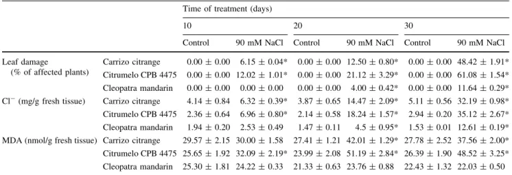
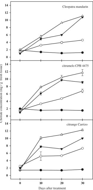
(4) Table 1 Damage, chloride concentration and malondialdehyde (MDA) content in leaves of intact plants of three citrus genotypes subjected to salt stress Time of treatment (days) 10 Control Leaf damage (% of affected plants) Cl (mg/g fresh tissue). 20 90 mM NaCl. Control. 30 90 mM NaCl. Control. 90 mM NaCl. Carrizo citrange. 0.00 ± 0.00. 6.15 ± 0.04*. 0.00 ± 0.00 12.50 ± 0.80*. 0.00 ± 0.00 48.42 ± 1.91*. Citrumelo CPB 4475. 0.00 ± 0.00 12.02 ± 1.01*. 0.00 ± 0.00 21.12 ± 3.29*. 0.00 ± 0.00 61.08 ± 1.54*. Cleopatra mandarin. 0.00 ± 0.00. 0.00 ± 0.00. 0.00 ± 0.00. 0.00 ± 0.00 11.64 ± 0.29*. Carrizo citrange. 4.14 ± 0.84. 6.32 ± 0.39*. 3.87 ± 0.65 14.47 ± 2.09*. 5.11 ± 0.56 32.19 ± 0.98*. Citrumelo CPB 4475. 2.36 ± 0.64. 6.96 ± 0.80*. 2.14 ± 0.58 18.24 ± 1.57*. 2.94 ± 0.20 35.12 ± 2.67*. 1.94 ± 0.20. 2.53 ± 0.49. 1.47 ± 0.11. 1.53 ± 0.01 12.61 ± 0.19*. Cleopatra mandarin MDA (nmol/g fresh tissue) Carrizo citrange. 29.57 ± 2.15 30.00 ± 1.58. 4.00 ± 0.42*. 4.5 ± 0.95*. 27.41 ± 1.21 42.01 ± 1.29* 27.78 ± 2.52 37.56 ± 2.00*. Citrumelo CPB 4475 25.65 ± 1.92 32.09 ± 2.19* 23.99 ± 2.08 51.19 ± 2.84* 26.39 ± 1.90 48.52 ± 3.25* Cleopatra mandarin. 25.30 ± 1.81 24.22 ± 0.33. 21.33 ± 0.63 23.76 ± 0.88. 22.43 ± 1.32 22.03 ± 0.50. * Symbols followed with an asterisk denote statistical significance at P \ 0.05. Data in insets are normalized mean values ± relative SE. 261 262 263 264 265 266 267 268 269 270 271 272 273 274 275 276 277. Cit. In this genotype, after 30 days of stress, affected plants were 61% of the total. Plants of CC also showed evident damage due to the increased concentration of NaCl from day 10, being 50% of the plants affected by the stress at day 30. Contrastingly, the percentage of CM plants affected by salt stress was only 11% over a 30-day period. Leaf Cl concentration mimicked damage and leaves of Cit and CC plants accumulated the highest concentration of the toxic ion whereas in leaves of CM, chloride content was much lower throughout the experimental period. Basal levels of Cl were lower in CM than in the rest of genotypes. Leaf MDA concentration (an indirect marker of salt stress induced oxidative damage) increased in the sensitive genotypes (Cit and CC) until a certain extent and then remained constant. In contrast, leaf MDA content in salt stressed plants of CM was similar to that in control plants throughout the experimental period.. 278. Adjustment of NaCl concentration in the in vitro system. 279 280 281 282 283 284 285 286 287 288 289 290 291 292 293. Shoots of the CC, Cit and CM cultivated in vitro were subjected to different saline treatments (30, 60 and 90 mM NaCl) and the pattern of Cl concentration followed over a 30 day period (Fig. 1). Chloride concentration in control shoots showed similar basal values among the three genotypes throughout the experimental period. After salinisation, chloride in shoots progressively increased in all genotypes and for all treatments, being the highest levels found in the most severe salt treatment. Although the studied citrus genotypes exhibited slightly different accu mulation patterns (chloride accumulation was faster in CC and Cit than in CM), all tended to similar maximum values (Fig. 1). For the subsequent experiments, 60 mM NaCl was set as the salt stress treatment because this intermediate concentration did not promote a high mortality (as that. 4. observed in shoots treated with 90 mM NaCl), but allowed an important and fast Cl accumulation (higher than in the 30 mM treatment) useful for further measurements.. 294 295 296. Effect of salt stress on in vitro cultured shoots of the different citrus genotypes. 297 298. It is well known that, under field conditions, salinity causes yellowing, bronzing, or browning of leaves and premature foliage drop. In our experimental system, damage caused by exposure of citrus shoots to 60 mM NaCl was evidenced by the apparition of characteristic leaf symptoms (Fig. 2). It was observed that leaf injury become more severe as the period of salt treatment progressed. After 10 days, light yellowing was observed in all genotypes. Leaf chlorosis increased after 20 days in all the cases and, at the end of the treatment (30 days), browning was evident in leaf tis sues of all genotypes. To quantify the occurrence of toxicity due to chloride ions, we considered as affected shoots those showing necrosis in at least 50% of their leaves. Figure 3 represents the percentage of CC, CM and Cit shoots affected by salinity (60 mM) after 0, 10, 20 and 30 days of treatment. Leaf damage increased very fast in Cit (60% of affected plants vs. 20% in CM and 17% in CC at day 10). After 20 days of salt treatment, 75 and 66% of CC and CM shoots were damaged respectively whereas almost all Cit shoots showed significant leaf damage. In all cases, and despite the slight different rates of leaf damage occurrence, shoots were in very bad conditions after 30 days of salt treatment (Figs. 2 and 3). When chloride concentration was determined in shoots of CC, CM and Cit after 10, 20 and 30 days of treatment, no differences were found among the three citrus geno types (Table 2). The accumulation of Cl ions took place. 299 300 301 302 303 304 305 306 307 308 309 310 311 312 313 314 315 316 317 318 319 320 321 322 323 324 325 326.
(5) 14. Cleopatra mandarin. 12 10 8 6 4 2. Choride concentration (mg / g fresh tissue). 0 14. 0. 10. 20. Días. 30. citrumelo CPB 4475. 12. 340 341 342 343 344 345 346 347 348 349 350 351 352 353. Early effects of salt stress on in vitro shoots of Carrizo citrange. 354 355. To investigate the early effects of salt stress, chloride concentration, MDA, ABA and SA contents were mea sured on in vitro cultured CC shoots 2, 5 and 10 days after the imposition of saline conditions (Table 3). Chloride content remained almost invariable throughout the exper imental period in control shoots. On the contrary, chloride concentration in salinized shoots progressively increased with time. Two days after the onset of the treatment, Cl concentration in salinized shoots was 1.5 times higher than that found in control ones, and achieved levels 4.6 times higher than in control shoots at day 10. No differences in MDA levels were found between control and salinized shoots after short saline treatment periods. However a slight increased of MDA took place at day 10; in comparison with the values obtained at day 2, in both, control (27.3 vs. 16.48 nmol/g fresh tissue) and salinized-shoots (29.74 vs. 20.79 nmol/g fresh tissue). Abscisic acid content in control shoots was similar to that measured in salinized ones; only after 5 days of saline treatment the differences between them were statistically significant. As observed before, ABA content in control shoots was much higher than in stressed ones (33.20 vs. 9.6 ng/g fresh tissue). No differences were found in the content of SA between control and salinized shoots after 2 and 5 days. After 10 days of stress, a slight although no significant increase in this hormone was recorded (Table 3).. 356 357 358 359 360 361 362 363 364 365 366 367 368 369 370 371 372 373 374 375 376 377 378 379 380 381 382. Effect of plant growth regulators. 383. To elucidate whether the addition of plant growth regula tors to the culture medium had some effect on the studied parameters, changes in foliar damage, chloride and MDA contents were measured after 20 days in CC shoots cul tured in medium with or without plant growth regulators. 384 385 386 387 388. 10 8 6 4 2 0 14. 0. 10. 20. 10. 20. 30 citrange Carrizo. 12 10 8 6 4 2 0 0. 30. Days after treatment. Fig. 1 Shoot chloride concentration in three citrus genotypes subjected to different concentrations of NaCl. In control (filled circle), 30 (open circle), 60 (filled inverted triangle), and 90 (open triangle) mM NaCl supplemented medium. Each point corresponds to the average ± standard error of four independent determinations. 327 328 329 330 331 332 333 334 335 336 337 338 339. 10 and 30 and a slightly significant increase at day 20 (1.2 fold above controls), suggesting a poor correlation between MDA content and leaf damage. Salt treatment did not induce significant ABA accumu lation in shoots of any genotype, regardless the extent of saline treatment (Table 2). ABA contents in control shoots were higher than in stressed ones in all genotypes throughout the experimental period except for a transient increase in salt-treated Cit shoots after 20 days. Elevated SA levels were observed in shoots of all studied genotypes after 10 days of treatment (Table 2), although it was no significantly different in the case of CC. This could evi dence an early signaling of SA as a consequence of salt stress.. progressively throughout the experimental period. After 10 days of treatment, values threefold above controls were recorded in salinized shoots (ranging from 6.80 to 8.89 mg/g in control shoots vs. 20.85–27.00 mg/g in salinized ones). At the end of the experiment, Cl- concentration in salinized shoots achieved values even higher (ranging from 6.55 to 13.20 mg/g in control shoots vs. 33.73–39.08 mg/g in sali nized ones). As an indicator of oxidative damage, MDA content was measured in shoots of citrus genotypes. No significant differences were found between salt-stressed and control shoots of Cit and CM (Table 2). However in CC, 60 mM NaCl treatment induced reductions in MDA content at day. 5.
(6) Fig. 2 Effect of salt stress in different citrus genotypes. Yellowing, browning and necrosis in leaves of Cleopatra mandarin (a), Citrumelo CPB 4475 (b) and citrange Carrizo (c) affected by salinity. In each picture and from left to right: control leaves and after 10, 20 and 30 days of saline treatment. d Necrotic shoot of Cleopatra mandarin after 30 days saline treatment (right) and control (left). 389 390 391 392 393 394 395 396 397 398 399 400 401 402 403 404 405 406 407. and with or without treatment with 60 mM NaCl (Table 4). Shoots grown on media, supplemented or not with plant growth regulators, showed the same behaviour. Healthy leaves (without evident damage) were observed in shoots cultured in both media (with or without plant growth reg ulators, Table 4). This suggests that no additional hor mones are required to maintain shoots in vitro for 20 days. Shoots growing in salt-stress conditions showed foliar damage (approximately a 70% of affected shoots) without significant differences due to the presence of plant growth regulators in the medium. Similar increases in Cl- concentration were observed in shoots growing in both salinized media (4.71 mg/g in media supplemented with plant growth regulators vs. 5.23 mg/g in media without them). In the same way, no significant variations in MDA content were observed between plants cultured with additional plant growth reg ulators or without them. In this case, as observed before, salt treatment did not modify MDA levels.. 408. Discussion. 409. The literature extensively describes how.
(7) intact plants), our system is oriented to elucidate common and specific response to ion toxicity among genotypes, avoiding the root filter. Salt stress tolerance has been correlated with an improved oxidative stress response in several crops (Shalata et al. 2001; Perl-Treves and Perl 2002). Increased MDA levels in leaves of intact plants of CC and Cit plants were also observed under salt treatment (Table 1). However, no significant differences were found between salt-stressed and control shoots of Cit and CM (Table 2) when cultured in vitro without root system. In the case of CC, a slight decrease in MDA content was found in some points. From these results, we can conclude that there is no correlation between foliar damage and oxidative stress: characteristic leaf symptoms caused by salinity (yellowing, browning, etc) were observed in all studied genotypes while no consistent MDA accumulation was detected. Abscisic acid plays a pivotal role in the adjustment of plants to abiotic stress conditions (Gómez-Cadenas et al. 1998; Christmann et al. 2006); it is the long-distance signal that communicates water stress from the root to the shoot. Evidence for root-derived ABA as a long-distance signal has been obtained from split-root experiments with whole plants in which only one part of the root system experienced water deficit (Dodd et al. 2008). Earlier studies showed that, in many crops, the leaf can accumulate ABA in response to salt stress (Montero et al. 1997; Sibole et al. 1998). In the case of citrus, previous reports indicate that the genotype CC responds to salinity by increasing ABA levels (Gómez-Cadenas et al. 1998). These experiments were performed with whole intact plants and it is possible that such salt treatment in the roots leads to a shoot water deficit that, in turn, triggers ABA accumulation. These results suggested the existence of an osmosensing mechanism and also the organ-specific nature of such a response. This suggestion is also supported by the fact that gene expression in response to salt stress usually is organ- or tissue-specific (Jia et al. 2002). When shoots were cultured in vitro, no accumulations of ABA were observed in any of the studied genotypes, which seem to discard an ABA dependent signaling. These results are similar to those obtained in maize by Jia et al. (2002), who observed that NaCl treatment only induced a small ABA accumulation in leaf tissues, whereas on the contrary, the same treatment of NaCl caused a significant ABA accumulation in root tissues. The transient increase of SA levels at 10 days of salt treatment could suggest a role for this hormone in the response of citrus to salinity. Although further work should be done to understand this effect, our data suggests a common signal not related to genotype tolerance as observed in all studied genotypes, independently of their tolerance to high salinity.. Cleopatra mandarin 100 80 60 40 20 0. citrumelo CPB 4475. Foliar damage (%). 100 80 60 40 20 0. citrange Carrizo 100 80 60 40 20 0. 0. 10 20 Days after treatment. 30. Fig. 3 Percentage of citrus shoots affected by salinity. Shoots were grown in control (filled cirlce) or 60 mM NaCl supplemented (open circle) medium. Each point corresponds to the average ± standard error of 18 independent determinations. 452 453 454 455 456 457 458 459 460 461 462 463 464 465. both salt-tolerant and salt-sensitive genotypes accumu lating similar amounts of chloride, which is really com plicated under field conditions. Therefore, the method proposed appears to be a good tool for studying bio chemical processes involved in the response of citrus to salt stress. Singh et al. (2004) cultured calli from dif ferent citrus rootstocks differing in their tolerance to salt stress. In this study, a good correlation between salt tolerance and ion accumulation was found. The differ ences found between both systems must be due to the totally different type of tissue cultured. Furthermore, while the system used by Singh et al. (2004) could be used as a practical tool to evaluate salt tolerance of citrus germoplasm (due to the similar behaviour of calli and. 7. 466 467 468 469 470 471 472 473 474 475 476 477 478 479 480 481 482 483 484 485 486 487 488 489 490 491 492 493 494 495 496 497 498 499 500 501 502 503 504 505 506 507 508 509 510 511 512 513 514 515 516 517 518.
(8) Table 2 Chloride, malondialdehyde (MDA), abscisic acid (ABA) and salicylic acid (SA) contents in shoots of three citrus genotypes subjected to salt stress (60 mM NaCl) Time of treatment (days) 10. 20. Control Cl. Control. 60 mM NaCl. Control. 60 mM NaCl. 7.70 ± 0.02. 27.00 ± 0.04*. 6.06 ± 0.02 30.25 ± 0.04*. 6.80 ± 0.01. 27.00 ± 0.09*. 7.90 ± 0.01 38.00 ± 0.03*. 9.09 ± 0.02 48.34 ± 0.04*. 8.89 ± 0.03. 20.85 ± 0.10*. 8.57 ± 0.08 43.14 ± 0.21*. 6.55 ± 0.04 39.08 ± 0.11*. Carrizo citrange. 37.09 ± 1.46. 24.74 ± 1.35*. 14.90 ± 0.85 18.42 ± 1.33*. 27.31 ± 2.92 16.93 ± 0.70*. Citrumelo CPB 4475. 21.99 ± 1.18. 21.55 ± 1.47. 22.04 ± 0.50 20.65 ± 0.51. 23.75 ± 0.84 20.45 ± 1.55. 21.34 ± 0.96 21.02 ± 0.76. 24.83 ± 0.47 25.21 ± 0.74 91.37 ± 8.31 33.13 ± 3.95*. Carrizo citrange (mg/g Citrumelo CPB 4475 fresh tissue) Cleopatra mandarin. MDA (nmol/g fresh tissue). 60 mM NaCl. 30. –. –. Cleopatra mandarin. 13.20 ± 0.02 33.73 ± 0.05*. 37.20 ± 9.35 ABA Carrizo citrange (ng/g Citrumelo CPB 4475 78.75 ± 7.51 fresh tissue) Cleopatra mandarin 104.50 ± 18.84. 29.05 ± 4.99. 53.55 ± 2.32 23.65 ± 3.84*. 52.05 ± 2.74*. 40.70 ± 1.85 78.42 ± 14.54* 157.40 ± 7.38 82.39 ± 3.40*. 90.50 ± 3.16. 26.00 ± 0.56 22.00 ± 2.30. 24.95 ± 2.30 13.30 ± 2.15*. 57.60 ± 11.03. 74.90 ± 9.05. 79.45 ± 6.05 75.20 ± 2.66. 108.15 ± 6.31 57.15 ± 18.54*. 33.25 ± 5.34. 55.35 ± 6.32*. 42.60 ± 4.98 54.25 ± 11.49. Carrizo citrange SA (ng/g Citrumelo CPB 4475 fresh tissue) Cleopatra mandarin. 67.20 ± 4.88. 176.45 ± 5.16* 116.10 ± 7.92 71.35 ± 5.02*. 60.15 ± 5.28 62.20 ± 11.59 79.60 ± 9.02 62.35 ± 7.56. * Symbols followed with an asterisk denote statistical significance at P \ 0.05. Data in insets are normalized mean values ± relative SE Table 3 Chloride, malondialdehyde (MDA), abscisic acid (ABA) and salicylic acid (SA) contents in Carrizo citrange shoots after 2, 5 and 10 days of saline treatment Time of treatment (days) 2. 5. Control Cl- (mg/g fresh tissue) MDA (nmol/g fresh tissue). 60 mM NaCl. 1.68 ± 0.03 16.48 ± 1.49. Control. 2.73 ± 0.04* 20.79 ± 0.99. 10. 1.72 ± 0.08 18.41 ± 2.08. ABA (ng/g fresh tissue). 25.20 ± 2.80. 30.07 ± 3.87. 33.20 ± 5.91. SA (ng/g fresh tissue). 34.60 ± 13.00. 27.73 ± 6.82. 28.93 ± 5.28. 60 mM NaCl 5.02 ± 0.09* 17.31 ± 1.38 9.60 ± 1.40* 37.80 ± 5.24. Control 1.15 ± 0.04 27.30 ± 1.35. 60 mM NaCl 5.38 ± 0.13* 29.74 ± 1.76. 8.40 ± 1.60. 11.33 ± 0.40. 35.00 ± 3.60. 56.73 ± 12.91. * Symbols followed with an asterisk denote statistical significance at P \ 0.05. Data in insets are normalized mean values ± relative SE. Table 4 Foliar damage, chloride and malondialdehyde (MDA) contents in Carrizo citrange shoots grown in medium supplemented or not with 0.2 mg/l of gibberellic acid and 6-benzylaminopurine. Data were recorded after 20 days of saline treatment Treatment ? Control Foliar damage (%) . Cl (mg/g fresh tissue) MDA (nmol/g fresh tissue). 0.00 ± 0.00. 60 mM NaCl 72.90 ± 1.10*. Control 0.00 ± 0.00. 60 mM NaCl 68.00 ± 0.20*. 1.67 ± 0.03. 4.71 ± 0.12*. 1.63 ± 0.07. 5.23 ± 0.12*. 28.06 ± 1.55. 33.24 ± 0.69*. 17.51 ± 1.67. 18.35 ± 1.98*. * Symbols followed with an asterisk denote statistical significance at P \ 0.05. Data in insets are normalized mean values ± relative SE. 519 520 521 522 523 524. taken place before 10 days of saline treatment, when first data of ABA concentration were recorded. To exclude this last possibility and to elucidate the early effects of salt stress on in vitro grown citrus shoots, the pattern of chlo ride concentration, MDA and hormone concentrations in CC shoots were determined after 2, 5 and 10 days of salt. The lack of ABA accumulation within in vitro cultured shoots of citrus genotypes under stress conditions could be due to the fact that the triggering signal for increasing biosynthesis must occur in the roots and the lack of this tissue in our system makes impossible for this early signal to occur. It was also possible that hormone-signalling had. 8. 525 526 527 528 529 530.
(9) 531 532 533 534 535 536 537 538 539 540 541 542 543 544 545 546 547 548. stress treatment. Chloride accumulation in shoots was again gradual throughout this short experimental period, but no differences were found in MDA, ABA or SA levels between control and salinized shoots after short saline treatment periods, which do not support a putative early ABA- or SA-dependent signalling pathway. After analyzing all data, we can conclude that when shoots are cultured without a root system, all genotypes accumulate the same levels of chloride and exhibit similar leaf damage as a consequence of the imposition of a salt stress treatment. The lack of an increase in MDA levels in all genotypes, and the common patterns of hormonal sig nalling, in both short and long periods of study indicate that, under the same salt conditions and with the same level of leaf chloride intoxication, no biochemical differences exist among tolerant and sensitive genotypes. This points to the roots as a key organ not only as a filter of chloride ions but also as a signalling system in citrus.. 549 550 551 552 553 554 555. Acknowledgments This work was supported by the Spanish Min isterio de Ciencia e Innovación and Universitat Jaume I/Fundació Bancaixa through grants no. AGL2007-65437-C04-03/AGR and P1IB2006-02, respectively. Hormone determinations were performed in the central facilities (Servei Central d’Instrumentació Cientı́fica, SCIC) of Universitat Jaume I. M. L-C was recipient of a fellowship from Universitat Jaume I.. 556. References. 557 558 559 560 561 562 563 564 565 566 567 568 569 570 571 572 573 574 575 576 577 578 579 580 581 582 583 584. Arbona V, Gómez-Cadenas A (2008) Hormonal modulation of Citrus responses to flooding. J Plant Growth Regul 27:241–250 Arbona V, Flors V, Garcı́a-Agustı́n P, Jacas J, Gómez-Cadenas A (2003) Enzymatic and non-enzymatic antioxidant responses of Carrizo citrange a salt-sensitive citrus rootstock, to different levels of salinity. Plant Cell Physiol 44:388–394 Arbona V, Hossain Z, Lopez-Climent MF, Pérez-Clemente RM, Gómez-Cadenas A (2008) Antioxidant enzymatic activity is linked to waterlogging stress tolerance in citrus. Physiol Plant 132:452–466 Bañuls J, Serna MD, Legaz M, Primo-Millo E (1997) Growth and gas exchange parameters of citrus plants stressed with different salts. J Plant Physiol 150:194–199 Chapman HD (1968) The mineral nutrition of citrus. In: Reuther W, Batchelor LD, Webber HD (eds) The Citrus Industry, vol II. University of California, Oakland USA, pp 127–289 Christmann A, Moes D, Himmelbach A, Yang Y, Tang Y, Grill E (2006) Integration of abscisic acid signalling into plant responses. Plant Biol 8:314–325 Dodd IC, Egea G, Davies WJ (2008) Accounting for sap flow from different parts of the root system improves the prediction of xylem ABA concentration in plants grown with heterogeneous soil moisture. J Exp Bot 59:4083–4093 Durgbanshi A, Arbona V, Pozo O, Miersch O, Sancho JV, Gómez Cadenas A (2005) Simultaneous determination of multiple phytohormones in plant extracts by liquid chromatography– electrospray tandem mass spectrometry. J Agric Food Chem 53:8437–8442. Gómez-Cadenas A, Tadeo FR, Primo-Millo E, Talon M (1998) Involvement of abscisic acid and ethylene in the response of citrus seedlings to salt shock. Physiol Plant 103:475–484 Gómez-Cadenas A, Arbona V, Jacas J, Primo-Millo E, Talon M (2002) Abscisic acid reduces leaf abscission and increases salt tolerance in citrus plants. J Plant Growth Regul 21:234–240 Hodges DM, DeLong JM, Forney CF, Prange RK (1999) Improving the thiobarbituric acid-reacting substances assay for estimating lipid peroxidation in plant tissues containing anthocyanin and other interfering compounds. Planta 207:604–611 Iglesias DJ, Levy Y, Gómez-Cadenas A, Tadeo FR, Primo-Millo E, Talon M (2004) Nitrate improves growth in salt-stressed citrus seedlings through effects on photosynthetic activity and chloride accumulation. Tree Physiol 24:1027–1034 Jia W, Youqun W, Shuqiu Z, Jianhua Z (2002) Salt-stress-induced ABA accumulation is more sensitively triggered in roots than in shoots. J Exp Bot 53:2201–2206 López-Climent MF, Arbona V, Pérez-Clemente RM, Gómez-Cadenas A (2008) Relationship between salt tolerance and photosynthetic machinery performance in citrus. Env Exp Bot 62:176–184 Montero E, Cabot C, Barcelo J, Poschenrieder C (1997) Endogenous abscisic acid levels are linked to decreased growth of bush bean plants treated with NaCl. Physiol Plant 101:17–22 Moya JL, Tadeo FR, Gómez-Cadenas A, Primo-Millo E, Talón M (2002) Transmisible salt tolerante traits identified through reciprocal grafos between sensitive Carrizo and tolerant Cleopatra citrus genotypes. J Plant Physiol 159:991–998 Moya JL, Gómez-Cadenas A, Primo-Millo E, Talón M (2003) Chloride absorption in salt-sensitive Carrizo citrange and salttolerant Cleopatra mandarin citrus rootstocks is linked to water use. J Exp Bot 54:825–833 Munns R, Tester R (2008) Mechanisms of salinity tolerance. Annu Rev Plant Biol 59:651–681 Murashige T, Skoog F (1962) A revised medium for rapid growth and bioassays with tobacco cultures. Physiol Plant 15:473–497 Perl-Treves R, Perl A (2002) Oxidative stress: an introduction. In: Inze D, Montagu MV (eds) Oxidative stress in plants. Taylor & Francis, London, pp 1–32 Romero-Aranda R, Moya JL, Tadeo FR, Legaz F, Primo-Millo E, Talon M (1998) Physiological and anatomical disturbances induced by chloride salts in sensitive and tolerant citrus: beneficial and detrimental effects of cations. Plant Cell Environ 21:1243–1253 Ruiz D, Martı́nez V, Cerdá A (1997) Citrus response to salinity: growth and nutrient uptake. Tree Physiol 17:141–150 Shalata A, Mittova V, Volokita M, Guy M, Tal M (2001) Response of the cultivated tomato and its wild salt-tolerant relative Lycopersicon pennellii to salt-dependent oxidative stress: the root antioxidative system. Physiol Plant 112:487–494 Sibole JV, Montero E, Cabot C, Poschenrieder CB (1998) Role of sodium in the ABA-mediated long-term growth response of bean to salt stress. Physiol Plant 104:299–305 Singh A, Saini ML, Behl RK (2004) In vitro screening of citrus rootstocks for salt tolerance. Indian J Genet Plant Breed 64:54–57 Walker RR, Sedgley M, Blesing MA, Douglas TJ (1984) Anatomy, ultrastructure and assimilate concentrations of roots of citrus genotypes differing in ability for salt exclusion. J Exp Bot 35: 1481–1494 Zhang YJ, Qian YQ, Mu X, Cai QG, Zhou YL, Wei XP (1998) Plant regeneration from in vitro-cultured seedling leaf protoplasts of Actinidia eriantha Benth. Plant Cell Rep 17(1998):819–821. 9. 585 586 587 588 589 590 591 592 593 594 595 596 597 598 599 600 601 602 603 604 605 606 607 608 609 610 611 612 613 614 615 616 617 618 619 620 621 622 623 624 625 626 627 628 629 630 631 632 633 634 635 636 637 638 639 640 641 642 643 644 645 646.
(10)
Figure


Documento similar