The effects of cholesterol on the nicotinic acetylcholine receptor : an update
23
0
0
Texto completo
(2) 2. Francisco J. Barrantes influence of cholesterol on receptor structural and functional properties. The mutual interaction of the two partners is discussed at various levels of organization, from the more general influences on the lipid phase to the more intimate relationship of the sterol with the annular and nonannular sites on the receptor.. Keywords: nicotinic receptor, membrane, lipid-protein interactions, membrane proteins, lipid domains, cholesterol, pentameric ligandgated ion channels. INTRODUCTION Neurotransmitter receptors can be subdivided into two main categories: a) the extensive superfamily of 7-transmembrane, G-protein coupled receptors (GPCR) or metabotropic receptors and b) the superfamily of pentameric ligand-gated ion channels (pLGIC), also termed ionotropic receptors. In the former case, binding of the neurotransmitter induces the coupling of the receptor with a G protein, triggering a relatively slow signaling cascade that takes the form of a metabolic process. In the case of ligand-gated ion channels, binding of the neurotransmitter induces the rapid opening of a channel that selectively allows certain ions to permeate into the postsynaptic target cell. In spite of the crucial functional (speed of the signaling process and mechanisms involved) and structural (7 versus 5 transmembrane segments) differences between the two superfamiles of receptors, both are made of transmembrane proteins with a large part of their mass embedded in the membrane lipid bilayer, and both superfamilies share common motifs to recognize lipids, and in particular, the neutral lipid cholesterol (Baier, Fantini et al., 2011, Fantini and Barrantes 2013, Fantini, Di Scala et al., 2016). The metabotropic GPCR employ transduction mechanisms involving the cAMP signal pathway and the phosphatidylinositol signal pathway. Upon ligand binding the GPCR undergo conformational changes that enable them to perform as guanine nucleotide exchange factors (GEF). The GPCR can then activate an associated G-protein by exchanging its bound GDP for a GTP molecule. The α-subunit of the G-protein, together with.
(3) The Effects of Cholesterol …. 3. the bound GTP, further proceed to dissociate from the β and γ subunits and involve downstream intracellular signaling proteins or target functional proteins directly, depending on the specific subtype of α-subunit with which they are associated. The neurotransmitter receptors included in the pLGIC superfamily comprise the nicotinic acetylcholine receptor (nAChR), -aminobutyric acid type A or C (GABAA/C) receptors, glycine receptors, the subtype 3 of the serotonin (5-HT3) receptors and glutamate-gated chloride channels (GluCl) (see reviews in (Nys, Kesters et al., 2013, Taly, Hénin et al., 2014, Sauguet, Shahsavar et al., 2015)). From a functional point of view, members of the superfamily can be divided into two distinct types: the cation-selective channels such as the nAChR and 5-HT3 receptor, and the anion-selective channels such as the glycine and the GABAA and GABAC receptors and invertebrate GluCl. These functionally different classes of ion channels mediate and/or modulate excitatory or inhibitory chemical signals, thanks not only to their distinct pharmacological properties, but also to their different anatomical and cellular localization and the connectivity of the neuronal cells involved, most especially in brain. The amino-terminal of all receptor subunits contains extracellular halves of a pair of disulfide-bonded cysteines separated by only 13 residues and is termed Cys-loop. Biochemical and X-ray crystallography studies have led to the identification of structural homologues of the eukaryotic pLGIC in Prokaryotes (Bocquet, Prado et al., 2007, Hilf and Dutzler 2008, Bocquet, Nury et al., 2009, Hilf and Dutzler 2009, Nury, Bocquet et al., 2010) reviewed in (Nys, Kesters et al., 2013, Cecchini and Changeux 2014, Olsen, Li et al., 2014, Sauguet, Shahsavar et al., 2014, Sauguet, Shahsavar et al., 2015). Although these bacterial channels do not possess a Cys-loop and their sequence identity with other members of the LGIC is rather low, their spatial (3-D) structural similarity with the eukaryotic LGIC is quite high. pLGIC are involved in chemical signaling between neuronal cells, and between neurons and muscle cells at the neuromuscular junction. Their alterations in the so-called “channelopathies” have been implicated in various disorders of the central nervous system, ranging from Alzheimer disease (Valles, Borroni et al., 2014), schizophrenia spectrum disorders (Lin, Hsu et al., 2014), Parkinson disease (Picciotto and Zoli 2008) and.
(4) 4. Francisco J. Barrantes. other severe neuropathological conditions. Consequently, pLGIC constitute targets for a great variety of pharmacological and clinically important drugs, including general anesthetics, anxiolytics, anticonvulsants, hypnotics, behavior- and mood-modifying substances, and a variety of drugs used in the effective treatment of epilepsies or the symptomatic amelioration of Parkinson and Alzheimer diseases.. NACHR The nAChR is the paradigm pLGIC. It is a transmembrane protein composed of five subunits organized around a central pore, the ion channel proper (Karlin 2002, Cecchini and Changeux 2015). Each nAChR subunit contains four transmembrane segments, 20–30 amino acids in length, named M1–M4. We have proposed that the interface between the protein and lipid moieties, comprising both the lipid-exposed transmembrane portions of the nAChR protein and the nAChR-vicinal lipid, behaves as an identifiable and evolutionarily conserved domain that regulates receptor function (Barrantes 2003, Barrantes 2004, Albuquerque, Pereira et al., 2009, Barrantes 2015, Barrantes and Fantini 2016). Muscle-type nAChRs are expressed in the peripheral nervous system and neuronal-type nAChRs in both peripheral and central nervous systems as well as in other non-neural cells such as immune cells, lymphocytes, lung epithelium and others. In brain, the nAChR is present in two principal forms: the heteropentameric receptor formed by 4 and 2 subunits and the homopentameric receptor formed exclusively by 7 subunits (Gotti 2006, Cecchini and Changeux 2014). The nAChR rapidly became the paradigm neurotransmitter receptor, and continues to be the prototype for the pLGIC superfamily. The 30-year improvement in the electron microscopy resolution of the Torpedo nAChR structure, currently at about 4 Å resolution (Klymkowsky and Stroud 1979, Zingsheim, Barrantes et al., 1982, Brisson and Unwin 1985, Unwin 1993, Unwin 2005, Unwin 2013) and the crystal structure of the water-soluble homologue of the nAChR extracellular domain (the molluscan ACh-binding protein, AChBP) by Xray diffraction techniques (Brejc, van Dijk et al., 2001, Celie, RossumFikkert et al., 2004), were also important landmarks in the structural.
(5) The Effects of Cholesterol …. 5. characterization of neurotransmitter receptors and structural homologues, and have had an important impact on our knowledge of pLGIC structure.. CHOLESTEROL At variance with the great variety of phospholipid species and variants, cholesterol is chemically unique (Barrantes 2016). Cholesterol concentration alters the physical properties of the bulk membrane and also promotes the formation of distinct membrane domains with a higher degree of order than the bulk lipid membrane, termed liquid-ordered (Lo) domains (Van Meer, Voelker et al., 2008). These domains enriched in cholesterol and sphingolipids have finite lifetimes, and are structurally and chemically different from the rest of the membrane, offering a platform with unique biophysical characteristics. The most important consequence of these transient lipid supramolecular assemblies is that they spatially and temporally confine signaling processes. Cholesterol is also capable of direct interactions with neurotransmitter receptors through consensus binding sequences like the so-called cholesterol recognition/interaction amino acid consensus motif (CRAC) or its inverted signature, CARC (Fantini and Barrantes 2009, Baier, Fantini et al., 2011, Fantini and Barrantes 2013, Fantini, Di Scala et al., 2016). The CARC motif exhibits a preference for the outer membrane leaflet and its mirror motif, CRAC, for the inner one. Some membrane proteins possess the double CARC-CRAC sequences within the same transmembrane domain. Thus, neurotransmitter receptors can be influenced by cholesterol through three non-mutually exclusive mechanisms: i) changes in bulk membrane fluidity that may influence the ability of receptors to undergo the conformational changes required to exert their function; ii) association/dissociation with/from cholesterol- and sphingolipid-rich Lo membrane domains and iii) specific interactions with cholesterol that can occur within or outside cholesterolrich domains, via cholesterol binding sites. Whereas modulation of the membrane’s physical properties by cholesterol appears to be a general mechanism affecting a broad range of membrane proteins and cell functions, direct binding of cholesterol to sites on the transmembrane regions of a protein and association with membrane domains are expected to be more specific and protein-dependent. I will next describe the intimate.
(6) 6. Francisco J. Barrantes. relationship of the nAChR with lipid domains and with cholesterol, and how these relationships modulate nAChR properties.. RELATIONSHIP OF THE NACHR WITH LIPID DOMAINS Model membrane studies showed that cholesterol–phospholipid mixtures at high cholesterol concentrations mimicked many aspects of the phase state displayed by biological membranes rich in cholesterol. These mixtures lack a defined lipid phase transition and instead are characterized by a single phase state, the liquid-ordered (Lo) phase (Ipsen, Karlström et al., 2003), with properties between the gel and the fluid lipid phases. For low cholesterol concentrations, solid-ordered (So) or liquid-disordered (Ld) phases are observed, depending on whether the system is above or below its gel-fluid transition temperature (Tm), respectively. When the binary lipid system is at intermediate cholesterol concentrations, there is phase coexistence of So and Lo (below) or Ld and Lo (above), depending on the temperature relative to Tm. The heterogeneity of model lipid systems is also observed in biological membranes, which are far from being homogeneous. The landscape of the plasmalemma at the mesoscopic (micron-level) and microscopic/ nanoscopic levels is characterized by lateral heterogeneities of different size and composition. The chemical composition of these structural domains is also heterogeneous, and characteristically rich in saturated fatty acids, sphingomyelins and cholesterol. Their chemistry is reflected in their physicochemical status, a characteristic Lo phase, different from that of the bulk membrane bilayer lipid, which is in a liquid-disordered Ld phase (Ing.l fsson, Melo et al., 2014, Krause and Regen 2014, Rao and Mayor 2014, Sevcsik and Schütz 2016). Lo domains form, transiently and reversibly, because of favorable enthalpic interactions between high melting temperature phospholipids with predominantly fully saturated or monounsaturated acyl chains, sphingomyelin and cholesterol. These nanodomains exhibit decreased conformational (trans-gauche) freedom and, consequently, viscoelastic properties different from those of the bulk lipid bilayer. Moreover, in compartmentalizing the membrane laterally, these nanodomains modify the diffusion of transmembrane proteins. The protein composition of Lo domains is also, albeit less characteristically,.
(7) The Effects of Cholesterol …. 7. distinct: Lo domains are predominantly enriched in glycosylphosphatidylinositol (GPI)-anchored proteins, as well as in proteins involved in signal transduction and intracellular trafficking (trimeric and small GTPases, Src family kinases) (Anderson 1998). Moreover, palmitoylation serves as a signal for the anchoring of several neurotransmitter receptors in Lo domains (Resh 2013). It should be emphasized that the existence of Lo domains has been clearly demonstrated experimentally in artificial membranes, but their presence in cell membranes is still an often controversial issue. The occurrence of ultranano-domains has recently been reported (Pathak and London 2015). They were difficult to detect due to their small size (<5 nm). Lipid domains in biological membranes not only exhibit spatial heterogeneity, but also display a wide distribution in the time domain, exhibiting lifetimes from the sub-millisecond to the tens of seconds. In the case of the nervous system, lipid domains have been reported to be involved in neuronal cell adhesion, axon guidance, synaptic transmission, growth factor-mediated signal transduction, vesicular trafficking, and membrane-associated proteolysis (Anderson 1998, Brown and London 1998, Tooze, Martens et al., 2001, Wolozin 2001, Aureli, Grassi et al., 2015). This makes Lo domains important for neural development and neuronal function and their diseases, as is the case with neurodevelopmental complex diseases like the schizophrenia spectrum disorders or the autistic spectrum disorders. The possible contribution of lipid domains to neuronal excitability stems from their reported participation in the clustering and regulation of neurotransmitter receptors on the one hand, and in the exocytic process of neurotransmitter release on the other (Martens, Navarro-Polanco et al., 2000, Bruses, Chauvet et al., 2001, Chamberlain, Burgoyne et al., 2001, Besshoh, Bawa et al., 2005, Nothdurfter, Tanasic et al., 2013, Sevcsik and Schütz 2016). Due to the essential structural role of cholesterol in the formation of liquid-ordered lipid domains, these aspects are discussed in more detail in the following sections of the review, addressing the specific roles of cholesterol on lipid platforms inhabited by nicotinic receptors. Using simple lipid model systems, we have used fluorescence techniques to study the propensity of cholesterol and other natural and synthetic sterols and steroids to promote the formation or disruption of Lo domains (Wenz and Barrantes 2003). We found that most compounds have.
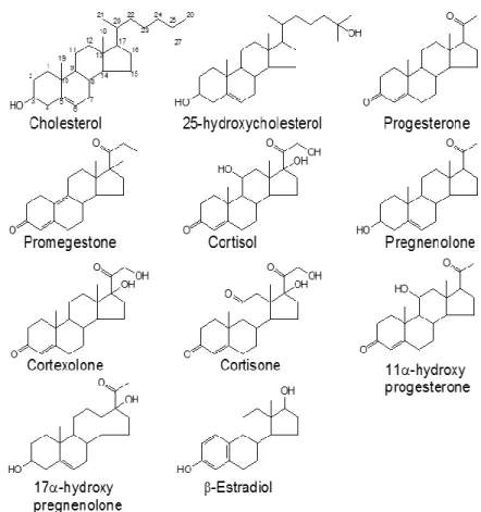
(8) 8. Francisco J. Barrantes. an ordered-phase disrupting activity, and we attributed this to the absence of carbon side chains (androstenol or C4 sterol) whereas cholesterol and 25-OH cholesterol, having such side chains, are Lo-promoting compounds (Figure 1). The side chains confer not only hydrophobicity to cholesterol and 25-OH cholesterol, but also the ability to achieve a better alignment, with their main molecular axis with a preferred orientation perpendicular to the membrane plane, and the 3-OH group at the opposite pole of the molecule at the level of the phospholipid polar head region. In living cells, we have focused our attention on the influence of cholesterol on the formation and stabilization of receptor assemblies at the plasmalemma. Cell-surface muscle type nAChRs are organized in micronand submicron-sized (nano)clusters, e.g., in CHO-K1/A5 cells and in resting C2C12 myotubes, respectively. It has not been straightforward to elucidate whether these receptor aggregates are associated with cholesterol-rich domains, as suggested by early experiments in which membrane patches rich in nAChR were isolated from muscle cells and their cholesterol composition subjected to biochemical analysis (Scher and Bloch 1991). Wide-field fluorescence microscopy of neuronal α7nAChR expressed in somatic spines of chicken ciliary neurons colocalized with GM1, a ganglioside reportedly associated with lipid Lo domains (Bruses, Chauvet et al., 2001). However, attempts to explore the possible association of the nAChR with cholesterol-rich Lo domains using widefield fluorescence microscopy and cholesterol-sensitive probes did not provide conclusive results (Scher and Bloch 1991, Kamerbeek, Borroni et al., 2013). The sensitivity and the spatial resolution of the techniques did not suffice to establish the association of nAChRs with a given type of lipid domain. When reconstituted in a sphingomyelin-cholesterol-POPC (1:1:1) model system, purified nAChR from Torpedo does not to appear to exhibit preference for Lo domains (Bermudez, Antollini et al., 2010). However, inclusion of sphingomyelin molecular species that generate bilayer asymmetry by enriching the sphingolipid content of the outer leaflet favor the partitioning of the nAChR in Lo domains (Perillo, Penalva et al., 2016). Using coarse-grained molecular dynamics simulations Sharp and Brannigan (Sharp and Brannigan 2017) have recently simulated the partitioning of nAChRs in domain-forming lipid mixtures. These authors indicate that, contrary to expectations, nAChR partitions into the Ld phase rich in n-3 polyunsaturated fatty acids and low in cholesterol. When.
(9) The Effects of Cholesterol …. 9. nAChR is partitioned into a cholesterol-poor liquid-disordered phase, binding of annular cholesterol is not observed, but cholesterol is stable in non-annular embedded sites for some cholesterol concentrations. These authors provided a structural explanation for this observation: the more flexible Ld phase can accommodate the deformation induced by the coneshaped nAChR. In summary, experimental and in silico data point to the plasticity of the nAChR relationship with lipid domains: the receptor can inhabit/be excluded from Lo domains (“rafts”) depending on the chemical composition of its lipid microenvironment.. Figure 1. Sterols that promote ordered lipid domain formation/stabilization (cholesterol and 25-hydroxycholesterol) possess an aliphatic chain at C-22. This side chain might be involved in the anchoring of the steroid in the bilayer (From ref. (Wenz and Barrantes 2003)..
(10) 10. Francisco J. Barrantes. ANNULAR AND NON-ANNULAR SITES ON THE NACHR If we zoom in on the nAChR-lipid interactions, we begin to focus on the relationship between two types of lipids in contact with the receptor macromolecule. Annular lipids (also referred to as shell lipids or boundary lipids) are a subset of lipid molecules preferentially located at the surface of membrane-embedded proteins, in a manner analogous to the solvent layer surrounding a water-soluble protein. The surface of a membrane protein contains shallow grooves and protrusions to which the fatty acyl chains of these annular lipids adapt to provide intimate contacts. Annular lipids constitute a shell, belt or annulus which is relatively immobile, in terms of translational and rotational motions, in comparison to bulk lipids. This approximation to a definition alludes to structural as well as dynamic aspects, and implicitly to thermodynamic features of annular lipids: they exhibit affinity for binding to the transmembrane regions of the proteins, and remain in their immediate microenvironment for periods longer than those experienced by the average thermally-driven lipid molecule in the bulk bilayer. The polar head regions of annular phospholipids and the polar region of cholesterol bind to the correspondingly more hydrophilic surfaces of the membrane protein, closer to the inner and outer interfaces of the bilayer with the extracellular and intracellular moieties. The hydrophobic fatty acyl chain region of the phospholipids or the more hydrophobic part of the cholesterol molecule establishes surface contacts with the hydrophobic core of the membrane-embedded polypeptide segments. Very few lipid molecules bound to membrane proteins have been resolved at atomic level in the scarce high-resolution structures of membrane proteins available to date. These crystallographic studies have provided evidence of the occurrence of lipid molecules between transmembrane α-helices, termed non-annular lipids. Binding of lipids to non-annular sites of some membrane proteins such as the potassium channel have been found to be functionally important (Levitan and Barrantes 2012, Rosenhouse-Dantsker, Noskov et al., 2013)..
(11) The Effects of Cholesterol …. 11. NICOTINIC ACETYLCHOLINE RECEPTOR (NACHR) AND CHOLESTEROL The most extensively studied neurotransmitter receptor in terms of lipid-protein interactions is the nAChR (Barrantes 2003, Barrantes 2004, Barrantes 2015). Using native membrane fractions enriched in the postsynaptic membrane of Torpedo electrocytes interrogated with electron spin resonance (ESR) techniques, nAChR-associated lipids were shown to be immobilized with respect to the bulk membrane lipid (Marsh and Barrantes 1978). Sterols were identified as part of these immobilized lipids. Other ESR studies (Bienvenüe, Rousselet et al., 1977, Marsh, Watts et al., 1981, Marsh, Watts et al., 1982, Ellena, Blazing et al., 1983, Arias, Sankaram et al., 1990, Horvath, Arias et al., 1990, Marcheselli, Daniotti et al., 1993) corroborated the occurrence of two distinct signals in experiments with native and reconstituted membranes containing nAChR at relatively high or low concentrations: one signal corresponded to the bulk membrane lipid and the other was interpreted as stemming from the protein-immobilized lipid. These direct interactions between protein and lipid moieties were observed with fatty acids, phospholipids, and sterols in the native membrane environment. Ellena et al., (Ellena, Blazing et al., 1983) confirmed our findings using reconstituted nAChR. Rousselet and coworkers (Bienvenüe, Rousselet et al., 1977, Rousselet, Devaux et al., 1979) found immobilization with fatty acids but not with phospholipids. The Lo-type phase of the nAChR immediate perimeter was further confirmed in native membrane using fluorescence (Antollini, Soto et al., 1996) or a combination of fluorescence and single-channel patch-clamp recordings (Antollini, Soto et al., 1996). Two cholesterol pools have been described in nAChR-rich membranes from Torpedo: an easily extractable fraction that influences the bulk fluidity of the membrane and a tightly bound nAChR-associated fraction (Leibel, Firestone et al., 1987). These studies from different laboratories demonstrated the occurrence of different lipid pools and nAChR protein-vicinal lipids that are relatively immobile with respect to the rest of the membrane lipids. They also pointed to the existence of phase lateral heterogeneity in nAChR-rich membrane lipids earlier than the concept of “rafts” came into use in the membrane field..
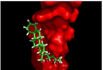
(12) 12. Francisco J. Barrantes. Consensus motifs for cholesterol binding are present in the transmembrane domain of all nAChR subunits, rendering a total of 15 cholesterol molecules per receptor (Baier, Fantini et al., 2011). In addition, in silico modeling studies led to the conclusion that in the total absence of cholesterol the nAChR structure would collapse (Brannigan, Henin et al., 2008). This model postulates the occurrence of cholesterol sites deep inside the transmembrane region of the nAChR, a premise on which until recently there was no conclusive experimental evidence (Brannigan, Henin et al., 2008). Cholesterol sites at the lipid belt region surrounding the nAChR macromolecule, in direct contact with the membrane-facing surfaces of the transmembrane segments (Baier, Fantini et al., 2011, Fantini, Di Scala et al., 2016) have been less elusive to characterize. The free energy of interaction between cholesterol molecules and the nAChR is about -510/-530 kJ.mol-1, i.e., more than -100 kJ.mol-1 per subunit. The particularly favorable fit between the “CARC-like” γTM4 segment from human nAChR (428RVCFLAML435) and cholesterol is noteworthy, with an energy of interaction of about -60 kJ.mol-1, i.e., ~60% of the total energy of interaction of the entire γ subunit, which exhibits the highest affinity for cholesterol among all nAChR subunits (Figure 2). The CARC motif generally exhibits more affinity for cholesterol than the CRAC motif (Fantini and Barrantes 2013). The significance of cholesterol for nAChR function has been extensively documented in experiments using purified nAChR from Torpedo reconstituted into membranes of various compositions, pointing to the clear influence this lipid –in addition to some glycerophospholipids (see reviews in (Barrantes 2003, Barrantes 2004, Barrantes 2015)) – has on the ability of the receptor to undergo the conformational transitions relevant to channel activation and desensitization. In living cells, acute cholesterol depletion has been reported to result in a transient ion channel gain-of-function in the cell line CHO-K1/A5 cells and in Torpedo nAChR expressed in Xenopus oocytes, whereas cholesterol enrichment has the opposite effect (Santiago, Guzman et al., 2001, Borroni, Baier et al., 2007). Subsequent studies provided a rationale for these observations: acute cholesterol depletion results in the marked acceleration of the rate of endocytosis of the receptor: the nAChR molecules remaining at the surface exhibit a moderate increase in the lifetime of their channel open state,.
(13) The Effects of Cholesterol …. 13. which we interpreted as a compensatory mechanism for the temporary loss of cell-surface receptors (Borroni, Baier et al., 2007).. Figure 2. CARC-like domain in human AChR γTM4 transmembrane segment. This hydrophobic transmembrane segment (Swiss Prot entry P02708, surface rendering) is located in the outer membrane leaflet. γTM4 is the transmembrane segment having the highest energy of interaction (-60 kJ/mol) with cholesterol (backbone rendering).. Figure 3. Changes in the orientation of I) γTM4 in POPC at 27°C, II) γTM4 in DPPC at 50°C, and III) γTM4 with the γTM1-TM2-TM3 bundle (grey) in DOPC at 50°C. From Antollini et al., (2005)..
(14) 14. Francisco J. Barrantes. Since the function of membrane proteins is influenced by the thickness of the surrounding lipid bilayer, there is crosstalk between protein and lipid moieties to ensure matching of their respective hydrophobic surfaces. The average thickness of the bilayer-embedded portion of a membrane protein is about 29 Å. A few proteins depart from this average thickness, and generate a hydrophobic mismatch with the surrounding lipid bilayer. Membrane proteins compensate changes in membrane thickness to minimize mismatch by tilting their α-helices and rotating their side chains at the ends of the helices. In the case of the nAChR, we have established the topography of the transmembrane portions of the receptor relative to that of surrounding lipids in the bilayer by means of Förster resonance energy transfer (FRET) and differential fluorescence quenching with spinlabeled lipid analogs, sterols included (Barrantes, Antollini et al., 2000). In another series of experiments, we specifically addressed the relative topography and tilt of the representative TM4 nAChR transmembrane chain and bilayer thickness, in the presence or absence of cholesterol (de Almeida, Loura et al., 2004) or in two different phosphatidylcholines (Antollini, Xu et al., 2005). The TM4 varied its angle with respect to the membrane normal depending on the thickness of the bilayer (POPC at 27° C or DPPC at 50°C) and whether the peptide was by itself or in the whole transmembrane bundle together with TM1, TM2 and TM3 (Antollini, Xu et al., 2005) (Figure 3); but it was the presence of cholesterol that was found to affect the bilayer thickness and consequently the orientation of the reconstituted TM4 peptide more profoundly (de Almeida, Loura et al., 2004).. CHOLESTEROL DEPENDENCE OF NACHR TRAFFICKING There is clear experimental evidence indicating that the size, stability and organization of the nAChR at the plasma membrane depend on cholesterol levels and actin cytoskeletal integrity (Barrantes 2007, Borroni, Baier et al., 2007, Barrantes 2012, Kamerbeek, Borroni et al., 2013). Exocytic trafficking of nAChRs to the plasmalemma as well as endocytic trafficking from the cell surface to the cell interior are both cholesteroldependent processes. In the exocytic pathway, receptors associate with cholesterol-rich domains as early as in the endoplasmic reticulum (ER) and.
(15) The Effects of Cholesterol …. 15. the Golgi apparatus (Marchand, Devillers-Thiery et al., 2002). Moreover, cholesterol depletion retains nAChRs in the Golgi complex, resulting in a decrease in cell-surface nAChR levels (Pediconi, Gallegos et al., 2004). Once nAChRs reach the cell surface, their stability is also highly dependent on cholesterol levels. Acute cholesterol depletion reduces the number of receptor domains by accelerating the rate of endocytosis and shifting the internalization of the nAChR to an alternate Arf6-dependent endocytic pathway (Borroni and Barrantes 2011) different from the constitutive endocytic pathway followed by the receptor under basal conditions (Kumari, Borroni et al., 2008). In summary, cholesterol depletion leads to nAChR conformational changes that alter its stability and its long-range dynamic association with other nAChR nanoclusters, accelerates its endocytosis, and transiently affects the channel kinetics of those receptors remaining at the surface (Borroni, Baier et al., 2007).. REFERENCES Albuquerque, E. X., E. F. Pereira, M. Alkondon and S. W. Rogers (2009). “Mammalian nicotinic acetylcholine receptors: from structure to function.” Physiol Rev 89(1): 73-120. Anderson, R. G. (1998). “The caveolae membrane system.” Annual review of biochemistry 67(1): 199-225. Antollini, S. S., M. A. Soto, I. Bonini de Romanelli, C. Gutierrez-Merino, P. Sotomayor and F. J. Barrantes (1996). “Physical state of bulk and protein-associated lipid in nicotinic acetylcholine receptor-rich membrane studied by laurdan generalized polarization and fluorescence energy transfer.” Biophys J 70(3): 1275-1284. Antollini, S. S., M. A. Soto, I. C. Bonini de Romanelli, C. GutierrezMerino, P. Sotomayor and F. J. Barrantes (1996). “Physical state of bulk and protein-associated lipid in nicotinic acetylcholine receptorrich membrane studied by laurdan generalized polarization and fluorescence energy transfer.” Biophys J. 70(3): 1275-1284. Antollini, S. S., Y. Xu, H. Jiang and F. J. Barrantes (2005). “Fluorescence and molecular dynamics studies of the acetylcholine receptor gammaM4 transmembrane peptide in reconstituted systems.” Mol Membr Biol 22(6): 471-483..
(16) 16. Francisco J. Barrantes. Arias, H. R., M. B. Sankaram, D. Marsh and F. J. Barrantes (1990). “Effect of local anaesthetics on steroid-nicotinic acetylcholine receptor interactions in native membranes of Torpedo marmorata electric organ.” Biochim. Biophys Acta 1027(3): 287-294. Aureli, M., S. Grassi, S. Prioni, S. Sonnino and A. Prinetti (2015). “Lipid membrane domains in the brain.” Biochimica et Biophysica Acta (BBA)-Molecular and Cell Biology of Lipids 1851(8): 1006-1016. Baier, C. J., J. Fantini and F. J. Barrantes (2011). “Disclosure of cholesterol recognition motifs in transmembrane domains of the human nicotinic acetylcholine receptor.” Sci Rep 1: 69. Barrantes, F. J. (2003). “Modulation of nicotinic acetylcholine receptor function through the outer and middle rings of transmembrane domains.” Curr Opin Drug Discov Devel 6(5): 620-632. Barrantes, F. J. (2004). “Structural basis for lipid modulation of nicotinic acetylcholine receptor function.” Brain Res Brain Res Rev 47(1-3): 7195. Barrantes, F. J. (2007). “Cholesterol effects on nicotinic acetylcholine receptor.” J Neurochem 103 Suppl 1: 72-80. Barrantes, F. J. (2012). “Regulation of the nicotinic acetylcholine receptor by cholesterol as a boundary lipid.” Cholesterol Regulation of Ion Channels and Receptors: 181-204. Barrantes, F. J. (2015). “Phylogenetic conservation of protein-lipid motifs in pentameric ligand-gated ion channels.” Biochim Biophys Acta 1848(9): 1796-1805. Barrantes, F. J. (2015). “Phylogenetic conservation of protein–lipid motifs in pentameric ligand-gated ion channels.” Biochimica et Biophysica Acta (BBA)-Biomembranes 1848(9): 1796-1805. Barrantes, F. J. (2016). “Cholesterol and nicotinic acetylcholine receptor: An intimate nanometer-scale spatial relationship spanning the billion year time-scale.” Biomed. Spectroscopy and Imaging 5: S67-S86. Barrantes, F. J., S. S. Antollini, M. P. Blanton and M. Prieto (2000). “Topography of nicotinic acetylcholine receptor membrane-embedded domains.” J Biol Chem 275(48): 37333-37339. Barrantes, F. J. and J. Fantini (2016). “From hopanoids to cholesterol: Molecular clocks of pentameric ligand-gated ion channels.” Prog Lipid Res 63: 1-13..
(17) The Effects of Cholesterol …. 17. Bermudez, V., S. S. Antollini, G. A. Fernandez Nievas, M. I. Aveldano and F. J. Barrantes (2010). “Partition profile of the nicotinic acetylcholine receptor in lipid domains upon reconstitution.” J Lipid Res 51(9): 2629-2641. Besshoh, S., D. Bawa, L. Teves, M. C. Wallace and J. W. Gurd (2005). “Increased phosphorylation and redistribution of NMDA receptors between synaptic lipid rafts and post-synaptic densities following transient global ischemia in the rat brain.” J Neurochem 93(1): 186194. Bienvenüe, A., A. Rousselet, G. Kato and P. F. Devaux (1977). “Fluidity of the lipids next to the acetylcholine receptor protein of Torpedo membrane fragments. Use of amphopholic reversible spin-labels.” Biochemistry 16: 841-848. Bocquet, N., H. Nury, M. Baaden, C. Le Poupon, J. P. Changeux, M. Delarue and P. J. Corringer (2009). “X-ray structure of a pentameric ligand-gated ion channel in an apparently open conformation.” Nature 457: 111-114. Bocquet, N., d. C. Prado, J. Cartaud, J. Neyton, C. Le Poupon, A. Taly, T. Grutter, J. P. Changeux and P. J. Corringer (2007). “A prokaryotic proton-gated ion channel from the nicotinic acetylcholine receptor family.” Nature 445(7123): 116-119. Borroni, V., C. J. Baier, T. Lang, I. Bonini, M. M. White, I. Garbus and F. J. Barrantes (2007). “Cholesterol depletion activates rapid internalization of submicron-sized acetylcholine receptor domains at the cell membrane.” Mol Membr Biol 24(1): 1-15. Borroni, V. and F. J. Barrantes (2011). “Cholesterol modulates the rate and mechanism of acetylcholine receptor internalization.” J Biol Chem 286(19): 17122-17132. Brannigan, G., J. Henin, R. Law, R. Eckenhoff and M. L. Klein (2008). “Embedded cholesterol in the nicotinic acetylcholine receptor.” Proc Natl Acad Sci US 105(38): 14418-14423. Brejc, K., W. J. van Dijk, R. V. Klaassen, M. Schuurmans, O. J. van Der, A. B. Smit and T. K. Sixma (2001). “Crystal structure of an AChbinding protein reveals the ligand-binding domain of nicotinic receptors.” Nature 411(6835): 269-276. Brisson, A. and P. N. T. Unwin (1985). “Quaternary structure of the acetylcholine receptor.” Nature 315: 474-477..
(18) 18. Francisco J. Barrantes. Brown, D. and E. London (1998). “Functions of lipid rafts in biological membranes.” Annual review of cell and developmental biology 14(1): 111-136. Bruses, J. L., N. Chauvet and U. Rutishauser (2001). “Membrane lipid rafts are necessary for the maintenance of the (alpha)7 nicotinic acetylcholine receptor in somatic spines of ciliary neurons.” J Neurosci 21(2): 504-512. Cecchini, M. and J. P. Changeux (2014). “The nicotinic acetylcholine receptor and its prokaryotic homologues: Structure, conformational transitions & allosteric modulation.” Neuropharmacology. Cecchini, M. and J. P. Changeux (2015). “The nicotinic acetylcholine receptor and its prokaryotic homologues: Structure, conformational transitions & allosteric modulation.” Neuropharmacology 96(Pt B): 137-149. Celie, P. H., S. E. Rossum-Fikkert, W. J. van Dijk, K. Brejc, A. B. Smit and T. K. Sixma (2004). “Nicotine and carbamylcholine binding to nicotinic acetylcholine receptors as studied in AChBP crystal structures.” Neuron 41(6): 907-914. Chamberlain, L. H., R. D. Burgoyne and G. W. Gould (2001). “SNARE proteins are highly enriched in lipid rafts in PC12 cells: implications for the spatial control of exocytosis.” Proceedings of the National Academy of Sciences 98(10): 5619-5624. de Almeida, R. F. M., L. M. S. Loura, M. Prieto, A. Watts, A. Fedorov and F. J. Barrantes (2004). “Cholesterol modulates the organization of the gamma M4 transmembrane domain of the muscle nicotinic acetylcholine receptor.” Biophysical Journal 86(4): 2261-2272. Ellena, J. F., M. A. Blazing and M. G. McNamee (1983). “Lipid-protein interactions in reconstituted membranes containing acetylcholine receptor.” Biochemistry 22: 5523-5535. Fantini, J. and F. J. Barrantes (2009). “Sphingolipid/cholesterol regulation of neurotransmitter receptor conformation and function.” Biochim Biophys Acta 1788(11): 2345-2361. Fantini, J. and F. J. Barrantes (2013). “How cholesterol interacts with membrane proteins: an exploration of cholesterol-binding sites including CRAC, CARC, and tilted domains.” Front Physiol 4: 31. Fantini, J., C. Di Scala, C. J. Baier and F. J. Barrantes (2016). “Molecular mechanisms of protein-cholesterol interactions in plasma membranes:.
(19) The Effects of Cholesterol …. 19. Functional distinction between topological (tilted) and consensus (CARC/CRAC) domains.” Chemistry and Physics of Lipids. Fantini, J., C. Di Scala, L. S. Evans, P. T. F. Williamson and F. J. Barrantes (2016). “A mirror code for protein-cholesterol interactions in the two leaflets of biological membranes.” Scientific Reports 6: 21907. Gotti, C. R., L.; S. Vailati, S.; Clementi, F. (2006). “Brain neuronal nicotinic receptors as new targets for drug discovery.” Curr. Pharm. Des 12: 407-428. Hilf, R. J. and R. Dutzler (2008). “X-ray structure of a prokaryotic pentameric ligand-gated ion channel.” Nature 452(7185): 375-379. Hilf, R. J. and R. Dutzler (2009). “Structure of a potentially open state of a proton-activated pentameric ligand-gated ion channel.” Nature 457(7225): 115-118. Horvath, L. I., H. R. Arias, H. O. Hankovszky, K. Hideg, F. J. Barrantes and D. Marsh (1990). “Association of spin-labeled local anesthetics at the hydrophobic surface of acetylcholine receptor in native membranes from Torpedo marmorata.” Biochemistry 29(37): 8707-8713. Ingólfsson, H. I., M. N. Melo, F. J. van Eerden, C. m. Arnarez, C. A. Lopez, T. A. Wassenaar, X. Periole, A. H. de Vries, D. P. Tieleman and S. J. Marrink (2014). “Lipid organization of the plasma membrane.” Journal of the American chemical society 136(41): 1455414559. Ipsen, J. H., Karlström, G, O. C. Mouritsen and M. J. Zuckermann (2003). Phase equilibria in the phosphatidylcholine-cholesterol system. Biochim. Biophys Acta. 905: 162-172. Kamerbeek, C. B., V. Borroni, M. F. Pediconi, S. B. Sato, T. Kobayashi and F. J. Barrantes (2013). “Antibody-induced acetylcholine receptor clusters inhabit liquid-ordered and liquid-disordered domains.” Biophys J 105(7): 1601-1611. Karlin, A. (2002). “Emerging structure of the nicotinic acetylcholine receptors.” Nat Rev Neurosci 3(2): 102-114. Klymkowsky, M. and R. M. Stroud (1979). “Immunospecific identification and three-dimensional structure of a membrane-bound acetylcholine receptor from Torpedo californica.” J. Mol. Biol. 128: 319-334. Krause, M. R. and S. L. Regen (2014). “The structural role of cholesterol in cell membranes: from condensed bilayers to lipid rafts.” Accounts of chemical research 47(12): 3512-3521..
(20) 20. Francisco J. Barrantes. Kumari, S., V. Borroni, A. Chaudhry, B. Chanda, R. Massol, S. Mayor and F. J. Barrantes (2008). “Nicotinic acetylcholine receptor is internalized via a Rac-dependent, dynamin-independent endocytic pathway.” J Cell Biol. 181(7): 1179-1193. Leibel, W. S., L. L. Firestone, D. C. Legler, L. M. Braswell and K. W. Miller (1987). “Two pools of cholesterol in acetylcholine receptor-rich membranes from Torpedo.” Biochim Biophys Acta 897(2): 249-260. Levitan, I. and F. J. Barrantes (2012). Cholesterol Regulation of Ion Channels and Receptors. Hoboken, NJ. Lin, H., F. C. Hsu, B. H. Baumann, D. A. Coulter, S. A. Anderson and D. R. Lynch (2014). “Cortical parvalbumin GABAergic deficits with α7 nicotinic acetylcholine receptor deletion: implications for schizophrenia.” Mol Cell Neurosci. 61: 163-175. Marchand, S., A. Devillers-Thiery, S. Pons, J. P. Changeux and J. Cartaud (2002). “Rapsyn escorts the nicotinic acetylcholine receptor along the exocytic pathway via association with lipid rafts.” J Neurosci 22(20): 8891-8901. Marcheselli, V., J. L. Daniotti, A. C. Vidal, H. Maccioni, D. Marsh and F. J. Barrantes (1993). “Gangliosides in acetylcholine receptor-rich membranes from Torpedo marmorata and Discopyge tschudii.” Neurochem. Res. 18(5): 599-603. Marsh, D. and F. J. Barrantes (1978). “Immobilized lipid in acetylcholine receptor-rich membranes from Torpedo marmorata.” Proc Natl Acad Sci US 75(9): 4329-4333. Marsh, D., A. Watts and F. J. Barrantes (1981). “Phospholipid chain immobilization and steroid rotational immobilization in acetylcholine receptor-rich membranes from Torpedo marmorata.” Biochim. Biophys Acta 645(1): 97-101. Marsh, D., A. Watts, R. D. Pates, R. Uhl, P. F. Knowles and M. Esmann (1982). “ESR spin-label studies of lipid-protein interactions in membranes.” Biophys J. 37: 265-274. Martens, J. R., R. Navarro-Polanco, E. A. Coppock, A. Nishiyama, L. Parshley, T. D. Grobaski and M. M. Tamkun (2000). “Differential targeting of Shaker-like potassium channels to lipid rafts.” Journal of Biological Chemistry 275(11): 7443-7446. Nothdurfter, C., S. Tanasic, B. Di Benedetto, M. Uhr, E.-M. Wagner, K. E. Gilling, C. G. Parsons, T. Rein, F. Holsboer and R. Rupprecht (2013)..
(21) The Effects of Cholesterol …. 21. “Lipid raft integrity affects GABA A receptor, but not NMDA receptor modulation by psychopharmacological compounds.” The International Journal of Neuropsychopharmacology 16(06): 1361-1371. Nury, H., N. Bocquet, C. Le Poupon, B. Raynal, A. Haouz, P. J. Corringer and M. Delarue (2010). “Crystal structure of the extracellular domain of a bacterial ligand-gated ion channel.” J Mol. Biol. 395(5): 11141127. Nys, M., D. Kesters and C. Ulens (2013). “Structural insights into Cysloop receptor function and ligand recognition.” Biochem Pharmacol 86(8): 1042-1053. Olsen, R. W., G. D. Li, M. Wallner, J. R. Trudell, E. J. Bertaccini, E. Lindahl, K. W. Miller, R. L. Alkana and D. L. Davies (2014). “Structural models of ligand-gated ion channels: sites of action for anesthetics and ethanol.” Alcohol Clin Exp Res. 38: 595-603. Pathak, P. and E. London (2015). “The Effect of Membrane Lipid Composition on the Formation of Lipid Ultrananodomains.” Biophys J 109(8): 1630-1638. Pediconi, M. F., C. E. Gallegos, E. B. De Los Santos and F. J. Barrantes (2004). “Metabolic cholesterol depletion hinders cell-surface trafficking of the nicotinic acetylcholine receptor.” Neuroscience 128(2): 239-249. Perillo, V. L., D. A. Penalva, A. J. Vitale, F. J. Barrantes and S. S. Antollini (2016). “Transbilayer asymmetry and sphingomyelin composition modulate the preferential membrane partitioning of the nicotinic acetylcholine receptor in Lo domains.” Arch Biochem Biophys 591: 76-86. Picciotto, M. R. and M. Zoli (2008). “Neuroprotection via nAChRs: the role of nAChRs in neurodegenerative disorders such as Alzheimer’s and Parkinson’s disease.” Front Biosci. 13: 492-504. Rao, M. and S. Mayor (2014). “Active organization of membrane constituents in living cells.” Current opinion in cell biology 29: 126132. Resh, M. D. (2013). “Covalent lipid modifications of proteins.” Current Biology 23(10): R431-R435. Rosenhouse-Dantsker, A., S. Noskov, S. Durdagi, D. E. Logothetis and I. Levitan (2013). “Identification of novel cholesterol-binding regions in Kir2 channels.” J Biol Chem 288(43): 31154-31164..
(22) 22. Francisco J. Barrantes. Rousselet, A., P. F. Devaux and K. W. Wirtz (1979). “Free fatty acids and esters can be immobilized by receptor rich membranes from Torpedo marmorata but not phospholipid acyl chains.” Biochem. Biophys. Res. Commun. 90: 871-877. Santiago, J., G. R. Guzman, L. V. Rojas, R. Marti, G. A. Asmar-Rovira, L. F. Santana, M. McNamee and J. A. Lasalde-Dominicci (2001). “Probing the effects of membrane cholesterol in the Torpedo californica acetylcholine receptor and the novel lipid-exposed mutation alpha C418W in Xenopus oocytes.” J Biol Chem 276(49): 4652346532. Sauguet, L., A. Shahsavar and M. Delarue (2015). “Crystallographic studies of pharmacological sites in pentameric ligand-gated ion channels.” Biochim Biophys Acta 1850: 511-523. Sauguet, L., A. Shahsavar, F. Poitevin, C. Huon, A. Menny, À. Nemecz, A. Haouz, J. P. Changeux, P. J. Corringer and M. Delarue (2014). “Crystal structures of a pentameric ligand-gated ion channel provide a mechanism for activation.” Proc Natl Acad Sci US 111: 966-971. Scher, M. G. and R. J. Bloch (1991). “The lipid bilayer of acetylcholine receptor clusters of cultured rat myotubes is organized into morphologically distinct domains.” Exp Cell Res 195(1): 79-91. Sevcsik, E. and G. J. Schütz (2016). “With or without rafts? Alternative views on cell membranes.” Bio Essays 38(2): 129-139. Sharp, L. M. and G. Brannigan (2017). “Interactions of Nicotinic Acetylcholine Receptors with Liquid-Disordered Domains Rich in n-3 Polyunsaturated Fatty Acids.” Biophys. J. 112: 225a. Taly, A., J. Hénin, J. P. Changeux and M. Cecchini (2014). “Allosteric regulation of pentameric ligand-gated ion channels: an emerging mechanistic perspective.” Channels (Austin) 8: 350-360. Tooze, S. A., G. J. Martens and W. B. Huttner (2001). “Secretory granule biogenesis: rafting to the SNARE.” Trends in cell biology 11(3): 116122. Unwin, N. (1993). “Nicotinic acetylcholine receptor at 9 Å resolution.” J. Mol. Biol. 229: 1101-1124. Unwin, N. (2005). “Refined structure of the nicotinic acetylcholine receptor at 4A resolution.” J Mol. Biol. 346(4): 967-989..
(23) The Effects of Cholesterol …. 23. Unwin, N. (2013). “Nicotinic acetylcholine receptor and the structural basis of neuromuscular transmission: insights from Torpedo postsynaptic membranes.” Q. Rev. Biophys. 46: 283-322. Valles, A. S., M. V. Borroni and F. J. Barrantes (2014). “Targeting Brain α7 Nicotinic Acetylcholine Receptors in Alzheimer’s Disease: Rationale and Current Status.” CNS Drugs. Van Meer, G., D. R. Voelker and G. W. Feigenson (2008). “Membrane lipids: where they are and how they behave.” Nature reviews molecular cell biology 9(2): 112-124. Wenz, J. J. and F. J. Barrantes (2003). “Steroid structural requirements for stabilizing or disrupting lipid domains.” Biochemistry 42(48): 1426714276. Wolozin, B. (2001). “A fluid connection: cholesterol and Aβ.” Proceedings of the National Academy of Sciences 98(10): 5371-5373. Zingsheim, H. P., F. J. Barrantes, J. Frank, W. Hanicke and D. C. Neugebauer (1982). “Direct structural localization of two toxinrecognition sites on an ACh receptor protein.” Nature 299(5878): 8184..
(24)
Figure
Documento similar
If the concept of the first digital divide was particularly linked to access to Internet, and the second digital divide to the operational capacity of the ICT‟s, the
In the “big picture” perspective of the recent years that we have described in Brazil, Spain, Portugal and Puerto Rico there are some similarities and important differences,
1. S., III, 52, 1-3: Examinadas estas cosas por nosotros, sería apropiado a los lugares antes citados tratar lo contado en la historia sobre las Amazonas que había antiguamente
In the preparation of this report, the Venice Commission has relied on the comments of its rapporteurs; its recently adopted Report on Respect for Democracy, Human Rights and the Rule
In the previous sections we have shown how astronomical alignments and solar hierophanies – with a common interest in the solstices − were substantiated in the
While Russian nostalgia for the late-socialism of the Brezhnev era began only after the clear-cut rupture of 1991, nostalgia for the 1970s seems to have emerged in Algeria
Díaz Soto has raised the point about banning religious garb in the ―public space.‖ He states, ―for example, in most Spanish public Universities, there is a Catholic chapel
teriza por dos factores, que vienen a determinar la especial responsabilidad que incumbe al Tribunal de Justicia en esta materia: de un lado, la inexistencia, en el
