Facultad de Odontología Vol. 18, No. 4 October-December 2014
pp 222-228
Revista Odontológica Mexicana
ORIGINAL RESEARCH
www.medigraphic.org.mx
* University Hospital, Monterrey, Nuevo Leon, Mexico.
§ Orthodontics Department, General Hospital «Dr Manuel Gea
González» UNAM, Mexico City. II Private practice.
¶ Metropolitan Autonomous University, Xochimilco, Mexico City. This article can be read in its full version in the following page: http://www.medigraphic.com/facultadodontologiaunam ABSTRACT
The present article describes an orthopedic appliance with pins used in the pre-surgical treatment of fully fi ssured unilateral cleft lip and palate before lip reparation. This appliance was designed with the aim of transversally correct palatal segments in the posterior area, align the smaller segment in the anterior section towards the facial midline, and decrease the cleft located near the lip so as to improve the shape of the nasal base fl oor and avoid later surgical compensations of the soft tissues.
Key words: Unilateral cleft palate, lip reparation, pre-surgical orthopedics.
Palabras clave: Paladar fi surado unilateral, reparación de labio, ortopedia prequirúrgica. RESUMEN
Este artículo describe un aparato ortopédico con pines utilizado en el tratamiento prequirúrgico para pacientes con labio y paladar unilateral completamente fi surado antes de la reparación del labio. Este aparato fue diseñado con el fi n de corregir transversalmente los segmentos palatinos en la parte posterior, alinear el segmento menor en la parte anterior hacia la línea media facial y reducir la fi sura cerca del labio para mejorar la forma del piso de la base na-sal y evitar compensaciones quirúrgicas posteriores de los tejidos blandos.
Pre-orthopedic appliance with pins used in alignment of
maxillary segments in patients with unilateral cleft lip and palate
Aparato preortopédico con pines utilizado en el alineamiento de los segmentos
maxilares en pacientes con labio y paladar unilateral fi surado
Luis González García,* Erika González Rodríguez,II Manuel Yudovich Burak,§ María de la Paz Aguilar Saavedra,II
Salvador García López,§,¶ Rosina E Villanueva Arriaga¶
INTRODUCTION
Cleft lip and palate are the most common malformations of the craniofacial region. These malformations affect one out of 700 newborns. Reports inform that frequency varies depending on the type of cleft, ethnicity and gender. Etiology of cleft lip and palate is still subject to debate. Nevertheless, in certain types of deformities clefts occur when mesenchymal connective tissues from the different embryonic structures do not fuse. This lack of fusion apparently takes place during the third month of fetal development. Deformities can be unilateral or bilateral and can extend up to the alveolar process.
Pre-surgical orthopedics consists on placing appliances a few days after birth in order to align maxillary segments. This process is undertaken before a plastic surgeon reconstructs lip and palate. Protocols for cleft treatment can be active or passive, as well as intra- or extra-oral.1 Some of the active
maxillary appliances are fixed with surgical pins, others are not. The appliance pre-determinedly displaces alveolar segments of the cleft, by means of
controlled forces. Results of this treatment are long-term, physical and financial, quantifiable benefits for the patient.2,3 Several publications have reported
benefits of early maxillary orthopedic treatment in cleft lip and palate patients.4-9 The aim of the present
article was to describe the designing process for an appliance with pins which could be successfully used during pre-surgical orthopedic treatment in patients with fully fi ssured unilateral lip and palate.
Case assessment
Traditionally, diagnosis of facial clefts is conducted at birth, during the first physical examination,
www.medigraphic.org.mx
Figures 1 and 2.
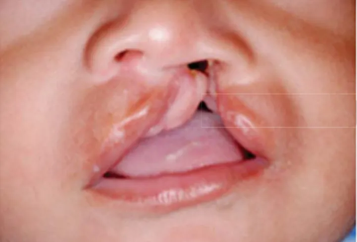
P a t i e n t b e f o r e t r e a t m e n t , s h o w i n g f u l l u n i l a t e r a l , anatomical displacement of lip with a 9 mm wide alveolar cleft in the palate.
w
w
w
w
w
w
w
w
w
w
w
w
w
w
w
w
w
w
w
w
w
w
w
w
w
w
w
w
w
w
ww
w
w
w
w
w
w
w
w
w
w
w
w
w
w
w
w
w
w
w
w
w
ww
w
w
w
w
w
w
w
w
w
w
w
w
w
w
w
w
w
w
w
w
w
w
w
w
w
w
w
w
w
w
w
w
w
w
w
w
w
w
w
w
w
w
w
w
w
w
w
w
w
w
w
w
m
me
m
m
m
m
m
m
m
m
m
m
m
m
m
m
m
m
m
m
m
m
m
e
e
e
e
e
e
e
e
e
e
e
e
e
e
e
e
e
e
e
e
e
e
e
ed
e
e
e
e
e
e
e
e
e
e
e
e
e
e
e
e
e
e
e
e
e
e
e
e
e
e
e
e
d
d
d
d
d
d
d
d
d
d
d
d
d
d
d
d
d
d
d
d
d
d
d
d
d
d
d
di
d
d
d
d
d
d
d
d
d
d
d
d
d
d
d
d
d
d
d
d
d
d
d
d
d
d
d
d
d
d
d
d
d
d
i
i
i
i
i
i
i
ig
i
i
i
i
i
i
i
i
i
i
i
i
i
i
i
i
i
i
i
i
i
i
i
i
i
i
i
i
g
g
g
g
g
g
g
g
g
g
g
g
g
g
g
g
g
g
g
g
g
g
g
g
g
g
g
g
g
g
gr
g
g
g
g
g
g
g
g
g
g
g
g
g
g
g
g
g
g
g
g
g
g
g
g
g
g
g
g
g
g
g
g
g
g
ra
r
r
r
r
r
r
r
r
r
r
r
r
r
r
r
a
a
a
a
a
a
a
a
a
a
ap
a
a
a
a
a
a
a
a
a
a
a
a
p
p
p
p
p
p
p
p
p
p
p
p
p
p
p
p
p
p
p
p
p
p
p
p
p
p
p
ph
p
p
p
p
p
p
p
p
p
p
p
p
p
p
p
p
p
p
p
p
p
p
p
p
p
p
p
p
p
p
p
p
p
p
h
hi
h
h
h
h
h
h
h
h
h
h
h
h
h
h
h
ic
i
i
i
i
i
i
i
i
i
c
c.
c
c
c
c
c
c
c
c
c
c
c
c
c
c
.
.o
.
.
.
o
o
o
o
oo
o
o
o
o
o
o
o
or
o
o
o
o
o
o
o
o
o
o
o
o
o
o
o
o
o
o
o
o
o
o
o
o
o
o
o
o
o
o
o
o
o
o
o
o
o
o
o
o
r
r
r
r
r
r
r
rg
r
r
r
r
r
r
r
r
g
g
g
g
g
g
g
g
g
g
g
g
g
g
g
g
g.
g
g
g
g
g
g
g
g
g
g
g
g
g
g
g
g
g
g
g
g
.m
.
.
.
.
.
.
.
.
m
m
m
m
m
m
m
m
m
m
m
m
m
m
m
m
m
m
m
m
mx
m
m
m
m
m
m
m
m
m
m
m
m
m
m
m
m
m
m
m
m
m
m
m
m
m
m
m
m
m
m
m
m
m
m
m
m
m
m
m
m
m
m
m
m
m
m
m
m
m
m
m
m
m
m
m
x
x
fed, with a large nozzle which can have a wider opening. Evidently, when parents discover that their child suffers a facial cleft, they become extremely anxious, therefore, timely and suitable handling of this situation must be initiated at an early stage.
Facial cleft treatment
Treatment of a newborn with facial cleft must bear the main objective of improving function, occlusion and appearance, even at the expense of an ideal occlusion, since the main target is to improve appearance and language. During the fi rst visit with the orthodontist, the patient is assessed by two departments: Plastic Surgery and Orthodontics, which will devise a suitable treatment plan. This plan usually involves placing appliances to align maxillary segments during the first days of the patient’s life. This is achieved with the help of pre-surgical orthopedics to facilitate surgical repair of the lip, with the purpose of achieving long-term facial growth. This procedure might not have a very promising future, nevertheless, social and personal interests must be taken into account.
All patients with cleft lip and palate must be individually assessed so that devised treatment will depend upon the type and severity of the cleft. The fi rst step is to expand the maxillary segments in the posterior region, later, they are leveled and the smaller segment is rotated towards the face’s midline. As a result, the cleft in the anterior segment is decreased by at least 1.5 mm. This procedure is undertaken before repairing the lip.
impression. The rims of the plaster study model must be very accurate so as to allow the appliance’s suitable adjustment and retention. The appliance is composed of two palatal plates, built with acrylic resin and linked by a .032 stainless steel wire frame (Dentaurum, Germany) which has a double omega bend to control expansion of the posterior segment and rotation of the smaller segment. Palatal plates count with embedded metallic buttons which are linked with an elastic chain so as to allow stability when the appliance is put into place. At the same time, they provide retention when the movement of the cleft is achieved (Figures 3A and
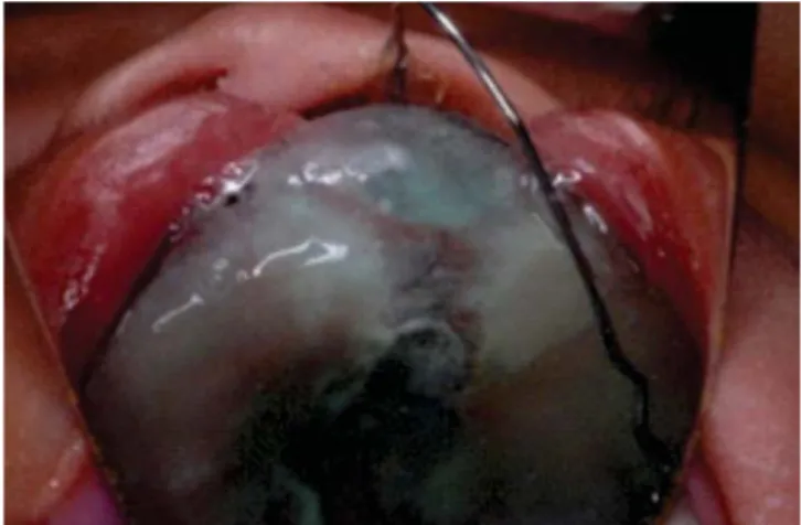
3B). The appliance is placed on the fissured palate
with .032 stainless steel wire pins, with a 45 degrees inclination so as to ensure they do not affect present tooth buds (Figure 4). Moreover, this inclination allows a lighter pressure in the retention of the appliance.
Surgical and orthopedic treatment
Orthodontists and plastic surgeons coordinate the procedure, which is conducted in an operating theater with the patient under general anesthesia. The appliance is put into place with a dental adhesive. Previously sterilized pins are installed with a band pusher, and acrylic resin is applied to the appliance so as to avoid its slippage (Figure 5).
Once the appliance is properly placed, its activation is initiated in the orthodontist’s offi ce. The number of required appointments varies according to the width of the cleft. The first step is to expand the palatal segments through activation of the wire located between the two omegas. For this purpose
three-www.medigraphic.org.mx
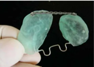
Figure 3A. Sections of the appliance: 1) Four stainless steel
wire pins, 2) A continuous elastic chain, 3) A corrector with an omega bend made of 0.32” stainless steel wire and 4) Adhesive lingual buttons.
1
2
3 4
Figure 3B. Pre-orthopedic appliance manufactured with
two acrylic resin plates linked at the anterior section with an elastic continuous chain on the lingual buttons, and in the posterior section, the wire with double omega bend.
Figure 4. The designed appliance is placed with dental
adhesive on the maxillary segments. Stainless steel 0.032” pins are placed at 45o in order to avoid damage to tooth buds.
Pins
Figure 5. The appliance placed in the patient’s mouth in the
operating theatre.
pronged straight pliers are used (Figures 6A and 6B). The smaller plate must be rotated with the lingual arch pliers by exerting pressure on the omega located closest to the plate. Once the appliance is widened, the plate can be fully and securely rotated (Figures
7A and 7B). In order to decrease the cleft with the
appliance , activation is applied to the middle section of the connector, for this purpose three-pronged straight pliers are used (Figures 8A and 8B). Appliance activation must be conducted every 3 days in the dental offi ce. Patient’s diet must remain unchanged at all stages of the treatment. This treatment will last for almost 4 weeks, so that segments can be positioned,
and the palatal segments can be re-organized, and therefore the anterior region cleft can be reduced to 1.5 mm (Table I). When the aforementioned results are achieved, the treatment is considered successful. In the case here presented, treatment was conducted without encountering any complications (Figure 9).
Clinical changes were observed when aligning the cleft. Among them we can mention shape improvement, since the smaller segment was forwardly displaced into the proper position, that is, the face’s midline. This process decreased by several
www.medigraphic.org.mx
Este documento es elaborado por Medigraphic
Figure 6A. Appliance activation with three-pronged straight
pliers on the corrector between the double omega bends for posterior expansion of plates.
Es Es Ess E
Eteteteteddddocococuoocoocumuuumuumumeumumumumumumumumumuumummmmmmmmmmmeneeeneneeenenenennnnnnntotottotototooooooooeeseeeeeeeeeees ss ssss s eleeleeleeleeeleleeeeelllababababababababbborbbbboororooradoorororooadaaadadadadddddooooo oopopopopopopopopopopooorooooor rr rrrrrrrrrrrrrrrMMeMeMeMeMeMeMeMeMeMMeMeMeMMMMeMeMeMeMeMMeMeeeeeeeeeeeeeeeeeeeeeeeeeeeeeeeeeeeeeeeeeeeeeeeeeeeeeeeeeeeeeeeeeeeeeeeeeeeeddiddddddddigrddddddddddddddiddddddddddddddiddddddigrgrgrgrgrapapapapapapapapappppppppppppppppppppppppppphpppppppppppppppppppppppppppppppppppppppppppppppppppppppppppppppppppppppppppphhichhihihihihhhihhhhhhihihihiiiicccccc
Figure 6B. Appliance after activation.
w
w
w
w
ww
w
w
w
w
w
w
w
w
w
w
w
w
w
w
w
w
w
w
w
w
w
w
w
w
w
w
w
w
w
w
w
w
w
w
w
w
w
w
w
w
w
w
w
w
w
w
w
w
w
w
w
ww
w
w
w
w
w
w
w
w
w
w
w
w
w
w
w
w
w
w
w
w
w
w
w
w
w
w
w
w
w
w
w
w
w
w
w
w
w
w
w
w
w
w
w
w
w
w
w
w
w
w
w
w
w
w
w
w
w
w
w
w
w
w
w
w
w
w
w
w
w
w
w
w
w
w
w
w
w
w
w
w
w
w
w
w
w
w
w
w
w
w
w
w
w
w
w
w
w
w
w
w
w
w
w
w
w
w
w
w
w
w
w
w
w
w
w
w
w
w
w
w
w
w
w
w
w
w
w
w
w
w
w
w
w
w
w
w
w
w
w
w
w
w
w
w
w
w
w
w
w
w
w
w
w
w
w
w
w
w
w
w
w.
w
w
w
w
w
w
w
w
w
w
w
w
w
w
w
w
w
w
w
w
w
w
w
w
w
w
w
w
w
w
w
w
w
w
w
w
w
w
w
w
w
w
w
w
w
w
w
w
w
w
w
w
w
w
w
w
w
w
w
w
w
w
w
w
w
w
w
w
w
w
w
w
w
w
w
w
w
w
w
w
w
w
w
w
w
w
w
w
w
w
w
w
w
w
w
w
w
w
w
w
w
w
w
w
w
w
w
w
w
w
w
w
w
w
w
w
w
w
w
w
w
.
.m
.
.
.
.
.
.
.
.
m
m
m
m
m
m
m
m
m
m
m
m
m
m
m
m
m
m
m
m
m
m
m
m
m
m
m
m
m
m
m
m
m
m
m
m
m
m
m
m
m
m
m
m
m
m
m
m
m
m
m
m
m
m
m
m
m
m
m
m
m
m
m
m
m
m
m
m
m
m
m
m
m
m
m
m
m
m
m
m
m
m
m
m
m
m
m
m
m
m
m
m
m
m
m
m
m
m
m
m
m
m
m
m
m
m
m
m
m
m
m
m
m
m
m
m
m
m
m
m
m
m
m
m
m
m
me
m
m
m
m
m
m
m
m
m
m
m
m
m
m
m
m
m
m
m
m
m
m
m
m
m
m
m
m
m
m
m
m
m
m
m
m
m
m
m
m
m
m
m
m
m
m
m
m
m
m
m
m
m
m
m
m
m
m
m
m
m
m
m
m
m
m
m
m
m
m
m
m
m
m
m
m
m
m
m
m
m
m
m
m
m
m
m
m
m
m
m
m
m
m
m
m
m
m
m
m
m
m
m
m
m
m
m
m
m
m
m
m
m
m
m
m
m
m
m
m
m
m
m
m
m
m
m
m
m
m
m
m
m
m
m
m
m
m
m
m
m
m
m
m
m
m
m
m
m
m
m
m
m
m
m
m
m
m
m
m
m
m
m
m
m
m
m
m
m
m
m
m
m
m
m
m
m
m
m
m
m
m
m
m
m
m
m
m
m
m
m
m
m
m
m
m
m
m
m
m
m
m
m
m
m
m
m
m
m
m
m
m
m
m
m
m
m
m
m
m
m
m
m
m
m
m
m
m
m
m
m
m
m
m
m
m
m
m
m
m
m
m
m
m
m
m
m
m
m
m
m
m
m
m
m
m
m
m
m
m
m
m
m
m
m
m
m
m
e
ed
e
e
e
e
e
e
e
e
e
d
di
d
d
d
ii
i
i
ig
gg
g
g
g
g
Figure 7A. Activation for small plate rotation.
Figure 7B. Appliance after activation.
p
p
p
p
p
p
p
p
p
p
p
p
ph
h
h
h
h
h
h
h
h
h
h
h
h
h
h
h
h
h
h
h
h
h
h
h
h
h
h
h
h
h
h
h
h
h
h
h
h
h
h
h
h
h
h
h
h
h
h
h
h
h
h
h
hi
h
h
h
h
h
h
h
h
h
h
h
h
h
h
h
h
h
h
h
h
h
h
h
h
h
h
h
h
h
h
h
h
h
h
h
h
h
h
h
h
h
h
h
h
h
h
h
h
h
h
h
h
h
h
h
h
h
h
h
h
h
h
h
h
h
h
h
h
h
h
h
h
h
h
h
h
h
h
h
h
h
h
h
h
h
h
h
h
h
h
h
h
h
h
h
i
i
i
i
i
i
i
ic
i
i
i
i
i
i
i
i
i
i
i
i
i
i
i
i
i
i
i
i
i
i
i
i
i
i
i
i
i
c
c
c
c
c
c
c
c
c
c
c
c
c
c
c
c
c
c
c
c
c.
c
c
c
c
c
c
c
c
c
c
c
c
c
c
c
c
c
c
c
c
c
c
c
c
c
.
.o
.
.
.
.
.
.
.
.
.
.
o
o
o
o
o
o
o
o
o
o
o
o
o
o
o
or
o
o
o
o
o
o
o
o
o
o
o
o
o
o
o
o
o
o
o
o
r
r
r
r
r
r
r
r
r
r
r
r
r
rg
r
r
r
r
r
r
r
r
r
r
r
r
r
r
r
r
r
r
r
g
g
g
g
g
g
g
g
g
g
g
g
g.
g
g
g
g
g
g
g
g
g
g
g
g
g
g
g
g
g
g
.
.m
.
.
.
.
.
.
.
.
.
m
m
m
m
m
m
m
m
m
m
m
m
mx
m
m
m
m
m
m
m
m
m
m
m
m
m
m
m
m
m
m
m
m
m
m
m
m
m
m
m
m
m
m
m
m
m
m
m
m
m
m
m
m
m
m
m
m
x
x
x
x
x
x
x
x
x
x
x
x
x
x
x
x
x
x
x
x
x
x
x
x
x
x
x
x
x
x
x
x
x
x
x
x
x
x
x
x
x
x
x
x
x
x
x
x
x
x
x
x
x
x
x
x
x
x
x
x
x
x
x
x
x
x
x
x
x
x
x
x
x
x
x
x
x
x
x
x
x
x
x
x
x
x
x
x
x
x
x
x
x
x
x
x
x
x
x
x
x
x
x
x
x
x
Orthodontics Department Appliance activation:
1. Expansion of posterior segments
2. Rotation of smaller segment to the anterior section of the midline of the face and reduction of the cleft close to the lip
3. Placement of Hotz appliance in order to achieve retention 4. Pre-orthopedic treatment. Three months average Reconstructive Plastic Surgery Department Lip reparation procedure
www.medigraphic.org.mx
Figure 8A. Activation of double omega bend to achieve
anterior region cleft reduction.
Figure 8B. Anterior section of acrylic resin plates must make
contact.
Figure 9. Fissure was signifi cantly reduced in the anterior
section with the use of pre-orthopedic appliance with pins.
w
w
w
w
w
w
w
w
w
w
w
w
w
w
w
w
w
w
w
w
w
w
w
w
w
w
w
w
w
w
w
w
w
w
w
w
w
w
w
w
w
w
w
w
w
w
w
w
w
w
w
w
w
w
w
w
w
w
w
w
w
w
w
w
w
w
w
w
w
w
w
w
w
w
w
w
w
w
w
w
w
w
w
w
w
w
w
w
w
w
w
w
w
w
w
w
w
w
w
w
w
w
w
w
w
w
w
w
w
w
w
w
w
w
w
w
w
w
w
w
w
w
w
w
w
w
w
w
w
w
w
w
w
w
w
w
w
w
w
w
w
w
w
w
w
w
w
w
w
w
w
w
w
w
w
w
w
w
w
w
w
w
w
w
w
w
w
w
w
w
w
w
w
w
w
w
w
w
w
w
w
w
w
w
w
w
w
w
w
w
w
w
w
w
w
w
w
w
w
w
w
w
w
w
w
w
w
w
w
w
w
w
w
w
w
w
w
w
w
w
w
w
w
w
w
w
w
w
w
w
w
w
w
w
w
w
w
w
w
w
w
w
w
w
w
w
w
w
w
w
w
w
w
w
w
w
w
w
w
w
w
w
w
w
w
w
w
w
w
w
w
w
w
w
w
w
w
w
w
w
w
w
w
w
w
w
w
w
ww
w
w
w
w
w
w
w
w
w
w
w
w
w
w
w
w
w
w
w
w
w
w
w
w
ww
w
w
w
w
w
w
w
w
w
w
w
w
w
w
w
w
w
w
w
w
w
w
w
w
w
w
w
w
w
w
w
w
w
w
w
w
w
w
w
w
w
w
w
w
w
w
w
w
w
w
w
w
w
w
w
w
w
w
w
w
w
w
w
w
w
w
w
w
w
w
w
w
w
w
w
w
w
w
w
w
w
w
w
w
w.
w
w
w
w
w
w
w
w
w
w
w
w
w
w
w
w
w
w
w
w
w
w
w
w
w
w
w
.m
.
.
.
.
m
m
m
m
m
m
m
m
m
m
m
m
m
m
m
m
m
m
m
m
me
m
m
m
m
m
m
m
m
m
m
m
m
m
m
m
m
m
m
m
m
m
m
m
m
m
m
e
e
e
e
e
ed
e
e
e
e
e
e
e
e
e
e
e
e
e
e
e
e
e
e
e
e
d
di
d
d
d
d
d
d
d
d
d
d
d
d
d
d
i
ig
i
i
i
i
i
i
i
i
i
i
i
i
g
g
g
g
g
g
g
g
g
g
g
g
g
g
g
g
g
g
g
g
g
g
g
g
g
g
g
g
g
g
g
g
g
g
g
g
g
Figure 10A. Before treatment there was a wide, 9 mm cleft
in the anterior section.
millimeters the width of the cleft (Figures 10A and
10B). The anterior section cleft was decreased from
9 to 1.5 mm. The expansion in the posterior section was performed with activation of the appliance. This procedure improved bone structure in preparation for later lip repair procedures. A Hotz-type retention appliance was placed in order to achieve nasoalveolar remodeling before lip closure (Figure 11).
Length of treatment and surgical procedure depend on the patient’s health as well as the surgeon’s experience. Different procedures can be used to repair the lip, the aim is to create a suitably long lip, not too tense and with satisfactory functions. When the maxillary segment’s pre-surgical orthopedic alignment is conducted, the lip is practically repaired in about three months (Figure 12).
Figure 10B. After four weeks of treatment, the fi ssure was
reduced to 1.5 mm and the anterior segment was aligned and rotated towards the face’s midline.
www.medigraphic.org.mx
Figure 11. After palatal segments alignment and cleft
reduction in the anterior section, a Hotz appliance was placed in order to shape the naso-alveolar area before undertaking lip repair.
Figure 12. Final result of upper lip repair procedure.
lip and palate was already known in the 50’s,4 great
diversity of active and passive appliances have since then been developed.7,12-15 Nevertheless, presently
there is some degree of controversy with respect to treatment of patients with unilateral cleft lip and palate16-18 as well as in time invested in treatments.19
Likewise, other clinical operators have stated there is no difference in facial growth in cleft lip and palate patients treated with pre-surgical orthopedics when compared to those who received no treatment prior to the primary gingivoperiosteoplasty20-22 as
was described by Drs Millard and Latham.23
Avant-garde clinical methods must be assessed with the help of prospective, multicenter studies in order to ascertain benefi ts achieved with pre-surgical pediatric orthopedics. These studies must per force be random and controlled in order to be able to judge therapy effectiveness24 based on the ability to achieve long
term positive results to improve balance between hard and soft tissues.25
CONCLUSIONS
The designed pre-orthopedic appliance achieved transverse and anterior-posterior correction of maxillary segments before lip repair. Results must be assessed in the long term.
REFERENCES
1. Huebener DV, Liu JR. Maxillary orthopedics. Clin Plast Surg. 1993; 20: 723-732.
2. Maull DJ, Grayson BH, Cutting CB, Brencht LL, Bookstein FL, Khorrambadi D et al. Long-term effects of naseoalveolar moulding on three-dimensional nasal shape in unilateral clefts. Cleft Palate Craniofac J. 1999; 36: 391-397.
3. Pfeifer T, Grayson BH, Cutting CB. Gingivoperiosteoplasty versus alveolar E bone graft: an outcome analysis of costs in the treatment of unilateral cleft alveolus. Presented at the 55th. Annual Meeting of the American Cleft Palate-Craniofacial Association; April 1998; Baltimore, MD: 1998.
4. McNeil CK. Orthodontic procedures in the treatment of congenital cleft palate. Dent Record. 1950; 79: 126-132.
5. Hotz M, Gnoinski W. Comprehensive care of cleft lip and palate children at Zurich University: a preliminary report. Am J Orthod. 1976; 70: 481-504.
6. Latham RA. Orthodontic advancement of the cleft maxillary segment: a preliminary report. Cleft Palate J. 1980; 17: 227-233. 7. Huebener DV, Marsh JL. Alveolar molding appliances in the
DISCUSSION
The appliance described in the present article was developed to be used in pre-surgical orthopedics in patients with full unilateral cleft lip and palate. The appliance was intended to align displaced maxillary segments, move them along soft tissue margins towards the face midline and thus decrease the size of the cleft in the anterior segment before the surgical repair of the lip. Alignment of the smaller segment is of the utmost importance, since, due to its size, it could become unstable and could rotate and collapse. This process entails functional consequences since, when some patients are operated on without previously having aligned segments of the palate, the result could be asymmetry in the nasal fl oor, causing thus
www.medigraphic.org.mx
treatment of cleft lip and palate patients. In: Bardach J MorrisHL, eds. Multidisciplinary management of Cleft Lip and Palate. Philadelphia: WB Saunders; 1990. 601-607.
8. Grayson BH, Cutting CB. Presurgical nasoalveolar orthopedic molding in primary correction of the nose, lip and alveolus of infants born with unilateral and bilateral Clefts. Cleft Palate Craniofac J. 2001; 38: 193-198.
9. Suri S, Thompson BD. A modifi ed muscle-activated maxillary orthopedic appliance for presurgical nasoalveolar molding in infants with unilateral cleft lip and palate. Cleft Palate Craniofac J. 2004; 3: 225-229.
10. Grayson BH, Cutting CB, Wood R. Preoperative columella lengthening in bilateral cleft lip and palate. Plast Reconstr Surg. 1998; 101: 630-639.
11. Grayson BH, Santiago P, Brecht L, Cutting CB. Presurgical naso-alveolar molding in patients with cleft lip and palate. Cleft Palate Craniofa J. 1999; 36: 486-498.
12. Burston WR. The early treatment of cleft palate conditions. Dent Pract. 1958; 9: 41-56.
13. Georgiade NG, Mladlick RA, Thorne FL. Positioning of the premaxilla in bilateral cleft lips by oral pinning and traction. Plast Reconstr Surg. 1968; 41: 240-243.
14. Georgiade NG, Latham RA. Maxillary arch alignment in the bilateral cleft lip and palate infant using the pinned coaxial screw appliance. Plast Reconstr Surg. 1975; 56: 52-60.
15. Figueroa AA, Reisberg DJ, Polley JW, Cohen M. Intraoral-appliance modifi cation to retract the premaxilla in patients with bilateral cleft lip. Cleft Palate Crabiofac J. 1996; 33: 497-500. 16. Winters JC, Hurtwitz DJ. Presurgical orthopedics in the surgical
management of unilateral cleft lip and palate. Plast Reconstr Surg. 1995; 95: 755-764.
17. Kuijpers-Jagtman AM, Long RE Jr. The infl uence of surgery and orthopedic treatment on maxillofacial growth and maxillary arch development in patients treated for orofacial clefts. Cleft Palate Craniofac J. 2000; 37: 527.
18. Prahl-Anderson B. Dental treatment of predental and infant patients with clefts and craniofacial anomalies. Cleft Palate Craniofacial J. 2000: 37; 528-532.
19. Gnoinski WM. Infant orthopedics and later orthodontic monitoring for unilateral cleft lip and palate patients in Zurich. In: Bardach J, Morris HL, eds. Multidisciplinary management of cleft lip and palate. Philadelphia: WB Saunders; 1990. 578-590.
20. Wood R, Grayson BH, Cutting CB. Gingivoperiosteoplasty and growth of the midface. Surg Forum. 1993; 16: 229.
21. Wood R, Grayson BH, Cutting CB. Gingivoperiosteoplasty and midfacial growth. Cleft Palate Craniofac J. 1997; 34: 17-20. 22. Lee C, Grayson BH, Lin WY, Cutting CB. Long term study of
midface growth in unilateral cleft lip and palate patients following gingivoperiosteoplasty. Presented at the American Cleft Palate-Craniofacial Association; April 1999; Scottsdale, AZ: 1999. 23. Millard DR, Latham RA. Improved primary surgical and dental
treatment of clefts. Plast Reconstr Surg. 1990: 86; 856-871. 24. Roberts CT, Semb G, Shaw WC. Strategies for the
advancements of surgical methods in cleft lip and palate. Cleft Palate Craniofac J. 1991: 28: 141-149.
25. Smith WP, Markus AF, Delaire J. Primary closure of the cleft alveolus: a functional approach Br J Oral Maxilo Fac Surg. 1995; 3: 156-165.
Mailing address: Salvador García López E-mail: [email protected]