Opposite Clinical Phenotypes of Glucokinase Disease:
Description of a Novel Activating Mutation and
Contiguous Inactivating Mutations in Human
Glucokinase (
GCK
) Gene
Fabrizio Barbetti, Nadia Cobo-Vuilleumier, Carlo Dionisi-Vici, Sonia Toni, Paolo Ciampalini, Ornella Massa, Pablo Rodriguez-Bada, Carlo Colombo, Lorenzo Lenzi, María A. Garcia-Gimeno, Francisco J. Bermudez-Silva,
Fernando Rodriguez de Fonseca, Patrizia Banin, Juan C. Aledo, Elena Baixeras, Pascual Sanz, and Antonio L. Cuesta-Mun˜oz
Bambino Gesu` Pediatric Hospital (F.B., C.D.-V., P.C., O.M., C.C.), Istituto di Ricovero e Cura a Carattere Scientifico, Rome 00164, Italy; Department of Internal Medicine (F.B.), University of Rome Tor Vergata, Rome 00134, Italy; Laboratory of Molecular Endocrinology and Metabolism (F.B.), S Raffaele Biomedical Park Foundation, Rome 00128, Italy; Center for the Study of Pancreatic-Cell Diseases (N.C.-V., P.R.-B., F.J.B.-S., F.R.d.F., J.C.A., E.B., A.L.C.-M.). Instituto Mediterráneo para el Avance de la Biotecnología y la Investigación Sanitaria Foundation and Carlos Haya Hospital, Ma´laga 29010, Spain; Regional Center for Juvenile Diabetes (S.T., L.L.), Meyer Pediatric Hospital, Florence 50132, Italy; Institute of Biomedicine of Valencia (CSIC) and CIBERER-ISCIII (M.A.G.-G., P.S.), Valencia 46010, Spain; Pediatric and Adolescent Unit (P.B.), S. Anna Hospital, Ferrara, Italy; and Molecular Biology and Biochemistry Department (J.C.A.). University of Ma´laga, Ma´laga 29071, Spain
Glucokinase is essential for glucose-stimulated insulin release from the pancreatic-cell, serving as glucose sensor in humans. Inactivating or activating mutations of glucokinase lead to different forms of glucokinase disease,i.e. GCK-monogenic diabetes of youth, permanent neonatal diabe-tes (inactivating mutations), and congenital hyperinsulinism, respectively. Here we present a novel glucokinase gene (GCK)-activating mutation (p.E442K) found in an infant with neonatal hypoglycemia (1.5 mmol/liter) and in two other family members suffering from recurrent hypo-glycemic episodes in their childhood and adult life. In contrast to the severe clinical presentation in the index case, functional studies showed only a slight activation of the protein (relative activity index of 3.3). We also report on functional studies of two inactivating mutations of theGCK(p.E440G and p.S441W), contiguous to the activating one, that lead to monogenic diabetes of youth. Interest-ingly, adult family members carrying theGCKpE440G mutation show an unusually heterogeneous and progressive diabetic phenotype, a feature not typical of GCK-monogenic diabetes of youth. In summary, we identified a novel activatingGCKmutation that although being associated with severe neonatal hypoglycemia is characterized by the mildest activation of the glucokinase enzyme of all previously reported.(Molecular Endocrinology23: 1983–1989, 2009)
G
lucokinase enzyme (GK) has unique functional and structural properties for acting as glucose sensor of the pancreatic -cell. GK thus plays a role in glucose-stimulated insulin release (GSIR) (1) and regulates the threshold for GSIR (GSIR-T) (1). The high control of GKon-cell function is best illustrated by the profound im-pact on GSIR-T of mutations of glucokinase gene (GCK). Indeed, GSIR-T in carriers of inactivating GCK muta-tions increases, leading to the mild fasting hyperglycemia in subjects with heterozygous mutations or to severe
dia-ISSN Print 0888-8809 ISSN Online 1944-9917 Printed in U.S.A.
Copyright © 2009 by The Endocrine Society
doi: 10.1210/me.2009-0094 Received February 25, 2009. Accepted August 24, 2009. First Published Online November 2, 2009
Abbreviations: AI, Activity index; FPIR, first phase insulin response;GCK, glucokinase gene; GK, glucokinase protein; GSIR, glucose-stimulated insulin release; GSIR-T, threshold for glucose-stimulated insulin release; IFG, impaired fasting glucose; IVGTT, iv glucose toler-ance test; OGTT, oral glucose tolertoler-ance test; WT, wild type.
betes as in the case of individuals with homozygous or compound heterozygous mutations. The resulting clinical phenotype of partial glucokinase deficiency is GCK-monogenic diabetes of youth (GCK-MDY, a newly pro-posed nomenclature), also known as maturity onset dia-betes of the young 2 (MODY2) (2), whereas complete glucokinase deficiency leads to permanent neonatal dia-betes mellitus (GCK-PNDM) (3, 4). On the contrary, the GSIR-T in carriers of activating mutations of GCK de-creases, causing hypoglycemia (GCK-HI) due to inappro-priate insulin secretion when plasma glucose is below the normal GSIR-T, featuring mild or severe forms of the disease (5– 8). These different GCK-linked disorders of glucose metabolism (GCK-MDY, GCK-PNDM, and GCK-HI) are the three different forms of glucokinase disease.
Glucokinase disease can be caused by missense GCK mutations located anywhere in the primary sequence with no major hot spots defined (9, 10). However, GCK-HI mutations cluster in the small domain of GK protein, where the allosteric activator site is located (11).
In this manuscript we present a novel activating GCK mutation that, in spite of presenting the lowest relative activity index (% AI) and the highest predicted GSIR-T of all naturally occurring GK activating muta-tions described so far, leads to severe neonatal hypo-glycemia (1.5 mmol/liter). Furthermore, we also report functional studies of contiguous inactivating mutations inGCKthat lead to the hyperglycemic form of the glu-cokinase disease (GCK-MDY).
Results
Identification of missense mutations in the GCK gene
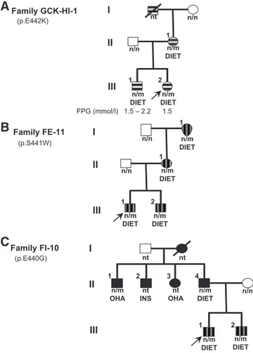
Family trees of the three patients carrying the GCK mutations studied in this report are depicted in Fig. 1. Denaturing gradient electrophoresis experiments showed abnormal patterns of PCR products of exon 10 of the GCKin all affected families’ members available for anal-ysis. We identified three missense mutations in the het-erozygous state. A novel mutation in codon 442, with lysine substituting for glutamic acid (p.E442K) in the pro-band of family GCK-HI-1 (subject III-2 in Fig. 1A), as well as in her affected brother and mother (subjects III-1 and II-1 in Fig. 1A). A single nucleotide change resulting in a tryptophan for serine substitution at codon 441 (p.S441W) was found in the proband of family FE-11 (subject III-1 in Fig. 1B), in his younger brother, his mother, and maternal grandmother (Fig. 1B). This muta-tion had been previously described in another Italian fam-ily (10). Finally, a novel mutation resulting in a
substi-tution of glutamic acid by glycine at codon 440 (p.E440G) was found in the proband of family FI-10 (subject III-1 in Fig. 1C), in his younger brother, in his father, and in a paternal uncle (Fig. 1C). None of the mutations were found in the chromosomes of 100 healthy chromosomes.
Metabolic features of families with GK disease
Family GCK-HI-1 (E442K)
The proband with hypoglycemia (Subject III-2 in Fig. 1A) was born at the 41st week of gestation and presented at d 1 of life with plasma glucose of 1.5 mmol/liter. Her birth weight was 2840 g, and the clinical examination was unremarkable. Family history disclosed that the maternal grandfather and the mother, as well as the eldest brother, presented with recurrent, symptomatic hypoglycemic-like episodes characterized by cold sweating, pallor, fa-tigue, hunger, and tachycardia; in addition, the mother and the brother of the index case showed fasting plasma
Family GCK-HI-1
A
1
1 nt n/n I
II
III 2
n/n
n/m DIET n/m DIET
n/m DIET (p.E442K)
FPG (mmol/l) 1.5 – 2.2 1.5
1
1 I
II
III 2
n/n
n/n
1
n/m DIET
n/m DIET
n/m DIET n/m DIET Family FE-11
B
(p.S441W)
Family FI-10
C
(p.E440G) I
II
III
nt OHA nt INS n/m
OHA DIETn/m
1
n/n
1 2 3 4
nt nt
n/m DIET DIETn/m
2
glucose of 2.2–3.0 mmol/liter on several occasions. At the age of 3 wk (weight, 4200 g) the proband was referred to the Metabolic Unit of Bambino Gesu` Pediatric Hospital for further investigation. At that time her plasma glucose ranged between 1.9 and 4.3 mmol/liter. She had normal plasma values of ammonia, lactate, triglycerides (122 mg/ dl), and total cholesterol (130 mg/dl). The urine excretion of␣-ketoglutarate, another marker of HI due to gain-of-function mutations of glutamate dehydrogenase gene (12), was normal, as well as blood acylcarnitines, amino acids, and serum transferrin isoelectric focusing. Abdom-inal and brain ultrasonography were also normal. After i.m. glucagon (1 mg), plasma glucose rose from 2.2 to 6.0 mmol/liter; simultaneous baseline evaluation of plasma cortisol (23 g/dl), IGF-I (237 ng/ml), and ACTH (35 pg/ml) were all normal. These clinical investigations com-bined excluded some causes of hypoglycemia. Low-dose diazoxide therapy (2 mg/kg䡠d), which normalized blood glucose, was started in the proband and was continued for 3 months. The patient, now 6 yr old and on diet therapy only, did not suffer from any other symptomatic hypo-glycemia; in contrast, the patient’s elder brother experi-enced, in the same time interval, two unexplained synco-pal episodes.
Family FE-11 (S441W)
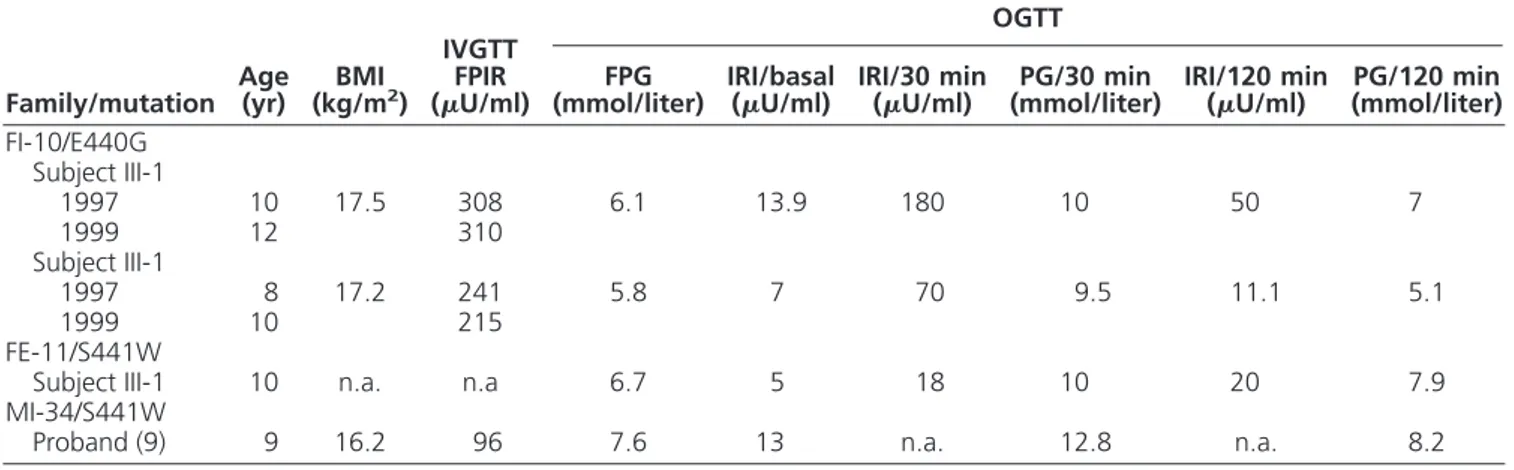
All the affected members of this family (Fig. 1B) pre-sented impaired fasting glucose (IFG) (6.5– 6.9 mmol/liter), and all were treated only with diet. The oral glucose toler-ance test (OGTT) carried out in the proband (Fig. 1B, sub-ject III-1) showed impaired glucose tolerance and low plasma insulin at 30 min (18 U/ml with corresponding plasma glucose of 10 mmol/liter) (Table 1), a result, which is typically found in GCK-MDY patients.
Family FI-10 (E440G)
The proband and his younger brother presented IFG (6.1 and 5.8 mmol/liter, respectively) (Fig. 1C, subjects III-1 and III-2). Intravenous glucose tolerance test (IVGTT)-derived first-phase insulin response (FPIR) of children carrying the p.E440G mutation showed unex-pectedly high figures of 308 and 241 U/ml (Table 1). These values, respectively, exceed the 97th (260.7U/ml) and the 90th (223.7U/ml) centile of normal Italian chil-dren of corresponding pubertal status (13). This high FPIR was observed again in both brothers when the IVGTTs were repeated 2 yr later (310 and 215U/ml, respectively) (Table 1). In addition, adult family mem-bers carrying the mutation showed a diabetic pheno-type, not typical of GCK-MDY, with the proband’s father presenting high fasting plasma glucose (10.4 mmol/liter) and a paternal uncle treated with oral hy-poglycemic agents; unfortunately, two other paternal uncles, one treated with oral hypoglycemic agents (OHA) and another with insulin, were not available for genetic analysis (Fig. 1).
Kinetic analysis of recombinant wild-type (WT) and mutant glucokinase
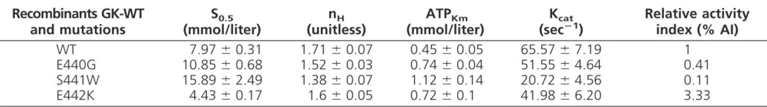
Kinetic properties of WT and mutant GK are shown in Table 2. Mutation GK-E442K showed a higher affinity for glucose (S0.5⫽4.43 mmol/liter) than GK-WT and a relative activity index (% AI) that was more than 3 times higher (Table 2), indicating that K442 is an activating mutation. On the contrary, mutations GK-E440G and GK-S441W showed a lower affinity for glucose, (S0.5⫽ 10.85 and 15.89 mmol/liter, respectively), as compared with GK-WT. The lowest affinity for the second substrate MgATP2⫺, catalytical activity and cooperativity index (Hill number⫺nH), were found in mutation GK-S441W
TABLE 1. Results of the metabolic studies performed in the probands with GCK-MDY
Family/mutation Age(yr) (kg/mBMI2)
IVGTT FPIR (U/ml)
OGTT
FPG
(mmol/liter) IRI/basal(U/ml) IRI/30 min(U/ml) (mmol/liter)PG/30 min IRI/120 min(U/ml) (mmol/liter)PG/120 min FI-10/E440G
Subject III-1
1997 10 17.5 308 6.1 13.9 180 10 50 7
1999 12 310
Subject III-1
1997 8 17.2 241 5.8 7 70 9.5 11.1 5.1
1999 10 215
FE-11/S441W
Subject III-1 10 n.a. n.a 6.7 5 18 10 20 7.9
MI-34/S441W
Proband (9) 9 16.2 96 7.6 13 n.a. 12.8 n.a. 8.2
(Table 2). The % AI of GK-E440G and GK-S441W were almost 60% and 90% lower than GK-WT. Consequently, the calculated GSIR-T of MDY-causing mutations GK-E440G and GK-S441W was higher than GK-WT (5.82 and 6.63 mmol/liter vs. 5 mmol/liter, respectively), whereas calculated GSIR-T of the activating mutation GK-E442K was lower (4.14 mmol/liter) than GK-WT (Fig. 2).
Prediction of structural effects of naturally occurring glucokinase mutations
We introduced the mutated residues into the closed active and super-open inactive glucokinasecell-specific structure models described by Kamataet al.(14) and then compared mutated GK-G440, GK-W441, and GK K442 with GK-WT. In Kamata’s model the side chain of Glu 442 (E442) is exposed to the solvent in both closed and super-open conformations. The mutant Lys residue in GK-HI-causing K442 would also be exposed to the sol-vent in both conformations (Fig. 3A). However, the pos-itive charge of the side chain of the Lys 442 residue may interact with the negative charge of Glu 216 residue (E216), stabilizing the closed conformation. In addition, in the super-open conformation (Fig. 3A), the side chain of Lys 442 would be closer to Ala 454 residue (A454), destabilizing the super-open conformation. As a result, the structure of K442 enzyme would favor the closed,
active conformation and would consequently lead to higher glucose affinity.
The change of Glu 440 residue (E440) by Gly residue (G440) (Fig. 3B) would destabilize the structure of the protein, because Gly residues are prone to modify the conformation of a particular structure. This interpreta-tion would explain the observed reducinterpreta-tion in glucose and
K440
E440 E440G
K414 Y413 K420
B
E440 G440
C
S441 S441W
C213
T209 α5-helix S441
W441
A
E442 E442K
A454
E216 E442
K442
α5 helix α13 helix
α5 helix
α13 helix
α13 helix
α5 helix
α5 helix
α13 helix
α13 helix
FIG. 3. Close-up of the structural model of the GK-E440, GK-S441, GK-E442 (WT), GK-E440G, GK-S441W (GCK-MDY naturally occurring mutation), and GK-E442K (HI mutation). The key␣5- and␣13-helixes of glucokinase are indicated in theleftstructures of panel A, (super-open conformation of glucokinase) (14) and panels B and C (closed conformation) (14). An enlargement of the region of interest (dotted square) is shown in each panel. Mutated residues are shown inred. The interacting residues are ingreen,cyan, andmagenta. Crystal coordinates from the closed active (1V4S) and super-open inactive (1V4T) conformation of GlkB (14) were visualized using the Pymol Molecular Graphics System version 0.97 (Delano Scientific LLC, Palo Alto, CA).
TABLE 2. Functional characteristics of the recombinants (GK-WT), and the naturally occurring mutations GK-E440G, GK-S441W, and GK-E442K
Recombinants GK-WT and mutations
S0.5 (mmol/liter)
nH (unitless)
ATPKm (mmol/liter)
Kcat (secⴚ1)
Relative activity index (% AI)
WT 7.97⫾0.31 1.71⫾0.07 0.45⫾0.05 65.57⫾7.19 1
E440G 10.85⫾0.68 1.52⫾0.03 0.74⫾0.04 51.55⫾4.64 0.41 S441W 15.89⫾2.49 1.38⫾0.07 1.12⫾0.14 20.72⫾4.56 0.11 E442K 4.43⫾0.17 1.6⫾0.05 0.72⫾0.1 41.98⫾6.20 3.33
Data are means⫾SEfrom three separate enzyme preparations. ATPKm, Michaelis constant for ATP; Kcat, catalytic constant; nH, Hill coefficient.
Threshold
for
GSIR (mmol/l)
Relative Activity Index (unitless) P.T.GSIR
S441W
WT E440G
E442K
4 4.5 5 5.5 6 6.5 7
0 0.5 1 1.5 2 2.5 3
MgATP2⫺ affinity. In the case of the S441W mutation (Fig. 3C, W441), the new bulkier tryptophan residue would be projected toward the inside of the protein, de-stabilizing it because of its interaction with the␣5 helix. This would cause a dramatic reduction in glucose affinity, as observed inin vitroexperiments.
Discussion
In this report we present a novel activatingGCKmutation (p.E442K) in a newborn with severe neonatal hypoglyce-mia. She is now 6 yr old, in good general condition, and treated with diet therapy only. Her older brother and mother, also bearing the same GCK mutation, did not present hypoglycemia in the neonatal period but repeated episodes of symptomatic hypoglycemia later in life. Func-tional studies of mutated protein showed an enzyme with almost a 2-fold increase in glucose affinity compared with GK-WT. The E442 residue is located in a loop between the13 and␣13 domains of GK. According to Kamata’s model (14), this loop plays an important role in the con-formational change of the GK from the super-open to the closed active conformation of the enzyme. Although E442 does not participate directly in the binding to the allosteric activator (compound A in Kamata’s model) (14), the K442 mutation favors the transition to the closed conformation of the enzyme, leading to increased glucose affinity (Fig. 3A). The structural analysis along with the calculated % AI and GSIR-T (3.3 and 4.14 mmol/liter, respectively) obtained from the experimental data, indicate that the activation of GK caused by the E442K mutation is the cause of hypoglycemia in the pro-band and in other members of the family carrying the mutation (Fig. 1A).
Recently, a de novo GCK-activating mutation (HI-GCK-M197I) has been described in a child that presented severe neonatal hypoglycemia (8). Interestingly, the in vitrostudies also showed a mild activation of GK-M197I (8). Therefore, our mutation HI-GCK-E442K is another good and confirmatory example of the discrepancy one may find between the severity of the clinical phenotype at presentation and the data resulting from the functional analysisin vitro of the mutated enzyme. Indeed, the re-sults of the kinetic (in vitro) analysis of mutation GK-E442K showed the lowest % AI as well as the highest GSIR-T of all naturally occurringGCKactivating muta-tions described to date (11, 15). This is in line with the relatively mild symptoms of hypoglycemia shown in af-fected members in childhood and adult life, but in con-trast to the severe clinical onset presented by the proband. We can only speculate that the proband could have some transient immaturity of glucoregulatory pathways that
contributed to lower her plasma glucose levels. However, we do not believe that this may be ascribed to the rela-tively low birth weight of the proband, which was in line with that reported in other patients with activating GCK mutations born from affected mothers (16).
We also described two inactivating GCK mutations (p.E440G, p.S441W) contiguous to the activating p.E442K. Patients carrying the mutation p.S441W present a phenotype highly characteristic of GCK-MDY patients,i.e. IFG combined with impaired glucose toler-ance with low values at tests evaluating (early) insulin release (17). In contrast, the two young and lean brothers carrying the mutation p.E440G showed high plasma in-sulin levels at 30 min of OGTT and in two independent IVGTT tests (Table 1). Like the family previously re-ported with GCK-MDY (mutation p.L184P) (18), we also observed some metabolic heterogeneity within the different E440G carriers of family FI-10 (Fig. 1). As a matter of fact, although functional and structural studies of GK-E440G (Table 2) fully explain the basic metabolic features of the proband and his brother, they are not sufficient to clarify the severe diabetic phenotype ob-served in adult family members carrying the mutation. Thus, the coexistence of other unknown genes implicated in glucose metabolism or unrecognized environmental factors should be considered in these kindred.
In summary, we identified a novel activatingGCK mu-tation that, although being associated with severe neona-tal hypoglycemia, is characterized by the mildest activa-tion of the glucokinase enzyme of all previously reported. In addition, the coexistence of unknown genes implicated in glucose metabolism or unrecognized environmental factors could be the cause of the metabolic heterogeneity presented by different carriers of the same inactivating GCK-E440G mutation.
Materials and Methods
Subjects
The proband with hypoglycemia (Subject III-2 in Fig. 1A) was referred to the Metabolic Unit of Bambino Gesu` Children’s Hospital for diagnostic workup. Routine laboratory examina-tions were all normal with the exception of insulin, which was inappropriately high (12U/ml) for the corresponding plasma glucose. Family history disclosed that her elder brother and mother (subjects III-1 and II-1 in Fig. 1A) suffered repeated episodes of symptomatic hypoglycemia, and the maternal grandfather (deceased, not tested) had hypoglycemia-like symp-toms (i.e. sweatiness, headache, hunger, and weakness).
Metabolic studies
Probands of families with (GCK-MDY) (subjects III-1 in Fig. 1, B and C) underwent two tests as part of the protocol for studying subjects with incidental hyperglycemia: a standard OGTT with serum insulin determinations and an IVGTT. IVGTT was performed injecting 0.5 g of glucose per kg/body weight. Blood samples were taken at⫺15, 0, and 1, 3, 5, and 10 min after the glucose injection for plasma insulin determination. FPIR was calculated as the sum of insulin immunoreactivity at min 1 and min 3, and the result was compared with those ob-tained in normal Italian children according to their pubertal stage (13). Insulin assay was not centralized. However, other IVGTT tests performed in the same center of family FI-10, on probands with mutations in the GCK, always elicited low FPIR (see Table 1). All tests were approved by the local institutional ethics committee, and a written informed consent was obtained from the parents of the probands.
Molecular genetic studies
Genomic DNA was extracted from peripheral lymphocytes. The complete coding sequence of theGCKwas amplified by the PCR and analyzed by denaturing gradient electrophoresis as previously described (10). In the proband clinically defined as having hyperinsulinemic hypoglycemia and his relatives, screen-ing ofGCKgene was performed first, based on the autosomal-dominant mode of inheritance of hypoglycemia (5, 11). We excluded the possibility of mutations of the glutamate dehydro-genase gene (GDH) because of the normal plasma ammonia concentrations (20). The PCR products showing abnormal elec-trophoretic pattern were subjected to direct sequencing by an ABI DNA sequencing apparatus 373A (PerkinElmer Applied Bio Systems, Foster City, CA). Mutations were confirmed in all affected family members available for analysis.
Site-directed mutagenesis
Mutations p.E440G, p.S441W, p.E442K onGCKwere in-troduced into the WT human pancreatic GCK using the QuikChange Site-Directed Mutagenesis kit from Stratagene (La Jolla, CA) (oligonucleotide sequences available upon request). Plasmid pUC-GlkB was used as a template in the PCRs. All plasmids were sequenced to confirm that only the desired mu-tation had been introduced. Mutated plasmids (pUC-GlkB-E440G, S441W, and E442K) were digested with EcoRI and
SalI, and the insert was subcloned into plasmid pGEX-6P-1 (Amersham Pharmacia, Piscataway, NJ), to allow its expression inEscherichia colias a glutathionylS-transferase fusion protein. Purified recombinant glutathionyl S-transferase-GK was rou-tinely screened for purity by SDS-PAGE.
Kinetic and structural analysis of the GK protein
Studies of the kinetic properties of and E440G, GK-S441W, and GK-E442K in the presence of 2 mmol of dithio-threitol per liter of reaction mixture were performed spectro-photometrically as described previously (21). At least three different preparations of GK-WT and GK-mutants were made and analyzed. We used nonlinear kinetics according to the Hill equation to determine the affinity of the enzyme for glucose and the Hill coefficient that characterizes the sigmoidal glucose de-pendency of GK. To measure the glucose phosphorylation ca-pacity of the enzyme, we used the relative activity index, which was calculated according to the formula previously reported
(11). The structural analysis of the activating mutation was carried out using the crystal structure of human GK (14).
Mathematical modeling
We used a minimal mathematical model (1, 11, 21) to assess the impact of GK mutations on GSIR-T. We determined the impact of blood glucose levels on GK expression for both WT and mutated alleles by using the expression coefficient for either allele:e⫽(SnH⫻2)/(SnH⫹S
0.5nH), whereSrefers to the glucose level at threshold, nH is the Hill coefficient for cooperativeness with glucose, the numerical value of 2 indicates that half-maximal induction is achieved at glucoseS0.5, andS0.5refers to the concen-tration of glucose needed to achieve the half-maximal rate of phos-phorylation (1, 11).
Acknowledgments
We thank all family members who participated in this study.
Address all correspondence and requests for reprints to: Fabri-zio Barbetti, M.D., Ph.D., Tor Vergata University Hospital, Laboratory Medicine, First floor, Section D, Room 118, Viale Oxford 81, 00134 Rome, Italy. E-mail: mody.2@libero.it or fabrizio.barbetti@spr-r.it; and Antonio Cuesta-Munoz, M.D., Ph.D., Center for the Study of Pancreatic -Cell Diseases. IMABIS Foundation and Carlos Haya Hospital. Avda. Carlos Haya 82, Pabello´n A-7a
planta 29010Ma´laga, Spain. E-mail: alcm@fundacionimabis.org.
F.B, S.T., and P.B. are members of the Diabetes Study Group of the Italian Society of Pediatric Endocrinology and Diabetol-ogy (SIEDP). This work was supported by grants (to A.L.C.-M. and N.C.-V.) from Ministerio de Ciencia e Innovacio´n, Direc-cio´n General de InvestigaDirec-cio´n Científica y Te´cnica (SAF2005-08014; SAF2006-12863) and Junta de Andalucía (SAS/PI-024/2007; SAS/PI-0236/2009).
Disclosure Summary: The authors have nothing to disclose.
References
1. Matschinsky FM2002 Regulation of pancreatic-cell glucokinase. From basics to therapeutics. Diabetes 51:S394 –S404
2. Vaxillaire M, Froguel Ph2008 Monogenic diabetes in the young, pharmacogenetics and relevance to multifactorial forms of type 2 diabetes. Endocr Rev 29:254 –264
3. Njølstad PR, Søvik O, Cuesta-Mun˜oz A, Bjørkhaug L, Massa O, Barbetti F, Undlien DE, Shiota C, Magnuson MA, Molven A, Matschinsky FM, Bell GI2001 Neonatal diabetes mellitus due to complete glucokinase deficiency. N Engl J Med 344:1588 –1592 4. Njølstad PR, Sagen JV, Bjørkhaug L, Odili S, Shehadeh N, Bakry D,
Sarici SU, Alpay F, Molnes J, Molven A, Søvik O, Matschinsky FM
2003 Permanent neonatal diabetes caused by glucokinase defi-ciency. Inborn error of the glucose-insulin signaling pathway. Dia-betes 52:2854 –2860
5. Glaser B, Kesavan P, Heyman M, Davis E, Cuesta A, Buchs A, Stanley CA, Thornton PS, Permutt MA, Matschinsky FM, Herold KC1998 Familial hyperinsulinism caused by an activating glucoki-nase mutation. N Engl J Med 338:226 –230
7. Cuesta-Mun˜oz AL, Huopio H, Otonkoski T, Gomez-Zumaquero JM, Na¨nto¨-Salonen K, Rahier J, Lo´pez-Enriquez S, García-Gimeno MA, Sanz P, Soriguer FC, Laakso M2004 Severe persistent hyper-insulinemic hypoglycemia due to a de novo glucokinase mutation. Diabetes 53:2164 –2168
8. Sayed S, Langdon DR, Odili S, Chen P, Buettger C, Schiffman AB, Suchi M, Taub R, Grimsby J, Matschinsky FM, Stanley CA2009 Extremes of clinical and enzymatic phenotypes in children with hyperinsulinism due to glucokinase activating mutations. Diabetes 58:1419 –1427
9. Gloyn AL2003 Glucokinase mutations in hyper- and hypoglyce-mia: maturity onset diabetes of the young, permanent neonatal diabetes and hyperinsulinemia of infancy. Hum Mutat 22:353–362 10. Massa O, Meschi F, Cuesta-Munoz A, Caumo A, Cerutti F, Toni S, Cherubini V, Guazzarotti L, Sulli N, Matschinsky FM, Lorini R, Iafusco D, Barbetti F; Italian Society of Paediatic Endocrinology and Diabetes (SIEDP)2001 High prevalence of glucokinase muta-tions in Italian children with MODY. Influence on glucose toler-ance, first-phase insulin response, insulin sensitivity and BMI. Dia-betologia 44:898 –905
11. Matschinsky FM2009 Assessing the potential of glucokinase acti-vators in diabetes therapy. Nat Rev Drug Discov 8:399 – 416 12. Meissner T, Mayatepek E, Kinner M, Santer R2004 Urinary
␣-ketoglutarate is elevated in patients with hyperinsulinism-hyperammonemia syndrome. Clin Chim Acta 341:23–26 13. Lorini R, Vanelli M1996 Normal values of first-phase insulin
re-sponse tom intravenous glucose in healthy Italian children and ad-olescents. Diabetologia 39:370 –371
14. Kamata K, Mitsuya M, Nishimura T, Eiki J, Nagata Y2004 Struc-tural basis for allosteric regulation of the monomeric allosteric en-zyme human glucokinase. Structure 12:429 – 438
15. Meissner T, Marquard J, Cobo-Vuilleumier N, Maringa M,
Rodríguez-Bada P, García-Gimeno MA, Baixeras E, Weber J, Olek K, Sanz P, Mayatepek E, Cuesta-Mun˜oz AL2009 Diagnostic difficulties in glucokinase hyperinsulinism. Horm Metab Res 41:320–326 16. Christesen HB, Tribble ND, Molven A, Siddiqui J, Sandal T, Brusgaard
K, Ellard S, Njølstad PR, Alm J, Brock Jacobsen B, Hussain K, Gloyn AL2008 Activating glucokinase (GCK) mutations as a cause of medically responsive congenital hyperinsulinism: prevalence in children and characterisation of a novel GCK mutation. Eur J En-docrinol 159:27–34
17. Byrne MM, Sturis J, Cle´ment K, Vionnet N, Pueyo ME, Stoffel M, Takeda J, Passa P, Cohen D, Bell GI1994 Insulin secretory abnor-malities in subjects with hyperglycemia due to glucokinase muta-tions. J Clin Invest 93:1120 –1130
18. Fajans SS, Bell GI2006 Phenotypic heterogeneity between different mutations of MODY subtypes and within MODY pedigrees. Dia-betologia 49:1106 –1108
19. Lorini R, Klersy C, d’Annunzio G, Massa O, Minuto N, Iafusco D, Bellane´-Chatelot C, Frongia AP, Toni S, Meschi F, Cerutti F, Barbetti F and the Diabetes Study Group of the Italian Society of Pediatric Endocrinology and Diabetology (ISPED)2009 Maturity-Onset Diabetes of the Young (MODY) in children with incidental hyperglycemia. A multicenter Italian study on 172 families. Diabe-tes Care 32:1864 –1866
20. Stanley CA2004 Hyperinsulinism/hyperammonemia syndrome: in-sights into the regulatory role of glutamate dehydrogenase in am-monia metabolism. Mol Genet Metab 81:S45–S51