FACULTAD DE MEDICINA UNIVERSIDAD DE CANTABRIA
GRADO EN MEDICINA
TRABAJO FIN DE GRADO
Magnetic nanoparticles in cancer diagnosis and
treatment.
Nanopartículas magnéticas en el diagnóstico y
tratamiento del cáncer
.
Autor:
D. Ignacio Piris Ruiz
Directoras:
Dª Mónica López Fanarraga,
Dª Elena Mª Navarro Palomares
TABLE OF CONTENTS
1. INTRODUCTION. IMPACT OF CANCER ON PUBLIC HEALTH. ... 3
2. FUNDAMENTALS OF MAGNETIC NANOPARTICLES ... 5
2.1. Introducing the concept of Nanotechnology and Nanomedicine ... 5
2.2. Magnetic Nanoparticles ... 6
2.3. Engineering magnetic nanoparticles ... 7
2.4. Pharmacokinetics of magnetic nanoparticles ... 10
2.5. Intracellular delivery and controlled release ... 12
2.6. Biodistribution and clearance ... 13
2.7. Potential hazards and toxicity of nanoparticles. Drawbacks. ... 13
3. MAGNETIC NANOPARTICLES IN CANCER DIAGNOSIS ... 15
3.1. Magnetic Resonance Imaging (MRI) fundamentals... 15
3.2. Achieving the desired magnetism of Magnetic Nanoparticles (MNP) ... 17
3.3. Applications of MNPs in MRI and other imaging modalities ... 17
3.3.1. Cellular and molecular imaging ... 17
3.3.2. Direct measuring of biomarkers ... 19
3.3.3. Specific ligands for more specific contrast agents: SPIONS tailored with targeting moieties ... 20
3.3.4. Multi-modal imaging using MNPs ... 22
4. MAGNETIC NANOPARTICLES IN CANCER TREATMENT ... 24
4.1. Hyperthermia for Cancer treatment ... 24
4.2. Cell Therapy with MNPs ... 28
4.3. Drug delivery with MNPs and controlled release ... 29
4.4. Inmunotherapy using MNPs ... 31
5. CONCLUSIONS ... 32
1
Abstract
Keywords: magnetic nanoparticles, cancer, diagnosis, treatment.
According to the World Health Organization (WHO), cancer is accountable for up to 8 million deaths worldwide every year, thus being a major cause of mortality and morbidity in all countries and regions.
As cancer arises as one of the major health issues, the concern about it being a matter of public health grows accordingly. Therefore, the efforts are now directed to both early diagnosis and efficient and targeted treatment of tumors, hoping to avoid the side effects of the classical approaches. For this purpose, magnetic nanoparticles appear as a promising tool. Their unique features, which will be discussed throughout this text, give them high versatility and diverse functionality options for both diagnosis and treatment of cancer, with several applications such as Magnetic Resonance Imaging (MRI), drug delivery or hyperthermia for the treatment of tumors.
Resumen
Palabras clave: nanopartículas magnéticas, cáncer, diagnóstico, tratamiento. Según la Organización Mundial de la Salud (OMS), el cáncer es responsable de hasta 8 millones de muertes en todo el mundo cada año, siendo por lo tanto causa importante de mortalidad y morbilidad en todos los países y regiones.
A medida que el cáncer se presenta como uno de los principales problemas de salud, la preocupación de que se trate de una cuestión de salud pública crece en consecuencia. Por lo tanto, los esfuerzos se dirigen ahora tanto al diagnóstico precoz como al tratamiento eficaz y específico de los tumores. Con este fin, las nanopartículas magnéticas se presentan como una herramienta prometedora. Sus características únicas, que se discutirán a lo largo de este trabajo, les dotan de una alta versatilidad y diversas opciones funcionales tanto para el diagnóstico como para el tratamiento del cáncer, con varias aplicaciones como la Resonancia Magnética Nuclear (MRI), el transporte de fármacos o la hipertermia para el tratamiento de tumores.
3
1. INTRODUCTION. IMPACT OF CANCER ON PUBLIC HEALTH.
Cancer is a major cause of morbidity and mortality, with approximately 14 million new cases and 8 million cancer-related deaths in 2012, affecting populations in all countries and regions. Prevalence estimates for 2012 show that there were 8.7 million people (older than 15 years) alive who had a cancer diagnosed in the previous year, 22 million with a diagnosis in the previous 3 years and 32.6 million with a diagnosis in the previous 5 years.
Breast cancer is, by far, the most prevalent cancer, with 6.3 million cases diagnosed in the previous 5 years, even when prevalence for both sexes is combined. It is followed in prevalence by prostate cancer (3.9 million) and colorectal cancer (3.5 million). Even though incidence according to type varies significantly depending on the geographic areas studied (i.e. colorectal cancer is more prevalent in developed countries while those malignancies related to contagious and transmittable agents, such as liver or stomach cancer, are more common in third world countries), there is not such a significant difference when talking about mortality, as age-standardized cancer mortality rates are nowadays rising in Asia and Africa while there is a decrease in the proportions occurring in the economically more developed regions of Europe and North America.
Figure 1. Global burden of cancer. Estimated number of cases of cancer and its relative percentage in 2012. Reproduced from Ref. 1.
4
Economically, the global burden of cancer is not dismissible either. The mentioned mortality and prevalence of the disease inflict a crushing burden of economic costs and human suffering. The estimated total annual economic cost of cancer was approximately 1.16 trillion US$ - in 2010. Between one third and one half of cancer deaths could be avoided with prevention, early detection and treatment. Research in cancer care and control could save up to 200 US$ billion by investing in prevention, early detection and effective treatment of the disease.
1
When analyzing the patterns of survival, age and stage in the moment of diagnosis of cancer have the greatest association on the absolute value of the 1-year survival and the adjusted excess mortality rate ratio for early mortality, whereas for sex, income deprivation and geographic area of residence the impact is smaller. It was also found that age and stage are the most predictive independent variables. This shows that the earlier the diagnosis (the lower stage the disease is diagnosed at), the better outcome is expectable in terms of survival. This is particularly true for lung cancer, being it the type that has the greater improvement in survival for earlier stages. For all cancer types studied, diagnosis before stage 4 substantially increases the 1-year survival. This shows
that research should be directed to improve the early diagnosis and treatment of cancer to improve the rates of mortality and the global burden of disease, moreover when this condition usually does not show symptoms until it is considerably advanced. Still, current cancer diagnosis methods are not sensitive enough as desirable to provide timely detection and curative therapy.1,2
Once diagnosed, cancer treatment is not trivial either. Chemotherapy may not always reach the malignant target tissue or might not be sufficiently specific, causing several adverse effects and damaging the healthy tissues of the host. Although surgery may seem a more precise tool for the treatment of tumors, it is not free of risks either, and not every patient can be benefited from this therapy. Seeing these problems, the need of a more specific and less damaging therapy seems urgent, and that is where nanomedicine could offer a reliable long term alternative.
5
2. FUNDAMENTALS OF MAGNETIC NANOPARTICLES
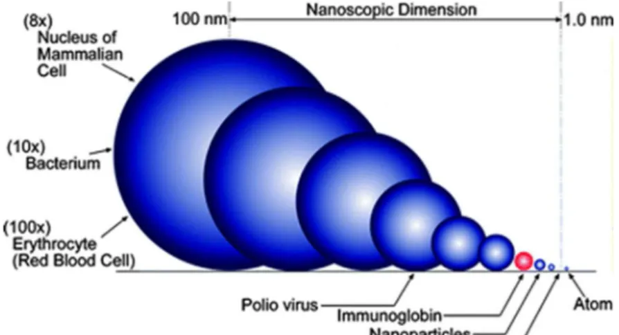
2.1. Introducing the concept of Nanotechnology and Nanomedicine The term “nanotechnology” applies to the creation, manipulation and application of materials which have a size between 1 and 100 nanometers, meaning that they are at the scale of molecules and atoms. Their particularly small size gives them fundamentally unique properties different to those of bulk and bigger materials. These differences are mostly due to the vastly increased surface area to volume ratio nanoparticles have. In comparison with the structures and biological forms they are designed to interact with, nanoparticles are of comparable size and even smaller, i. e. a cell’s magnitude is around the µm, viruses are usually bigger than 20 nm and proteins’ size orbits around 2-4nm.
3,4
Figure 2. Comparison of size of different biological entities and nanoparticles. Reproduced from ref. 5. 5
The mentioned size of nanoparticles and its relation to that of biological entities sums up in the concept of “nanomedicine”, being it the use of nanoscale technology in health, diagnosis, therapeutics… It is in 1960 when Richard Feynman introduced, when answering a colleague in a conference, how interesting would be “in surgery, to swallow the surgeon” and how miniaturization would lead to promising new applications in several technique fields.6
Summing up, the benefits of using nanomaterials include as follows: - Smaller devices are less invasive
- They can be implanted inside the body - Biochemical reaction time is much shorter
- Devices are faster and more sensitive than typical drug delivery - The use of less quantity of drug given the specificity of nanomedicine 7
There are several nanomaterials with potential use in different branches of life sciences, as it is summed up in Table 1.
6 Table 1. Nanomaterials with potential applications in biomedicine. Modified from ref. 7.
Particle Class Materials Application
Natural materials or derivatives
Gelatine
Liposomes
Starch
Drug / Gene delivery
Dendrimers Branched polymers Drug delivery
Fullerenes Carbon based carriers Photodynamics Drug delivery
Polymer carriers
Polyethyleinemine
Block copolymers Drug /gene delivery
Ferrofluids SPIONS
USPIONS
Imaging (MRI)
Quantum dots Cd / Zn – selendies Imaging
In vitro diagnosis
Various
Silica-nanoparticles
Mixtures of above Gene delivery
2.2. Magnetic Nanoparticles
The use of magnetism is not new in the field of Medicine, going back to even ancient Egypt and the use of milk with magnetite particles to treat intoxication after the accidental swallowing of rust. More modernly, magnets have had an increasing impact mainly in the surgical field, catheters and in the evolving field of magnetic resonance imaging. Bringing together the concepts of nanotechnology, nanomedicine and applying magnetism to medicine, magnetic nanoparticles (MNPs) arise as a new and promising tool for diagnosis and treatment of several diseases, especially for cancer. 8
MNPs, due to their unique properties, are being investigated as the next generation of magnetic resonance imaging (MRI) contrast agents as well as promising carriers for targeted drug delivery. Even though the early research in the field of nanotechnology dates back several decades, it is more recently when the increasing interest in this field has meaningfully expanded the wideness of MNP research. Along these lines, their broad range of potential applications make
7
them a very interesting tool to be investigated and to play a significant role in the healthcare needs of the future. 9
Given the extensive research carried out in the past decades in the field of nanoparticles, it is clear that a significant effort, both in terms of time and financial investment, has been made. In Europe, the European Commission (in the framework of the FP7 program in the past, and nowadays in Horizon2020) has contributed in several research projects including MNPs, in different areas of medicine such as Alzheimer’s disease, rheumatoid arthritis and osteoarthritis… but, most of all, in cancer diagnosis and therapeutics. 6
2.3. Engineering magnetic nanoparticles
Several forms and chemical compositions have been proposed and evaluated for medical and biomedical applications to take advantage of magnetic phenomena. Composition, size, morphology and surface chemistry can be nowadays tailored by numerous engineering processes in order to improve the characteristics of the nanoparticle and also affect its behavior both in vitro and in vivo.
Down to its simplest form, an MNP which is going to be used in a biomedical application is composed of a biocompatible coating surface that covers an inorganic nanoparticle core. The biocompatible layer will give the nanoparticle its ability to penetrate and interact the physiological medium without being rapidly taken away from blood circulation, and it can also be the base to bind functional ligands. 9
This introduces the concept of “multilayer”. When talking about a medical platform (in this case, the MNP), the multilayer concept implies its capability to have various properties from the detection to the cure of diseases. This way, the prototype of MNP includes a magnetic core, a biocompatible layer that gives it the ability to stay in the physiological enviroment, and a functional coating to give it its diagnostic or therapeutic properties. 6
Colloidal iron oxide nanoparticles (such as super paramagnetic iron oxide -SPIO- and ultrasmall superparamagnetic iron oxide -USPIO-) are the most extensively studied MNPs for biomedical applications, since they have an excellent biocompatibility profile and are easily synthetized. This SPIO and USPIO are typically formed by a nanocrystalline magnetite or maghemite core covered by a variable, biocompatible coating. The magnetic properties of this particles rely on the interaction between the Fe2+ and Fe3+, in between which the
electrons hop and produce the magnetization. The widespread use of this particles does not only rely on their magnetic properties, but also in their favorable dynamics once inside the body. Upon metabolism, iron ions are added to the organism’s iron deposits and eventually incorporated as hemoglobin to the erythrocytes, allowing the safe use of this nanoparticles in vivo. The SPIO and USPIO utilized or under investigation for clinical application (predominantly as
8
MRI contrast agents), are currently synthesized by an aqueous co-precipitation process in the presence of the coating material, but are liable to be synthesized by a variety of processes which range from traditional wet-chemistry solution to more complex techniques like laser pyrolysis or chemical vapor deposition. Different synthesis processes can cause a vast variation in the magnetization of the particle, even with those of similar size, since impurities can incorporate to the crystal structure of the nanoparticle. 10
At small sizes (of the order of tens of nanometers), ferri- or ferro-magnetic materials (such as MNPs) become a single magnetic domain and therefore maintain one large magnetic moment. This superparamagnetic property, marked by the lack of remnant magnetization after removal of external fields, enables the particles to maintain their colloidal stability and avoid aggregation making it suitable for their use in biomedical applications.9
Bi-metallic or metal alloy nanoparticles can also exhibit superparamagnetic properties, making them plausible candidates as MRI contrast agents or carriers for drug delivery. FePt is between the most known ones, since the interaction between the two chemical species present in the alloy lead to greater chemical stability than other high moment metallic nanoparticles. Besides, the surface chemistry of these nanoparticles is susceptible of having carboxylate- and amine-based surfactants to improve their water solubility. 9
But not only the core defines the nanoparticle, and it is as or even more important to define the best surface coatings for them, since it is the external layer the one that will be in contact with the physiological system and will define the functionalization of the magnetic nanoparticle.
Although MNPs are not attracted magnetically to each other, they still tend to agglomerate as a result of their high surface energy, due to their superparamagnetic properties. Also, upon injection to the plasmatic compartment, “naïve” MNPs are not inert and they are subjected to adsorption of plasma proteins, or opsonization and the first step in their clearance by the reticulo-endothelial system (the pharmacokinetics of the MNPs will be discussed widely later). This surrounding of physiological elements is called “protein corona”, and it is formed from all the opsonization components of the native immune response. The coatings that surround the magnetic core are the ones that provide means to tailor the properties of MNPs such as surface charge and chemical functionality. Critical aspects compromising the performance of MNPS, regarding the polymeric surface, include their structure (and the intrinsic hydrophilicity/hydrophobicity, biodegradation feature), the size/molecular weight of polymer, its conformation, etc.
One of the most widely utilized coatings, and successful in terms of in vivo applications, has been the polysaccharide dextran. Polyethylene glycol (PEG) has also been broadly used as it shows reduced nanoparticle uptake by macrophages and, therefore, a longer circulation time in the blood. These biodegradable molecules are defined as capsules when consisting of a central
9
liquid cavity surrounded by a polymer wall or as spheres when made of a solid polymer framework. Compared to liposomes, polymeric nanoparticles are more stable when in contact with biological fluids and they also exhibit the advantage of the possibility of modulating the drug release profile (the release of the carried drug can depend either on the diffusion rate through the polymer framework or the erosion rate of the biodegradable matrix itself). When talking about polymer coatings, they are divided in two generations depending on whether they can pass through capillaries: first generation ones are those which are not able to (being fundamentally used for chemoembolization by arterial injection); while second generation consists in nanoparticles able to pass through capillaries and then suitable for systemic use. 6,8
Liposomes, as drug delivery carriers, can also be considered one of the first shapes of nanomedicine. These vesicles, formed by phospholipid bilayers, have been used for the delivery of small molecules such as MR imaging contrast agents, DNA and peptides. One of the advantages of liposome encapsulation is their well known in vivo behavior due to processes of PEGylation that result in a longer life of the molecule in the bloodstream. Also, liposomes can encapsulate a large number of MNPs that are delivered together so the dilution is avoided. All these mentioned features are also applicable to multifunctional micelles formed with amphiphilic molecules. The preparation procedure consists on dissolving appropriate amounts of phospholipids in an organic solvent, evaporating it and subsequently disrupting the dry lipid layer with an excess of water or buffer, which leads to the spontaneous formation of multilamellar liposomes of various sizes. In these structures, drugs can be located either in the inner aqueous phase or in the lipid bilayer, depending on their hydrophilicity/hydrophobicity ratio. 8
Inert coatings, also called shells, formed by biocompatible gold or silica can also perform the role of the surface layer of MNPs, and have become an attractive approach for developing MRI contrast agents or magnetic targeted carriers for drug delivery. Their inert profile gives both protection against the chemical degradation of the core and prevent from the release of potentially toxic components. Furthermore, functionalization chemistries are generally better established with these materials than those that comprise MNPs. Silica shells are especially attractive, since they are easy to synthesize and have good stability under aqueous conditions. Gold has also several advantages as a coating material for MNP due to its low chemical reactivity profile, although this could be a drawback, causing a difficulty in forming gold shells over MNPs.
At last, and with a greater interest from the functional point of view, functional ligands can be used as coating of MNPs. This includes permeation enhancers, optical dyes, targeting agents and therapeutic agents that can be bound to the surface or be incorporated to the molecular structure to improve its functionality (the functionalization potential of MNPs will be widely discussed later on). 9
10 Figure 3. Multilayer structure of a magnetic nanoparticle. The scheme shows a simplified view of the potential elements building an MNP, including a magnetic core, a biocompatible coating, a therapeutic layer and a targeting ligand. Reproduced from ref. 6.
2.4. Pharmacokinetics of magnetic nanoparticles
Intravenous (IV) administration is, at first glance, the most effective method to reach target organs and tissues all over the body, since every cell receives blood supply. For this purpose, the already mentioned colloidal drug carriers (liposomes and polymeric particles) are crucial, since their features will affect the fate of the MNP once it contacts the bloodstream and all the active agents that are present in it. In order to do so, and compared to liposomes, polymeric nanoparticles can modulate the drug release profile and are more stable in biological fluids.
Targeting routes
There are two possible targeting routes for MNPs: passive targeting and active targeting (Figure 4).
Pathophysiological features of tumors and their enviroment have been exploited for passive targeting, particularly the Enhance Permeability and Retention effect (EPR effect), which promotes the accumulation of nanodrugs in the tumor. The EPR effect takes advantage of the leaky tumoral vascularization, whose capillaries have wider fenestrations in their endothelium (up to around 800 nm) allowing nanoparticles through these gaps. Then, by convection and diffusion processes, passive targeting of nanomedicine therapies into tumors can occur without any specific ligand attached to the surface of the carrier. Nevertheless, it is widely accepted that passive targeting, based on the EPR effect, is not enough to fully exploit the benefits of targeted delivery and control the side effects of cytotoxic drugs. Also, the heterogenicity of the tumor and its stroma (such as a hypoxic gradient) can strongly impact on the efficacy of drugs delivered by passive targeting, resulting in an inefficient or uneven transport of the compound into the tumor. Also, and more recently, it was shown that iron oxide nanoparticles themselves (as well as other nanoparticles) induce gaps of
11
tens microns in size between endothelial cells, augmenting the permeability of the vessels. 11,12
On the other hand, and when more selective uptake is desired, for creating an active targeting route, there are two requirements: long-circulating nanoparticles and ligand conjugation.
To prolong the half-life of nanoparticles in the bloodstream, the classical approach is the injection of large doses of a placebo molecule in order to saturate or impair the phagocytic activity of macrophages; another potential mode of bypassing the reticuloendothelial system is the transient destruction of liver and spleen macrophages by prior administration of gadolinium chloride particles or liposomes with entrapped clodronate, which induce apoptosis, although this protocol has little justification since it compromises the essential defense system of the body. Nevertheless, the most satisfactory strategy to extend the circulating time of nanoparticles is the use of macrophage-evading molecules with a plasma half-life as long as possible, thus the probability of attaining to the desired target is increased. Taking into account the physicochemical factors determining the opsonization process, the conclusion that the smaller, the more neutral and the more hydrophilic the carrier surface is, the longer plasma half-life will be. Also, it is assumed that, for this purpose, the structure of the surface is more important than the core’s, since that is the one which is in direct contact with the plasma enviroment. Among the macromolecules studied for the enveloping of MNPs, poloxamines are specially interesting, and their use is widely spread in Galenical pharmacology to extend the circulation lifetime, bioavailability, and decrease the immunogenicity, renal clearance rate and dosing frequency, by a process of PEGylation. These molecules allow the particle to avoid phagocytosis.
At last, and giving the MNPs the major specificity possible, there is the possibility of ligand conjugation to guarantee active targeting. This consists in the labelling of the MNP surface with ligands that specifically bind to surface epitopes or receptors on the target sites (in a molecular recognition process similar to the antigen-antibody interaction), and it will also require the presence of “stealth carriers” to avoid macrophage recognition and endocytosis. In case of cancer therapy, active targeting potentially allows the selective recognition of malignant cells even when they have become metastatic. Nevertheless, in the engineering of these specific ligands, there are two major drawbacks: their size (for example antibodies are around 20 nm) and their potential immunogenicity. 8,13
12 Figure 4. Targeting routes for nanoparticles. A: extravasation through fenestrated vasculature leads to passive targeting of tumor tissue; B: active targeting of cancer cells (1) or human endothelial cells (2). Modified from ref. 11.
Summing up, there are three potential levels of sophistication applicable to the carriers used:
- Colloidal carriers (the simplest of levels), consisting of liposomes or the naked nanoparticles, which would be passively adopted by the reticuloendothelial system and then target the organs of this system. - The “stealth carriers”, the ones that avoid macrophage endocytosis,
whose features allow to be used as drug reservoirs, blood-pool imaging and passive targeting via vascular defects (like the ones seen in tumor’s capillaries)
- Ligand-decorated carriers which allow active targeting towards specific cells, tissues and organs. 8
2.5. Intracellular delivery and controlled release
Obviously, the function of the MNP does not take place until it is in direct contact with the cellular components, so the internalization and the subsequent release of the therapeutic agents of the nanoparticle are essential. There are various mechanisms of entrance proposed, including receptor-mediated endocytosis and internalization by caveolae structures. The size and surface properties of the nanoparticle, once again, play a fundamental role in moving across the plasma membrane. Nanoparticles of a size smaller than 50 nm, and those coated with lipophilic polymers (like PEG), efficiently diffuse through cell membranes. Moreover, permeation enhancing molecules (that increase the passage of the nanoparticle through the lepidic bilayer) can be attached to the nanoparticle.
Once the MNP is inside the cell, the next goal is targeting the subcellular organelles, prior to being trafficked to lysosomes, where their biological activity may be nulled. To serve the purpose of endosomal release after cellular internalization, several strategies have been proposed: tailoring of cleavable linkers responsive to pH, osmolarity or enzymatic activity, or integration of
13
cationic polymers to induce osmotic swelling and subsequent escape from endosome. 9
2.6. Biodistribution and clearance
The biodistribution of nanoparticles involves fundamentally the reticuloendothelial system, also known as the mononuclear phagocyte system, which is defined as the group of cells including bone marrow progenitors, blood monocytes and tissue macrophages (i. e. liver’s Kupffer cells). The function of these cells is to remove senescent cells, invading micro-organisms or particles that they recognize, and are widely distributed throughout the different organs and systems. For this process, the first step is the opsonization of the particle that is going to be cleared. Opsonins nonspecifically join to the surface of the strange entity and then these molecules (immunoglobulins, fibronectin, complement factors…) are recognized by the monocyte-macrophage’s specific receptors. The second step is the endocytosis/phagocytosis of the opsonized particle by circulating monocytes or fixed macrophages and the consequent elimination from the systemic circulation and their accumulation in the organs with high phagocytic activity. The rate and site of clearance of the particle from the blood compartment depends on different surface characteristics, size and morphology which will determine the different array of opsonins attracted. Therefore, after IV administration, colloidal drug carriers are cleared up within minutes from the bloodstream, and their typical final biodistribution is 80-90% in the liver, 5-8% in the spleen and 1-2% in the bone marrow, depending mainly on the size (since these are the tissues that show the highest density of monocyte-macrophage cells, conforming the reticuloendothelial system). Therefore, the mononuclear phagocyte system-mediated clearance is crucial in the biodistribution of the nanoparticle and its presence is, at first, not compatible with long-lasting circulation times. However, it itself can be a target, and it gives a chance of delivery to the organs comprising the system. For example, poly(alkylcyanoacrylate) nanospheres loaded with doxorubicin may be used for the treatment of hepatic metastases. 6,8
2.7. Potential hazards and toxicity of nanoparticles. Drawbacks.
The accurate estimation of cytotoxicity by conventional methods is complex, due to the variability within the individual parameters of each nanoparticle, making it difficult to stablish a standardized and comparative method of assessment. The development of such a method is fundamental and a priority for the European Medical Agency who states that “specific guidance on quality, toxicology, clinical development and monitoring aspects may be developed in the future, once sufficient scientific experience has been gained for specifically identified sub-technologies within the field of nanomedicines”
14
Normally, seeking approval of a medicinal product by the FDA or EMA takes two steps: in vitro screening to rule out significant adverse events and to ensure sufficient biocompatibility and posterior in vivo screening. There are several areas of specific toxicity that should be measured and outlined for the safer use of MNPs (and any product that is going to be used broadly in vivo), which are:
- Immunotoxicity: compromising of both the innate and adaptive immune system, generation of reactive oxygen species, cytokine release, hypersensitivity…
- Formation of agglomerates - Reproductive toxicity - Genotoxicity
- Carcinogenic potential 6
A number of nanomedicine products have been approved by the FDA and EMA, fulfilling the current safety requirements sets forth by these agencies. FDA has a draft of a guidance document published back in December 2017 concerning industry, and related to all those drug products, including biological products that contain nanomaterials; while the EMA published in March 2015 a consensus document about iron-based nanomaterials and their pre-clinical features14,15
When reflecting about the major drawbacks for the widespread use of nanoparticles in the field of medicine, the greatest doubts arise from the lack of clinical experience of their use, and being the knowledge mostly based on in vitro and animal models. Some of the potential disadvantages of nanoparticles are as follow:
- The excessive costs of nanotechnology and the research in the field, as well as the pricy manufacture processes stablished nowadays. - Their surface properties, that make nanoparticles unique when
compared to bulky materials, can also have an impact on their toxicity, and this impact is still not well known.
- The different physicochemical properties of nanoparticles can result in unexpected behavior in pharmacokinetics.
- Environmentally, their small size can be cause of inhalation hazards with results still unknown, and they may be potentially alike those problems a person gets from inhaling asbestos particles. 7
15
3. MAGNETIC NANOPARTICLES IN CANCER DIAGNOSIS 3.1. Magnetic Resonance Imaging (MRI) fundamentals
Magnetic Resonance Imaging (MRI) is a non-invasive, tomographic imaging modality which is based on the manipulation of the inherent nuclear magnetic moment of endogenous nuclei.
In the early seventies of the 20th century, Lauterbur and colleagues
showed that nuclear magnetic resonance (NMR) (which is the absorption of electromagnetic radiation by a nucleus having a magnetic moment when in an external magnetic field) can be made spatially selective. By applying a magnetic field gradient in one of the spatial domains and measuring an NMR signal, spatial information is obtained, and this can be done in the three directions offering a three-dimensional spatial picture. Hence, NMR shows the spatial distribution of protons (i.e. water molecules’ hydrogen atoms) in an object.
In MRI, images are obtained by exposing nuclei to a static magnetic field and, within that static field, perturbing the steady-state equilibrium with time and space variation of the magnetic fields. After ceasing the perturbation, all nuclei suffer relaxation by two mechanisms called T1 (spin-lattice or longitudinal
relaxation) and T2 (spin-spin or transverse relaxation), which are then
reconstructed into gray scale images. The endogenous MRI contrast in soft tissue comes from local differences in the proton density (water concentration) resulting in different values of T1 and T2. On this basis, endogenous contrast depends on
the chemical and physical nature of the tissues. Despite the relatively high quality of such images of soft tissues, in some cases there is not enough image contrast to diagnose the pathology of interest. An important alternative is to use exogenous contrast agents. When in presence of an MRI contrast agents, the T1
and the T2 of the protons of the nearby tissues change, creating a modification
that can be detected by the MRI machine. Relaxation rates are the reciprocals of the corresponding relaxation time. 16,17
By changing the pulse sequences of NMR (the radiofrequency pulses created in an exactly defined time), the obtained images show difference depending on their weighting (T1-weighted or T2-weighted), allowing different
perspectives of a same object and its components, depending on what is interesting for the test. For T1-weighted MRI images, a faster T1 relaxation rate
gives a brighter contrast, while a faster T2 relaxation rate yields a darker contrast
for T2-weighted images. MRI contrast agents can accelerate these relaxation
rates; for example, gadolinium-based contrast agents lead to a reduction in T1
relaxation time and then to a positive (brighter) contrast in T1-weighted images
while contrast agents based on iron oxides shorten the T2 relaxation time,
16
Although there are numerous contrast agents or new applications for the already existing ones, the most used ones are three: gadolinium (Gd)-based contrast agents, hyperpolarized molecules and iron oxide magnetic nanoparticles:
- Gd-based compounds, in spite of their nephrotoxicity, are widely used as MRI contrast agents to enhance images in medical procedures.
- Hyperpolarized molecules are less popular, having a more recent history in MRI. The main molecules are gases (hyperpolarized Helium and Xenon) which allow imaging of lungs and the obtention of information about pulmonary diseases.
- Magnetic nanoparticles, including ferromagnetic iron oxide particles and ultrasmall superparamagnetic iron oxide particles are also being already used in clinical diagnosis as MRI contrast agents. 6
Before elaborating about the features and potential uses of magnetic nanoparticles, a brief explanation of different concepts of magnetism is considered crucial. These concepts to explain are paramagnetism, ferromagnetism and superparamagnetism:
- Paramagnetism: each atom of a paramagnetic material behaves as an individual, non-interacting and randomly orientated molecular magnet with a magnetic dipole moment (µA). The mentioned dipoles are oriented
randomly when in absence of an external magnetic field, but when this field is applied to the paramagnetic particles, the molecules start to align their magnetic dipoles with the direction of the external magnetic field, proportionally to the intensity of the external field until it is strong enough to align all the molecular magnets. This complete alignment of the molecular magnets to the external magnetic field is known as magnetic saturation, and it can only occur when whether the temperature is very low, or the external magnetic field is of a very high strength (for example, paramagnetic gadolinium chloride starts to saturate at approximately 50 T). Paramagnetic materials do not retain any magnetization once the external field ceases to act on them.
- Ferromagnetism is characterized by a strong exchange interaction energy that leads to a regular orientation of the magnetic moments. The ferromagnetic particle is organized in Weiss domains in which every magnetic dipole is oriented in the same direction, and they will remain the same as long as no external magnetic field is applied. Differently to paramagnetism, once an external magnetic field is applied to a ferromagnetic material, it retains magnetization, showing magnetic remanence and potentially acting as a permanent magnet. The number of Weiss domains depends on the particle’s size and, if the size of the particle’s core (Dc) is smaller than a critical size (Dsd), the multiple domain
state is lost for the benefit of an energetically favorable single domain state in which all the internal dipoles point to the same direction. To these particles with a single Weiss domain, magnetic saturation occurs to much
17
smaller external fields compared to paramagnetic nanoparticles (around 1 or 2,5T).
- If the size of the ferromagnetic single domain particle is further reduced, being Dc smaller than a second critical size (called Dsp),
superparamagnetism is achieved. In these nanoparticles, the thermal fluctuations of the enviroment will outcompete the dipole-dipole interactions and cause the magnetization to flip randomly even in the absence of an external magnetic field. Thus, the energy barrier which separates the ferromagnetic single domain state from the superparamagnetic state is proportional to the volume of the particle, the anisotropy constant and to the observation time. 16
3.2. Achieving the desired magnetism of Magnetic Nanoparticles (MNP) To avoid agglomeration and to obtain stable colloidal suspension, the bare metal core of the magnetic nanoparticle must meet some stability requirements. There are two major obstacles to overcome in this aspect: the magnetic dipole-dipole interactions and the van der Waals forces (defined as attractive or repulsive interactions between different molecules due to an intramolecular bond – ionic, metallic or covalent bond). Other minor aspects affecting the stability of magnetic nanoparticles (when referring to magnetism) are their stability against setting in the gravitational field and the stability in a magnetic field gradient, being both able to be achieved by reducing the size, something that goes hand in hand with stabilization against magnetic interaction.
The main compounds with the ideal magnetic properties, in the field of MNPs are as follow:
- Gadolinium based paramagnetic agents
- Manganese based paramagnetic nanoparticles
- Iron oxide polymorphs, being Magnetite (Fe3O4) the one with greater
interest in biomedical applications. 16
3.3. Applications of MNPs in MRI and other imaging modalities 3.3.1. Cellular and molecular imaging
It was found that the relaxation rate R2 (the reciprocal of T2) increases
upon internalization of the MNPs by the cells compared to MNPs free in solution. In an experiment performed by Weissleder and colleagues it was showed that cells loaded with nanoparticles led to a different signal loss in T2-weighted MRI
compared to free particles, not incorporated in the cells. Also, for ultra-small SPIOs (USPIO), it was proven a greater signal altering effect when intracellular
18
than when free in solution. Thus, this signal altering effect can be potentially handy to track cells in vivo using high-resolution MRI. 18
Macrophages phagocytose USPIO, and this process can be taken advantage of for the visualization of inflammatory and degenerative processes using MRI. Once administered, USPIO are internalized by phagocytes, attracted due to inflammation processes. Several pathologic situations produce inflammation and can be therefore indirectly visualized by detecting macrophages loaded with this USPIO: focal ischemic lesions in the cardiovascular system, atherosclerosis, multiple sclerosis, infections… Also, since macrophages are accumulated in the lymph nodes, millimeter sized metastasis can be revealed by MRI before the node enlarges.
But more importantly, there are several biomarkers that conditionate the future and prognosis of different cancer cells and are liable to being targeted and detected by the combined use of MNPs and MRI. These specific biomarkers known from the field of oncology are HER2 (Human Epidermal growth factor Receptor), VEGF (Vascular Endothelial Growth Factor), and even intracellular markers like Ki-67, p53 or EGFR (Epidermal Growth Factor Receptor). 16
Once a malignant tumor grows up to a critical size (approximately one cubic millimeter), its cells cannot longer rely on receiving their blood supply from distant blood vessels in a passive way, needing direct blood supply from new vasculature to be able to proliferate and prosper. The process by which new blood vessels are developed in order to guarantee the mentioned blood supply is called angiogenesis, and it depends on several proteins (angiogenic factors), in between which the better known ones are VEGFs (vascular endothelial growth factors) that cause the endothelial cells from the existing vessels to grow, proliferate and migrate following the VEGF concentration gradient, i.e., towards the growing tumor. Virtually all tumors are dependent upon angiogenesis for their survival, and there are several chemotherapeutic drugs already designed to interfere with this process, so it would be potentially useful for clinical oncology to have a method to quantitatively asses the process of angiogenesis.
Although, ideally, the method for visualizing angiogenesis would be direct labeling the VEGFs to detect their levels of tissue expression, MRI provides a less specific but very informative method of assessing the vascular changes that occur in tumors. As explained before in this text, the vascular framework of tumors has particular features such as wider intercellular unions of the endothelium, being more fragile and leaky. These incomplete and defective vessels may be exploited in order to study the tumors neovasculature and, subsequently, its development state. Dynamic contrast enhanced magnetic resonance imaging (DCE-MRI) is a modality of MRI that measures the integrity, density and leakiness of tissue vasculature; changes in the parameters obtained by DCE-MRI can then be used to assess how a tumor is responding to treatment. For this, this method uses mathematical models of how the paramagnetic tracer diffuses through such vessels. Then, it is reasonable to think that healthy tissue
19
and its vascularization can be characterized by a series of parameters measuring vessel permeability, blood flow and tissue volume fractions (meaning the fractions of a given portion of tissue that can be attributed to intervascular or extravascular space) in contrast to the parameters of a tumor, which will be different given the particularities of its vascularization.
By DCE-MIR, serial MR images of the tissue of interest (the tumor in this case) are acquired before, during and after the intravenous injection of a paramagnetic contrast agent. By the ulterior processing of these images, several study parameters are measured. One of interest is Ktrans, which represents the
product of the permeability and the vascular epithelial surface area from which the agents leaks into tissue. Both healthy and pathologic tissues show their own parameters in this aspect. Furthermore, because these parameter values are probes of tissue status, they may be used to even differentiate malignant from benign tumors, aid in tumor staging and monitor response to treatment. 19
Figure 5. DCE imaging of human breast cancer. In the top row, Ktrans maps overlaid on T1-weighed SPGRE image acquired before (panel a), after one cycle (panel b) and after chemotherapy (panel c), in a patient showing clinical response. In the bottom row, similar data from a patient with progressive disease. In the top row, it is shown a general decrease in Ktrans values from pre to post-one cycle of therapy (panels a and b), meanwhile in the bottom row there is a general increase in Ktrans, correlating with disease burden at the time of therapy. Modified from ref. 19.
3.3.2. Direct measuring of biomarkers
Cancer cells exhibit substantial molecular heterogenicity, and methods to detect numerous key targets in size-limited clinical samples is increasingly becoming essential to assess the spatial and temporal status of the pathophysiology of tumors and to realize the goal of personalized medicine.
20
Nanomaterials offer unique physical properties that make them ideal biosensors for small cell populations, and they can be an alternative to traditional molecular detection methods (such as Western blotting, flow cytometry, immunofluorescence, etc.) that require prohibitively large cell numbers or lack from multiplexing capability. Nanomaterial-based detection platforms can provide advantages over these approaches, in terms of signal sensitivity, stability, and capability for multiplexing given their unique features. There has been substantial interest in developing, by using nanomaterials functionalized with targeting ligands, sensitive and robust methods to detect biomarkers in the surface of cells of tissues. 20
Successful development of ultrasensitive molecular imaging nanoprobes for the detection of targeted biological items is a challenging task. Magnetic nanoprobes have the potential to perform such a role, but the results obtained with probes that are currently available were far from optimal. Artificial engineering approaches where used by Lee et al. to develop innovative magnetic nanoprobes that, when conjugated with antibodies, showed enhanced magnetic resonance imaging sensitivity for the detection of cancer markers compared to currently available probes.
Molecular imaging nowadays, with techniques such as Positron Emission Tomography (PET), is highly dependent on the molecular imaging agents whom, if efficient, can precisely detect early-stage diseases, monitor the responses to drug therapies and track cell migrations. Due to their small size and functional versatility, nanoparticles are now emerging as suitable agents to be used in PET. The signal enhancement caused by conventional iron oxide nanoparticles is still unsatisfactory compared to that obtained by other imaging modalities such as PET. Lee et al. overcame the limitations of nanoparticle-based agents by using an innovative approach to develop magnetism-engineered iron oxide (MEIO) nanoparticles with exceptional magnetism features. For this, they made a screening process in other to obtain the best nanoparticle and it was shown that MnFe2O4 was the ideal material due to its magnetic susceptibility and
characteristics. Later, this MnFe2O4 was conjugated with the cancer-targeting
antibody Herceptin, which specifically binds to the HER2/neu marker overexpressed in breast and ovarian cancer. After proving that these particles where biologically nontoxic in cytotoxicity studies, comparison between MEIO-herceptin conjugates and cross linked iron oxide-MEIO-herceptin conjugates was performed, and ultra-sensitive detection capabilities where demonstrated, which took to the in vivo testing. With nude mice it was also showed superior sensitivity by MnFe2O4. 3
3.3.3. Specific ligands for more specific contrast agents: SPIONS tailored with targeting moieties
Second generation SPIONS are those functionalized with different targeting moieties or ligands, such as monoclonal antibodies (mAbs), allowing them to specifically bind to cellular receptors. This causes the MNPs to
21
accumulate more intensely in tumors and also, since they are more specific, to cause less side effects.
The ligands proposed for the enhancement of the specificity of SPIONS for MRI are as follows:
- Monoclonal antibodies (mAb), which are highly specific but have several drawbacks including their potential immunogenicity and their big size (causing MNPs to be less “nano”, crippling their specific capabilities). Using fragments of monoclonal antibodies (Fab, peptides…) was proposed to bypass mAbs’ weaknesses, but they have rarely been used because of their production complexities.
- Aptamers (synthetic single-stranded DNA or RNA oligomers), which have smaller molecular size and entail easier manufacturing processes compared to mAbs. Their specificity reaches levels high enough to differentiate between subtypes of small-cell lung cancer.
- Lectins, molecules that bind to the glycoprotein and glycolipid moieties of glycans of the cell membrane. Glycans are crucial to cell-cell communication, adhesion, differentiation and development, processes overexpressed in cancer. However, lectins commonly lack specificity towards a determined cell type.
- Cell penetrating peptides, like those naturally expressed in HIV virus 1, which increase endocytosis process, but also with low specificity to cell types.
- Folic acid, given it is pivotal to cancer cell proliferation, but it can also be found physiologically in several tissues such as brain, thyroid and lung. - Methotrexate, a chemotherapeutic drug which specifically kills cancer cells
overexpressing the folate receptor can play a similar role. 21
SPIONS labelled with specific ligands have already been proposed for MRI imaging of several types of cancers such as liver cancer: Kupffer cells (hepatic macrophages) are the ones that internalize SPIONs in healthy hepatic tissues and liver tumors are mostly devoid of these macrophages. This causes negative enhancement on T2 weighted MRI sequences in tumor lesions, facilitating
detection of liver tumors or metastases of small sized (up to 2-3 mm). In a more specific way, anti-alfa-fetoprotein and anti-glypican 3 antibodies can be used for targeting hepatocellular carcinoma, and USPIONs conjugate with those were shown to perform shortening T1 and T2 relaxation for the MRI detection of this
malignancy. Also for brain tumors, SPIONS can be conjugated with ligands specific for various central nervous system (CNS) targets such as gliomas, which are among the most common brain tumors. For targeting gliomas, chlorotoxin, a modified small peptide isolated from a scorpion, is used, since these tumors have a receptor for this peptide in their chloride channel. 21–24
Recently, various types of NPs have been proposed for the detection of circulating tumor cells (understood as the first step in the formation of metastases). Given the similarities between healthy and metastatic lymph nodes,
22
creating a non-invasive method for the detection of the malignant ones is challenging. Mannan-coated SPIONS have been used for this, since it has been proposed that malignancy swamped lymph nodes lack the antigen presenting cell-related functions to internalized these SPIONs to the extent that normal lymph nodes do, leading to differences in MRI imaging. 21,25
3.3.4. Multi-modal imaging using MNPs
Owing to the need for accurate imaging capable of detecting small biological targets in complex environments, compensating the weaknesses of one imaging modalities with the strengths of other arises as a potential answer. For example, imaging modalities such as PET offer high sensibility but low anatomic definition, while others like MRI show anatomical details with high specificity. Hence, combining MRI with PET or optical modalities gives a method of acquiring both sensible and specific information compared to one modality alone. Serving this purpose, several multi-modal imaging methods are prosed.
T1 and T2-weighted images made one
Typically, contrast agents show enhanced signal in either T1-weighted or
T2-weighed images. Dual-mode contras agents provide complementary T2 and
T2- weighted images that enable self-confirmation of the signals from the contrast
agents, avoiding problems such as discrepancies and mismatch between T1 and
T2-weighted images. This can be done by combining elements that have T1
properties (like Gd or Mn-based chelates) and those with T2 properties (metal
ferrite magnetic nanoparticles).
Other possibility is DMCA: dual-mode contrast agents, which offer the ability to perform an AND logic gate algorithm for accurate interpretation of biological information by eliminating MRI artifacts. This method can be a useful tool for enhancing the accuracy of MRI of small biological targets such as tumors in early stages and lymph node affectation.
MRI-PET/SPECT dual-mode imaging agents
Nuclear imaging techniques, which utilize gamma rays emitted from decaying radioisotopes, generally offer high sensitivity, but their poor spatial resolution is a limitation. Thus, the combination of those with MRI would offer tomographic images with high spatial resolution alongside strong sensitivity and accurate quantification of biological information, such as biodistribution, pharmacokinetics, physiological and pathological performance, etc. 26
To reach these imaging processes, MNPs modified with radioisotopes via chemical conjugation have been synthetized, acquiring fused images capable of delineating different types of small sentinel lymph nodes of only a few millimeters in diameter, for example, with Mn-doped ferrite nanoparticles bound to 124I. 27
23 Figure 6. MRI-PET dual imaging. Left: schematic image of a radioisotope-labeled Mn-doped ferrite nanoparticle. Right: Nanoparticles delineate the sentinel lymph nodes (axillary and brachial nodes) combining adequate PET signals and the anatomical MRI imaging. Reproduced from ref. 26.
Others
Other imaging methods which can take advantage of using MNPs to acquire images of increased sensitivity and anatomical definition are as follow:
- MPI-MRI dual imaging: magnetic particle imaging combined with MRI. This adds the quantitative lecturing capacity of MPI to the image and, also, the elimination of background signals since the magnetic nature of the human tissues do not emit interference to MPI
- Magnetic nanoparticle-assisted multi-modal ultrasound (US) imaging: utilizing magnetic nanoparticles to improve the sensitivity of ultrasound, which is poor.
- MMUS-US: magneto-motive ultrasound imaging. In MMUS, a focused high intensity pulsed magnetic field is applied to the magnetic nanoparticles which are induced motion that can be detected with US imaging. Hence, it is possible to assess the distribution of MNPs in biological systems, and MMUS-US could rise as an imaging approach for cellular and molecular level dynamic imaging.
- MPA (magneto-photoacoustic imaging)-US dual-modal imaging: in photoacoustic imaging, tissues absorb laser light and expand generating ultrasound waves. Magnetic nanoparticles, when under a magnetic field, generate vibrational motion that can be detected by PA (this combination of MNP and PA is what is called MPA). This can be utilized in order to detect tumor cells. 26
24
4. MAGNETIC NANOPARTICLES IN CANCER TREATMENT
The undesired secondary effects and reduction of quality of life of current cancer treatments force research into finding more selective and less harmful therapies. Regarding magnetic nanoparticles, several approaches have been proposed: hyperthermia, directed delivery, cell therapy, immunotherapy…
4.1. Hyperthermia for Cancer treatment
Nowadays, cancer treatments can be categorized, generally, into chemotherapy, radiotherapy and surgical tumor extirpation. Even though the effectiveness in terms of survival from these approaches has been historically proven, they are not always sufficient to completely eradicate the disease. Besides, chemotherapy and radiotherapy can also cause several debilitating side effects, deteriorating the patients’ quality of life (and even being a cause of refusing further treatment).
Given these facts, there is a need for a localized and efficient treatment that allows the patients to be more comfortable during the therapy, and this is where hyperthermia may be the answer. 10
The notion of hyperthermia, e.g. the use of heat for the treatment of diseases and body dysfunctions, is ancient, being heat mentioned as a potential treatment for breast cancer more than 5,000 years ago. Today, hyperthermia remains a form of cancer treatment, and this is based in the tumor cells’ higher sensitivity to heat in the range of 42 ºC to 45 ºC. Compared to chemo- and radiotherapy, heat can be applied locally with no systemic effects and less side effects. Unfortunately, the mentioned temperature to which tumors are especially sensitive is not that different from that one of healthy cells. To overcome this, the attempts have been directed to the combination of hyperthermia and other treatment modalities such as irradiation or cytotoxic drugs. This approach takes advantage of the environmental conditions of cancer cells, which are known to be more resistant to radiation given their hypoxic conditions; therefore, it is interesting to combine therapies in order to acquire better results. The superiority of the combination of radiation and chemotherapy, which has been already clinically proven, is based in the malfunction of DNA repair induced by heat. The different modalities of overheating cancer cells to induce clinical response are summed up in Table 2. 8
25 Table 2. Modalities of cancer treatment using hyperthermia. The table summarizes the strategies followed to treat cancer with hyperthermia depending on the area of effect. Modified from ref. 8.
STRATEGY Whole Body Organ REGION Tumor Tumor cell
Contact with hot water Hot bath, air, wax…
Isolated organ perfusion Direct injection of hot water Ultrasound applicator Scanned focused ultrasound monitored by MRI Electromagnetic hyperthermia Radioactive applicator Water-filtrated infrared exposure Interstitial laser photocoagulation Capacitive applicator Radiofrequency capacitance hyperthermia Focalized microwave Rf-antennas, interstitial microwave antennas Inductive applicator (magnetic hyperthermia) Magnetic interstitial implants, arterial embolization hyperthermia Intracellular hyperthermia through magnetic particles
Magnetic Fluid Hyperthermia
Radiative and capacitive application of heat, although capable to induce apoptosis and reduce cancer tissue, are limited by the ability of their heat sources to penetrate body tissues; on the other hand, magnetic fluid hyperthermia (MFH) can be the alternative, since it involves injecting a fluid containing magnetic nanoparticles to reach tumors. Then, when placed under an alternating magnetic field (AMF) the nanoparticles would dissipate heat and destroy the tumor (as shown in Figure 7). This heat is produced due to hysteresis losses in multidomain ferro- or ferrimagnetic materials and by rotational processes (called Brownian relaxation) in superparamagnetic ones.
26 Figure 7. Schematic representation of magnetic fluid hyperthermia. By introducing MNPs into a tumor and then placing them in an alternating magnetic field, MNPs will dissipate energy in the form of heat leading to tumor cell death. Modified from ref. 10.
The engineering of MNPs for MFH directs its efforts to obtaining nanoparticles with optimal magnetic properties for heat production. In order to do so, the magnetic core can be doped with various metals in order to alter features such as the Curie temperature and the Specific Absorption Rate (SAR), being the SAR the specific capacity of a material to heat and the Curie temperature the temperature in which a ferromagnetic material loses its magnetic properties. Altering the SAR (which depends on several parameters of the material such as size, chemical composition of particles, distribution…), e.g. using materials with higher heating capacity, the dosage of the MNP required for MHF would be smaller, leading to a reduction in its potential toxicity and side effects; for this purpose, it has been proposed the use of thermally blocked materials, such as cobalt ferrite, which have higher SAR compared to magnetite. 8,10
There is a need of controlling and monitoring the temperature during treatment, since, if left unchecked, magnetic nanoparticles embedded in a tumor can dissipate enough heat to rise the temperature higher than the desired rank (42 ºC-45 ºC). Here, the Curie temperature takes importance. As said, once a material is overheated further than its Curie point, it will lose its magnetic properties, since, above the curie point, there are no magnetized domains or align magnetic moments. To avoid overheating above the desired therapeutic temperature, the ideal material for MHF would have a Curie temperature just above the mentioned range, so it would stop dissipating heat once the desired temperature is reached. Several materials, such as manganese/zinc ferrites, alloys of iron and platinum or substituted manganese oxides, have been suggested as candidates for this purpose.
27
In vitro background for MFH
In order to prove the safety of this theoretically promising approach, in vitro studies needed to take place. For this purpose, in 1996 Jordan et al. showed that dextran-coated magnetite nanoparticles were nontoxic for human colonic adenocarcinoma at concentrations of up to 5 mg/ml. Also the effectivity was showed, since it was demonstrated cell death by MFH at temperatures between 43 ºC and 45 ºC.28
In a more directed study, Bergey and colleagues labeled magnetite nanoparticles with luteinizing hormone releasing hormone (LHRH) to selectively target lyse cells that overexpressed LHRH receptor when MFH was applied. 29
These and other basic science studies led to the application in vivo of MFH for the treatment of cancer. 10
In vivo results of MFH up to date
Clinical studies of magnetic hyperthermia started in 2007 by the evaluation of feasibility and tolerability of magnetic nanoparticles for glioblastoma multiforme by Jordan et al., showing good tolerance by all patients with minor side effects; and, in 2009, van Landeghem et al. presented the first post-mortem study of the neuropathology of three patients with glioblastoma who had undergone magnetic hyperthermia treatment. 30
Regarding prostate cancer, there has also been studies using MFH for minimally invasive treatment of this malignancy. In a clinical phase I trial, at a median follow-up of 17.5 months, no systemic toxicity was observed, treatment-related morbidity was moderate and quality of life was only temporally affected, showing that magnetic hyperthermia was feasible and well tolerated in patient with locally recurrent and prostate cancer, in protocols performed by Johannsen et al. in 2007 and 2010. 31,32
The recent success in clinics of these procedures has led to approval in Europe for the treatment of brain tumors, and the advanced clinical trials of magnetic hyperthermia for treating prostate tumor are currently under investigation. 33
The delivery route affects nanoparticles’ concentration in tumor and the efficacy of treatment. The main approaches are intratumoral injection and intravenous effusion. Direct intratumoral injection has been effectively applied to anatomically accessible tumors with sufficient dosage for hyperthermia to successfully occur, but this approach cannot be applied to those tumors who lie deep in the body. In these last cases, intravenous injection may be used instead, but the main issue with this approach is that the amount of compound that accumulates in tumor site is too less to generate heat for hyperthermia treatment, then the nanoparticles must be tailored to reach their optimal magnetization heating capabilities. 34
28
4.2. Cell Therapy with MNPs
Cell-based therapy exploits modified human cells to treat diseases. Using cells as vehicles, several therapeutic agents can be carried, including proteins, viruses and even modified stem cells; but targeting cells to the specific site of desired action has been problematic, since these therapies have to be administered systemically.
To solve the problem of targeting these therapies, MNPs offer an alternative: using magnetic resonance targeting (MRT) and creating magnetic field gradient coils to steer ferromagnetic particles (or cells containing them) to the target site. Also, it is known that macrophages accumulate in large numbers in avascular hypoxic/necrotic areas (common in malignant growths), so the group of Muthana and colleagues took advantage of this situation to carry oncolytic viruses in primary and metastatic tumors in mice.
They showed that an MRI scanner can be used to non-invasively attract cells to both primary and secondary tumors within the body, leading to a significant improvement in the therapeutic outcome. The method includes the utilization of magnetic nanoparticles internalized into macrophages of the patient/host themselves and their posterior injection in the bloodstream; later, the subject is placed in the center of an MRI scanner which applies a magnetic gradient to the body in order to attract the “magnetic macrophages” charged with oncolytic viruses to the areas of interest, as shown in Figure 8. This experiment took place in murine individuals, and the major concern about its feasibility in humans falls on the ability to provide the needed targeting force by using a clinical MRI scanners. 35
Also, in the field of cell therapy, human mesenchymal stem cells (hMSCs) labelled with ferucarbotran (an FDA-approved iron oxide nanoparticle) induces epidermal growth factor receptor (EGFR) overexpression in hMSCs, which can be lured by tumorous epidermal growth factor (EFG) and more effectively migrate towards tumor than unlabeled cells. Then, the overexpressed EGFR can bind the tumor’s EGF, blocking tumor growth, angiogenesis and VEGF expression, resulting in a potent antitumor activity. 36
29 Figure 8. Proposed method of cell therapy with macrophages loaded with MNPs and oncolytic viruses. Modified from ref. 35.
4.3. Drug delivery with MNPs and controlled release
As it has been mentioned several times in this text, the intrinsic limits (cytotoxicity, poor water solubility, drug resistance and poor specificity) of traditional chemotherapeutic agents usually make them cause systemic side effects, given the high dose that has to be given to produce their effects. The side effects and toxicity of chemotherapies relies mostly on their non-specificity and, to avoid these undesirable effects, targeting therapies to the malignant tissue is the ideal to pursue.
By binding MNPs to therapeutic agents, several advances have been done in the field of targeting therapies. For example, Lübbe et al. proposed magnetic fluid loaded cytokines that could be directed to specific organs by exposing the subject to high energy magnetic fields; magnetic nanoparticles bound with mitoxantrone (an anthracenedione antineoplastic agent) were also used in targeting squamous cell carcinoma in rabbits with magnetically-controlled drug delivery. 37
30
To make even more perfect this targeted drug delivery systems, the ideal scenario would allow zero release before reaching the targeted sites and, once reached, a stimuli-responsive system would be responsible for releasing the therapeutic agent (for example, highly toxic antitumor drugs).There are several microenvironmental features of tumor tissue that can be taken advantage of for this purpose: pH, redox enviroment… and also, externally, magnetic signals or pulses can be sent to activate the MNPs. In this area, Yu et al. reported a pH sensitive doxorubicin-loaded SPION which would release the majority of the drug when in a medium of pH 5.1. 38
It is also known that a high frequency magnetic field (HFMF) can accelerate the rotation of magnetic nanoparticles and subsequently induce the creation of heat energy that enlarges the structure of the nanomaterial (the silica matrix, in this case) formatting pores and enabling the carried therapeutic agent to be released. 37
By coating the nanoparticle in a thermosensitive polymer, a similar phenomenon occurs: inductive heat is generated due to a HFMF, the polymer around the nanoparticle collapses, resulting in drug release. 34
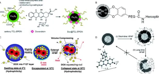
Figure 9 sums up the different options of functionalization of MNPs in terms of targeted drug delivery and controlled release.
Figure 9. Functionalization of MNPs for therapeutic use. The schematic figure shows: Doxorrubicin loaded MNP (A), Herceptin bound to a MNP (B), encapsulated DOX (C) and controlled release upon HFMF (D). Modified from ref. 34.