1
PREPARATION AND CHARACTERIZATION
OF COMPOSITE MATERIALS FOR MEDICAL
APPLICATIONS
MASTER THESIS
CHEMICAL ENGINEERING
2016/2017
SARA ORTEGA ARROYO
Under the guidance of
Assoc. Professor Agnieszka Sobczak-Kupiec, PhD
CRACOW UNIVERSITY OF TECHNOLOGY
2
INDEX
I. Introduction... 3
II. Purpose and scope ... 4
III. Theoretical part ... 5
3.1. Biomaterials ... 5 3.1.1. Definition of biomaterials ... 5 3.1.2. Classification of biomaterials ... 6 3.1.3. Applications of biomaterials ... 9 3.2. Hydroxyapatite... 10 3.2.1. Characteristics of hydroxyapatite ... 10
3.2.2. Preparation methods of hydroxyapatite ... 11
3.2.3. Biomedical applications of hydroxyapatite ... 14
3.3. Drug delivery systems ... 15
3.3.1. Definition and examples ... 15
3.3.2. Hydroxyapatite as a drug carrier ... 19
IV. Experimental part ... 21
4.1. Schematic diagram of work ... 21
4.2. Materials ... 22
4.3. Hydroxyapatite synthesis ... 23
4.4. Characterization of hydroxyapatite ... 24
4.4.1. Ca/P molar ratio ... 24
4.4.2. FT-IR ... 25
4.4.3. XRD ... 27
4.4.4. UV-VIS ... 28
4.5. Preparation of simulated body fluids ... 29
4.6. Study of infusion/effusion of active substance to/from hydroxyapatite ... 31
V. Conclusions... 36
VI. Abstract ... 37
3
I.
Introduction
Biomaterials are materials designed to act with biological systems in order to evaluate, treat, increase or replace some tissue, organ or function of the body. Such functions may be relatively passive, like being used for a heart valve, or may be bioactive with a more interactive functionality such as hydroxyapatite coated hip implants. Biomaterials are also used every day in dental applications, surgery, and drug delivery [1].
The biomaterials industry will in the near future be a potential market due to the progressive aging of the population. It is estimated that in the near future, one in every two women and one in five men over 65, will suffer some type of fracture, so that the market for joint replacement products will grow between 5 and 10 per cent annually in the coming decades [1].
Currently, about 2,700 types of medical devices have been developed that are considered biomaterials. In the United States, the world's leading power in the sector, 3 million prostheses are implanted annually, generating a market of more than 100 million dollars. In Europe, around 40,000 cardiac prostheses and 275,000 hip prostheses are implanted annually [1].
Drug delivery systems are technologies designed for the targeted delivery and / or controlled release of therapeutic agents [2]. Drugs have been used for a long time to improve health and lengthen life. The practice of drug administration has changed dramatically in recent decades and even greater changes are anticipated in the near future. Biomedical engineers have contributed significantly to our understanding of the physiological obstacles to efficient drug delivery, such as transport in the circulatory system and movement of drugs through cells and tissues; they have also contributed to the development of several new drug delivery models that are already used in clinical practice [2].
However, with all this progress, many drugs, including those that have been discovered by the most advanced strategies in molecular biology, have unacceptable side effects due to the drug's interaction with healthy tissues that are not the intended site of the drug. Side effects limit the ability to design optimal drugs for many diseases such as cancer, neurodegenerative diseases and infectious diseases. Drug delivery systems control the rate of drug release and the place in the body where it is released and some systems can control both [2].
4
II.
Purpose and scope
The main objective of this project was the preparation of synthetic hydroxyapatite (HA, HAp), physicochemical characterization of the obtained material and initial evaluation of the drug delivery potential.
The work includes the following steps:
- Synthesis of hydroxyapatite by the simple wet chemical method using varied substrates;
- Characterization of the obtained material using different characterization techniques:
o Determination of the calcium and phosphorous molar ratio (Ca/P), o Fourier transform infrared spectroscopy (FT-IR),
o Ultraviolet-visible spectroscopy (UV-Vis), o X-ray powder diffraction (XRD);
- Drug delivery tests: study of infusion/effusion of active substance (clindamycin) to/from hydroxyapatite in the form of pressed tablets.
5
III. Theoretical part
3.1. Biomaterials
3.1.1. Definition of biomaterials
Biomaterial can be described as a combination of substances originating from natural, inorganic or organic materials that is biocompatible in exactly or partially contact with the body for healing time. They are used to be incorporated or implanted within a living organism to replace or restore some function by remaining in permanent or intermittent contact with body fluids. Biomaterials involve complete or part of a living organism or biomedical device which performs, augments or replacements any natural function [3].
Biomaterial is a nonviable substance used in a medical device intended to interact with biological systems. Their usage within a physiologic medium needs the characteristic features such as efficient and reliable. These characteristic features have provided with a suitable combination of chemical, mechanical, physical and biological properties [3].
In general, it is assumed that the material can not endanger the health or life of the patient. The most important characteristics of an implant material are:
1) Biocompatibility, i.e. the ability of the material to cause an acceptable host response;
2) No toxicity and minimal effects on the immune system, which means that the material must be chemically inert and not cause inflammatory reactions and excessive tissue reaction;
3) Biofunctionality, which is a measure of the ability of a material to take over the function of tissues and organs;
4) Ability of the implant surface to adhere directly to the soft or hard tissue without forming the intermediate layer constructed from a modified tissue [3, 4].
Nowadays, biomaterials are commonly used in various medical devices and systems; synthetic skin; drug delivery systems; tissue cultures; hybrid organs; synthetic blood vessels; artificial hearts; cardiac pacemakers; screws, plates, wires and pins for bone treatments; total artificial joint implants; skull reconstruction; dental and maxillofacial applications [3, 4]. Biomaterials can be derived either from nature or synthesized in
6 the laboratory using a variety of chemical approaches utilizing metallic components, polymers, ceramics or composite materials [5, 6].
3.1.2. Classification of biomaterials
- Classification based on biocompatibility
Biocompatibility is concerned with the acceptance of an artificial implant by the surrounding tissues and by the body as a whole.
a) Bioinert: any material that once placed in the human body has minimal
interaction with its surrounding tissue. Examples of these are stainless steel, titanium, alumina, partially stabilized zirconia, and ultra-high molecular weight polyethylene;
b) Bioactive: refers to a material, which upon being placed within the human
body interacts with the surrounding bone and in some cases, even soft tissue;
c) Bioresorbable: refers to a material that upon placement within the human
body starts to dissolve (resorbed) and slowly replaced by advancing tissue (such as bone) [6, 7, 8].
- Classification based on material source a) Biological: natural origin;
7 Figure 1.Classification based on material source [10]
Based on the application in the medical field biomaterial are classified into [10]:
b.1) Ceramics: The class of ceramics used for repair and replacement of
diseased and damaged parts of musculoskeletal systems are termed bioceramics. They are used in the manufacture of implants that do not have to carry loads. Bioceramics have become a diverse class of biomaterials presently including three basic types:
- Bioinert high strength ceramics, such as Aluminia (Al2O3), Zircona (ZrO2)
and carbon;
- Bioactive ceramics which form direct chemical bonds with bone or even with soft tissue of a living organism, such as bioglass and glass ceramics;
- Various bioresorbable ceramics that actively participate in the metabolic processes of an organism with the predictable results. Calcium phosphate ceramics are categorized as bioresorbable.
Bioceramics became an accepted group of materials for medical applications, mainly for implants in orthopaedics, maxillofacial surgery and for dental implants. [7, 8, 9]
Bioceramics are attractive as biological implants for their biocompatibility. The analysis of the results of animal experiments and the evaluation of clinical follow up studies show that alumina ceramic with high mechanical strength
8 show minimal or no tissue reaction, nontoxic to tissues and blood compatability tests were also satisfactory. Zirconia ceramic revealed its bioinertness and noncytotoxicity. Carbon with similar mechanical properties of bone is an exciting candidate, for it elicits blood compatibility, no tissue reaction and nontoxicity to cells. None of the three-bioinert ceramics exhibited bonding with the bone. However, the bioactivity of the bioinert ceramics can be achieved by forming composites with bioactive ceramics [16].
b.2) Metals: Are used when load is required, as in hip prostheses, for which is
used cobalt (Co) alloys with chromium (Cr), or titanium (Ti) alloys with aluminum (Al) and vanadium (V) [7].
b.3) Polymers: Polymer biomaterials are widely used in clinics, both in surgical
implants and in protective membranes, drug dosing systems or acrylic bone cements [8,9].
b.4) Composites: Composites are engineering materials that contain two or
more physical and/or chemical distinct, properly arranged or distributed constituent materials that have different physical properties with an interface separating them. They consist of mixtures of polymers, metals and ceramics to form materials such as fiberglass, a mixture of glass fibers coated with a
polymeric matrix [7, 8, 9].
In Figure 3 are shown the advantages, disadvantages and some examples of the biomaterials:
9
3.1.3. Applications of biomaterials
Biomaterials are making a breakthrough in many fields of medicine because they are getting better and more compatible with humans [11].
Figure 3.Medical applications of biomaterials
As there is a great variety of biomaterials, they can be used for a wide variety of applications:
- The total hip prosthesis - Knee Implant - Valves of heart - Dental implants - Spine - Tissue Engineering - Degradable sutures
- Gastrointestinal segments & Tracheal tubes - Breast implants
- Bone cement & Intra ocular lenses - Diagnostic and in vivo Imaging - Drug delivery [11, 12].
10
3.2. Hydroxyapatite
3.2.1. Characteristics of hydroxyapatite
Hydroxyapatite, also called hydroxylapatite (HA, HAp), is the hydroxyl endmember of the complex apatite group. Is a naturally occurring mineral form of calcium apatite with the formula Ca5(PO4)3(OH), but it is usually written
Ca10(PO4)6(OH)2. It represents a deposit of 99% of body calcium and 80% of total
phosphorus. Pure hydroxylapatite powder is white [13].
An important characteristic of hydroxyapatite is its stability when compared to other calcium phosphates. Thermodynamically, hydroxyapatite is the most stable calcium phosphate compound under physiological conditions as temperature, pH and composition of the body fluids [13].
HAp structure is formed by a tetrahedral arrangement of phosphate (PO43-), which
constitute the "skeleton" of the unit cell. Two of the oxygens are aligned with the c axis and the other two are in a horizontal plane. Within the unit cell, phosphates are divided into two layers, with heights of 1/4 and 3/4, respectively, resulting in the formation of two types of channels along the c axis, denoted by A and B [13].
Figure 4.Crystalline structure of hydroxyapatite [13]
The monoclinic form of HAp is more ordered and thermodynamically stable and is formed at high temperatures, but have never had evidence of its presence in calcified tissues. Despite being taken to the stoichiometric hydroxyapatite as a model, it is noteworthy that hydroxyapatites produced biologically are much more complicated, they are not stoichiometric, have an atomic ratio Ca/P <1.67 and does not contain only ions and radicals of the HAp but also traces of CO3, Mg, Na, F and Cl. These amounts
11 vary according at the specific type of tissue, which is related to the properties and bioactivity of it [13, 14].
One aspect that is important to note is that, the closer the value of Ca/P to 1.67, the greater the stability of the material inside the human body as they tend to be inert, and on the other hand, if this value decreases (deficient HAp), the better the bioactivity [13, 14].
Another aspect we must consider is the degree of crystallinity. It has been observed that the crystallinity in the tissues for the tooth enamel is very high, while in the cases corresponding to dentin and bone, it is very poor. This means that the reactivity depends on the degree of crystallinity, since the reactivity in dentin and bone is higher than in tooth enamel [13, 14].
In order to manufacture articles made of hydroxyapatite, it is necessary to take into account, besides the above, that differences in structure and composition of apatites also depend on the different processing techniques, as well as temperature and atmosphere in which are made [13, 14].
3.2.2. Preparation methods of hydroxyapatite
Due to the existence of a myriad number of phosphate compounds the calcium phosphate system is highly complex. This is further complicated by the sensitive stability of phosphates to minor changes in: composition, pH, and reaction conditions (e.g. temperature). It is important to note that the purity and particle characteristics of the final synthesized powder can affect the bioactivity, mechanical and biological dissolution properties. [15] These characteristics ultimately determine the medical application of the material, thus making it imperative to develop a synthesis method that enables the control of: crystal morphology, chemical composition, crystallinity, particle size distribution, and agglomeration. A variety of synthesis techniques have been published for HAp [13]:
- Precipitation technique: is the most popular and widely researched technique for synthesis of HAp. This technique is also called as wet precipitation or chemical precipitation or aqueous precipitation. Because, relatively large amount of HAp can be produced by precipitation technique in absence of organic solvents at a reasonable cost. Preparation of the synthetic HA is based on precipitation of the sludge from the aqueous solution. The formation of HA involves interaction of the calcium acetate and the phosphorus salts. The wet precipitation was carried out at ambient or elevated temperature according to an example following reaction [17]:
12 10 Ca(CH3COO)2 + 6 Na2HPO4 + 2 H2O → Ca10(PO4)6OH2 + 12 NaCH3COO +
8 CH3COOH
- Sol-gel approach: recently, sol-gel techniques have attracted much attention due to the inherent associated advantages of this method; homogeneous molecular mixing, low processing temperatures (<400ºC), and the ability to generate nano-sized particles. However, the energy saving gained from the low temperatures used is offset by the high cost of reactants. In comparison to other low temperature methods, sol-gel techniques have very limited scalability due to the sensitivity of the process [13, 15].
The first stage of this method is to form a “sol”; a dispersion of solid particles, otherwise known as colloids, in a liquid. Precursor materials are mechanically mixed in a solvent at a pH that prevents precipitation. Typically, metal alkoxides and metal salts are used. Hydrolysis and polycondensation reactions occur to link these monomer units and form M-O-M bonds within the sol causing the viscosity to increase; this process is termed gelation. The result is a “gel”, which can be defined as a diphasic system consisting of a solid and interstitial liquid phase. The next step is to remove the liquid phase via a drying process; this is usually accompanied by a significant amount of shrinkage and densification. To avoid cracking in a target 3-D monolith structure it may be necessary to age the gel before drying. Alternatively, cracking can be accommodated if the goal is to create a fine sol-gel powder for further processing as a granulate or shaped product. Lastly, a material specific sintering protocol is employed; in practise this step can be time-consuming [15].
- Hydro/solvo- thermal: Hydro- and solvo-thermal processing involves the use of a solvent (with precursor soluble ions), which is heated in a sealed vessel. In the case of hydrothermal synthesis the solvent used is water [13]. The temperature of the solvent can be brought above boiling point as the autogenous pressure within the vessel exceeds the ambient pressure. The change in solvent and reactant properties (e.g. solubility) at these elevated temperature mean that experimental variables can be controlled to a higher degree. This makes the reaction more predictable as crystal nucleation, growth, and ageing can be regulated [13, 15].
- Solid state: This procedure relies on the solid diffusion of ions amongst powder raw materials and thus requires relatively inefficient high temperature processing (< 1250°C) to initiate the reaction. Even though the technique is comparatively simple there are a number of processes involved. To ensure homogeneity and sufficiently small particle sizes, starting materials must firstly be ball milled for approximately 16 hours. Commonly calcium and phosphate sources are mixed with additives (e.g. silicon dioxide, alumina, and hydrofluoric acid), a binder (e.g. PVA) and an organic vehicle (e.g. acetone) to form slurry before milling. The slurry must then be dried. Pellets can then be formed from
13 the resulting powder using either hot or cold pressing at pressures of up to 135 MPa. Finally, sintering is performed at up to 1250°C to crystallize the product [15, 17].
- Self-propagating combustion synthesis: Self propagating combustion synthesis (SPCS) has recently been proposed as a viable simple, quick energy saving synthesis option for HA. The success of SPCS is dependent on the intimate mixing of constituents in an aqueous medium: calcium and phosphate sources, a suitable fuel, and an oxidizing agent. The solution temperature is then increased causing a vigorous exothermic reaction to occur between the fuel and oxidizer; the gaseous products of this reaction spontaneously combust. This gives rise to a very high local temperature that causes the formation of a solid calcium phosphate powder. The heat energy evolved from the reaction is dependent on the fuel used as well as the ratio of fuel to oxidizer. These parameters can be varied, resulting in the formation of different calcium phosphate phases and/or particle morphologies. Furthermore, depending on the fuel used the product formed may be either crystalline or amorphous. Both require a calcination step; to remove organic residues and crystallize the phase formed, respectively [15, 16].
- Emulsion/Micro-emulsion: Emulsions are heterogeneous mixtures of at least one immiscible liquid dispersed in another in the form of droplets. These systems are often described as either water-in-oil (W/O) or oil-in-water (O/W). Depending on the size of the aqueous drops, this technique can be referred to as emulsion or microemulsion.
A reaction takes place when two different droplets containing the reactants collide with each other. Nano- and micro-sized particles of HAp can be formed via microemulsion and emulsion, respectively. Furthermore, microemulsion techniques have been reported to reduce particle agglomeration of HAp. One specific application of this synthesis process is the formation of porous spherical HAp granules for drug delivery [18].
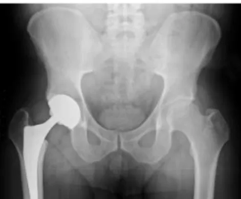
Figure 5.Calcium hydroxyapatite used to encourage and artificial hip to fuse with the bone
14 Table 1 shows detailed comparison of mentioned HAp preparation methods.
Table 1.Comparison of HAp preparation methods [15]
3.2.3. Biomedical applications of hydroxyapatite
Hydroxyapatite can be found in teeth and bones within the human body. Thus, it is commonly used as a filler to replace amputated bone or as a coating to promote bone ingrowth into prosthetic implants. Many modern implants, e.g. hip replacements, dental implants and bone conduction implants are coated with hydroxyapatite [13].
- Dental applications: The dental enamel is a cover formed by hydroxyapatite that covers the dental pieces, when this structure suffers some damage the body does not have the capacity to regenerate.
- Prosthesis applications
- Ophthalmology applications: Hydroxyapatite is used in ophthalmology as a material in orbital implants; in those cases where a person suffers the total or partial loss of the eyeball, an ocular implant can be placed instead.
15
3.3. Drug delivery systems
3.3.1. Definition and examplesDrug delivery systems (DDS) are engineered technologies for the targeted delivery and/or controlled release of therapeutic agents. A DDS is defined as a formulation or a device that enables the introduction of a therapeutic substance in the body and improves its efficacy and safety by controlling the rate, time and place of release of drugs in the body [20].
This process includes the administration of the therapeutic product, the release of the active ingredients by the product, and the subsequent transport of the active ingredients across the biological membranes to the site of action [21].
Advantages of advanced drug delivery systems over traditional systems are: - The ability to deliver a drug more selectively to a specific site; - Easier, more accurate, less frequent dosing;
- Decreased variability in systemic drug concentrations;
- Absorption that is more consistent with the site and mechanism of action; and - Reductions in toxic metabolites.
- Oral drug delivery systems: formulations range from simple tablets to newer modified control release tablets. It involves use of various polymers and hydrogel based formulations [23].
16 Figure 7.Oral drug delivery system [23]
- Injection based drug delivery systems: provide fast systemic effects bypassing first-pass metabolism. Drugs can be administered in unconscious or comatose patients. Drugs having short half-life can be infused continuously [23].
Figure 8.Injection based drug delivery systems [23]
- Transdermal drug delivery systems: adhesive patches containing the drug are applied on the skin. The drug crosses the skin surface by diffusion by percutaneous absorption and goes into systemic circulation [23].
17 - Carrier based drug delivery system: Colloidal drug carrier systems such as micellar solutions, vesicle and liquid crystal dispersions, as well as nanoparticle dispersions consisting of small particles of 10–400 nm diameter show great promise as drug delivery systems. When developing these formulations, the goal is to obtain systems with optimized drug loading and release properties, long shelf-life and low toxicity. The incorporated drug participates in the microstructure of the system, and may even influence it due to molecular interactions, especially if the drug possesses amphiphilic and/or mesogenic properties[22].
Figure 10.Pharmaceutical carriers [23]
Current research on drug delivery systems can be described in four broad categories: routes of delivery, delivery vehicles, cargo and targeting strategies.
- Routes of delivery: Medications can be taken in a variety of ways—by swallowing, by inhalation, by absorption through the skin, or by intravenous injection. Each method has advantages and disadvantages, and not all methods can be used for every medication. Improving current delivery methods or designing new ones can enhance the use of existing medications [22].
18 Figure 11.Routes of drug delivery [22]
- Delivery vehicles: Biotechnology advances are leading to improved medications that can target diseases more effectively and precisely. Researchers have begun to reformulate drugs so they may be more safely used in specific conditions [22, 23].
- Cargo: Perhaps the most obvious route to improving disease treatment would be to focus on the medications themselves. In addition to drugs and novel vaccines, researchers are also exploring the use of genes, proteins, and stem cells as treatments [22].
- Targeting strategies: Working backwards is sometimes an effective way to solve a problem. In drug delivery research, this means starting with a delivery method [24].
19
3.3.2. Hydroxyapatite as a drug carrier
Currently, nanosized materials are extensively used in design of the optical devices, catalysts, biosensors, imaging agent, drugs and gene delivery, etc. There are a large number of nanoparticles including gold nanoparticles, polymeric nanoparticles, quantum dots, bioceramic based nanoparticles and so on that are applied as carriers in the drug delivery systems. Definitely, dominant physicochemical properties of these materials such as small size and high surface to volume ratio lead to improvement of their effectiveness as a suitable carrier in drug and gene delivery [13-15].
In the recent decades, the production of bioceramics with nanostructures has attracted much attention for biomedical applications. Hydroxyapatite (HAp) is one of the attractive bioceramics which is widely used in various fields of science such as tissue engineering, drug delivery systems and chromatographic purification [13-15] Hap can incorporate the drug molecules either physically or chemically so that the drug retains intact until it reaches to the target site. It could also gradually degrade and then deliver the drug in a controlled manner over time. So therefore, this bioceramic is an excellent candidate for targeted drug delivery and a promising bio scaffold in tissue engineering [13-15].
An important technique to treat disease would be to use controlled drug- release vectors to ensure that drugs are specifically released at a constant rate. Hydroxyapatite is the most important inorganic constituent of human teeth and bones [1,2], and has attracted considerable attention in various biomedical areas such as gene and drug delivery [3,4], bone repair and tissue engineering owing to its excellent biocompatibility, biodegradability and bioactivity [5,6]. Recently, many studies have been carried out to investigate using hydroxyapatite materials as a controlled drug release system [30].
Figure 13 shows a schematic illustration of pharmaceutical applications of hydroxyapatite:
20 Figure 13.Schematic illustration of pharmaceutical applications of hydroxyapatite [18]
21
IV. Experimental part
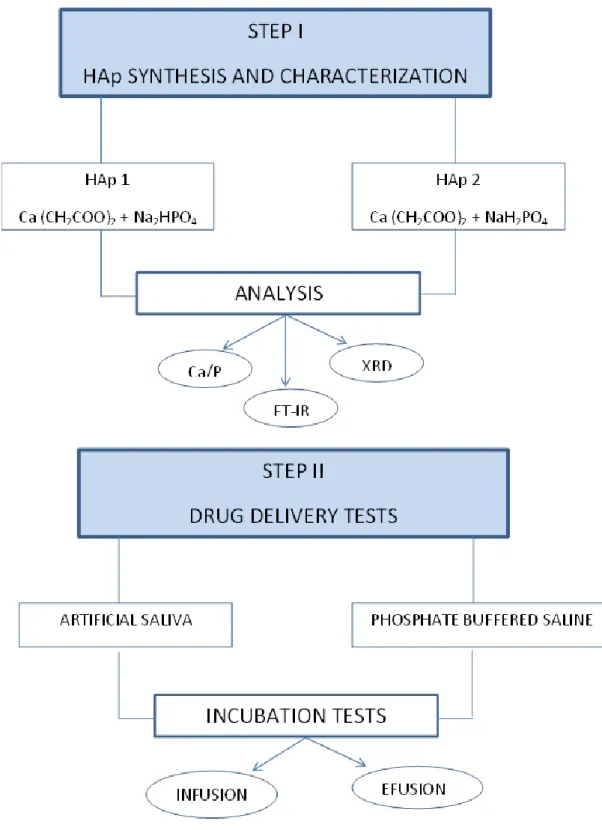
4.1. Schematic diagram of work
The steps to follow in the laboratory during this project are shown in the diagram below:
22
4.2. Materials
The materials used in the laboratory were the following ones:
- Sodium hydrogen phosphate, Na2HPO4: purchased by the company POCh S.A.
- Sodium dihydrogen phosphate, NaH2PO4: purchased by the company POCh S.A.
- Calcium Acetate, Ca(CH3COO)2: purchased by the company POCh S.A.
- Distilled water: used for preparing the solutions.
- Clindamycin hydrochloride: it contains not less than 84.0 per cent and not more than 93.0 per cent of clindamycin calculated with reference to the anhydrous substance. It contains a variable quantity of water. Is a white or almost white, crystalline powder, very soluble in water and very slightly soluble in alcohol [25].
Figure 15.Skeletal structure of clindamycin
Clindamycin is used primarily to treat anaerobic infections caused by susceptible anaerobic bacteria, including dental infections, and infections of the respiratory tract, skin, and soft tissue, and peritonitis. In people with hypersensitivity to penicillins, clindamycin may be used to treat infections caused by susceptible aerobic bacteria, as well. It is also used to treat bone and joint infections, particularly those caused by Staphylococcus aureus. Topical application of clindamycin phosphate can be used to treat mild to moderate acne [26].
23
4.3. Hydroxyapatite synthesis
For the synthesis of hydroxyapatite, several steps were followed. First, three different solutions were elaborated:
- 1000 ml of 0.2 M Na2HPO4
- 1000 ml of 0.2 M NaH2PO4
- 2000 ml of 0.08 M Ca(CH3COO)2
Then, the synthesis of hydroxyapatite with Na2HPO4 was carried out to obtain HAp 1:
The amount of 650 cm3 of the doubly distilled water and 100 cm3 of the 0.2 M Na2HPO4 was poured into round bottomed flask, and repeated this for three round
bottomed flasks. Several pieces of ceramic were inserted and the equipment was assembled as shown in the Figure 15. The solutions were heated to boiling. [17]
Figure 16.Equipment for synthesis
Then, 250 cm3 of the 0.08M Ca(CH3COO)2 was added dropwise to the solution with the
speed of 1 drop min keeping the temperature at boiling point. Subsequently, disconnect the equipment and the attained suspension was magnetically stirred and the mixture was aged for 24 hours. Ultimately, the precipitate was separated from the solution by filtration and dried at 105 ° C. [17]
24 Figure 17.Equipment for vacuum filtration
The final product is shown in Figure 17. This process was repeated 3 times to obtain the required amount of HAp for the project and was also repeated with NaH2PO4
product to obtain HAp 2. [17]
Figure 18.HAp final obtained product
4.4. Characterization of hydroxyapatite (Ca/P, FT-IR, XRD)
4.4.1. Ca/P molar ratioThe determination of the calcium content in Ca(OH)2 is based on polish standard
denoted as PN-97/R-64803 [17]. The principle of the method consists in dissolving the sample in nitric acid, precipitating the phosphates and then determining the calcium content by the complexometric titration of disodium edetate (EDTA) in the presence of a mixture of calcein and thymolphelin [17].
The procedure consists in dissolving 0.1 g of mineralized sample in 10 ml of 3 M nitric acid. Subsequently, the mixture was boiled for 10 min. 20 ml of doubly distilled water was added to the primary solution and boiled for another 5 min. The attained solution was transferred to the 100 ml volumetric flask and mixed with 6.25 of 0.4 M bismuth
25 nitrate. The remaining volume of flask was filled with doubly distilled water to attain 100 ml and then filtrated with double paper. 25 ml of the solution was retrieved from the flask and mixed with 25 ml of distilled water, 3 ml of 25% triethanoloamine and 20% of potassium hydroxide to reach finally pH from the range of 5-7. The solution was titrated with 0.025 M EDTA with the presence of calcein and thymolphtalein as indicators [17].
The determination of the phosphorus in the samples was achieved by means of differential photometric procedure. The samples prior to analysis were prepared according to polish standard termed as PN-80/C-87015 [17]. At the primary stage, 0.02 g of mineralized HAp powder was dissolved in 2.5 ml of concentrated hydrochloric acid and 7.5 ml of concentrated nitric acid at the boiling point. Subsequently, the attained solution was transferred to 100 ml volumetric flask and the remaining volume was filled in with distilled water. Accordingly, 10 ml of the mixture was transferred to another 50 ml volumetric flask and mixed with 20 ml of solution termed as D and the remaining volume of flask was filled in with distilled water. The whole mixture was aged for 15 min. Consequently, the spectrophotometric measurement was performed employing 430 nm wavelength.
This procedure was repeated for the two HAps obtained, with three measurements for each material.
The reaction for the synthesis of hydroxyapatite is:
10 Ca(CH3COO)2 + 6 Na2HPO4 + 2 H2O → Ca10(PO4)6OH2 + 12 NaCH3COO + 8 CH3COOH
The molar ratio between Ca/P of hydroxyapatite should be 1.67.
The results for the spectrophotometric measurement give the following Ca/P molar ratio: 1.58 for HAp1 and 1.64 for HAp2. These values are extremely close for the
theoretical value but are not the same. The Ca/P molar ratio obtained may differ from the theoretical because in some cases the load deficit caused by the substitution of the PO43- groups is compensated by the elimination of the Ca2+ ions from the crystalline
structure, resulting in the reduction of the Ca/P molar ratio.
4.4.2. FT-IR
Infrared spectroscopy (IR spectroscopy or Vibrational Spectroscopy) involves the interaction of infrared radiation with matter.
It covers a range of techniques, mostly based on absorption spectroscopy. As with all spectroscopic techniques, it can be used to identify and study chemicals. Sample may be solid, liquid, or gas. The method or technique of infrared spectroscopy is conducted
26 with an instrument called an infrared spectrometer (or spectrophotometer) to produce an infrared spectrum. An IR spectrum is essentially a graph of infrared light absorbance (or transmittance) on the vertical axis vs. frequency or wavelength on the horizontal axis. Typical units of frequency used in IR spectra are reciprocal centimeters (sometimes called wave numbers), with the symbol cm−1 [27].
A common laboratory instrument that uses this technique is a Fourier transform infrared (FTIR) spectrometer. Fourier transform infrared (FTIR) spectroscopy is a measurement technique that allows one to record infrared spectra. Infrared light is guided through an interferometer and then through the sample (or vice versa). A moving mirror inside the apparatus alters the distribution of infrared light that passes through the interferometer. The signal directly recorded, called an "interferogram", represents light output as a function of mirror position. A data-processing technique called Fourier transform turns this raw data into the desired result (the sample's spectrum): Light output as a function of infrared wavelength (or equivalently, wavenumber). The sample's spectrum is always compared to a reference [27, 29]. The FTIR spectra shown in Figure 20 were used to identify the functional groups of the samples. According to the literature reports, the most characteristic vibrational modes in the FTIR spectrum of synthesized HA are PO43-, OH-, CO32- and HPO42-, respectively
that characterize non-stoichiometric HAp. Notably, PO43- groups form two intensive
bands at approximately 600-500 and 950-850 cm-1 for both HAps. HPO42- group form a
weak peak at 800-830 cm-1 which can be seen remarkably in HAp2. Finally hydroxyl
group OH- appears at 2714 cm-1 and 650-600 for HAp1 and at approximately 2800 cm-1
and 650-600 for HAp2, with a greater slope for HAp1.
27 Figure 20.FT-IR absorbance spectra of hydroxyapatite formed using various substrates
4.4.3. XRD
X-ray powder diffraction (XRD) is a rapid analytical technique primarily used for phase identification of a crystalline material and can provide information on unit cell dimensions. The analyzed material is finely ground, homogenized, and average bulk composition is determined [28].
X-ray diffractometers consist of three basic elements: an X-ray tube, a sample holder, and an X-ray detector. X-rays are generated in a cathode ray tube by heating a filament to produce electrons, accelerating the electrons toward a target by applying a voltage, and bombarding the target material with electrons. When electrons have sufficient energy to dislodge inner shell electrons of the target material, characteristic X-ray spectra are produced [28].
These X-rays are collimated and directed onto the sample. As the sample and detector are rotated, the intensity of the reflected X-rays is recorded. When the geometry of the incident X-rays impinging the sample satisfies the Bragg Equation, constructive interference occurs and a peak in intensity occurs. A detector records and processes this X-ray signal and converts the signal to a count rate which is then output to a device such as a printer or computer monitor [28].
X-ray powder diffraction is most widely used for the identification of unknown crystalline materials (e.g. minerals, inorganic compounds). Determination of unknown
28 solids is critical to studies in geology, environmental science, material science, engineering and biology [28].
Other applications include:
Characterization of crystalline materials.
Identification of fine-grained minerals such as clays and mixed layer clays that are difficult to determine optically.
Determination of unit cell dimensions.
Measurement of sample purity [28].
Figure 21 shows the XRD pattern of the typical samples, which are consisted of a well-crystalline phase with hexagonal-structured HAps. No peaks from impurities such as CaHPO4 were observed.
Figure 21.XRD patterns of hydroxyapatite formed using various substrates
4.4.4. UV-VIS
Clindamycin phosphate inhibits bacterial protein synthesis by binding to the 50S ribosomal subunit. Clindamycin also affects the peptide chain initiation step in protein synthesis.
29 Chemically it is methyl 7 -chloro-6,7,8-trideoxy-6-(1-methyl-trans-4-propyl-L-2-pyrrolidinecarboxamido)-1-thio-L-threo-α-D-galacto-octopyranoside-2-dihydrogen phosphate.
Several spectrophotometric, HPLC,HPTLC and LC-MS methods have been reported so far for determination of Clindamycin phosphate alone and its combination with other drugs.
Therefore, it was thought worthwhile to develop simple, accurate and reliable spectrophotometric method for estimation of Clindamycin phosphate in bulk dosage form using water and phosphate buffer of saline pH 6.75 as a solvent. All the chemicals used were of analytical grade. Spectral and absorbance measurement were made on Shimadzu UV-Visible Spectrophotometer.
The most striking feature of this method is its simplicity and rapidity, non- requiring- consuming sample preparations such as extraction of solvents, heating, degassing which are needed for HPLC procedure. These are new and novel methods and can be employed for routine analysis in quality control analysis. This method gives accurate and precise results for determination of Clindamycin phosphate.
In the present investigation we have developed a simple precise and accurate UV Spectrophotometric method for the determination of Clindamycin hydrochloride. The method was developed using in both water and phosphate buffer saline solution using pH 6.75. The detection was carried out at 210nm. The developed method was found to be appropriate for the determination of Clindamycin hydrochloride in bulk and in dosage forms. Beer’s law was found to be obeyed in the range of 5- 30 μg/ml in distilled water and in the range of 5-30μg/ml in phosphate buffer saline pH 6.75). The stability of Clindamycin hydrochloride and was done in both distilled water and in phosphate buffersaline pH 6.75 and was ascertained over a period of 72 hrs. Analysis of variance (ANOVA) of the mean absorbance values of both the solutions of different concentrations at various time intervals revealed that there was no significant difference between the readings [31].
4.5. Preparation of simulated body fluids
For next studies in this project of infusion/effusion of active substance to/from hydroxyapatite some simulated body fluids are required.
The first one is artificial saliva. 1L of this will be required. Human saliva is a complex fluid comprising 99.5% water, the remainder being organic compounds, inorganic salts and microorganisms. Artificial saliva solution was used in this study and was prepared according to the following Table 2:
30 Table2. Composition for artificial saliva
Component Quantity (g) NaCl 0.8 KCl 0.8 CaCl2•H2O 1.3682 NaH2PO4•H2O 1.3566 Na2S•9H2O 0.01
For this preparation, the ingredients have to be mixed in the order shown in the table. The next ingredient must to be added after complete dissolution of the previous one and pH should be in the range 5-7.
Another substance that will be necessary for the studies is phosphate buffered saline. Phosphate-buffered saline (abbreviated PBS) is a buffer solution commonly used in biological research. It is a water-based salt solution containing phosphate, sodium and, in some formulations, potassium chloride and potassium dihydrogen phosphate. PBS has many uses because it is isotonic and non-toxic to most cells. These uses include substance dilution and cell container rinsing. PBS with EDTA is also used to disengage attached and clumped cells.
There are many different ways to prepare PBS solutions. The most common composition is shown in Table 3:
Table 3.Most common composition of PBS
Salt Concentration (mmol/L) Concentration (g/L)
NaCl 137 8.0
KCl 2.7 0.2
Na2HPO4 10 1.42
KH2PO4 1.8 0.24
It is needed to start with 800 mL of distilled water to dissolve all salts. Then, the pH has to be adjusted to 7.4 with HCl. Finally, distilled water is added to a total volume of 1 liter.
31
4.6. Study of infusion/effusion of active substance to/from
hydroxyapatite
For this study, different pills were made:
Firstly, 10 pills of each HAp were prepared for the infusion test. For this, 0.5 g of each sample was weight and pills were made in a manual press machine.
For the effusion test, pills were made of 0.48 g of HAp and 0.02 of clindamycin.
Figure 24.Pills for infusion/effusion test
32 To carry on with the test, different solutions were prepared.
For the infusion test, three solutions were made with 20 ml of artificial saliva and another three with 20 ml of PBS. To these solutions were added 10 mg of clindamycin and one pill of HAp 1 in each can. Another six samples were made for HAp 2 pills.
Figure 25.Solutions for infusion test
For the effusion test, three solutions were made with 20 ml of artificial saliva and another three with 20 ml of PBS. To these solutions were added one pill of HAp 1 in each can. Another six samples were made for HAp 2 pills.
With the infusion test, it was checked the possibility to incorporate the clindamycin into the HAps synthesized. With the effusion test, it was checked the possibility of released of clindamycin from the HAps synthesized.
All samples were incubated for different times and the absorbance was measured to calculate the average concentration. In the following Tables are shown the results for the measurements for different times.
Figure 26 was used to obtain the clindamycin concentration for artificial saliva and PBS solutions.
33 Table 4.Infusion test in phosphate buffered saline – incubation of pure HAp fittings in fluid containing 10 mg of clindamycin
Table 5.Infusion test in artificial saliva – incubation of pure HAp fittings in fluid containing 10 mg of clindamycin
INFUSION TEST in ARTIFICIAL SALIVA 30 min incubation
Sample Average absorbance Average drug content in solution [mg/20 ml]
Average drug content incorporated in sample [%] HAp1 2,38 9,43 0 HAp2 2,37 9,38 0,45 control 2,35 9,21 - 90 min incubation HAp1 2,40 9,46 0 HAp2 2,38 9,62 0 control 2,35 9,21 -
INFUSION TEST in PHOSPHATE BUFFERED SALINE 30 min incubation
Sample Average absorbance
Average drug content in solution [mg/20 ml]
Average drug content incorporated in sample [%] HAp1 2,35 9,10 0,15 HAp2 2,39 9,35 0,74 control 2,40 9,42 - 90 min incubation HAp1 2,41 9,46 0 HAp2 2,45 9,62 0 control 2,40 9,42 -
34 Table 6.Effusion test in artificial saliva – incubation of HAp fittings containing 10 mg of drug in pure fluid
EFFUSION TEST in ARTIFICIAL SALIVA 30 min incubation
Sample Average absorbance Average drug content in solution [mg/20 ml]
Average drug content released from sample [%]
HAp1 0,19 0,37 3,66 HAp2 0,21 0,44 4,36 control 0 - - 90 min incubation HAp1 1,19 4,44 44,4 HAp2 1,82 7,04 70,4 control 0 - -
Table 7.Effusion test in phosphate buffered saline – incubation of HAp fittings containing 10 mg of drug in pure fluid
EFFUSION TEST in PHOSPHATE BUFFERED SALINE 30 min incubation
Sample Average absorbance Average drug content in solution [mg/20 ml]
Average drug content released from sample [%]
HAp1 0,26 0,69 6,88 HAp2 0,40 1,25 12,51 control 0 - - 90 min incubation HAp1 1,46 5,60 56,0 HAp2 1,75 6,76 67,6 control 0 - -
35 Figure 27.The calibration curve of clindamycin dissolved in selected simulated body fluids [mg of drug/ml of solution]
As it can be shown in Tables 4, 5, 6 and 7, for the infusion test the average drug content incorporated in the sample is very small and for incubation time of 90 minutes the drug content is nil.
For the effusion test at first the average drug content released from the sample is very low, but as the incubation time increases, the released average drug content increases. This means that HAps synthesized could be used as drug carrier.
36
V.
Conclusions
Pure HAp was obtained by a wet synthesis method. It can be corroborate with the FTIR and XRD methods. FTIR spectra were used to identify the functional groups of the samples. The most characteristic vibrational modes in the FTIR spectrum of synthesized HAp are PO43-, OH-, CO32- and HPO42-. XRD showed that the samples were
consisted of a well-crystalline phase with hexagonal-structured Haps, where no peaks from impurities such as CaHPO4 were observed. Furthermore, the spectrophotometric
measurement was used to calculate Ca/P molar ratio. The obtained values were extremely close to the theoretical value; reason why it is possible to affirm that obtained HAp is pure.
Infusion and effusion studies were used to check if the HAps synthesized are good candidates for drug delivery system. Infusion study was used to check the possibility to incorporate a drug used in mouth inflammation into the biomaterial synthesized and effusion study was used to check the possibility of release of drug to body from the biomaterial synthesized.
The result for these studies was that the average drug content incorporated in the sample is very small and as time increase the average drug content incorporated is nil. Also, it was showed that the average drug content released from the sample increases as incubation time increases.
For all this, it can be concluded that HAps synthesized in this project can be used as drug carrier.
37
VI. Abstract
A biomaterial is defined as a substance that has been engineered to take a form which, alone or as part of a complex system, is used to direct, by control of interactions with components of living systems, the course of any therapeutic or diagnostic procedure. Biomaterials can be used for a wide variety of medical applications.
Hydroxyapatite, HAp, was synthesized and submitted to some studies to check if it is a pure biomaterial and if can be used in drug delivery systems.
The determination of the calcium content in Ca(OH)2 consists in dissolving the sample
in nitric acid, precipitating the phosphates and then determining the calcium content by the complexometric titration of disodium edetate (EDTA) in the presence of a mixture of calcein and thymolphelin. The determination of the phosphorus in the samples was achieved by means of differential photometric procedure.
The crystal phase and molecular structure of the samples were characterized by XRD and FTIR.
A drug delivery system is defined as a formulation or a device that enables the introduction of a therapeutic substance in the body and improves its efficacy and safety by controlling the rate, time and place of release of drugs in the body. Clindamycin was used as a drug to check if hydroxyapatite can be used as a drug carrier.
Spectrophotometric method (UV-VIS) was used for the determination of Clindamycin hydrochloride. Hydroxyapatite and clindamycin were submitted to infusion and effusion studies with body fluids to check the possibility of release drug in simulate body fluids or incorporation of the drug to the biomaterial from the fluid, which affirms if it can be an application in drug delivery system.
38
VII. References
[1] Schieker M, Seitz H, Drosse I, Seitz S, Mutschler W (2006) Biomaterials as scaffold for bone tissue engineering. European Journal of Trauma, 32(2): p. 114-124.
[2] Department of Health & Human Services, October 2016, Spain, Sistemas de Administración de Fármacos: Administración Localizada de Fármacos de Manera Controlada,
https://www.nibib.nih.gov/espanol/temas-cientificos/sistemas-de-administraci%C3%B3n-de-f%C3%A1rmacos-administraci%C3%B3n-localizada-de
[3] Boretos JW, Eden M (1984) Contemporary Biomaterials, Material and Host Response, Clinical Applications, New Technology and Legal Aspects. Noyes Publications, Park Ridge, NJ, pp. 232–233.
[4] Niinomi M (2002) Recent Metallic Materials for Biomedical Applications. Metal. Mater. Transac. A. 33 A: 477-486.
[5] A. Binnaz Hazar Yoruç and B. Cem Şener (2012). Biomaterials, A Roadmap of
Biomedical Engineers and Milestones, Prof. Sadik Kara (Ed.), ISBN: 978-953-51-0609-8, InTech, Available from: http://www.intechopen.com/books/a-roadmap-of-biomedical-engineers-and-milestones/biomaterials
[6] Clickmica, ¿Qué son los biomateriales?, [06/06/2017]
https://clickmica.fundaciondescubre.es/conoce/100-preguntas-100-respuestas/que-son-los-biomateriales/
[7] Ele Uri, Biomaterials, [30/05/2017],
http://www.ele.uri.edu/courses/bme462/handouts/Intro_Biomaterials.pdf
[8] slideshare, Application of biomaterials, [30/05/2017],
https://es.slideshare.net/vishnumenon332/biomaterials-vishnu1
[9] Zom, Biomaterials - Classifications and Behaviour of Different Types of Biomaterials,
http://www.azom.com/article.aspx?ArticleID=2630
[10] Journal of the Chemical Society, Dalton transactions, 2002, Issue 24, Page 4527 to 4754
[11] Polymers as biomaterial, [25/05/207]
https://www.naefrontiers.org/File.aspx?id=44699
[12] BIOMATERIALES: CARACTERÍSTICAS Y APLICACIONES, [25/05/2017],
https://franciscoalavez.wordpress.com/2007/12/19/biomateriales-caracteristicas-y-aplicaciones/
39 [13] Hydroxyapatite-Based Materials: Synthesis and Characterization, [25/05/2017],
https://www.intechopen.com/books/biomedical-engineering-frontiers-and-challenges/hydroxyapatite-based-materials-synthesis-and-characterization
[14] Fluidinova, Hydroxyapatite, [30/05/2017]
http://www.fluidinova.pt/hydroxyapatite-properties-uses-and-applications
[15] Synthesis method of hydroxyapatite, [31/05/2017],
https://www.researchgate.net/profile/Sophie_Cox/publication/281089917_Synthesis_ methods_of_hydroxyapatite/links/55d44a0d08aec1b0429f54a1/Synthesis-methods-of-hydroxyapatite.pdf
[16] Biological Evaluation of Bioceramic Materials - A Review, Trends Biomater. Artif. Organs, Vol 18 (1), pp 9-17 (2004)
[17] Physicochemical characterization of zinc-substituted calcium phosphates, DOROTA WALCZYK, DAGMARA MALINA, MILENA KRÓL, KLAUDIA PLUTA and AGNIESZKA
SOBCZAK-KUPIEC∗ Institute of Inorganic Chemistry and Technology, Cracow University of Technology, 31-155 Kraków, Poland, MS received 18 August 2015; accepted 29 October 2015.
[18] Lim, G. K., et al., Processing of hydroxyapatite via microemulsion and emulsion routes. Biometerials, 1997. 18: p. 1433 – 1439.
[19] Synthetic hydroxyapatite in pharmaceutical applications Joanna Kolmas, Sylwester Krukowski, Aleksandra Laskus, Maria Jurkitewicz, Medical University of Warsaw, Faculty of Pharmacy and Laboratory Medicine, Department of Inorganic and Analytical Chemistry, ul. Banacha 1, 02-097 Warsaw, Poland.
[20] Drug Delivery Systems: Getting Drugs to Their Targets in a Controlled Manner. [04/06/2017], https://www.nibib.nih.gov/science-education/science-topics/drug-delivery-systems-getting-drugs-their-targets-controlled-manner
[21] Drug delivery systems: An updated review [04/06/2017]
https://www.ncbi.nlm.nih.gov/pmc/articles/PMC3465154/
[22] Recent Advances in Novel Drug Delivery Systems,
[06/06/2017],http://www.azonano.com/article.aspx?ArticleID=1538
[23] Novel drug delivery systems [06/06/2017],
https://www.slideshare.net/zionpattres/new-drug-delivery-systems
[24] Drug targeting, [06/07/2017], https://www.slideshare.net/anvitaj1/drug-targeting-42888705
40 [25] Council of Europe, 67075 Strasbourg Cedex, France, 2001, European
pharmacopoeia, 2001, Fourth edition, ISBN: 92-871-4587-3, Page 940. [26] Clindamycin, [09/06/02017], https://en.wikipedia.org/wiki/Clindamycin
[27] Infrared spectroscopy, https://en.wikipedia.org/wiki/Infrared_spectroscopy#FTIR [28] XRD techniques, [09/06/2017]
https://serc.carleton.edu/research_education/geochemsheets/techniques/XRD.html
[29] Hierarchically nanostructured hydroxyapatite: Hydrothermal synthesis,
morphology control, Ming-Guo Ma, Beijing Forestry University, International Journal of Nanomedicine · April 2012
[30] Hydrothermal fabrication of porous hollow hydroxyapatite microspheres for a drug delivery system , Wen Lai , Cen Chen, Xiaoyuan Ren , In-Seop Lee , Guohua Jiang , Xiangdong Kong, Materials Science and Engineering C, 22 January 2017.
[31] UV SPECTROPHOTOMETRIC METHOD DEVELOPMENT FOR ESTIMATION OF CLINDAMYCIN PHOSPHATE IN BULK AND DOSAGE FORM, K.S.Nataraj1,
G.N.V.Surya.Narasimha Raju2, Bevara.Anusha3, IJPBS |Volume 3| Issue 4


![Figure 4.Crystalline structure of hydroxyapatite [13]](https://thumb-us.123doks.com/thumbv2/123dok_es/6844212.281913/10.892.228.661.628.877/figure-crystalline-structure-of-hydroxyapatite.webp)
![Table 1.Comparison of HAp preparation methods [15]](https://thumb-us.123doks.com/thumbv2/123dok_es/6844212.281913/14.892.72.854.202.670/table-comparison-of-hap-preparation-methods.webp)
![Figure 6.Types of drug delivery [23]](https://thumb-us.123doks.com/thumbv2/123dok_es/6844212.281913/15.892.246.705.719.963/figure-types-of-drug-delivery.webp)
![Figure 8.Injection based drug delivery systems [23]](https://thumb-us.123doks.com/thumbv2/123dok_es/6844212.281913/16.892.320.620.101.352/figure-injection-based-drug-delivery-systems.webp)
![Figure 10.Pharmaceutical carriers [23]](https://thumb-us.123doks.com/thumbv2/123dok_es/6844212.281913/17.892.227.723.331.705/figure-pharmaceutical-carriers.webp)
![Figure 12.Drug targeting strategies [24]](https://thumb-us.123doks.com/thumbv2/123dok_es/6844212.281913/18.892.193.755.107.429/figure-drug-targeting-strategies.webp)