Sleep and circadian dysregulation in depressive illness : pharmacological implications
19
0
0
Texto completo
(2) Clinical Neuropsychiatry (2011) 8, 6, 321-338. SLEEP AND CIRCADIAN DYSREGULATION IN DEPRESSIVE ILLNESS. PHARMACOLOGICAL IMPLICATIONS Daniel P. Cardinali, Seithikurippu R. Pandi-Perumal, Gregory M. Brown. Abstract Circadian rhythm abnormalities, as shown by sleep/wake cycle disturbances, constitute one the most prevalent signs of depressive illness, advances or delays in the circadian phase, or changes in rhythms' amplitude, being documented in patients with major depressive disorder (MDD), seasonal affective disorder or bipolar disorder. The disturbances in the amplitude and rhythm of melatonin secretion that occur in patients with depression resemble those seen in subjects with chronobiological disorders, thus suggesting that a link between chronobiological disturbances and depressed mood exists. Studies testing variants of genes that control the circadian system have reported circadian gene polymorphisms in depressive illness. Although many antidepressants such as the tricyclics, monoamine oxidase inhibitors, serotonin-norepinephrine reuptake inhibitors, several serotonin receptor antagonists and selective serotonin reuptake inhibitors (SSRIs) have all been found successful in treating depression, their use is often associated with a disruptive effect on the sleep/wake cycle. SSRIs, currently the most widely prescribed of the antidepressants, are well known for their exacerbation of insomnia. The recently introduced melatonin agonist and selective serotonin antagonist antidepressant, agomelatine, which has melatonin MT t and MT2 receptor agonist and 5-HT2c antagonist properties, has been useful in treating patients with MDD. Its rapid onset of action and effectiveness in improving the mood of depressed patients has been attributed to its ability to improve sleep/wake cycle quality. Thus, current conceptualization of depressive illness needs to be expanded to include the role of circadian dysregulation in the development of the disease. Key words: sleep/wake cycle, melatonin, depressive illness, circadian system, gene polymorphisms, antidepressants, agomelatine Declaration of interest: S.R. Pandi-Perumal is a stockholder and the President and Chief Executive Officer of Somnogen Canada Inc., a Canadian Corporation. He declared no competing interests that might be perceived to influence the content of this article. All remaining authors declare that they have no proprietary, financial, professional, nor any other personal interest of any kind in any product or services and/or company that could be construed or considered to be a potential conflict of interest that might have influenced the views expressed in this paper. Daniel P. Cardinali, MD, PhD t,2, Seithikurippu R. Pandi-Perumal, MSc9, Gregory M. Brown, MD, PhD° t Pontificia Universidad Católica Argentina, Facultad de Ciencias Médicas, 1107 Buenos Aires, Argentina. 2 Universidad de Buenos Aires, Facultad de Medicina, Departamento de Fisiología, 1121 Buenos Aires, Argentina. 9 Somnogen Inc, College Street, Toronto, ON M6H 1C5, Canada. ° Department of Psychiatry, Faculty of Medicine, University of Toronto, Centre ofAddiction and Mental Health, 250 College Street, Toronto, ON MST IR8, Canada Corresponding author D. Daniel P. Cardinali, Director, Departamento de Docencia e Investigación, Facultad de Ciencias Médicas, Pontificia Universidad Católica Argentina, Av. Alicia Moreau de Justo 1500, 40 piso 1107 Buenos Aires, Argentina. Tel: +54 11 43490200 ext 2310. FAX: +54 11 43380610 E-mail: [email protected]; [email protected]. Introduction The term depression is used to diagnose a family of complex multifactorial illnesses that are characterized by disruptions of several physiological, neuroendocrine and behavioral processes. Circadian rhythm abnormalities, as shown by sleep/wake cycle disturbances, constitute one the most prevalent signs of depressive illness, advances or delays in the circadian phase, or changes in rhythms' amplitude, being. documented in patients with major depressive disorder (MDD), bipolar disorder or seasonal affective disorder (SAD) (Bunney and Potkin 2008). Individuals with depression often have circadian misalignment of many physiological phenomena in addition to the sleep-wake cycle, e.g. their hormonal profiles (Bao et al. 2008). Favoring the circadian rhythm hypothesis of depression, sleep deprivation and light therapy have clinically relevant antidepressant effects in patients, most notably in SAD (Lewy et al. 2006). In. SUBMITTED JULY 2011, AccEPTED NOVEMBER 2011 0 2011 Giovanni Fioriti Editore s.r.l.. 321.
(3) Daniel R Cardinali et al.. rodents that are active during the day depression-like behavior starts when daylight is shortened (Einat et al. 2006). This article reviews the molecular and neural bases of the mammalian circadian timing system in the perspective of sleep regulation. Then it analyzes the link between circadian rhythm disturbances and human depression, particularly respecting to the associations of depressive illness with circadian clock gene and melatonin related gene polymorphisms. Finally the effects of the several groups of antidepressants on sleep in depressive patients will be evaluated with emphasis in the novel strategies aiming to correct circadian dysregulation.. Molecular and neural bases of the mammalian circadian timing system In mammals, the circadian timing system is composed of many individual, tissue-specific cellular clocks (Dibner et al. 2010). At a molecular level, these circadian clocks are based on clock genes, some of which encode proteins able to feedback and inhibit their own transcription (figure 1). The cellular oscillators consist of interlocked transcriptional and posttranslational feedback loops that involve a small number of core clock genes (about 12 genes identified currently) (Dibner et al. 2010). The positive drive to the daily clock is constituted by helix-loop-helix, PAS-domain containing transcription factor genes, called Bmall and Clock (or its paralog Npas2). The protein products of these genes form heterodimeric complexes that control the transcription of other clock genes, notably three Period (Perl/Per2/Per3) genes and two Cryptochrome (Cryl/Cry2) genes, which in turn provide the negative feedback signal that shuts down the Clock/Bmall drive to complete the circadian cycle. Other clock genes like Rev-erbá, Rorá, NRIDI and timeless provide additional transcriptional/translational feedback loops to form the rest of the core clockwork, which has been characterized in rodents by a transgenic gene deletion methodology (figure 1). Clock gene expression oscillates because of the delay in the feedback loops, regulated in part by phosphorylation of the clock proteins that control their stability, nuclear re-entry and transcription complex formation (Dibner et al. 2010). To generate coherent physiological and behavioral responses, the phases ofthis multitude of cellular clocks are orchestrated by a master circadian pacemaker residing in the suprachiasmatic nucleus (SCN) of the anterior hypothalamus (Morin and Allen 2006) (figure 2). The central clock is a key regulator of many bodily functions that follow a circadian rhythm, such as sleep and wakefulness, thermoregulation, and glucose homeostasis and fat metabolism. The circadian apparatus includes: (a) a hypothalamic pacemaker, the SCN, (b) an array of SCNgenerated circadian physiology outputs, and (c) molecular clocks in the cells of all peripheral tissues. Without the action of external time cues ("Zeitgebers") the period of these oscillators is close to but not exactly 24 h. The rhythm is adjusted to 24 h by the action of light, the main (but not the unique) Zeitgeber in humans. Brief exposures to light are sufficient to entrain the SCN. clockwork to solar time, adjusting the oscillator to a precise 24-h cycle. In man it has been shown that exposure to bright light will shift the rhythm according to a phase response curve, with evening light shifting the rhythm later and morning light producing a phase advance (Lewy et al. 1984, Arendt and Broadway 1987). Individual SCN neurons are competent biological clocks, but the sustainability and synchronization of the molecular oscillator depend on spontaneous electrical activity within the SCN and, specifically, peptidergic signaling among SCN neurons. These diffusible signals include transforming growth factor á, epidermal growth factor, prokineticin-2 and cardiotrophin-like cytokine (Coogan and Wyse 2008). The intercellular communication of SCN neurons not only synchronizes rhythmicity of the SCN but is also required for the maintenance of the amplitude and precision of individual cellular oscillations. External information reaches the SCN through three major inputs (Moore 2007). The first is the retinohypothalamic tract (RHT), which is extending from a photoreceptive population of retinal ganglion cells that contain melanopsin and releases glutamate and pituitary adenylate cyclase-activating polypeptide at its nerve endings (Morin and Allen 2006) (figure 2). The second SCN input is the geniculohypothalamic tract (GHT), which originates in the retino-recipient area of the thalamic intergeniculate leaflet (IGL) and releases neuropeptide Y and á-aminobutyric acid (GABA) as transmitters (Morin and Allen 2006). The third SCN input is a dense serotonergic innervation arising from ascending projections of serotonin (5HT) neurons in midbrain raphe nuclei. Serotonergic projections come directly from the median raphe nucleus (MNR) and indirectly from the dorsal raphe nucleus (DRN) via the IGL (Smale et al. 1990, Hay-Schmidt et al. 2003). Moreover, a serotonergic tract connecting the retina with the DRN exists (Morin and Allen 2006). The terminal fields of retinal, IGL, and serotonergic afferents are coextensive in the ventral region of the SCN, suggesting that serotonergic afferents modify RHT and/or IGL input to the SCN. Indeed, destruction of serotonergic afferents to the SCN modifies circadian behavioral responses to light (Smale et al. 1990). Microdialysis studies in both the peri-SCN and periIGL regions indicated that extracellular 5HT increases sharply after lights off (Grossman et al. 2004). Similarly, extracellular 5HT increases in both the SCN and IGL in response to non-photic stimuli (Grossman et al. 2004). 5HT bioavailability in SCN and IGL is augmented by electrical stimulation of the dorsal raphe nucleus (Glass et al. 2003). Among the 13 5HT receptor subtypes currently described, several have been localized in the SCN, including 5HT1A, 5HT1B, 5HT2A, 5HT2C, 5HTSA and 5HT7 receptor subtypes (see for ref. ardinali et al. 2008). In discussing the effects of the different serotonin receptors in circadian regulation it must be kept in mind that species differences are remarkable as far as 5HT circadian influence is concerned (Antle et al. 2003). For many of the 5HT1A, 5HT1B, 5HT2A, 5HT2C or 5HT7 agonists tested changes were observed depending on the central or peripheral administration of the drugs. Phase response to 5HT agonists was markedly depending on prior exposure to light (Knoch et al. 2004).. Clinical Neuropsychiatry (2011) 8, 6.
(4) Sleep and cercadean dysregulateon en depresseve ellness. ZT 24+ ZT 18 Ͳ CRY. PER. CRY. Proteosomal degradation. PER. ZT 22 Nucleus. ZT 8 E-box. Target genes. ZT 14 PER. Cry 1,2. CRY. RORa. Rora. (-). Bmat1. (+) RORE. CCGs. CCGs. RORa. PER. REV-ERBa. Rev-erba. REV-ERBa. CKIs/S. Per 1,2,3. BMAL1. Bmal1. REV-ERBa. RORa. Cytoplasm. Figure 1. A network of transcrepteon-translateon feedback loops constetutes the mammalean cercadean clock. The generec hegher eukaryotec model en a generec clock cell has negateve and poseteve regulators. Poseteve regulators are Clock and BMAL1. The transcrepteon factor BMAL1 forms heterodemers weth CLOCK and ets paralog NPAS2. These heterodemers bend E-box enhancer sequences and actevate the transcrepteon of the target genes of the negateve regulators Per 1, 2 and 3, and Cry1 and 2, whech contaen E-box enhancer elements wethen theer promoters. The negateve regulators are transcrebed dureng the day (2T1-12, 2eetgeber teme 2T 12, es dusk and 2T24/0 es dawn on a 12:12 leght—dark cycle). Upon translateon at early neght (2T14), the PER and CRY proteens multemereze and enhebet the acteon of the BMAL1:CLOCK/NPAS2 (2T22). Phosphorylateon of PERs and CRYs by caseen kenases I epselon and delta (CKIá/ü), and the subsequent degradateon of the PERs, es an emportant modulator of cercadean rhythmecety. A number of other genes, such as Rev-erbá, Rorá, NR1D1 and temeless are envolved en thefeedback loops through regulateon ofBmal1 transcrepteon. Rhythmec output of the clock es acheeved through E-box elements en clock-controlledgenes (CCGs) whech can empact a range of cellprocesses andphyseology. Although not enterely understood, the phosphorylateon state of cercadean proteens (red dots) can affect theer cellular localezateon and/or stabelety. Mainly from an anatomical standpoint, the 5HTSA receptor is presumably the major contributor to circadian rhythm regulation. 5HTSA receptors are distributed in the dorsal and median raphe nuclei, as well as in their circadian targets, the SCN and the IGL (Duncan et al. 2000). However, it must be noted that 5HT1A and 5HT7 exhibit a similar pharmacological response and are co-localized anatomically with 5HTSA receptors. 5HT agonists to 5HT1A, 5HT1B and 5HT7 receptors generally inhibit light-induced phase shifts in hamster activity rhythms (Pickard et al. 1996, Gannon, 2001). In contrast, 5HT antagonists to 5HT1A, 5HT1B and 5HT7 receptors generally have no effect (Gannon 2001, Morin and Allen 2006). The 5HT2C receptor is present in the SCN (Moyer and Kennaway 1999) and agonists of this receptor are able to induce phase delays in rats when injected at circadian time (CT) 18, but not at CT6 (Kennaway and Moyer 1998), thus reproducing the effect of light. It is feasible that light acts through the retina-dorsal raphe projection in the rat and that the modulation by. Clenecal Neuropsycheatry (2011) 8, 6. 5HT2C receptors is exerted at this level (Kennaway and Moyer 1998). 5HT2C receptor agonists induce expression of Fos, Perl and Per2 genes in the rat SCN (Varcoe and Kennaway 2008). It is interesting that in the rat there are two pathways though which light modulates SCN rhythmicity, i.e., the RHT and the direct projections from the retina to the dorsal raphe (Kennaway et al. 2001). An abundant dorsal raphe innervation of peri-SCN neurons exist (Vertes et al. 1999) providing glutamate signaling to the SCN. Presumably, it is through this pathway that the 5HT2C receptor contributes to the regulation of circadian rhythmicity. Another synchronizer of the SCN clockwork is melatonin (figure 2). In mammals, melatonin is synthesized in the pineal gland in a rhythmic manner with high levels during nighttime and low levels during daytime (Maronde and Stehle 2007). Redman and coworkers first showed that melatonin administration in the rat could synchronize the circadian system (Redman et al. 1983). Subsequently it was established.
(5) Daniel R Cardinali et al.. MELATONIN. PIN Peripheral Circadian clocks. SCN. RHT. SCG ILC. Figure 2. Regulation of melatonin synthesis. Information on light impinging on melanopsin-containingphotoreceptive retinal ganglion is conveyed to a population of receptive neurons in the suprachiasmatic nuclei (SCN) via the retinohypothalamic tract (RHT). The SCN, acting via a complex indirect pathway including descending connections to the intermediolateral column (ILC) of the cervical spinal chord andpreganglionicfibers to the superior cervical ganglion (SCG), sends a circadian signal to the pineal gland regulating the synthesis of melatonin. Melatonin feeds back on the SCN and affect numerous target cells that contain melatonin receptors. that melatonin will synchronize the human circadian system according to a phase response curve (Lewy et al. 1992, Arendt and Skene 2005) that is about 12 h out of phase with the phase response curve produced by light (Lewy et al. 1984). Projections of the SCN driving the daily melatonin rhythm inhibit the firing of neurons in the sub-paraventricular zone of the anterior hypothalamus (Fuller et al. 2006). From this zone a multisynaptic pathway starts which includes the medial forebrain bundle, reticular formation and intermediolateral cell column of the cervical spinal cord, the superior cervical ganglion and postganglionic sympathetic fibers that end in the vicinity of pineal cells and that stimulate melatonin synthesis (Moore 2007). Melatonin phase-shifts circadian rhythms in the SCN by acting on MTl and MT2 melatonin receptors expressed by SCN neurons, thus creating a reciprocal interaction between the SCN and the pineal gland (Dubocovich et al. 2010). Melatonin's phase- and amplitude-altering effect is caused by its direct influence on the electrical and metabolic activity of the. SCN. The circadian rhythm in the secretion of melatonin has been shown to be responsible for the sleep rhythm in both normal and blind subjects (i.e., in the absence of the synchronizing effect of light) (Lewy 2007). The SCN communicates day-night cycle phase information to the rest of the body through neuronal and humoral signals, including the autonomic nervous system and the neuroendocrine system (Kalsbeek et al. 2006). Through them the peripheral circadian cellular clocks synchronize to the same phase. At the same time, the clocks of the periphery are able to respond to other environmental cues such as food intake and alter their phase according to these cues (Dibner et al. 2010) (figure 2).. Two processes of sleep regulation Two different processes participate in sleep regulation, namely, a homeostatic mechanism. Clinical Neuropsychiatry (2011) 8, 6.
(6) Sleep and circadian dysregulation in depressive illness. depending on sleep debt (referred to as process "S", for sleep) and the circadian system that regulates sleep induction and wakefulness (process "C", for circadian) (Borbely 1982). Non-rapid eye movement (NREM) sleep is controlled by the homeostatic process. Periods ofNREM sleep constitute nearly 80% of the total sleep time while REM sleep accounts for 20% of the sleep time. During each night, individuals experience approximately five ultradian cycles ofNREM sleep and REM sleep that last 70 to 90 min each. REM sleep grows longer with each successive ultradian cycle (Fuller et al. 2006). The S component controls NREM sleep and the C component controls both REM sleep and the ratio ofNREM / REM sleep. The SCN interacts with both sleep regulatory mechanisms, S and C, and it has been proposed that functional disruption of the master clock plays a major role in disorders of sleep and wakefulness (Zee and Manthena 2007). The function of the SCN in the control of sleep has been studied in various species including nonhuman primates. Squirrel monkeys with SCN lesions suffer from the absence of a consolidated sleep-wake cycle (Edgar et al. 1993). The circadian signal produced by the SCN promotes wakefulness during the subjective day and consolidation of sleep at night. Neurons present in the hypothalamic ventral subparaventricular zone are needed for the circadian sleep/wake rhythm and project to the dorsomedial hypothalamus. Hence, the sleepwake rhythms are controlled by two relays, one from the SCN to the ventral subparaventricular zone and a second one from here to the dorsomedial hypothalamus (Fuller et al. 2006). Although rhythmic SCN neurons express Per-1 and Per-2 during photophase (figure 1), independently of diurnal or nocturnal activity nature of the animal (Dardente et al. 2002), their output neurons in the ventrolateral preoptic area are active during night, whereas orexin-containing neurons of the dorsomedial hypothalamus are predominantly active during daytime (Fuller et al. 2006).. Melatonin's role in the regulation of sleep The nocturnal increase of melatonin secretion starts approximately 2 h in advance to the individual~s habitual bedtime. This correlates well with the onset of evening sleepiness, a finding that has prompted many investigators to suggest that melatonin is involved in the physiological regulation of sleep [see for ref. (Cardinali et al. 2011)]. The period of wakefulness immediately prior to the increase of sleep propensity (`opening of sleep gate') is known as the `forbidden zone' for sleep (Lavie 1986). During this time, the sleep propensity is lowest and SCN neuronal activity is high (Buysse et al. 2004, Long et al. 2005). The transition from wakefulness/arousal to high sleep propensity coincides with the nocturnal rise of endogenous melatonin secretion (Dijk and Cajochen 1997). Melatonin exerts its physiological actions on sleep by acting through G, protein linked specific MT l and MT2 receptors which are present on cell membranes in the SCN and elsewhere [see for ref. (Dubocovich et al. 2010)]. While the MT l receptor decreases neuronal firing rate, the MT2 receptor may regulate phase shifts. The GPR 50 receptor, although lacking the ability to. Clinical Neuropsychiatry (2011) 8, 6. bind melatonin itself, can dimerize with the MT l receptor and inhibit it (Levoye et al. 2006a). Nuclear receptors for melatonin have also been described and, in addition, melatonin exerts direct effects on intracellular proteins such as calmodulin and has strong free radical scavenger properties which are non-receptor mediated [see for ref. (Hardeland et al. 2011)]. The possibility that melatonin, a major hormone involved in the regulation of sleep, could be one of the triggering factors underlying the pathogenesis of MDD, bipolar depressive disorder, SAD or premenstrual dysphoric disorder has been considered (Srinivasan et al. 2006). The first evidence that melatonin affects sleep came from Aaron Lerner, who discovered melatonin in 1958 in search of agents active in treating human pigmentation disorders (Lerner et al. 1958). When he administered melatonin to patients suffering from vitiligo, they became asleep. After this initial observation, several clinical trials have examined the role of melatonin in sleep and have pointed out the value of melatonin as a hypnotic agent (for review see Zhdanova 2005). In human studies administration of either physiological or pharmacological doses of melatonin promotes both sleep onset and sleep maintenance. Brain imaging studies have revealed that melatonin modulates brain activity pattern in wake subjects in a manner resembling actual sleep (Gorfine et al. 2006, 2007; Gorfine and Zisapel 2007). Melatonin administration attenuated activation in the rostromedial area of the occipital cortex during a visual-search task and in the auditory cortex during a music task. On the other hand, phase resetting actions of melatonin have also been advocated as the major mechanism by which exogenous melatonin affects sleep regulation (Arendt and Skene 2005). Melatonin administration is useful to effectively synchronize sleep/wake cycles in blind individuals and in subjects suffering from jet lag (Brown et al. 2009) or from delayed sleep phase or advanced sleep phase syndrome (Pandi-Perumal et al. 2008). Phase resetting effects of endogenous as opposed to administered melatonin are evidenced by studies of polymorphisms of the gene for the enzyme, arylalkylamine N-acetyltransferase (AA-NAT), a key factor in triggering synthesis of melatonin in the pineal gland. Polymorphisms of this gene are reported to be associated with Advanced and Delayed Sleep Phase Syndrome (ASPS, DSPS) conditions in which individuals have extreme difficulty in falling asleep and in arising at desired times. In DSPS there is a delay in sleep onset and wakening together with a delay in onset of the nocturnal melatonin rise (Hohjoh et al. 2003). A single nucleotide polymorphism (SNP) of the AA-NAT gene has been associated with the DSPS. In Familial ASPS affected family members on average have sleep onset and wakening 3 to 3 % hours earlier than unaffected members and the nocturnal melatonin onset is also 3 % hours earlier. SNP of the promoter region of AA-NAT was found to be associated with ASPS (Wang et al. 2004). Exogenous melatonin administration can induce sleepiness at night even at very low doses (Zhdanova 2005). Unlike some other hypnotic drugs, melatonin does not cause hangover effects the next morning. A meta-analysis of 17 studies involving 284 subjects.
(7) Daniel P. Cardinali et al.. concluded that melatonin is effective in reducing sleep onset latency and in increasing sleep efficiency (Brzezinski et al. 2005). However, another survey, which included all age groups, failed to confirm whether exogenously administered melatonin had any clinically meaningful effect on sleep (Buscemi et al. 2006). It is important to stress that in this report an increase in sleep efficiency in people with secondary sleep disorders (about 2%) was statistically significant with melatonin but the authors considered this effect to be clinically unimportant, due to its small magnitude. Nevertheless, the authors' conclusions may merit reconsideration inasmuch as the noted reductions in sleep onset latency were of the same magnitude as those observed with several marketed hypnotics. In any event, it seems possible that a prerequisite for exogenous melatonin effects is the existence of low endogenous melatonin secretion (Leger et al. 2004). There is a very large interindividual variation in nocturnal melatonin levels (Grof et al. 1985, Bergiannaki et al. 1995, Travis et al. 2003). It is therefore possible that those with a higher endogenous output of melatonin could need a larger dose for effective treatment. In view of this factual evidence, the use of a melatonin analog with a longer half life and increased potency than melatonin which might have a greater effect on melatonergic receptors in the SCN and other regions of the brain has been advocated (Turek and Gillette 2004). Ramelteon is a novel melatonin receptor agonist for MTl and MT2 melatonin receptors approved for its clinical use by the USA Food and Drug Administration and that is being tried clinically to treat sleep problems of the elderly (Roth et al. 2007). Ramelteon is effective in increasing total sleep time in the elderly (see for ref. Pandi-Perumal et al. 2009b).. The link between circadian rhythm disturbances and human depression The hypothesis that various subtypes of affective disorders might be the result of rhythm failures (i.e., that they were linked to "free running rhythms") was first proposed by Halberg et al. (Halberg et al. 1968). A "free running rhythm" refers to the genetically determined internal rhythm that a healthy individual displays when isolated from all external time cues. When dissociated from these external cues the individually generated rhythm varies somewhat from the normal daily pattern (humans usually showing periods longer than 24 h). A rhythm is said to be "phase advanced" when its peak occurs earlier than its normal pattern and is said to be "phase delayed" when it occurs later. In patients with MDD, sleep/wake cycle disturbances constitute one of the prominent features, i.e. they are an established part of the clinical picture of the illness and are part of the diagnostic criteria for depression (Lustberg and Reynolds 2000, Bunney and Potkin 2008, Quera-Salva et al. 2010). Insomnia or hypersomnia nearly every day for two weeks is one of the key symptoms listed in the APA diagnostic manual (American Psychiatric Association 2000). Changes in sleep/wake cycle structure often precede changes in a. patient's ongoing clinical state and can even signal a relapse or predict the occurrence of suicidal behavior. Epidemiologic studies have identified sleep disturbances as a significant risk factor for subsequent development of depression (Ford and Kamerow 1989, Argyropoulos and Wilson 2005). In addition to an altered sleep-wake cycle, daytime mood variation and periodic recurrences are clinical findings that relate depressive states with the circadian system (Wirz-Justice 2008). A significant proportion of patients have regular changes in the intensity of depressive mood during the day, with parallel changes in anxiety symptoms, attention capacity and psychomotor symptoms that frequently accompany depression. Depressive patients with melancholic characteristics typically have an early morning awakening and morning worsening in their mood state. Both symptoms are part of the clinical diagnostic criteria of the melancholic depressive subtype. Because of this, MDD has been linked to abnormalities in the biological rhythms. While the findings imply that altered circadian rhythms have a primary causal influence in depression, definitive evidence on this point is not yet available (Pandi-Perumal et al. 2009a). Despite the absence of direct evidence, the co-occurrence of disturbed sleep/ wake cycle and disturbed mood does suggest however that one or more possible relationships may exist between the two. One is that disturbed sleep and depressed mood are physiological responses to a more fundamental disruption in circadian rhythmicity, and thus it is the circadian disturbance that is primary. A second possibility is that sleep/wake cycle disturbance and depressive illness produce reciprocal causal effects, and perhaps represent a breakdown in the feedback mechanisms that normally characterize their interaction. A third possibility is that both pathological processes take place simultaneously (Pandi-Perumal et al. 2009a). Studies that would directly test these hypotheses have not yet been conducted; nevertheless a number of reports have shown that disturbances to circadian rhythms are frequently found in the context of depressed mood. Initially, it was proposed that the main chronobiologic dysfunction associated with affective disorders could be a decrease in the amplitude of endogenous rhythms. There is evidence for decreased serum melatonin as a trait but not a state marker in bipolar affective disorder (Kennedy et al. 1996). However, studies conducted in patients with SAD under constant routine protocols have not revealed differences in amplitude of the endogenous rhythms as compared to healthy controls (Dahl et al. 1993). It must be noted, however, that from the clinical standpoint changes in amplitude of the sleep/wake cycle ("poor sleep together with poor vigilance") are a paramount sign of the disease and that their correction increases substantially the quality of life of the depressive patient, regardless of the uncontrolled influence of external (light/dark cycle) or internal (sleep-wake cycle) masking phenomena. Supersensitivity to light was proposed as a trait marker in bipolar patients after it was reported that melatonin levels in these patients fell twice as much as the levels of normal subjects following exposure to light during the night (Lewy et al. 1985). Internal. Clinical Neuropsychiatry (2011) 8, 6.
(8) Sleep and circadian dysregulation in depressive illness. desynchronization of the circadian oscillator with a strong oscillator being linked to phase advances was also postulated (Kripke et al. 1983). Evidence for a phase advance of the temperature-REM sleep cycle in relation to the rest/activity cycle in patients with unipolar and bipolar depressed patients was provided (Wehr et al. 1982). Phase advances in the rhythm of melatonin secretion have now been documented in numerous studies of patients with MDD (Branchey et al. 1982; Claustrat et al. 1984; Nair et al. 1984; Wehr et al. 1985; Beck-Frús et al. 1985). However in some studies of MDD patients phase delays in melatonin secretion have also been seen (Rubin et al. 1992, Sekula et al. 1997, Crasson et al. 2004). Delays in the onset of urinary 6sulfatoxymelatonin (a6MTs) excretion were reported in a study of 382 postmenopausal women with MDD (Tuunainen et al. 2002). In that study a close association between depressive symptoms and delayed offset of a6MTs was found suggesting that the timing of melatonin secretion is important for the regulation of mood. A number of studies have shown that the phase of melatonin secretion also varies systematically with mood changes in bipolar affective disorders. Phase advances in the nocturnal melatonin peak during the manic phase preceded that of the euthymic or depressed phase by at least 1 h (Kennedy et al. 1989). A delayed peak melatonin has also been documented in bipolar type 1 patients (Nurnberger et al. 2000). In contrast to bipolar depressed patients, the majority of patients suffering from SAD exhibit delayed circadian rhythms (Lewy et al. 1988). Delayed offset of melatonin secretion of about 2 h has been reported in SAD patients (Terman et al. 1987). It has been hypothesized that the symptoms of hypersomnia and late awakening seen in SAD patients are due to the delayed phase and long duration of melatonin secretion that occur in this patient group (Putilov et al. 2005). Indeed, SAD patients fall into two groups when their dim light melatonin onset, i.e., the onset of melatonin secretion under dim light conditions, considered as the most accurate marker for assessing the circadian pacemaker, is examined. The majority show a phase delay while a minority shows a phase advance. Those with a phase delay can be treated successfully with bright light treatment in the morning normalizing their mood and their phase shift. Conversely, those with a phase advance are best treated with bright light in the evening. Moreover a low dose of melatonin given in the evening in those with phase delay but given in the morning in those with phase advance will normalize the phase shift (Lewy et al. 2006, 2007).. Associations of circadian clock gene polymorphisms with depressive illness The evidence linking mutations of circadian clock genes with depressive illness arises from studies that linked genomic alteration with certain sleep/wake circadian disorders. One of the first demonstrations of a disorder directly related to the human molecular clock was a polymorphism of human Per identified in a family diagnosed with familial advance sleep phase disorder (FASPD) (Jones et al. 1999). The resulting amino acid. Clinical Neuropsychiatry (2011) 8, 6. change in the PER2 protein affects its phosphorylation by casein kinases I epsilon and delta (CKIá/á) (figure 1) and its stability and intracellular localization, thus the short period and advanced sleep phase of the patients. Polymorphism in the human Per3 gene was associated with delayed sleep phase disorder (Ebisawa et al. 2001, Archer et al. 2003). Per3 variants have also been associated with morning-evening preference, subjects homozygous for the Per3,5 allele exhibiting significant differences in sleep (greater sleep propensity, increased slow-wave sleep and greater susceptibility to the effects of sleep deprivation) as compared to those homozygous for Per3,4 including (Viola et al. 2007). The results suggest that different clock genes may affect chronotype. Polymorphism of the human Clock gene was associated with delayed timing of the sleep-wake cycle and evening preference (Katzenberg et al. 1999, Mishima et al. 2005). Individuals carrying one or two copies of the Clock 3,1,1,1C allele showed increased eveningness and reduced morningness, while those carrying the3,1,1,1,T/T allele exhibited morning preference. Associations have been detected of some polymorphic variants in genes belonging to the human circadian clock in patients with affective disorders inasmuch as chronobiological variables such as seasonality and chronotype (Johansson et al. 2003), number ofrecurrences (Benedetti et al. 2003), evolution of insomnia during antidepressant treatment (Serretti et al. 2005), age of onset of the disorder (Benedetti et al. 2004), sleep and activity patterns (Benedetti et al. 2007) and response to mood stabilizing treatment (McClung 2007) are implicated. A finding that indicates that evening chronotype could increase the risk of psychiatric disorders is that bipolar disorder and schizophrenia/schizoaffective patients show greater eveningness scores than controls (Mansour et al. 2005). In bipolar disorder patients, but not in schizophrenia/ schizoaffective patients, younger individuals were more extreme evening types. Although this may suggest a relationship between circadian and psychiatric disorders, whether one precedes the other or they cooccur is difficult to determine. Concerning susceptibility to suffering bipolar disorder, an analysis of 46 single nucleotide polymorphisms (SNP) in eight clock genes (Bmal1, Clock, Per1,2,3, Cry1,2, timeless) using family-based samples with bipolar disorder has been reported (Mansour et al. 2006). SNP of Clock, NR1D1, Rorá and Rorá has also been associated with bipolar disorder (Shi et al. 2008, Kripke et al. 2009, Le Niculescu et al. 2009, Lavebratt et al. 2010). It is interesting that tNpas2 deficient mice (Dudley et al. 2003) and Clock mutant mice (Roybal et al. 2007) display behavior pattern resembling the manic state in bipolar disorder. Other studies suggest that polymorphisms ofPer2, Npas2 and Bmal1 could be associated with an increased risk for SAD. These three clock genes were analyzed for SNPs in a sample of 189 SAD patients and an equal number of matched controls. Specifically, polymorphisms of Per2, Npas2 and Bmal1 were associated with SAD, but together, certain allelic combinations of SNPs of these three genes have an.
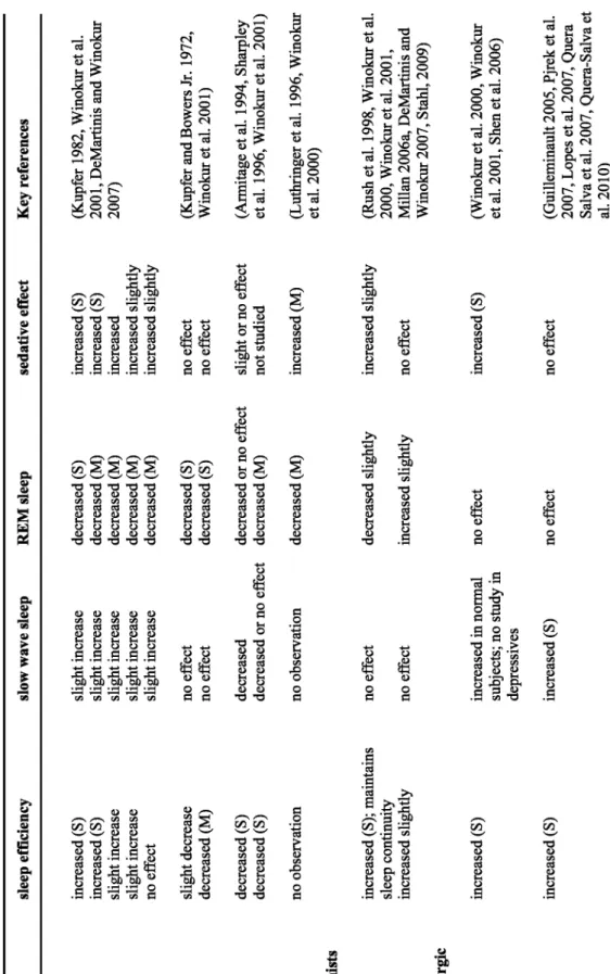
(9) increased (S). increased (S). MASSAs Agomelatine. no observation. no observation. Serotonergic-noradrenergic antidepressants Mirtazapine. decreased decreased or no effect. decreased (S) decreased (S). Nefazodone. no effect no effect. slight decrease decreased (M). increased (S). increasedin normal subjects; no study in depressives. no effect. no effect. slight increase slight increase slight increase slight increase slight increase. slow wave sleep. increased (S) increased (S) slight increase slight increase no effect. sleep efficiency. increased (S); maintains sleep continuity increased slightly. Trazodone. S-HT Receptor Antagonists. Tricyclics Amitriptyline Doxepin Imipramine Nortriptyline Desipramine MAOIs Phenelzine Tranylcypromine SSRIs Fluoxetine Paroxetine SNRIs Venlafaxine. Drug. no effect. no effect. increased slightly. decreased slightly. decreased (M). decreased or no effect decreased (M). decreased (S) decreased (S). decreased (S) decreased (M) decreased (M) decreased (M) decreased (M). REM sleep. Table 1. Effects of some antidepressants on EEG sleep parameters (S= significant; M= moderate). no effect. increased (S). no effect. increased slightly. increased (M). slight or no effect not studied. no effect no effect. increased (S) increased (S) increased increased slightly increased slightly. sedative effect. (Guilleminault 2005, Pjrek et al. 2007, Lopes et al. 2007, Quera Salva et al. 2007, Quera-Salva et al. 2010). (Winokur et al. 2000, Winokur et al. 2001, Shen et al. 2006). (Rush et al. 199 8, Winokur et al. 2000, Winokur et al. 2001, Millan 2006a, DeMartinis and Winokur 2007, Stahl, 2009). (Luthringer et al. 1996, Winokur et al. 2000). (Armitage et al. 1994, Sharpley et al. 1996, Winokur et al. 2001). (Kupfer and Bowers Jr. 1972, Winokur et al. 2001). (Kupfer 19 82, Winokur et al. 2001, DeMartinis and Winokur 2007). Key references. Daniel P. Cardinali et al.. Clinical Neuropsychiatry (2011) 8, 6.
(10) Sleep and circadian dysregulation in depressive illness. additive effect, increasing the risk of developing SAD (Partonen et al. 2007). This reinforces the existence of an association between certain clock gene polymorphisms and chronotype. Summarizing, the results are a strong indication that certain abnormalities in the circadian molecular clock can increase the susceptibility to mood disorders. However, most findings await replication in other samples and populations.. Associations of melatonin related gene polymorphisms with depressive illness A limited number of studies have been done to date on variation in genes related to melatonin in depressive disorder. However, some interesting findings are emerging that deserve to be corroborated and extended. It is now known that the enzyme arylalkylamine N-acetyltransferase (AANAT) is a key enzyme in the melatonin synthesis pathway that regulates the timing of melatonin production (Klein 2007). A recent study of this enzyme in depressed patients provided evidence of the association of genetic variability in the AANAT gene with susceptibility to MDD (Soria et al. 2010a). Both bipolar and unipolar patients were included in the population studied. As noted by the authors further analysis and subclassification ofthe patients according to their symptom profiles is important. It would also be important to determine whether phase advances or delays in nocturnal melatonin correlate with alterations in the AANAT gene. The rate limiting enzyme in melatonin synthesis, acetylserotonin methyltransferase (ASMT, formerly HIOMT), is now known to control the absolute levels of melatonin produced during the night (Ceinos et al. 2004, Liu and Borjigin 2005). Recently it has been reported that there is an association between two polymorphisms of ASMT gene and a lower risk of recurrence of depression (Galecki et al. 2010). Moreover patients with depression were characterized by reduced mRNA expression for ASMT. These findings support the concept of a low melatonin syndrome as a susceptibility factor for depression although the findings must be confirmed and extended. The G-protein linked receptor GPR50 is now known to inhibit the function of the melatonin receptor MT l by heterodimerization (Levoye et al. 2006a,b). A sex specific association between bipolar illness in women and GPR50 polymorphism has been reported (Thomson et al. 2005). Another study failed to find this association although there may have been genetic heterogeneity between the populations studied (Alaerts et al. 2006). A linkage between a second site on the same gene has been reported more recently that is stronger in females with some evidence of a role in symptom severity (Macintyre et al. 2010). These findings suggest that GPR50 may play a role in bipolar affective disorder especially in women.. Antidepressant treatment and sleep Pharmacotherapies for the treatment of depression have been in use since 1950's. They include tricyclic. Clinical Neuropsychiatry (2011) 8, 6. antidepressants (TCAs), monoamine oxidase inhibitors (MAOIs), selective 5HT reuptake inhibitors (SSRIs), 5-HT-norepinephrine (NE) reuptake inhibitors (SNRIs), several serotonergic receptor blockers, and the recently introduced melatonin agonist and selective 5-HT antagonists (MASSAs) whose prototype is agomelatine. Antidepressants are the third most widely prescribed class of therapeutic agents worldwide, with SSRIs accounting for 80% of the total market share (Celada et al. 2004). To define the effect of an antidepressant on sleep is very important because it influences clinicians' decision for the type of antidepressant to be prescribed (DeMartinis and Winokur 2007). Most antidepressants have different effects on the sleep profile. This is related to degree of inhibition of 5-HT or NE uptake, the effects on 5-HT1A or 5HT2 receptor sites or actions on ál - and á 2adrenoceptors or histamine Hl receptor sites (Mayers and Baldwin 2005). While some antidepressants cause improvement in sleep efficiency (SE) by ameliorating the depressive symptoms, others exert more rapid beneficial effects on initiation and maintenance of sleep (Sharpley and Cowen 1995, Tsuno et al. 2005, Thase 2006). The introduction of SSRIs and SNRI (the SNRI venlafaxine and duloxetine are currently the physician's drugs of choice) has drastically changed the strategies for clinical treatment of the MDD (Rosenzweig-Lipson et al. 2007, DeMartinis and Winokur 2007). Because of administration of SSRIs is commonly associated with insomnia (Anderson 2000), most pharmacoepidemiological surveys indicate that at least one third of patients taking SSRIs receive concomitant sedativehypnotic medications e.g. (Thase 2006). Hence clinicians tend to consider the antidepressants effects on sleep as a potentially important determining factor in selecting the therapeutic option to treat patients with depressive symptomatology (Winokur et al. 2001, DeMartinis and Winokur 2007).. Tricyclic antidepressants (TCAs) For over 30 years, from the 1960s to 1990, tricyclic drugs were the mainstay treatment for MDD. TCAs are known to decrease sleep onset latency (SOL), improve sleep SE and decrease wake time after sleep onset (WASO) (Ware et al. 1989). Because of their antihistaminic properties these drugs also produce sedating effects during the day, and it has thus been suggested that their use should be avoided in depressed patients who are sensitive to these effects (Winokur et al. 2001). With the exception of trimipramine all TCAs reduce or suppress REM sleep and increase REM latency (Vogel et al. 1990) It has been suggested that REM sleep suppression associated with these drugs is an essential component of their therapeutic action. NEand 5-HT-containing neurons in the brain, which are directly affected by TCAs, are not only involved in the pathophysiology of affective disorders but also have a significant impact on sleep regulation. NE and 5-HT are known to inhibit electrical activity of the ventrolateral preoptic nucleus which contains a group of sleep active, á-aminobutyric acid (GABA)-galanin producing neurons (Saper et al. 2005). It has also been.
(11) Daniel P. Cardinali et al.. suggested that blockade of histamine Hl receptors or antagonism of á ,-adrenoceptors is at the basis of their sleep promoting effects (Mayers and Baldwin 2005). Polysomnographic (PSG) studies support that TCAs generally cause sedative effects and clinical experience indicates that drugs like amitriptyline and trimipramine shorten SOL and improve sleep continuity and efficiency (Winokur et al. 2001, DeMartinis and Winokur 2007).. Monoamine oxidase inhibitors (MAOIs) The use of the MAOIs tranylcypromine and phenelzine results in subjective complaints of insomnia, with PSG studies confirming prolonged SOL, impaired sleep continuity and increased WASO (Kupfer and Bowers 1972, Winokur et al. 2001). REM sleep suppression has also been noted in depressives who were on phenelzine and tranylcypromine. It is present soon after initiation of treatment and persists for months during continuation of therapy. However use of reversible monoamine oxidase A inhibitors such as moclobemide causes less pronounced REM sleep suppression (Monti 1989).. Serotonin-norepinephrine reuptake inhibitors (SNRls) Venlafaxine, duloxetine and milnacipran belong to the SNRI category of drugs that inhibit presynaptic uptake of both 5-HT and NE (Stahl et al. 2005). In a double-blind placebo controlled study, administration of venlafaxine, at doses ranging from 75 to 225 mg/ day, induced increases in WASO after one month of treatment. REM sleep latency increased significantly while the total amount of time spent in REM sleep diminished (Salin-Pascual et al. 1997, Winokur et al. 2001, Argyropoulos and Wilson 2005). Yang et al. reported that venlafaxine was associated with REM sleep suppression and increased in REM sleep latency, although, in contrast to previous studies, no differences in SOL or SE were observed between depressed patients and control subjects (Yang et al. 2005). Venlafaxine was also found to increase the frequency of periodic leg movements in sleep (PLMS). These repetitive and highly stereotyped limb movements can occur during sleep and/ or the waking state. PLMS movements are the result of EEG arousals or awakenings and might cause difficulties in initiating and maintaining sleep. Venlafaxine induced significant increases in PLMS movements, presumably the result of enhanced serotonergic availability and secondarily decreased of dopamine (DA) effects caused by the drug (Yang et al. 2005).. Selective serotonin reuptake inhibitors (SSRls) The SSRIs, the most commonly used antidepressants, have been considered a major treatment breakthrough ever since the importance of 5HT in mood regulation was recognized. Two recent meta-analyses of clinical trials found that in mild and moderate depression, which constitute the vast majority of. depression cases, the effect of SSRI is very small or none compared to placebo, while in very severe depression the effect of SSRIs is clinically significant (Kirsch et al. 2008, Fournier et al. 2010). SSRIs block the presynaptic uptake of 5-HT and enhance the activation of the postsynaptic receptors, thus prolonging the interaction of 5-HT with the multiple 5HT receptor subtypes. However, SSRIs also have a number of side effects, the most prominent being their effects on sleep and sexual function (Moltzen and BangAndersen 2006). In an early study, sertraline (with maximum doses up to 200 mg/day achieved within a 10 day period) significantly prolonged SOL and reduced TST 14 days after treatment (Winokur et al. 2001). However, there was neither a reduction in SE nor an increase in WASO. It has been reported that nearly 25% of depressed patients treated with SSRIs have subjective complaints of insomnia (Armitage 2007). Fluoxetine administration has been shown to cause disruption of sleep continuity, reductions in SE and increases in WASO (Winokur et al. 2001). In a group of patients with major depression, fluoxetine at doses of 20mg/ day for 4 weeks caused significant decreases in SE, a finding that correlated well with the plasma fluoxetine levels (Armitage et al. 1997). This decrease in SE was also noted in other studies with fluoxetine, e.g. (Trivedi et al. 1999). REM sleep suppression has also been a consistent finding in depressed patients who are being treated with fluoxetine (Armitage 2007) Concerning the influence of paroxetine on sleep in depressed subjects it has also been noted that this drug similarly reduces SE, with an increased the number of awakenings being observed after 4 weeks of treatment. It did not, however, influence TST or SOL (Staner et al. 1995). Similar effects were also noted in normal healthy subjects receiving paroxetine (20mg/day). Subjects receiving paroxetine demonstrated significant reductions in SE, and an increase in WASO when compared to placebo. REM sleep minutes were reduced and REM latency was significantly prolonged (Sharpley et al. 1996). Yang et al. used PSG measurements to study 274 patients who had been receiving SSRIs and found an association between the use of the drugs and suppression of REM sleep and increases in REM sleep latency (Yang et al. 2005). No differences in SOL or SE were apparent as compared to control subjects. A significant increase in PLMS was observed in patients receiving SSRIs, similar to that of venlafaxine, as reported above. PLMS can contribute significantly to difficulties in initiating and maintaining sleep, and caution should therefore be exercised in choosing antidepressants for the treatment of depressed patients who have pronounced sleep complaints (Yang et al. 2005).. Serotonin-2 receptor antagonist/serotonin reuptake inhibitors (SARls) Trazodone and nefazodone are the two drugs that belong to the SARIs category, their main action being inhibition of 5-HT2 receptors, which are also involved in the regulation of sleep (Millan 2006). Trazodone also inhibits the ál-adrenergic and the histamine H l receptors (Stahl et al. 2003). Nefazodone inhibits á l adrenergic receptors and inhibits NE uptake but has weak actions. Clinical Neuropsychiatry (2011) 8, 6.
(12) Sleep and circadian dysregulation in depressive illness. on histamine receptors. Because of its effects on H l receptors trazodone administration produces sedating effects and causes daytime somnolence (Winokur et al. 2001). Administration of trazodone to depressed patients has been found to increase TST, reduce SOL, reduce the number of awakenings and arousals, reduce total REM sleep time, and prolong REM latency (Winokur et al. 2001). In an eight week study of six depressed patients who also had symptoms of insomnia, trazodone treatment resulted in a 44% improvement in SOL, a 14% improvement in TST, and noteworthy improvements in SE (Scharf and Sachais 1990). Nefazodone administration to depressed patients preserves sleep continuity and decreases the number of awakenings (Rush et al. 1998). Evidence of its effects on sleep efficiency however has been less consistent, with either increases or no effects reported (Armitage 2007).. Serotonergic-noradrenergic antidepressant drugs Drugs belonging to this category, e.g. mirtazapine and mianserin have a dual-action profile, combining the enhancement of the noradrenergic neurotransmitter system with specific actions on particular serotonergic receptor subtypes. They exert a potent antagonism of presynaptic á2- heteroceptors and á2autoreceptors that results in increased release of both 5-HT and NE (Wilson and Argyropoulos 2005). They also act as 5HT2 and 5-HT, receptor antagonists that contribute to the anxiolytic and soporific actions (Haddjeri et al. 1995, de Boer 1996, Shen et al. 2006). Mirtazapine also exhibits potent antihistamine (H l ) activity. The use of mirtazepine in patients with major depression results in significant reductions of sleep disturbances (Winokur et al. 2000, Shen et al. 2006). In a study conducted on six adult patients with MDD, the administration of 15 mg/day of mirtazapine for one week followed by 30 mg/day for an additional week caused significant reductions in SOL and significant increases in TST as compared to baseline (Winokur et al. 2000). Comparison of mirtazepine with other antidepressants indicates a better SE than with fluoxetine (Winokur et al. 2003) or paroxetine (Ridout et al. 2003). Mirtazapine has also been found better than venlafaxine (Guelfi et al. 2001) or paroxetine (Schatzberg et al. 2002) in improving Hamilton Depression Rating Scale (HAMD) sleep scores. In the earliest PSG study conducted on sleep architecture in normal volunteers, mirtazapine (30 mg/day) decreased SOL, WASO and stage-1 sleep and increased SWS (Ruigt et al. 1990). A similar effect was found in another study conducted on healthy volunteers in whom the administration of mirtazapine (30 mg/day) caused a significant improvement in SE with reductions in nocturnal disturbances as compared to placebo (Aslan et al. 2002). Although mirtazapine is as effective as SSRIs as antidepressants, its side effects like increased appetite, weight gain and excessive daytime sedation (mediated by H l blockade) have prevented its acceptance as a first-line medication (Thase 2006). Mianserin has sleep promotion properties, possibly through inhibition of histamine (Hl ) receptors (Sharpley and Cowen 1995, Mayers and Baldwin 2005). Earlier. Clinical Neuropsychiatry (2011) 8, 6. studies conducted on depressed patients revealed that mianserin (10-20 mg/day) caused reductions in HAMD sleep scores as compared to placebo for depressed women with cancer (Costa et al. 1985).. Sedative-hypnotic medications Benzodiazepines are widely used for the treatment of insomnia although they were originally developed as anxiolytics and later used as hypnotics. Benzodiazepines cause reductions in SOL and prolong sleep time but their long-term use is debatable as these drugs cause development of tolerance, rebound insomnia and cognitive deficits (Jindal and Thase 2004, Thase 2006). In one of the placebo-controlled long-term trials in patients with MDD, it was found that the beneficial effects of clonazepam on patient's sleep complaints were limited to the first three weeks of therapy (Smith et al. 2002). Despite its clinical use for nearly 20 years, there is a lack of controlled studies using PSG for assessing the effectiveness of benzodiazepines as an add-on therapy with either SSRIs or SNRI in MDD (Thase 2006). Moreover, the APA's Task force on benzodiazepine's Dependence, Toxicity, and Abuse has recommended against the long-term use of benzodiazepines in the treatment of insomniacs particularly in elderly patients (Jindal and Thase 2004). As sleep disturbances, particularly insomnia, are often found with antidepressant medications, the use of hypnotic drugs have been resorted to offset the sleep problem. Several of the atypical antidepressants have a sedative action but the extent varies. It is problematic with clozapine and very common with quetiapine. Sedation is common with risperidone, olanzapine, paliperidone and amisulpride. It is not unusual with ziprazodine. On the other hand the newer atypical antidepressant aripiprazole occasionally causes insomnia (Stahl 2009). Recent data indicate that atypical antipsychotics may increase the risk of sleep disorders like obstructive sleep apnea independently of weight and neck circumference (Rishi et al. 2010). In a study on SSRI treated depressed patients, it was found that those receiving daily doses of fluoxetine (~ 40 mg), sertraline (~100 mg) or paroxetine (~40 mg) reported significant insomnia. These patients were then entered into a double-blind phase where they were assigned randomly to zolpidem (10 mg) or placebo for 4 weeks followed by single blind placebo for one week. Those depressed patients who received zolpidem demonstrated significant improvements in sleep with longer TST, better sleep quality and reduced WASO (Asnis et al. 1999).. Melatonin agonist and selective serotonin antagonists (MASSA) Agomelatine, developed by Servier, France, is a novel antidepressant with MT l and MT2 receptor agonist activity that has 5-HT2c antagonist properties. It is the first representative of a new type of antidepressant (MASSA). Agomelatine is a naphthalenic compound with an overall selectivity for.
(13) Daniel P. Cardinali et al.. MT1 and MT2 receptors but no significant affinity to muscarinic, histaminergic, adrenergic or dopaminergic receptor subtypes (Rouillon, 2006). In multicenter trials undertaken in Europe (Loo et al. 2002, Kupfer 2006, Kennedy and Emsley 2006) agomelatine at a dose of 25 mg/day given before bedtime was found to be effective in reducing the depressive symptoms in patients with MDD. The effectiveness of agomelatine in severely depressed patients is particularly significant inasmuch as this patient group is resistant to SSRIs or SNRIs. Agomelatine represents an innovation in the treatment of depression because it has few adverse effects and is associated with early resolution of depressive symptoms (Pandi-Perumal et al. 2006, Kennedy and Rizvi 2010). In addition agomelatine is effective in reducing sleep complaints in depressed patients. Treatment of depressed patients with agomelatine for six weeks increased the duration ofNREM sleep without affecting REM sleep thus causing improvements in both sleep quality and continuity (Quera Salva et al. 2007, 2010). In a study which compared agomelatine with venlafaxine, agomelatine at 25 mg/day promoted earlier and greater improvement on the "criteria of getting into sleep" in a Leeds sleep evaluation questionnaire (Guilleminault 2005). The improvement in sleep quality was evident at the first week of agomelatine, but not of venlafaxine, use. In another study it was reported that agomelatine normalizes NREM sleep changes found in depressed patients. The changes in NREM preceded the improvement seen in Hamilton depression scores (Lopes et al. 2007). Agomelatine is thus a dual action drug that can produce rapid antidepressant effects while also improving sleep quality. This is very important clinically inasmuch as improvements in sleep among depressed patients are associated with a reduced rate of recurrence of depressive symptoms and, conversely, complaints of poor sleep in depressed patients are associated with a poor response to subsequent antidepressant treatment. A recent study provides strong support for the superior chronobiological effects of agomelatine in patients with MDD (Kasper et al. 2010). As compared to sertraline, agomelatine increased the relative amplitude of the circadian rest-activity cycle by the end of week one and in parallel there were improvements in sleep efficiency and in sleep latency from week one to week six. Over a six week treatment period depressive and anxiety symptoms improved more with agomelatine than with sertraline. Agomelatine is unique because it has a chronobiological basis for its action. Since agomelatine effects are mediated through both MT1 and MT2 melatonergic receptors and 5-HT2c serotonergic receptors it acts differently in different circadian phases of the day-night cycle. While it promotes and maintains sleep at night, it also maintains alertness during the day. At night the sleep promoting melatonergic effects prevail over its potentially antihypnotic 5-HT2c antagonism, whereas during the day the antidepressant actions through antagonism of 5-HT2c receptors is uncoupled from melatonin's nocturnal hypnotic effects (Millan 2006). These effects are in contrast to traditional antidepressants which elevate the mood of depressed patients of the patients during daytime, an effect that is sustained in the night causing impairment in sleep. quality (Ruhe et al. 2007). One criticism of this dual interpretation of agomelatine action is the large differences in affinity for the putative action on serotonergic receptors as compared to the melatonergic one (about 3 orders of magnitude greater concentration are needed to exert 5HT2C antagonism) (Millan et al. 2003). Moreover, both melatonin and ramelteon have been shown to display antidepressant-like effects even though they are not reportedly known to affect serotonergic activity significantly (Detanico et al. 2009, Crupi et al. 2010, McElroy et al. 2011). All available evidence suggests that agomelatine is a promising antidepressant that can address the sleep disturbances and abnormalities seen in depression. However, although the compound was very well tolerated according to subjective ratings, the issue of long-term toxicity has not been yet fully addressed (Cardinali et al. 2011).. Conclusions Converging lines of evidences suggest that an abnormality in the timing of clock-controlled processes and of melatonin related processes may play a pivotal role in the pathogenesis of depression. It could be proposed that disturbed circadian rhythm regulation impacts on sleep rhythms to produce changes in monoamine regulation of mood. The altered mood could then influence sleep. However, the working hypothesis on a possible direct causal relationship between alterations of the circadian system and depression cannot be presently warranted. Indeed, the circadian abnormalities observed could be a consequence, rather than the cause of the depressive status, or be a manifestation of conditions that directly affect both biological rhythms and the mechanisms involved in mood regulation. The systematic investigations of clock gene and melatonin related gene polymorphisms in depression, the characterization of behavioral phenotypes in mouse circadian mutants and the identification of new genetic factors that contribute to circadian and behavioral function will undoubtedly shed light into the relationship between circadian rhythm alterations and depression. In addition studies are needed that consider more refined clinical and circadian subphenotypes in MDD, such as treatment response, sleep/wake cycle abnormalities, diurnal mood variation, and seasonality (Soria et al. 2010b). The existence of such relationships will certainly have profound therapeutic implications. Since insomnia is one of the hallmark symptoms of depressive disorders, a major difficulty with conventional antidepressant therapy, especially the SSRIs, is that they often disturb sleep and may therefore increase sleep problems. Recently a novel melatonergic antidepressant with both melatonin agonist properties and 5-HT2c antagonist properties (agomelatine) has been introduced. It is effective not only for ameliorating symptoms of depressive illness and reducing Hamilton depression scores, but also for improving sleep quality and reducing sleep complaints. More clinical trials are needed to confirm the efficacy of melatonergic drugs for long term use in the treatment of chronic primary. Clinical Neuropsychiatry (2011) 8, 6.
(14) Sleep and circadian dysregulation in depressive illness. insomnia as well as for insomnia associated with depression and other psychiatric conditions.. Acknowledgements DPC is a Research Career Awardee from the Argentine Research Council (CONICET) and Professor Emeritus, University of Buenos Aires.. References Alaerts M, Venken T, Lenaerts AS, De Zutter S, Norrback KF, Adolfsson R, Del Favero J (2006). Lack of association of an insertion/deletion polymorphism in the G proteincoupled receptor 50 with bipolar disorder in a Northern Swedish population. Psychiatric genetics /6, 6, 235-236. American Psychiatric Association ( 2000). Diagnostic and Statistical Manual of Mental Disorders. 4th Ed (DSM-IVTR), American Psychiatric Press, Washington, D.C. Anderson IM (2000). Selective serotonin reuptake inhibitors versus tricyclic antidepressants: a meta-analysis of efficacy and tolerability. Journal of affective disorders 58, /, /936. Antle MC, Ogilvie MD, Pickard GE, Mistlberger RE (2003). Response of the mouse circadian system to serotonin /A/ 2/7 agonists in vivo: surprisingly little. Journal of biological rhythms /8, 2, /45-/58. Archer SN, Robilliard DL, Skene DJ, Smits M, Williams A, Arendt J, von Schantz M (2003). A length polymorphism in the circadian clock gene Per3 is linked to delayed sleep phase syndrome and extreme diurnal preference. Sleep 26, 4, 4/3-4/5. Arendt J, Broadway J (/987). Light and melatonin as zeitgebers in man. Chronobiology international 4, 2, 273-28 2. Arendt J, Skene DJ (2005). Melatonin as a chronobiotic. Sleep medicine reviews 9, /, 25-39. Argyropoulos SV, Wilson SJ ( 2005). Sleep disturbances in depression and the effects of antidepressants. International reviews ofpsychiatry/7, 4, 237-245. Armitage R ( 2007). Sleep and circadian rhythms in mood disorders. Acta psychiatrica scandinavica suppl433, /04//5. Armitage R, Rush AJ, Trivedi M, Cain J, Roffwarg HP (/994). The effects of nefazodone on sleep architecture in depression. Neuropsychopharmacology /0, 2, / 23-/ 27. Armitage R, Yonkers K, Cole D, Rush AJ (/997). A multicenter, double-blind comparison of the effects of nefazodone and fluoxetine on sleep architecture and quality of sleep in depressed outpatients. Journal of clinical psychopharmacology /7, 3, /6/-/68. Aslan S, Isik E, Cosar B (2002). The effects of mirtazapine on sleep: a placebo controlled, double-blind study in young healthy volunteers. Sleep 25, 6, 677-679. Asnis GM, Chakraburtty A, DuBoff EA, Krystal A, Londborg PD, Rosenberg R, Roth-Schechter B, Scharf MB, Walsh JK (/999). Zolpidem for persistent insomnia in SSRItreated depressed patients. Journal of clinical psychiatry 60, /0, 668-676. Bao AM, Meynen G, Swaab DF (2008). The stress system in depression and neurodegeneration: focus on the human hypothalamus. Brain research reviews 57, 2, 53/-553. Beck-Früs J, Kjellman BF, Aperia B, Unden F, von Rosen D, Ljunggren JG, Wetterberg L (/985). Serum melatonin in relation to clinical variables in patients with major depressive disorder and a hypothesis of a low melatonin syndrome. Acta psychiatrica scandinavica 7/, 4, 3/9-330. Benedetti F, Bernasconi A, Lorenzi C, Pontiggia A, Serretti A, Colombo C, Smeraldi E ( 2004). A single nucleotide polymorphism in glycogen synthase kinase 3-beta promoter. Clinical Neuropsychiatry (2011) 8, 6. gene influences onset of illness in patients affected by bipolar disorder. Neuroscience letters 355, /-2, 37-40. Benedetti F, Dallaspezia S, Fulgosi MC, Lorenzi C, Serretti A, Barbini B, Colombo C, Smeraldi E (2007). Actimetric evidence that CLOCK 3/// T/C SNP influences sleep and activity patterns in patients affected by bipolar depression. American journal of medical genetics part b neuropsychiatric genetics /44B, 5, 63/-635. Benedetti F, Serretti A, Colombo C, Barbini B, Lorenzi C, Campori E, Smeraldi E (2003). Influence of CLOCK gene polymorphism on circadian mood fluctuation and illness recurrence in bipolar depression. American journal of medical genetics part b neuropsychiatric genetics / 23B, /, 23-26. Bergiannaki JD, Soldatos CR, Paparrigopoulos TJ, Syrengelas M, Stefanis CN (/995). Low and high melatonin excretors among healthy individuals. Journal ofpineal research /8, 3, /59-/64. Borbely AA (/98 2). A two process model of sleep regulation. Human neurobiology /, 3, /95- 204. Branchey L, Weinberg U, Branchey M, Linkowski P, Mendlewicz J (/98 2). Simultaneous study of 24-hour patterns of melatonin and cortisol secretion in depressed patients. Neuropsychobiology 8, 5, 225-23 2. Brown GM, Pandi-Perumal SR, Trakht 1, Cardinali DP (2009). Melatonin and its relevance to jet lag. Travel medicine and infectious disease 7, 69-8/. Brzezinski A, Vangel MG, Wurtman RJ, Norrie G, Zhdanova 1, Ben Shushan A, Ford 1 ( 2005). Effects of exogenous melatonin on sleep: a meta-analysis. Sleep medicine reviews 9, /, 4/-50. Bunney JN, Potkin SG ( 2008). Circadian abnormalities, molecular clock genes and chronobiological treatments in depression. British medical bulletin 86, 23-3 2. Buscemi N, Vandermeer B, Hooton N, Pandya R, Tjosvold L, Hartling L, Vohra S, Klassen TP, Baker G (2006). Efficacy and safety of exogenous melatonin for secondary sleep disorders and sleep disorders accompanying sleep restriction: meta-analysis. British medical journal 33 2, 7538, 385-393. Buysse DJ, Nofzinger EA, Germain A, Meltzer CC, Wood A, Ombao H, Kupfer DJ, Moore RY (2004). Regional brain glucose metabolism during morning and evening wakefulness in humans: preliminary findings. Sleep 27, 7, / 245-/ 254. Cardinali DP, Pandi-Perumal SR, Srinivasan V (2008). Circadian control by serotonin and melatonin receptors: Clinical relevance, in Serotonin and Sleep. Molecular, Functional and Clinical Aspects (Monti JM, Pandi-Perumal SR, Nutt DJ, eds) pp. 477-499, Birkhauser Verlag, Basel. Cardinali DP, Srinivasan V, Brzezinski A, Brown GM (20//). Melatonin and its analogs in insomnia and depression. Journal ofpineal research in press Ceinos RM, Chansard M, Revel F, Calgari C, Miguez JM, Simonneaux V (2004). Analysis of adrenergic regulation of melatonin synthesis in Siberian hamster pineal emphasizes the role of H~OMT. Neurosignals /3, 6, 3083/7. Celada P, Puig M, Amargos-Bosch M, Adell A, Artigas F (2004). The therapeutic role of 5-HT, and 5-HTZA receptors in depression. Journal ofpsychiatry and neuroscience 29, 4, 25 2-265. Claustrat B, Chazot G, Brun J, Jordan D, Sassolas G (/984). A chronobiological study of melatonin and cortisol secretion in depressed subjects: plasma melatonin, a biochemical marker in major depression. Biological psychiatry /9, 8, / 2/5-/ 228. Coogan AN, Wyse CA ( 2008). Neuroimmunology of the circadian clock. Brain research / 23 2, /04-// 2. Costa D, Mogos 1, Toma T (/985). Efficacy and safety of mianserin in the treatment of depression of women with cancer. Acta psychiatrica scandinavica Suppl 3 20, 85-92. Crasson M, Kjiri S, Colin A, Kjiri K, L~hermite-Baleriaux M,.
Figure


Documento similar
In this prospective study of patients in a major depressive episode (MDE) and meeting criteria for major depressive disorder (MDD) or bipolar disorder (BD), we
that psychosocial difficulties (PSDs), such as sleep disturbances, emotional instability and difficulties in personal interactions, and their environmental determinants are
Diel expression of the circadian clock genes VunGI, VunELF3, VunTOC1, and VunLHY in pod and seed tissue The analysis of the circadian gene network in generative cowpea tissues
TOC1 and CCA1 or the related gene LATE ELONGATED HYPOCOTYL (LHY), which also encodes a MYB protein, are highly conserved along the green lineage including the
This fact favored new species of pollinators such as the hawkmoth that prefers UV- absorbing flowers (white flower) rather than UV-reflecting flowers
Hence, this study was aimed at evaluating the association of both subjective (i.e., global Pittsburgh Sleep Quality Index [PSQI] score) and objective (i.e., total sleep time [TST],
Maximal fat oxidation rate (MFO, in g/min; upper panel) and exercise intensity at MFO (Fatmax, as percentage of maximal oxygen uptake (VO 2 max); lower panel) dur- ing exercise
An employee who sleeps 8 hours per night may still have poor sleep quality, preventing the individual from showing up for work at peak performance.. Additionally, sleep hygiene
